Embed Size (px)
Citation preview
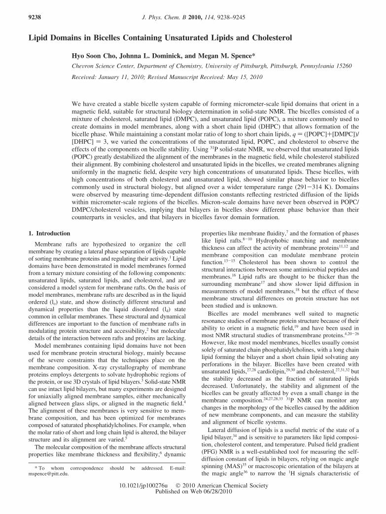
Lipid Domains in Bicelles Containing Unsaturated Lipids and Cholesterol
Hyo Soon Cho, Johnna L. Dominick, and Megan M. Spence*CheVron Science Center, Department of Chemistry, UniVersity of Pittsburgh, Pittsburgh, PennsylVania 15260
ReceiVed: January 11, 2010; ReVised Manuscript ReceiVed: May 15, 2010
We have created a stable bicelle system capable of forming micrometer-scale lipid domains that orient in amagnetic field, suitable for structural biology determination in solid-state NMR. The bicelles consisted of amixture of cholesterol, saturated lipid (DMPC), and unsaturated lipid (POPC), a mixture commonly used tocreate domains in model membranes, along with a short chain lipid (DHPC) that allows formation of thebicelle phase. While maintaining a constant molar ratio of long to short chain lipids, q ) ([POPC]+[DMPC])/[DHPC] ) 3, we varied the concentrations of the unsaturated lipid, POPC, and cholesterol to observe theeffects of the components on bicelle stability. Using 31P solid-state NMR, we observed that unsaturated lipids(POPC) greatly destabilized the alignment of the membranes in the magnetic field, while cholesterol stabilizedtheir alignment. By combining cholesterol and unsaturated lipids in the bicelles, we created membranes aligninguniformly in the magnetic field, despite very high concentrations of unsaturated lipids. These bicelles, withhigh concentrations of both cholesterol and unsaturated lipid, showed similar phase behavior to bicellescommonly used in structural biology, but aligned over a wider temperature range (291-314 K). Domainswere observed by measuring time-dependent diffusion constants reflecting restricted diffusion of the lipidswithin micrometer-scale regions of the bicelles. Micron-scale domains have never been observed in POPC/DMPC/cholesterol vesicles, implying that bilayers in bicelles show different phase behavior than theircounterparts in vesicles, and that bilayers in bicelles favor domain formation.
1. Introduction
Membrane rafts are hypothesized to organize the cellmembrane by creating a lateral phase separation of lipids capableof sorting membrane proteins and regulating their activity.1 Lipiddomains have been demonstrated in model membranes formedfrom a ternary mixture consisting of the following components:unsaturated lipids, saturated lipids, and cholesterol, and areconsidered a model system for membrane rafts. On the basis ofmodel membranes, membrane rafts are described as in the liquidordered (lo) state, and show distinctly different structural anddynamical properties than the liquid disordered (ld) statecommon in cellular membranes. These structural and dynamicaldifferences are important to the function of membrane rafts inmodulating protein structure and accessibility,2 but moleculardetails of the interaction between rafts and proteins are lacking.
Model membranes containing lipid domains have not beenused for membrane protein structural biology, mainly becauseof the severe constraints that the techniques place on themembrane composition. X-ray crystallography of membraneproteins employs detergents to solvate hydrophobic regions ofthe protein, or use 3D crystals of lipid bilayers.3 Solid-state NMRcan use intact lipid bilayers, but many experiments are designedfor uniaxially aligned membrane samples, either mechanicallyaligned between glass slips, or aligned in the magnetic field.4
The alignment of these membranes is very sensitive to mem-brane composition, and has been optimized for membranescomposed of saturated phosphatidylcholines. For example, whenthe molar ratio of short and long chain lipid is altered, the bilayerstructure and its alignment are varied.5
The molecular composition of the membrane affects structuralproperties like membrane thickness and flexibility,6 dynamic
properties like membrane fluidity,7 and the formation of phaseslike lipid rafts.8-10 Hydrophobic matching and membranethickness can affect the activity of membrane proteins11,12 andmembrane composition can modulate membrane proteinfunction.13-15 Cholesterol has been shown to control thestructural interactions between some antimicrobial peptides andmembranes.16 Lipid rafts are thought to be thicker than thesurrounding membrane17 and show slower lipid diffusion inmeasurements of model membranes,18 but the effect of thesemembrane structural differences on protein structure has notbeen studied and is unknown.
Bicelles are model membranes well suited to magneticresonance studies of membrane protein structure because of theirability to orient in a magnetic field,19 and have been used inmost NMR structural studies of transmembrane proteins.4,20-26
However, like most model membranes, bicelles usually consistsolely of saturated chain phosphatidylcholines, with a long chainlipid forming the bilayer and a short chain lipid solvating anyperforations in the bilayer. Bicelles have been created withunsaturated lipids,27,28 cardiolipin,29,30 and cholesterol,27,31,32 butthe stability decreased as the fraction of saturated lipidsdecreased. Unfortunately, the stability and alignment of thebicelles can be greatly affected by even a small change in themembrane composition.24,27,28,33 31P NMR can monitor anychanges in the morphology of the bicelles caused by the additionof new membrane components, and can measure the stabilityand alignment of bicelle systems.
Lateral diffusion of lipids is a useful metric of the state of alipid bilayer,34 and is sensitive to parameters like lipid composi-tion, cholesterol content, and temperature. Pulsed field gradient(PFG) NMR is a well-established tool for measuring the self-diffusion constant of lipids in bilayers, relying on magic anglespinning (MAS)35 or macroscopic orientation of the bilayers atthe magic angle36 to narrow the 1H signals characteristic of
* To whom correspondence should be addressed. E-mail:[email protected].
J. Phys. Chem. B 2010, 114, 9238–92459238
10.1021/jp100276u 2010 American Chemical SocietyPublished on Web 06/28/2010
anisotropic molecular systems. Using PFG NMR, distinctdiffusion constants have been observed for lipids in the lo andld phases, and the onset of domain formation has been measuredfor domain forming systems.18,37,38 The size of lipid domainshas been measured by using PFG NMR experiments to observethe lipid diffusion behavior as a function of diffusion time, withthe time-dependent diffusion constant offering a measure of thelength scale of membrane inhomogeneity.39,40
2. Experimental Section
2.1. Sample Preparation. 1,2-dimyristoyl-sn-glycero-3-phosphatidylcholine (DMPC, 99%) in powder form, 1,2-dihexanoyl-sn-glycero-3-phosphatidylcholine (DHPC, 99%) in
chloroform, and 1-palmitoyl-2-oleoyl-sn-glycero-3-phosphati-dylcholine (POPC) in chloroform were purchased from AvantiPolar Lipids (Birmingham, AL). Lipids were used aspurchased without further purification. Cholesterol (ovinewool, >98%) in powder form, 4-(2-hydroxyethyl)-1-pipera-zineethanesulfonic acid (HEPES, g99.5%) and sodium azidewere purchased from Sigma-Aldrich (Allentown, PA). Deu-terium oxide was purchased from Cambridge Isotopes Lab(Woburn, MA).
The lipids were dissolved in chloroform and combined in amolar ratio of 2.6/0.4/1 and 1.5/1.5/1 molar ratios of DMPC/POPC/DHPC with and without 13 mol % cholesterol (calculatedwith respect to moles of long chain lipids) to create unsaturated
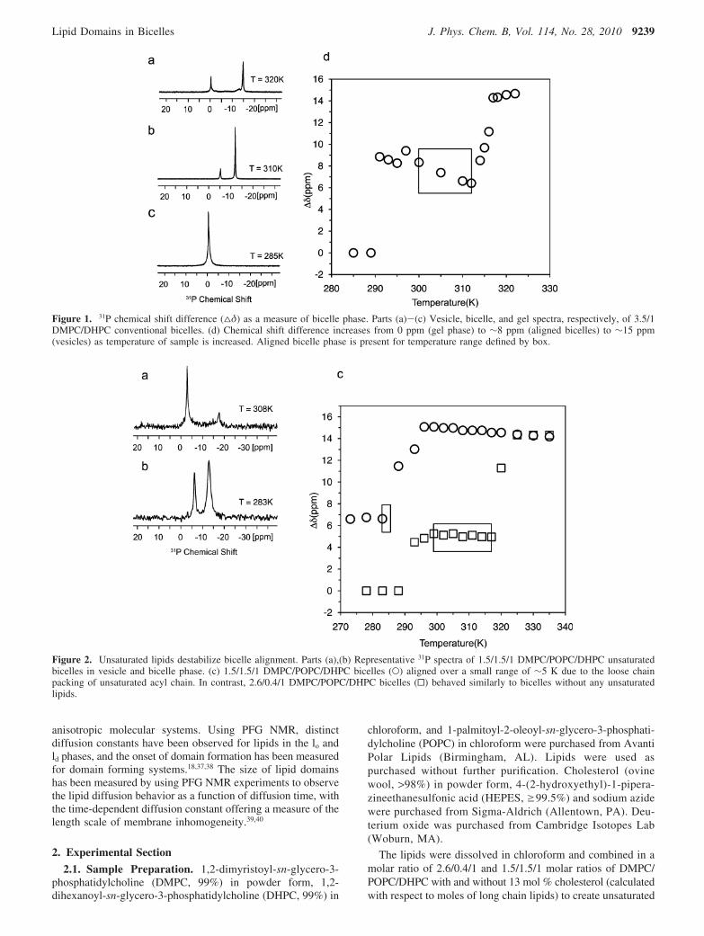
Figure 1. 31P chemical shift difference (4δ) as a measure of bicelle phase. Parts (a)-(c) Vesicle, bicelle, and gel spectra, respectively, of 3.5/1DMPC/DHPC conventional bicelles. (d) Chemical shift difference increases from 0 ppm (gel phase) to ∼8 ppm (aligned bicelles) to ∼15 ppm(vesicles) as temperature of sample is increased. Aligned bicelle phase is present for temperature range defined by box.
Figure 2. Unsaturated lipids destabilize bicelle alignment. Parts (a),(b) Representative 31P spectra of 1.5/1.5/1 DMPC/POPC/DHPC unsaturatedbicelles in vesicle and bicelle phase. (c) 1.5/1.5/1 DMPC/POPC/DHPC bicelles (O) aligned over a small range of ∼5 K due to the loose chainpacking of unsaturated acyl chain. In contrast, 2.6/0.4/1 DMPC/POPC/DHPC bicelles (0) behaved similarly to bicelles without any unsaturatedlipids.
Lipid Domains in Bicelles J. Phys. Chem. B, Vol. 114, No. 28, 2010 9239

bicelles. A conventional bicelle sample (no cholesterol orunsaturated lipids) was composed of DHPC and DMPC dis-solved in chloroform with molar ratio, q ) [DMPC]/[DHPC],equal to 3.5. The lipid mixtures were placed under a nitrogengas flow for ∼20 min to remove chloroform and then placedon a vacuum line for at least 4 h until the sample was reducedto a powder or powder/film mixture. A mixture of 10%deuterium oxide and water was added to each unsaturated bicellesample to a concentration of 30% w/w. A 20 mM HEPES buffer(pH ) 7.1) was used to redissolve the conventional bicellemixture to a concentration of 30% w/w. Three cycles of thefollowing: heating at 40 °C for 15 min, vortexing for 2 min,cooling at 0 °C for 15 min and again vortexing for 2 min, wereperformed to form bicelle mixtures.
2.2. NMR Measurements. All spectra were acquired withan 11.7 T magnet with a Bruker Avance console (BrukerBiospin, Billerica, MA) and BCU05 Variable TemperatureControl Unit. All sample rotors were 200 µL Bruker 4 mm ZrO2
magic-angle spinning (MAS) rotors.A 4-mm HXY MAS probe, was used to acquire the 31P NMR
spectra. All 31P experiments were performed under staticconditions. The variable temperature 31P experiments at 201.98MHz used FLOPSY at 6 kHz41 for proton decoupling. Thetemperature was varied from 273 to 335 K, in increments of 5or 3 K with the sample equilibrating in the probe for 10 to 15min at each temperature. The 31P chemical shifts are referencedto 85% phosphoric acid.
A high-resolution HCN HR-MAS probe (Bruker Biospin,Billerica, MA) was used to acquire the 1H NMR spectra. All
1H experiments were performed under MAS at 5kHz in orderto obtain isotropic spectra. 1H spectra were acquired aftertemperature equilibration for 30 min. 1H diffusion NMR spectrawere recorded at 499.81 MHz with a stimulated echo bipolarpulsed field gradient diffusion experiments using WATERGATEfor water suppression.42,43 The maximum gradient strength ofthe magic angle gradient coil was 0.513 T/m. The intensitiesof the acyl peak (1.2 ppm) were used to calculate the diffusioncoefficient. The 1H chemical shifts are referenced to H2O.
3. Results and Discussion
3.1. Creating Magnetically Alignable Lipid Domains.3.1.1. 31P Spectra As a Metric for Alignment and Phase. The31P chemical shift anisotropy of phospholipids is a sensitiveprobe of the lipid phase and membrane morphology. Alignedbicelles show a characteristic 31P spectrum with two narrow,symmetric lines (Figure 1b) separated by ∼8 ppm.44 Thechemical shift difference reflects the width of the chemical shift
Figure 3. Cholesterol increases alignment temperature range of bicelles. Parts (a)-(c) Representative 31P spectra of 1.5/1.5/1/13 mol % DMPC/POPC/DHPC/cholesterol bicelles in vesicle, bicelle, and gel phase. (d) Bicelles containing unsaturated lipid and cholesterol (0) show increasedstability over conventional bicelles and bicelles containing only unsaturated lipid (O) due to condensing effect of cholesterol on the lipid membrane.
TABLE 1: Alignment Behavior of Bicelles ContainingUnsaturated Lipids and Cholesterol
[DMPC]/[POPC]/[DHPC]/mol %
cholesterol Talign (K)∆Talign alignment
range (K)
3.5/0/1/0 mol % (conventional bicelle) 300 300-3141.5/1.5/1 283 283-2881.5/1.5/1/13 mol % 291 291-3142.6/0.4/1 299 299-3202.6/0.4/1/13 mol % 317 >325a
a Temperatures above 325 K were not sampled.
Figure 4. The presence of lipid domains is possible in 1.5/1.5/1/13mol % molar ratios of DMPC/POPC/DHPC/cholesterol bicelles.Alignment of 1.5/1.5/1/chol DMPC/POPC/DHPC bicelles begins at 291K, between the two Tm of DMPC (296 K) and POPC (270 K), sodomains between 291 and 296 K should orient in magnetic field.
9240 J. Phys. Chem. B, Vol. 114, No. 28, 2010 Cho et al.
anisotropy pattern of the phosphatidylcholine headgroup, andthe two peaks arise from the different orientations of lipids inthe plane of the membrane and the lipids solvating the holes inthe membrane. Three 31P spectra of conventional bicelles (q )3.5) at different temperatures are shown in Figure 1 to markthe different phases of the lipid mixture. To summarize the phasebehavior of the lipid mixture, we use the chemical shiftdifference, ∆δ, between the upfield and downfield peaks in thespectra. In the gel phase at 285 K, only one 31P peak is presentso the chemical shift difference is 0 ppm (Figure 1c). Whenthe bicelles align at 310 K the two peaks are separated by ∼8ppm (Figure 1b) and at 320 K the peaks are approximately 15ppm apart (Figure 1a), reflecting the transition to vesicles.Plotting ∆δ as a function of temperature (Figure 1d) shows themajor changes in lipid phase as reflected in the 31P spectrum.The stability and phase behavior of bicelles can be describedby the following two parameters: Talign, the temperature at whichtwo symmetric peaks appear in the 31P spectrum, and ∆Talign,the temperature range over which the bicelles align, indicatedby a box in the phase diagram (Figure 1d).
The incorporation of unsaturated lipids or membrane componentssuch as cholesterol increases are necessary for domain formationbut can also strongly alter the phase behavior and alignmenttemperature of a bicelle system. To explore this, we made modelmembranes containing saturated long chain DMPC and short chainDHPC lipids as well as unsaturated long chain lipid POPC(palmitoyl-2-oleoyl-sn-glycero-3-phosphatidylcholine). In addition,in order to look at the effect of cholesterol in the model membrane,we added 13 mol % of cholesterol with respect to long chain lipidsinto bicelles containing POPC. The phase transitions of the modelmembranes between different morphologies were detected by solidstate 31P NMR spectroscopy and these spectra were used to createphase diagrams like that of Figure 1d.
3.1.2. Effect of Unsaturated Long Chain Lipid on Bicelles.In order to examine the effect of unsaturated long chain lipidson the ability of the membrane to align in the magnetic field,
we combined DMPC and DHPC with two different molaramounts of unsaturated lipid. One sample contained a smallamount of unsaturated lipid, with molar ratios of 2.6/0.4/1 ofDMPC/POPC/DHPC, while another contained equal amountsof saturated and unsaturated long chain lipids (similar tobiological membranes45), with a 1.5/1.5/1 molar ratio of DMPC/POPC/DHPC. Figure 2a,b shows the 31P NMR spectra of the1.5/1.5/1 DMPC/POPC/DHPC bicelles at main phase transitiontemperatures between 270 and 325 K. The spectrum in Figure2b exhibits the two symmetric lines characteristic of membranealignment in the magnetic field. The width of the peak at -13.2ppm likely reflects a distribution in the orientation of the bilayernormal. The spectrum in Figure 2a is consistent with theformation of vesicles above 288 K observed in previous bicellestudies.5 We also measured 31P NMR spectra of the 2.6/0.4/1molar ratios of DMPC/POPC/DHPC unsaturated lipid bicellesat temperatures between 273 and 325 K and created phasediagrams of both model membranes (Figure 2c). The 2.6/0.4/1DMPC/POPC/DHPC bicelles show that small amounts ofunsaturated lipid do not significantly change the alignmenttemperature, Talign, or alignment temperature range, ∆Talign, ofbicelles. However, bicelles containing equal amounts of satu-rated and unsaturated long chain lipids aligned at a much lowertemperature (Talign ) 283 K) and aligned over a very smalltemperature range (∆Talign < 5 K). While the large componentof unsaturated lipids makes this bicelle system a good modelfor cellular membranes, the small alignment range makes it apoor model membrane for structural and biophysical studies.
3.1.3. Effect of Cholesterol on Bicelles. In order to look atthe effect of cholesterol in the model membrane, we added 13mol % of cholesterol to both unsaturated bicelle samples,reflecting the cholesterol content of the endoplasmic reticulum.46
Cholesterol is able to restrict the motion of the lipid chains andincrease the lipid membrane stability,33 possibly counteractingthe effect of unsaturated lipid on the alignment of bicelles.27
Representative 31P NMR spectra of the 1.5/1.5/1/13 mol %DMPC/POPC/DHPC/cholesterol bicelles are shown in Figure3a-c for main phase transition temperatures between 273 and325 K. At 278 K, the lipids are in the gel state (Figure 3c),transitioning to aligned bicelles at 291 K (Figure 3b), and tovesicles at 315 K (Figure 3a). As shown before, we created aphase diagram of model membranes containing 1.5/1.5/1DMPC/POPC/DHPC with and without cholesterol, shown inFigure 3d. In the case of the 1.5/1.5/1 DMPC/POPC/DHPCbicelles, cholesterol increased the alignment temperature, Talign,from 283 to 291 K, and increased the ∆Talign to 20 K (see Table1).
Among these model membranes, the 1.5/1.5/1/13 mol %DMPC/POPC/DHPC/cholesterol unsaturated bicelles show thegreatest similarity to biological membranes and the greateststability, reflected in the large alignment temperature range.
3.2. Restricted Diffusion Indicates Lipid Domains. Theformation of domains has been noted in many systems com-bining saturated and unsaturated lipids, arising from the differentmain chain phase transition temperatures, Tm, for the twocomponents.47 Below the Tm of the unsaturated component, thelipids are miscible in the gel state. In the presence of cholesterol,when the sample is heated above the transition temperature forthe unsaturated component, the saturated lipids remain in theordered state, lo, but the unsaturated chains form domains ofliquid-disordered (ld) phase.10 Once the sample is heated abovethe Tm for the saturated lipid, both components are in the ld
phase and mix uniformly again. Domains can be present betweenthe transition temperatures of the two components. According
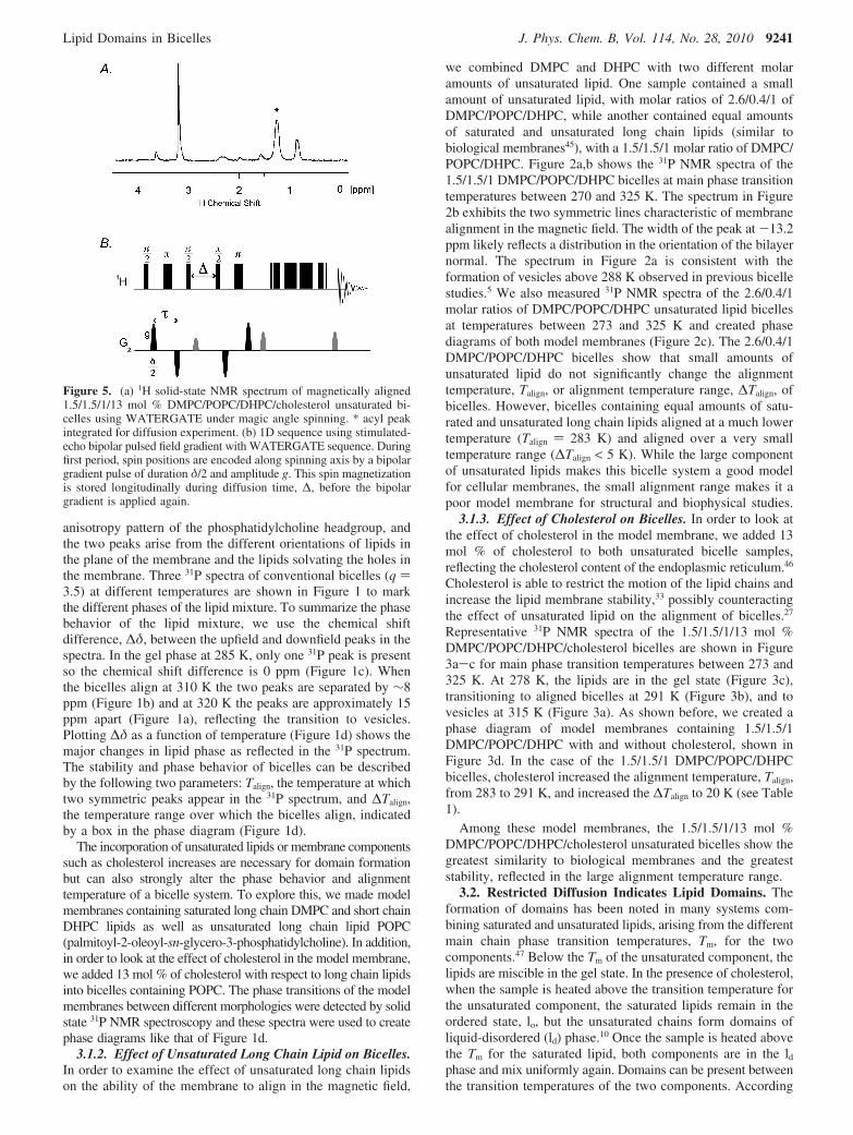
Figure 5. (a) 1H solid-state NMR spectrum of magnetically aligned1.5/1.5/1/13 mol % DMPC/POPC/DHPC/cholesterol unsaturated bi-celles using WATERGATE under magic angle spinning. * acyl peakintegrated for diffusion experiment. (b) 1D sequence using stimulated-echo bipolar pulsed field gradient with WATERGATE sequence. Duringfirst period, spin positions are encoded along spinning axis by a bipolargradient pulse of duration δ/2 and amplitude g. This spin magnetizationis stored longitudinally during diffusion time, ∆, before the bipolargradient is applied again.
Lipid Domains in Bicelles J. Phys. Chem. B, Vol. 114, No. 28, 2010 9241
to this model, domain formation in the 1.5/1.5/1/13 mol %sample is expected between 270 and 296 K, Tm(POPC) andTm(DMPC) respectively, and the uniaxial orientation of thedomains in the magnetic field would exist between 292 and 296K (Figure 4). To measure domain formation in the bicelles, wecarried out pulsed-field gradient measurements of the lipid self-diffusion constant. In the absence of domains, the diffusionconstant should be independent of the diffusion time of themolecules. If domains are present and restrict the lipid diffusion,then the diffusion constant will decrease as the lipid diffusiontime increases because the lipid diffusion is confined.35,40
3.2.1. Measuring Lipid Diffusion. Figure 5a shows the 1HNMR spectrum of 1.5/1.5/1/13 mol % DMPC/POPC/DHPC/cholesterol bicelles with the choline peak appearing at 3.2 ppmand two acyl chain peaks at 1.2 ppm and 0.8 ppm. The allylicproton peak from POPC appears at 2.0 ppm and WATERGATEhas been used to suppress the water peak at 4.7 ppm. For thediffusion measurements, we combined a stimulated-echo pulsed-field bipolar gradient diffusion experiment48 with water sup-pression, spinning the sample at the magic angle to obtain well-resolved, isotropic 1H spectra. The acyl peak at 1.2 ppm (markedwith an asterisk) was integrated to monitor the lipid signal.
In this experiment, the spin location is encoded by a pair ofsine-shaped bipolar gradients of duration δ and amplitude g,
bracketing a diffusion time ∆. Diffusion is measured along thedirection of the spinning axis with a magic-angle gradient. Theintensity of the lipid signal is modulated by the gradient lengthand amplitude, reflecting the self-diffusion of the lipids. Thevariation of the signal strength, I, with the experimentalparameters can be described by the Stejskal Tanner equation49
where I0 is the acyl peak integral in the absence of gradients, τis the spacing of the bipolar gradient pulses, and γ is gyromag-netic ratio for the observed nucleus (in this experiment 1H). Themeasured self-diffusion constant, Dmeas, can be extracted froma plot of ln(I/I0) versus k (shown in Figure 6). Under the magicangle spinning, the aligning force of the bicelles is lost and themembranes adopt a random distribution of the membrane normalwith respect to the magnetic field.50 Therefore, Dmeas should bemultiplied by 3/2 to compensate for the powder pattern to obtainthe apparent lateral diffusion constant, Dapp.51
In the absence of confinement, lipids show classical diffusionin two dimensions, with a diffusion constant that is independent
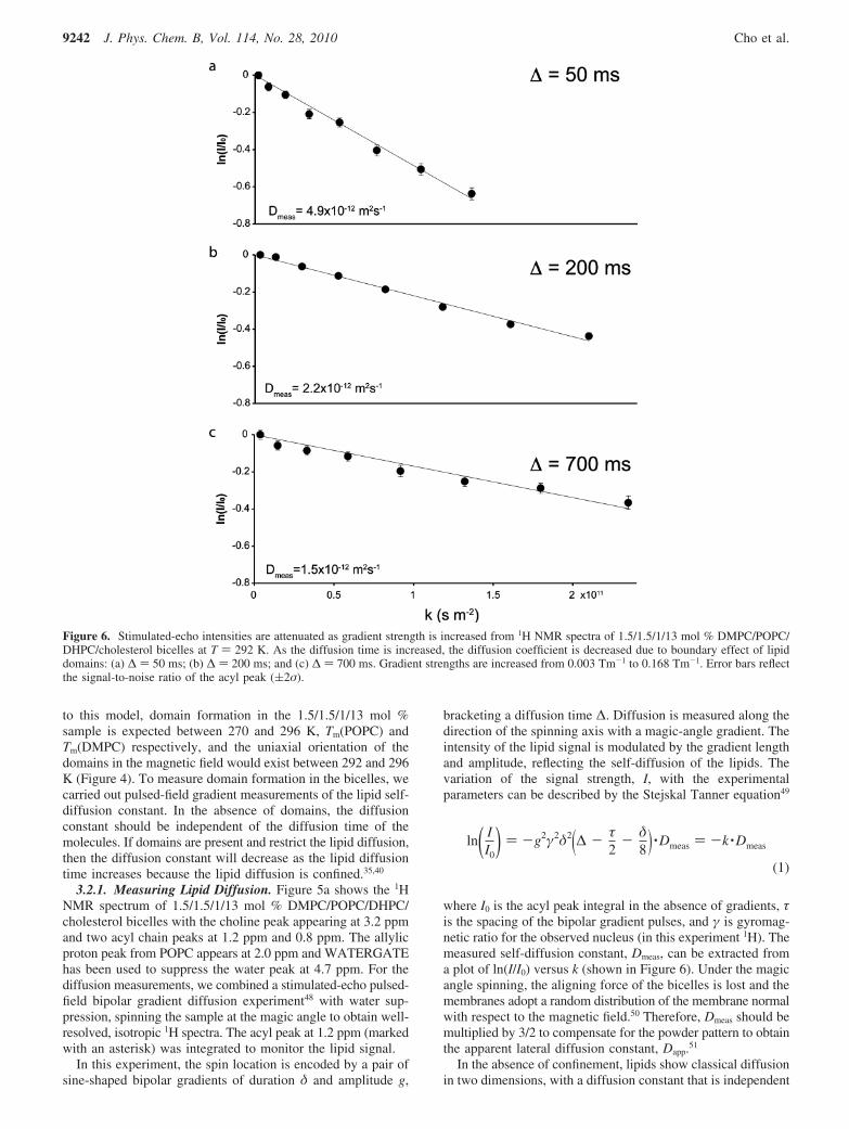
Figure 6. Stimulated-echo intensities are attenuated as gradient strength is increased from 1H NMR spectra of 1.5/1.5/1/13 mol % DMPC/POPC/DHPC/cholesterol bicelles at T ) 292 K. As the diffusion time is increased, the diffusion coefficient is decreased due to boundary effect of lipiddomains: (a) ∆ ) 50 ms; (b) ∆ ) 200 ms; and (c) ∆ ) 700 ms. Gradient strengths are increased from 0.003 Tm-1 to 0.168 Tm-1. Error bars reflectthe signal-to-noise ratio of the acyl peak ((2σ).
ln( II0
) ) -g2γ2δ2(∆ - τ2- δ
8) ·Dmeas ) -k ·Dmeas
(1)
9242 J. Phys. Chem. B, Vol. 114, No. 28, 2010 Cho et al.

of diffusion time. Displacement in free diffusion is describedby a Gaussian distribution with the root-mean-square displace-ment of a lipid increasing with diffusion time, ∆, according tothe following equation:
Conventional bicelles containing only DMPC and DHPC areunable to form domains. The long chain lipids in these samplesexhibit free diffusion in which the displacement varies linearlywith the square root of diffusion time, shown in Figure 7. Eachpoint on this graph represents an apparent diffusion constantmeasured for a given diffusion time, ∆.
When confined diffusion occurs, the measured diffusionconstant decreases with diffusion time, as more moleculesencounter the boundary. Figure 6a shows the diffusion measure-ment of lipids in 1.5/1.5/1/13 mol % bicelles at 292 K, in whichthe diffusion time, ∆, was 50 ms. As the diffusion time increasesto 700 ms (Figure 6a-c), the measured self-diffusion constantis reduced from 4.9 × 10⁻¹² m2s-1 to 1.5 × 10⁻¹² m2s-1,demonstrating lipid confinement.
In Figure 8, the apparent displacement, ⟨xapp2 ⟩1/2 ) �4Dj app∆,
is plotted as a function of the square root of diffusion time forthree different temperatures. At 300 K, above the expectedmiscibility transition, the displacement varies linearly with thesquare root of diffusion time, indicating free diffusion with noconfinement or boundaries as observed in the conventionalbicelles (Figure 7). At 295 K, the displacement does not increaselinearly with the square root of diffusion time, but rather plateausat ∼3.0 µm, indicating that the lipids are confined within amicrometer-scale region. At 292 K, the plateau is at ∼2.6 µm,indicating that the areas of confinement decreased with tem-
perature (Figure 8). The changes in diffusion at differenttemperatures are fully reversible and diffusion measurementsmade on other samples of the same composition gave resultsconsistent with those presented here.
To extract the true diffusion constant and the domain size,the displacement at 292 and 295 K can be fit to the followingequation:52
which reduces to the expression for free diffusion when thedomain size, r, is much larger than the displacement (eq 2).The true diffusion constant at 292 K is 4.3 ( 1.8 × 10⁻¹² m2s-1
and 7.3 ( 2.6 × 10⁻¹² m2s-1 at 295 K (Table 2).The composition of the domains is likely ld. The size of the
domain increases with temperature, consistent with ld domains,and the domains disappear above the Tm of DMPC. Interestingly,our 1H NMR spectrum of 1.5/1.5/1/13 mol % bicelles showsnarrow (∼45 Hz) lipid resonances that increase in intensity from273 to 310 K as the bicelles align. Previous 1H MAS NMRspectroscopy of similar ternary lipid mixtures (DPPC, DOPC,and cholesterol) showed narrow acyl peaks (∼50 Hz) for ld
domains, similar to our spectra, showing the same basictemperature dependence. However, we observed no signal fromthe lo phase, while they observed broad (∼1 kHz) peaks for lo
domains.53
This POPC/DMPC bicelle system exhibited micrometer-scalelipid domains, while vesicular systems of POPC/DMPC haveshown no domains.54 In previous work, it was speculated thatthe asymmetry of the POPC legs decreased its rotational
Figure 7. Conventional bicelles (DMPC/DHPC, q ) 3.5) show a linear relationship between lipid displacement and the square root of diffusiontime, consistent with free diffusion. Error bars reflect the fit uncertainty ((2σ) of the diffusion constant for each point.
⟨x2⟩1/2 ) √4D∆ (2)
⟨x2⟩1/2 ) rsin(√4Dtrue∆
r ) (3)
Lipid Domains in Bicelles J. Phys. Chem. B, Vol. 114, No. 28, 2010 9243

mobility, and hindered formation of the lo phase.54 Bicellesexhibit greater lateral and rotational fluidity of the bicelle bilayerthan vesicles,55 apparent in the scaled 31P chemical shiftanisotropy of the phosphatidylcholine headgroup, which mightaffect or favor the formation of domains. The presence of theamphiphile DHPC could play a role in the phase separationdirectly by partitioning into the bilayer, or the properties of themembrane as a whole simply by stabilizing perforations in thebilayer. Further work characterizing the domain formation inthis model membrane, particularly the role of DHPC in domainformation, is underway.
4. Conclusions
In this work, we created a uniaxially aligned membranecontaining both saturated and unsaturated lipids, as well ascholesterol, capable of phase separating into lipid domains.Among the various model membranes we created, the 1.5/1.5/1/13 mol % molar ratios of DMPC/POPC/DHPC/cholesterolbicelles showed strong alignment over a large range of tem-peratures, and exhibited micrometer-scale phase separation intolo and ld domains, raising the possibility of structural studies ofmembrane proteins in rafts and outside of rafts. Structuralbiology studies of this sort could clarify the molecular actionof membrane rafts in cellular biology.
Acknowledgment. We thank Neil Donovan for many helpfuldiscussions and Ad Bax for the conversation sparking this work.We gratefully acknowledge funding support from Eli Lilly andOak Ridge Associated Universities, as well as funds from theUniversity of Pittsburgh.
References and Notes
(1) Edidin, M. Annu. ReV. Biophys. Biomol. Struct. 2003, 32, 257.(2) Lucero, H. A.; Robbins, P. W. Arch. Biochem. Biophys. 2004, 426,
208.(3) Caffrey, M. J. Struct. Biol. 2003, 142, 108.(4) Marassi, F. M.; Opella, S. J. Curr. Opin. Struct. Biol. 1998, 8, 640.(5) Triba, M. N.; Warschawski, D. E.; Devaux, P. F. Biophys. J. 2005,
88, 1887.(6) Boesze-Battaglia, K.; Schimmel, R. J. Exp. Biol. 1997, 200, 2927.(7) van Meer, G.; Voelker, D. R.; Feigenson, G. W. Na.t ReV. Mol.
Cell. Biol. 2008, 9, 112.(8) Wassall, S. R.; Brzustowicz, M. R.; Shaikh, S. R.; Cherezov, V.;
Caffrey, M.; Stillwell, W. Chem. Phys. Lipids 2004, 132, 79.(9) Bakht, O.; Pathak, P.; London, E. 2007, 93, 4307.
(10) Silvius, J. R. Biochim. Biophys. Acta, Biomembr. 2003, 1610, 174.(11) Lee, A. Biochim. Biophys. Acta 2004, 1666, 62.(12) Park, S. H.; Opella, S. J. J. Mol. Biol. 2005, 350, 310.(13) Turnheim, K.; Gruber, J.; Wachter, C.; Ruiz-Gutierrez, V. Am. J.
Physiol. Cell Physiol. 1999, 277, C83.(14) Schmidt, D.; Jiang, Q.-X.; MacKinnon, R. Nature 2006, 444, 775.(15) van den Brink-van der Laan, E.; Antoinette Killian, J.; de Kruijff,
B. Biochim. Biophys. Acta, Biomembr. 2004, 1666, 275.(16) Ramamoorthy, A.; Lee, D.-K.; Narasimhaswamy, T.; Nanga,
R. P. R. Biochim. Biophys. Acta, Biomembr., In Press, Corrected Proof.(17) Tokumasu, F.; Jin, A. J.; Feigenson, G. W.; Dvorak, J. A. 2003,
84, 2609.(18) Filippov, A.; Oradd, G.; Lindblom, G. 2007, 93, 3182.(19) Marcotte, I.; Auger, M. Conc. Magn. Reson. A 2005, 24A, 17.(20) Ramamoorthy, A.; Kandasamy, S. K.; Lee, D.-K.; Kidambi, S.;
Larson, R. G. Biochemistry 2007, 46, 965.(21) Kandasamy, S. K.; Lee, D.-K.; Nanga, R. P. R.; Xu, J.; Santos,
J. S.; Larson, R. G.; Ramamoorthy, A. Biochim. Biophy. Acta, Biomembr.2009, 1788, 686.
Figure 8. The average displacements in 1.5/1.5/1/chol DMPC/POPC/DHPC unsaturated lipid system with and without rafts as a function ofdiffusion time at three temperatures. At 300 K (9), the displacement increased as the square root of time, consistent with diffusion. In contrast, attemperatures 295 K (b) and 292 K (2), the lipid displacements are limited and showed plateaus, indicating confined diffusion within a lipiddomain. Error bars reflect the fit uncertainty ((2σ) of the diffusion constant for each point.
TABLE 2: Diffusion Constants and Domain Sizes in1.5/1.5/1/13 mol % Bicelle
sampletemperature
(K)diffusion constant,Dtrue (10-12 m2s-1)
domain size,r (µm)
292 4.3 ( 1.8 2.6 ( 0.4295 7.3 ( 2.6 3.0 ( 0.3300 8.8 ( 0.3
9244 J. Phys. Chem. B, Vol. 114, No. 28, 2010 Cho et al.

(22) De Angelis, A. A.; Howell, S. C.; Nevzorov, A. A.; Opella, S. J.J. Am. Chem. Soc. 2006, 128, 12256.
(23) De Angelis, A. A.; Nevzorov, A. A.; Park, S. H.; Howell, S. C.;Mrse, A. A.; Opella, S. J. J. Am. Chem. Soc. 2004, 126, 15340.
(24) De Angelis, A. A.; Opella, S. J. Nat. Protocols 2007, 2, 2332.(25) De Angelis, A. A. Methods Enzymol. 2005, 394, 350.(26) Xu, J.; D,rr, U. H. N.; Im, S.-C.; Gan, Z.; Waskell, L.; Ramamoor-
thy, A. Angew. Chem., Int. Ed. 2008, 47, 7864.(27) Minto, R. E.; Adhikari, P. R.; Lorigan, G. A. Chem. Phys. Lipids
2004, 132, 55.(28) Triba, M. N.; Devaux, P. F.; Warschawski, D. E. Biophys. J. 2006,
91, 1357.(29) Parker, M. A.; King, V.; Howard, K. P. Biochim. Biophys. Acta,
Biomembr. 2001, 1514, 206.(30) Czerski, L.; Sanders, C. R. Anal. Biochem. 2000, 284, 327.(31) Sasaki, H.; Fukuzawa, S.; Kikuchi, J.; Yokoyama, S.; Hirota, H.;
Tachibana, K. Langmuir 2003, 19, 9841.(32) De Meyer, F. d. r.; Smit, B. Proc. Natl. Acad. Sci. 2009, 106, 3654.(33) Lindblom, G.; Johansson, L. B. A.; Arvidson, G. Biochemistry 1981,
20, 2204.(34) Edidin, M. Annu. ReV. Biophys. Bioeng. 1974, 3, 179.(35) Gaede, H. C.; Gawrisch, K. Biophys. J. 2003, 85, 1734.(36) Lindblom, G.; Oradd, G. Prog. Nucl. Magn. Reson. Spectrosc. 1994,
26, 483.(37) Filippov, A.; Oradd, G.; Lindblom, G. Biophys. J. 2004, 86, 891.(38) Oradd, G.; Westerman, P. W.; Lindblom, G. Biophys. J. 2005, 89,
315.
(39) Polozov, I. V.; Bezrukov, L.; Gawrisch, K.; Zimmerberg, J. Nat.Chem. Biol. 2008, 4, 248.
(40) Polozov, I. V.; Gawrisch, K. Biophys. J. 2004, 87, 1741.(41) Kadkhodaie, M.; Rivas, O.; Tan, M.; Mohebbi, A.; Shaka, A. J. J.
Magn. Reson. 1991, 91, 437.(42) Latour, L. L.; Li, L. M.; Sotak, C. H. J. Magn. Reson., Ser. B 1993,
101, 72.(43) Tanner, J. E. J. Chem. Phys. 1970, 52, 2523.(44) Sanders, C. R.; Schwonek, J. P. Biochemistry 1992, 31, 8898.(45) Gennis, R. B. Biomembranes: Molecular Structure and Function;
Springer-Verlag: New York, 1989.(46) Yeagle, P. L. Biochim. Biophys. Acta, ReV. Biomembr. 1985, 822,
267.(47) London, E. Curr. Opin. Struct. Biol. 2002, 12, 480.(48) Cotts, R. M.; Hoch, M. J. R.; Sun, T.; Markert, J. T. J. Magn.
Reson. 1989, 83, 252.(49) Stejskal, E. O.; Tanner, J. E. J. Chem. Phys. 1965, 42, 288.(50) Zandomeneghi, G.; Williamson, P. T. F.; Hunkeler, A.; Meier, B. H.
J. Biomol. NMR 2003, 25, 125.(51) Pampel, A.; Karger, J.; Michel, D. Chem. Phys. Lett. 2003, 379,
555.(52) Gaede, H. C.; Gawrisch, K. Biophys. J. 2003, 85, 1734.(53) Polozov, I. V.; Gawrisch, K. Biophys. J. 2006, 90, 2051.(54) Veatch, S. L.; Keller, S. L. Biophys. J. 2003, 85, 3074.(55) Sanders, C. R.; Prestegard, J. H. Biophys. J. 1990, 58, 447.
JP100276U
Lipid Domains in Bicelles J. Phys. Chem. B, Vol. 114, No. 28, 2010 9245