Embed Size (px)
Citation preview
Case ReportLimb Pain as Unusual Presentation of a ParietalIntraparenchymal Bleeding Associated with CrackCocaine Use: A Case Report
Alan Lucerna ,1 James Espinosa ,1 Taimur Zaman,2 Risha Hertz,3 and Douglas Stranges1
1Department of Emergency Medicine, Rowan University SOM/Jefferson Health, Stratford, NJ, USA2Department of Neurology, Jefferson Health, Stratford, NJ, USA3Penn Medicine, Gibbsboro, NJ, USA
Correspondence should be addressed to Alan Lucerna; [email protected]
Received 14 February 2018; Revised 19 April 2018; Accepted 24 April 2018; Published 31 May 2018
Academic Editor: Peter Berlit
Copyright © 2018 Alan Lucerna et al. This is an open access article distributed under the Creative Commons Attribution License,which permits unrestricted use, distribution, and reproduction in any medium, provided the original work is properly cited.
Limb pain as a presenting feature of an ischemic or hemorrhagic stroke is extremely rare. Here we present a case of a 65-year-old male with complaints of left arm pain and allodynia (specifically light touch to any part of the left arm produced significantdiscomfort) who was found to have a right parietal lobe intraparenchymal bleed after smoking crack cocaine. Acute central painis mainly associated with parietal, thalamic, and brainstem lesions. It has been proposed that acute limb pain from a parietal lobestroke is due to the disconnection of the parietal cortex from the thalamus secondary to the interruption of the pathways betweenthe hemisphere and thalamus/basal ganglia.
1. Introduction
Acute limb pain as a stroke presentation is extremely rare.Central causes of pain are well accepted and understood,explained by physiologic and neuroanatomical principles[1, 2]. Central pain syndrome is a neurological conditionsecondary to damage or dysfunction of the central nervoussystem (CNS), which is comprised of the brain, brainstem,and spinal cord. In addition to stroke, central pain syndromehas also been associated with multiple sclerosis, tumors,epilepsy, brain or spinal cord trauma, and Parkinson’s disease[3].
2. Case Report
A 65-year-old right hand dominant, African American malepresented to the ED via emergency medical service. He hadjust finished smoking crack cocaine when he developed leftarm pain that he described as “cramping”. He reported thatthe pain was so intense that he became weak causing him tofall onto the ground. The pain made him feel like “jumpingout of the window.” He denied any head injury and he had
no loss of consciousness (LOC). The patient had no chest,shortness of breath, or dyspnea on exertion. He denied anyneck, back, or abdominal pain.
The patient’s past medical history included diabetes,hypertension, hepatitis C, sick sinus syndrome, paroxys-mal atrial fibrillation, hyperlipidemia, deep vein thrombo-sis, chronic kidney disease, hilar mediastinal adenopathy,diastolic heart failure, valvular heart disease, and cardiacarrhythmia of nonsustained ventricular tachycardia with apermanent pacemaker. The patient admitted to intermit-tent cocaine abuse. His medications include atorvastatin,furosemide, isosorbide mononitrate, acetaminophen withcodeine, apixaban, hydralazine, metformin, albuterol sulfate,amlodipine, and tamsulosin.
Vital signs were essentially within normal limits with theexception of a blood pressure of 142/83 mmHg.
The patient had a strong left radial pulse and brisk cap-illary refill of the left hand with no tenderness or deformity.The patient was noted to have left arm weakness and whatlooked like choreiform or clumsy left arm movements. Hisleft leg was also noted to be weak. There was no numbness.
HindawiCase Reports in Neurological MedicineVolume 2018, Article ID 9598675, 4 pageshttps://doi.org/10.1155/2018/9598675
2 Case Reports in Neurological Medicine
Interestingly, light touch to any part of the left arm producedsignificant discomfort to the point where he did not wantanything touching the left arm. He was noted to havedecreased rapid alternating movements on the left upperextremity as well as mild difficulty with fine motor control.His left arm and left leg motor strength was 4/5. His cranialnerves II to XII were grossly intact.Therewere no visual fieldscuts noted. Extraocular motility was intact. The grimace wassymmetric. There was no evidence of double simultaneousextinction.
There were no pulsatile abdominal masses on exam andthe bilateral radial pulses were equal. The patient was unableto tell the exact time of onset of his symptoms. The patient’sleft arm pain improved with morphine 4 mg intravenously.
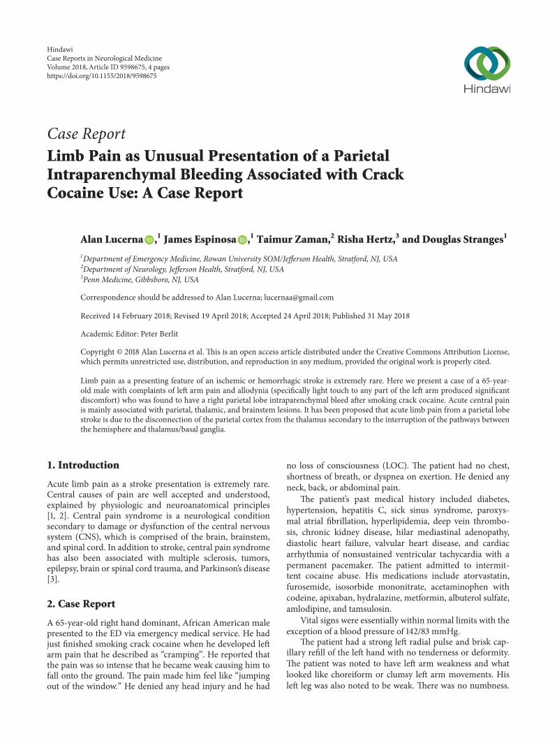
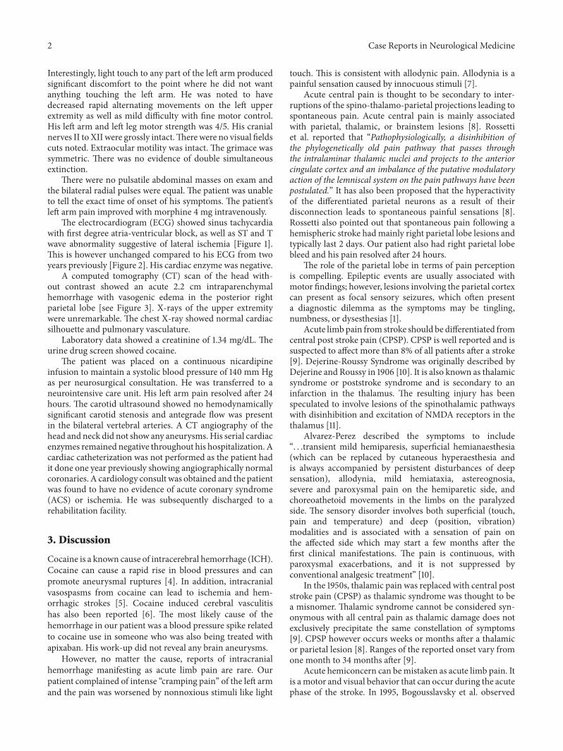
The electrocardiogram (ECG) showed sinus tachycardiawith first degree atria-ventricular block, as well as ST and Twave abnormality suggestive of lateral ischemia [Figure 1].This is however unchanged compared to his ECG from twoyears previously [Figure 2]. His cardiac enzyme was negative.
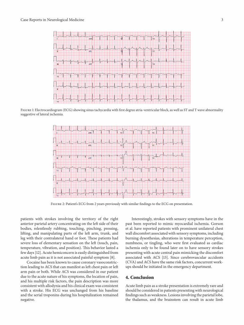
A computed tomography (CT) scan of the head with-out contrast showed an acute 2.2 cm intraparenchymalhemorrhage with vasogenic edema in the posterior rightparietal lobe [see Figure 3]. X-rays of the upper extremitywere unremarkable. The chest X-ray showed normal cardiacsilhouette and pulmonary vasculature.
Laboratory data showed a creatinine of 1.34 mg/dL. Theurine drug screen showed cocaine.
The patient was placed on a continuous nicardipineinfusion to maintain a systolic blood pressure of 140 mm Hgas per neurosurgical consultation. He was transferred to aneurointensive care unit. His left arm pain resolved after 24hours. The carotid ultrasound showed no hemodynamicallysignificant carotid stenosis and antegrade flow was presentin the bilateral vertebral arteries. A CT angiography of thehead and neck did not show any aneurysms.His serial cardiacenzymes remained negative throughout his hospitalization. Acardiac catheterization was not performed as the patient hadit done one year previously showing angiographically normalcoronaries. A cardiology consult was obtained and the patientwas found to have no evidence of acute coronary syndrome(ACS) or ischemia. He was subsequently discharged to arehabilitation facility.
3. Discussion
Cocaine is a known cause of intracerebral hemorrhage (ICH).Cocaine can cause a rapid rise in blood pressures and canpromote aneurysmal ruptures [4]. In addition, intracranialvasospasms from cocaine can lead to ischemia and hem-orrhagic strokes [5]. Cocaine induced cerebral vasculitishas also been reported [6]. The most likely cause of thehemorrhage in our patient was a blood pressure spike relatedto cocaine use in someone who was also being treated withapixaban. His work-up did not reveal any brain aneurysms.
However, no matter the cause, reports of intracranialhemorrhage manifesting as acute limb pain are rare. Ourpatient complained of intense “cramping pain” of the left armand the pain was worsened by nonnoxious stimuli like light
touch. This is consistent with allodynic pain. Allodynia is apainful sensation caused by innocuous stimuli [7].
Acute central pain is thought to be secondary to inter-ruptions of the spino-thalamo-parietal projections leading tospontaneous pain. Acute central pain is mainly associatedwith parietal, thalamic, or brainstem lesions [8]. Rossettiet al. reported that “Pathophysiologically, a disinhibition ofthe phylogenetically old pain pathway that passes throughthe intralaminar thalamic nuclei and projects to the anteriorcingulate cortex and an imbalance of the putative modulatoryaction of the lemniscal system on the pain pathways have beenpostulated.” It has also been proposed that the hyperactivityof the differentiated parietal neurons as a result of theirdisconnection leads to spontaneous painful sensations [8].Rossetti also pointed out that spontaneous pain following ahemispheric stroke hadmainly right parietal lobe lesions andtypically last 2 days. Our patient also had right parietal lobebleed and his pain resolved after 24 hours.
The role of the parietal lobe in terms of pain perceptionis compelling. Epileptic events are usually associated withmotor findings; however, lesions involving the parietal cortexcan present as focal sensory seizures, which often presenta diagnostic dilemma as the symptoms may be tingling,numbness, or dysesthesias [1].
Acute limbpain from stroke should be differentiated fromcentral post stroke pain (CPSP). CPSP is well reported and issuspected to affect more than 8% of all patients after a stroke[9]. Dejerine-Roussy Syndrome was originally described byDejerine and Roussy in 1906 [10]. It is also known as thalamicsyndrome or poststroke syndrome and is secondary to aninfarction in the thalamus. The resulting injury has beenspeculated to involve lesions of the spinothalamic pathwayswith disinhibition and excitation of NMDA receptors in thethalamus [11].
Alvarez-Perez described the symptoms to include“. . .transient mild hemiparesis, superficial hemianaesthesia(which can be replaced by cutaneous hyperaesthesia andis always accompanied by persistent disturbances of deepsensation), allodynia, mild hemiataxia, astereognosia,severe and paroxysmal pain on the hemiparetic side, andchoreoathetoid movements in the limbs on the paralyzedside. The sensory disorder involves both superficial (touch,pain and temperature) and deep (position, vibration)modalities and is associated with a sensation of pain onthe affected side which may start a few months after thefirst clinical manifestations. The pain is continuous, withparoxysmal exacerbations, and it is not suppressed byconventional analgesic treatment” [10].
In the 1950s, thalamic pain was replaced with central poststroke pain (CPSP) as thalamic syndrome was thought to bea misnomer. Thalamic syndrome cannot be considered syn-onymous with all central pain as thalamic damage does notexclusively precipitate the same constellation of symptoms[9]. CPSP however occurs weeks or months after a thalamicor parietal lesion [8]. Ranges of the reported onset vary fromone month to 34 months after [9].
Acute hemiconcern can bemistaken as acute limb pain. Itis amotor and visual behavior that can occur during the acutephase of the stroke. In 1995, Bogousslavsky et al. observed
Case Reports in Neurological Medicine 3
Figure 1: Electrocardiogram (ECG) showing sinus tachycardia with first degree atria-ventricular block, as well as ST and T wave abnormalitysuggestive of lateral ischemia.
Figure 2: Patient’s ECG from 2 years previously with similar findings to the ECG on presentation.
patients with strokes involving the territory of the rightanterior parietal artery concentrating on the left side of theirbodies, relentlessly rubbing, touching, pinching, pressing,lifting, and manipulating parts of the left arm, trunk, andleg with their contralateral hand or foot. These patients hadsevere loss of elementary sensation on the left (touch, pain,temperature, vibration, and position). This behavior lasted afew days [12]. Acute hemiconcern is easily distinguished fromacute limb pain as it is not associated painful symptom [8].
Cocaine has been known to cause coronary vasoconstric-tion leading to ACS that can manifest as left chest pain or leftarm pain or both. While ACS was considered in our patientdue to the acute nature of his symptoms, the location of pain,and his multiple risk factors, the pain description was moreconsistent with allodynia and his clinical examwas consistentwith a stroke. His ECG was unchanged from his baselineand the serial troponins during his hospitalization remainednegative.
Interestingly, strokes with sensory symptoms have in thepast been reported to mimic myocardial ischemia. Gorsonet al. have reported patients with prominent unilateral chestwall discomfort associatedwith sensory symptoms, includingburning dysesthesias, alterations in temperature perception,numbness, or tingling, who were first evaluated as cardiacischemia only to be found later on to have sensory strokespresenting with acute central pain mimicking the discomfortassociated with ACS [13]. Since cerebrovascular accidents(CVA) and ACS have the same risk factors, concurrent work-ups should be initiated in the emergency department.
4. Conclusion
Acute limb pain as a stroke presentation is extremely rare andshould be considered in patients presentingwith neurologicalfindings such asweakness. Lesions involving the parietal lobe,the thalamus, and the brainstem can result in acute limb
4 Case Reports in Neurological Medicine
Figure 3: CT scan of the brain without contrast showing an acute2.2 cm intraparenchymal hemorrhage with vasogenic edema in theposterior right parietal lobe.
pain. Other syndromes should be distinguished from acutelimb pain from a stroke and include central post stroke pain,which takes time to manifest, and acute hemiconcern, thatwhile acute, it is not associated with pain syndromes. Lastly,strokes with sensory symptoms can mimic ACS. A carefulneurological exam is therefore important in patients thoughtto have a cardiac emergency. Additionally, a concurrentcardiac and neurological work-up should also be considered.
Conflicts of Interest
The authors declare that they have no conflicts of interest.
References
[1] J. Michael, Aminoff., and K. Arthur, Harrisons Neurology inClinical Medicine, 15, 3 edition.
[2] A. Malavera, F. A. Silva, F. Fregni, S. Carrillo, and R. G. Garcia,“Repetitive Transcranial Magnetic Stimulation for PhantomLimb Pain in LandMine Victims: A Double-Blinded, Random-ized, Sham-Controlled Trial,”The Journal of Pain, vol. 17, no. 8,pp. 911–918, 2016.
[3] https://www.ninds.nih.gov/disorders/all-disorders/central-pain-syndrome-information-page.
[4] S. Martin-Schild, K. C. Albright, H. Hallevi et al., “Intracerebralhemorrhage in cocaine users,” Stroke, vol. 41, no. 4, pp. 680–684,2010.
[5] P. Garcıa-Bermejo, C. Rodrıguez-Arias, E. Crespo, S. Perez-Fernandez, J. F. Arenillas, and M. Martınez-Galdamez, “Severecerebral vasospasm in chronic cocaine users during neuroint-erventional procedures: A report of two cases,” InterventionalNeuroradiology, vol. 21, no. 1, pp. 19–22, 2015.
[6] E. Pieterse and J. van der Vlag, “Cracking the pathogenesis ofcocaine-induced vasculitis,” Rheumatology, vol. 56, no. 4, pp.503–505, 2017.
[7] S. Lolignier, N. Eijkelkamp, and J. N. Wood, “Mechanicalallodynia,” Pflugers Archiv - European Journal of Physiology, vol.467, no. 1, pp. 133–139, 2014.
[8] A. O. Rossetti, J. A. Ghika, F. Vingerhoets, J. Novy, and J.Bogousslavsky, “Neurogenic pain and abnormal movementscontralateral to an anterior parietal artery stroke,” JAMA Neu-rology, vol. 60, no. 7, pp. 1004–1006, 2003.
[9] J. L. Henry, C. Lalloo, and K. Yashpal, “Central poststroke pain:An abstruse outcome,”Pain Research&Management, vol. 13, no.1, pp. 41–49, 2008.
[10] F. J. Alvarez-Perez, “Dejerine-Roussy-Like Syndrome in aPatient with an Ischemic Lesion of the Right Dorsal Protu-berance: A Different Cause of Central Neuropathic Pain,” SMJournal of Case Reports, vol. 2, no. 2, 2016.
[11] M. N. Siddiqui, F. Furgang, S. M. Siddiqui, and J. Sue Ranas-inghe, “Dejerine roussy syndrome: The role of methadone inthe treatment of a central pain syndrome,”The Internet Journalof Pain, Symptom Control and Palliative Care, vol. 2, no. 1, pp.XV–XVI, 2001.
[12] J. Bogousslavsky, E. Kumral, F. Regli, G. Assal, and J. Ghika,“Acute hemiconcern: A right anterior parietotemporal syn-drome,” Journal of Neurology, Neurosurgery & Psychiatry, vol.58, no. 4, pp. 428–432, 1995.
[13] K. C. Gorson, M. S. Pessin, L. D. DeWitt, and L. R.Caplan, “Stroke with sensory symptoms mimicking myocardialischemia,” Neurology, vol. 46, no. 2, pp. 548–551, 1996.

Stem Cells International
Hindawiwww.hindawi.com Volume 2018
Hindawiwww.hindawi.com Volume 2018
MEDIATORSINFLAMMATION
of
EndocrinologyInternational Journal of
Hindawiwww.hindawi.com Volume 2018
Hindawiwww.hindawi.com Volume 2018
Disease Markers
Hindawiwww.hindawi.com Volume 2018
BioMed Research International
OncologyJournal of
Hindawiwww.hindawi.com Volume 2013
Hindawiwww.hindawi.com Volume 2018
Oxidative Medicine and Cellular Longevity
Hindawiwww.hindawi.com Volume 2018
PPAR Research
Hindawi Publishing Corporation http://www.hindawi.com Volume 2013Hindawiwww.hindawi.com
The Scientific World Journal
Volume 2018
Immunology ResearchHindawiwww.hindawi.com Volume 2018
Journal of
ObesityJournal of
Hindawiwww.hindawi.com Volume 2018
Hindawiwww.hindawi.com Volume 2018
Computational and Mathematical Methods in Medicine
Hindawiwww.hindawi.com Volume 2018
Behavioural Neurology
OphthalmologyJournal of
Hindawiwww.hindawi.com Volume 2018
Diabetes ResearchJournal of
Hindawiwww.hindawi.com Volume 2018
Hindawiwww.hindawi.com Volume 2018
Research and TreatmentAIDS
Hindawiwww.hindawi.com Volume 2018
Gastroenterology Research and Practice
Hindawiwww.hindawi.com Volume 2018
Parkinson’s Disease
Evidence-Based Complementary andAlternative Medicine
Volume 2018Hindawiwww.hindawi.com
Submit your manuscripts atwww.hindawi.com