Embed Size (px)
Citation preview
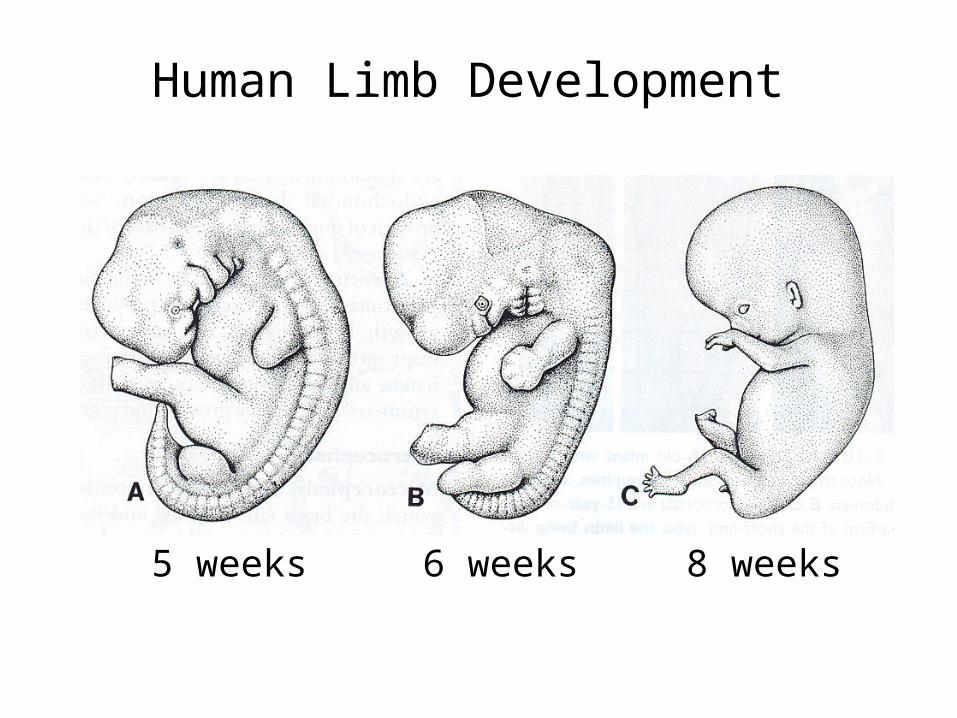
Human Limb Development
5 weeks 6 weeks 8 weeks
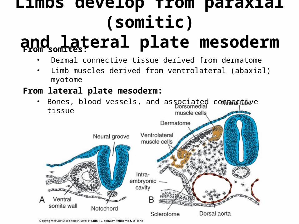
Limbs develop from paraxial (somitic)and lateral plate mesoderm
From somites:• Dermal connective tissue derived from dermatome• Limb muscles derived from ventrolateral (abaxial) myotome
From lateral plate mesoderm:• Bones, blood vessels, and associated connective tissue
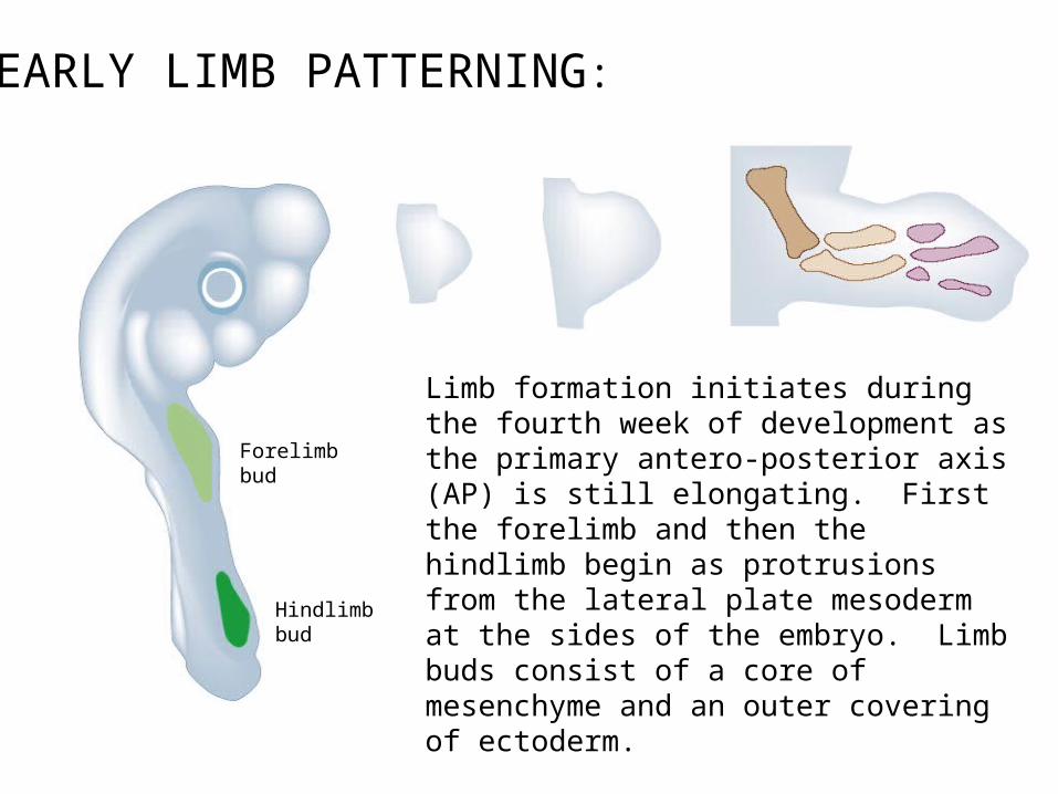
EARLY LIMB PATTERNING:
Limb formation initiates during the fourth week of development as the primary antero-posterior axis (AP) is still elongating. First the forelimb and then the hindlimb begin as protrusions from the lateral plate mesoderm at the sides of the embryo. Limb buds consist of a core of mesenchyme and an outer covering of ectoderm.
1 in 200 live human births display limb defects.
Forelimb bud
Hindlimb bud
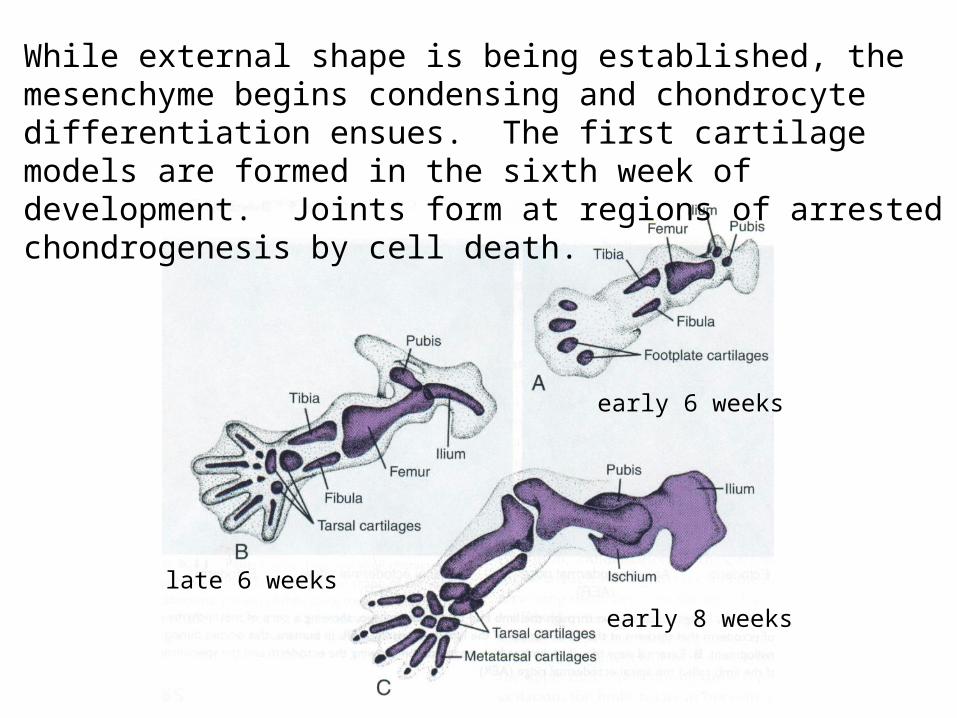
While external shape is being established, the mesenchyme begins condensing and chondrocyte differentiation ensues. The first cartilage models are formed in the sixth week of development. Joints form at regions of arrested chondrogenesis by cell death.
early 6 weeks
early 8 weeks
late 6 weeks
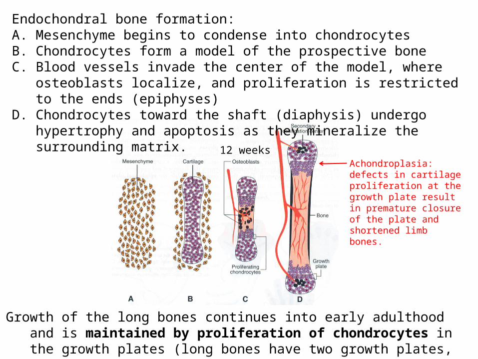
Endochondral bone formation:A. Mesenchyme begins to condense into chondrocytesB. Chondrocytes form a model of the prospective boneC. Blood vessels invade the center of the model, where osteoblasts localize, and
proliferation is restricted to the ends (epiphyses)D. Chondrocytes toward the shaft (diaphysis) undergo hypertrophy and apoptosis
as they mineralize the surrounding matrix.
Growth of the long bones continues into early adulthood and is maintained by proliferation of chondrocytes in the growth plates (long bones have two growth plates, in smaller bones (phalanges), there is only one at the tip)
12 weeksAchondroplasia: defects in cartilage proliferation at the growth plate result in premature closure of the plate and shortened limb bones.
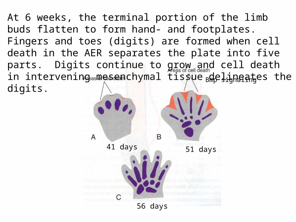
At 6 weeks, the terminal portion of the limb buds flatten to form hand- and footplates. Fingers and toes (digits) are formed when cell death in the AER separates the plate into five parts. Digits continue to grow and cell death in intervening mesenchymal tissue delineates the digits.
41 days 51 days
56 days
Bmp signaling
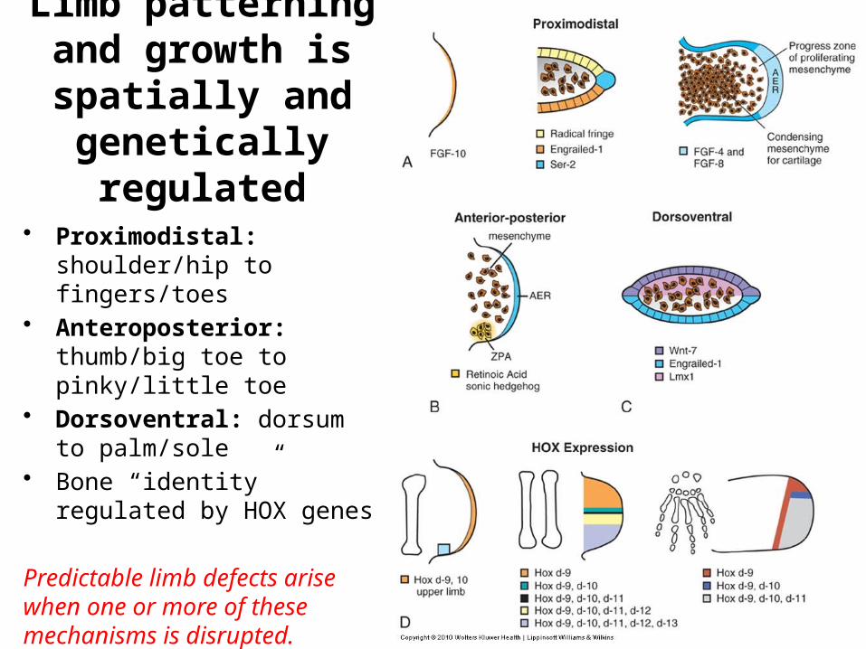
Limb patterning and growth is spatially and genetically regulated
• Proximodistal: shoulder/hip to fingers/toes
• Anteroposterior: thumb/big toe to pinky/little toe
• Dorsoventral: dorsum to palm/sole
• Bone “identity” regulated by HOX genes
Predictable limb defects arise when one or more of these mechanisms is disrupted.
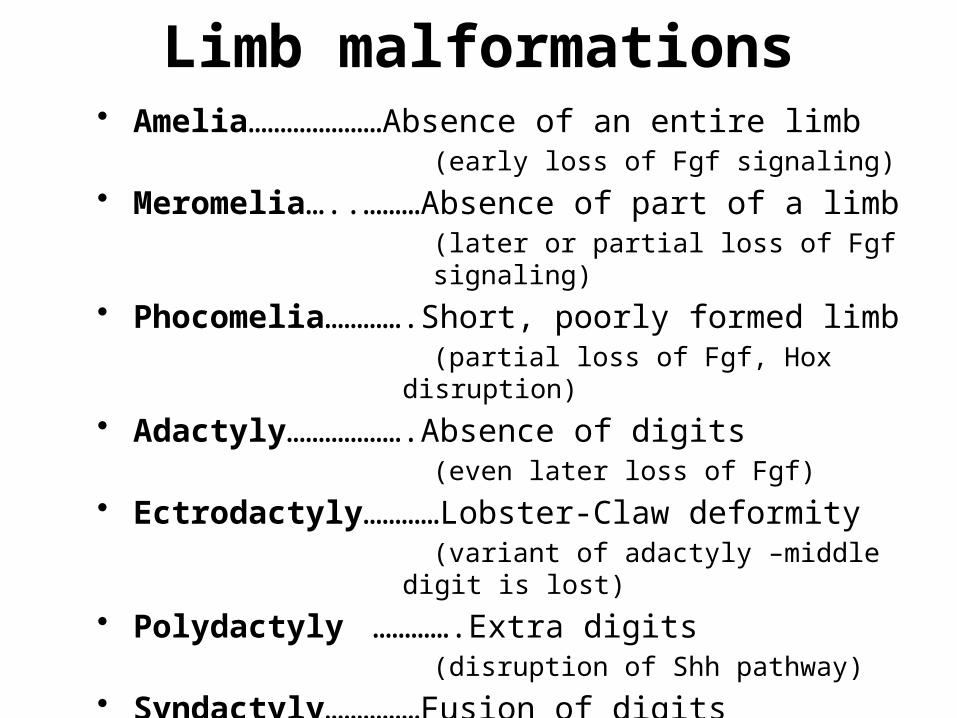
• Amelia…………………Absence of an entire limb(early loss of Fgf signaling)
• Meromelia…..………Absence of part of a limb(later or partial loss of Fgf signaling)
• Phocomelia………….Short, poorly formed limb(partial loss of Fgf, Hox disruption)
• Adactyly……………….Absence of digits (even later loss of Fgf)
• Ectrodactyly…………Lobster-Claw deformity(variant of adactyly –middle digit is lost)
• Polydactyly ………….Extra digits(disruption of Shh pathway)
• Syndactyly……………Fusion of digits(BMP disruption)
Limb malformations
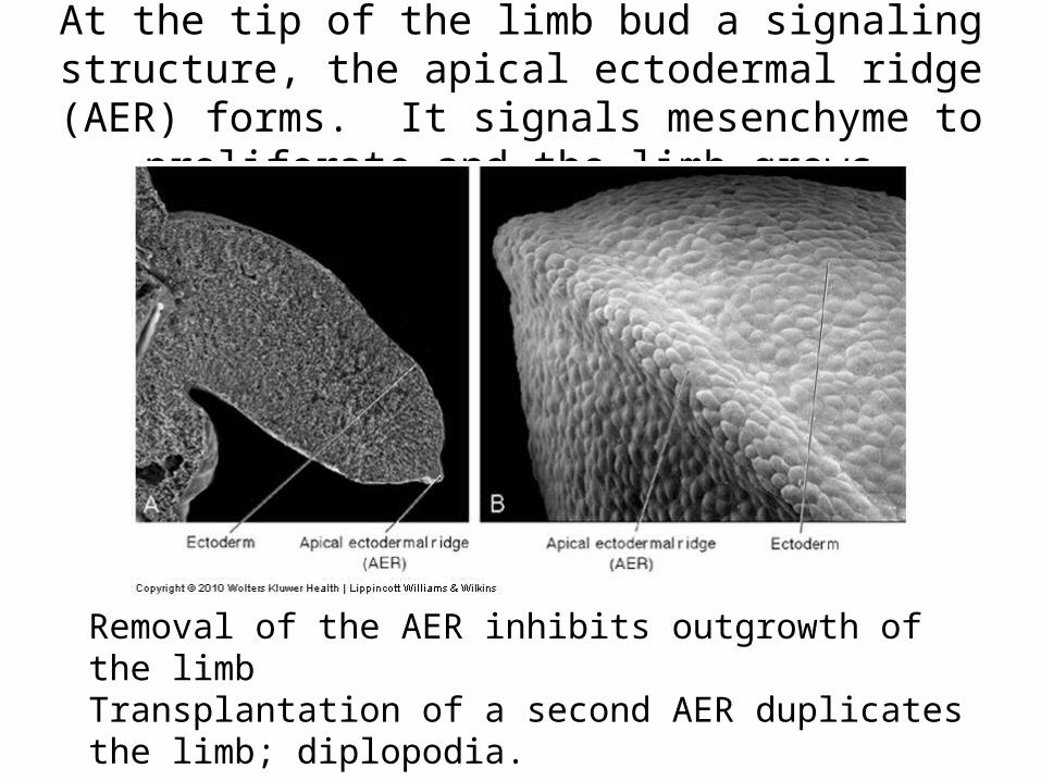
At the tip of the limb bud a signaling structure, the apical ectodermal ridge (AER) forms. It signals mesenchyme to
proliferate and the limb grows.
Removal of the AER inhibits outgrowth of the limbTransplantation of a second AER duplicates the limb; diplopodia.
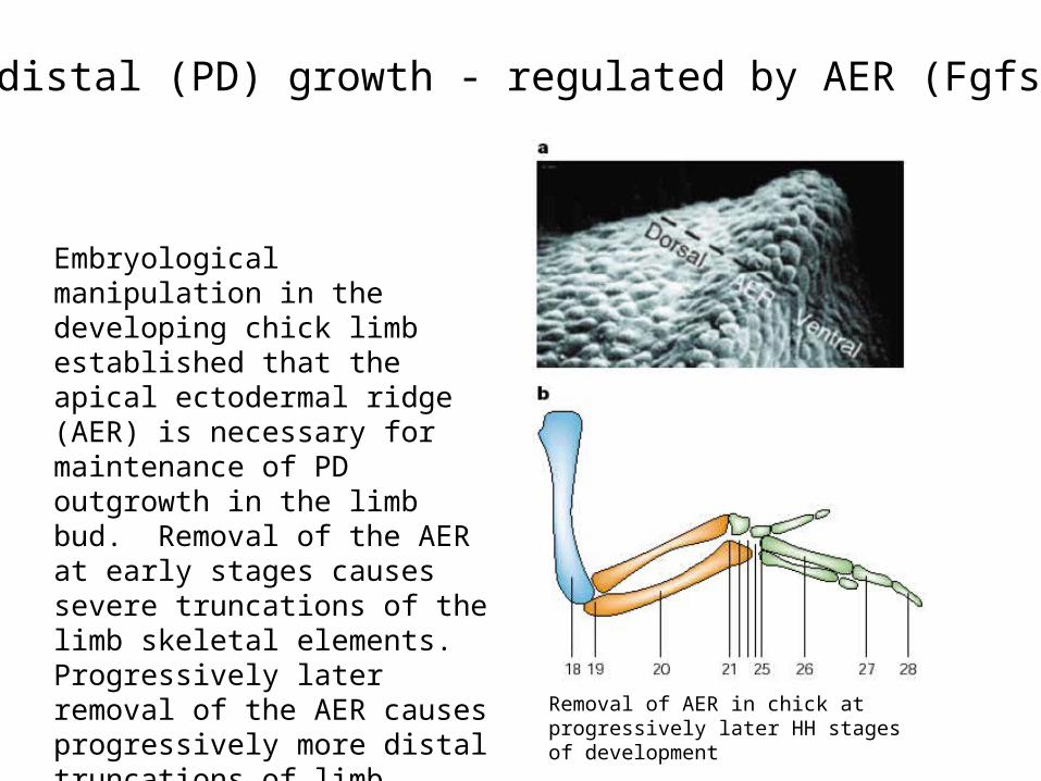
Proximodistal (PD) growth - regulated by AER (Fgfs):
Embryological manipulation in the developing chick limb established that the apical ectodermal ridge (AER) is necessary for maintenance of PD outgrowth in the limb bud. Removal of the AER at early stages causes severe truncations of the limb skeletal elements. Progressively later removal of the AER causes progressively more distal truncations of limb elements.
Removal of AER in chick at progressively later HH stages of development

Removal of AER: arrests limb development at stage at which it was removed
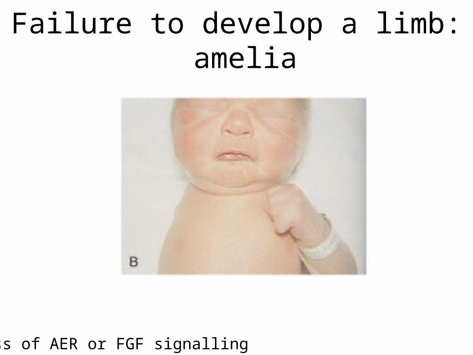
Failure to develop a limb: amelia
Loss of AER or FGF signalling
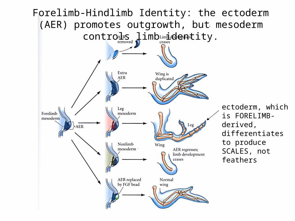
Forelimb-Hindlimb Identity: the ectoderm (AER) promotes outgrowth, but mesoderm controls limb identity.
ectoderm, which is FORELIMB-derived, differentiates to produce SCALES, not feathers
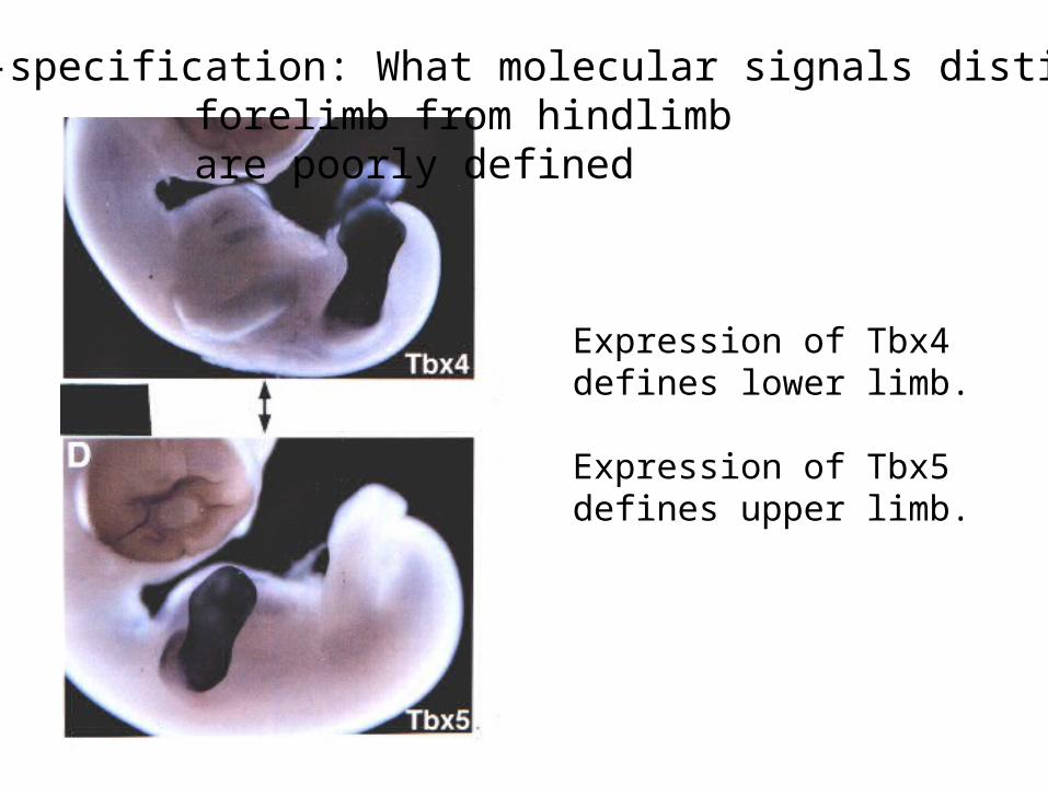
Expression of Tbx4defines lower limb.
Expression of Tbx5defines upper limb.
Post-specification: What molecular signals distinguish forelimb from hindlimb are poorly defined
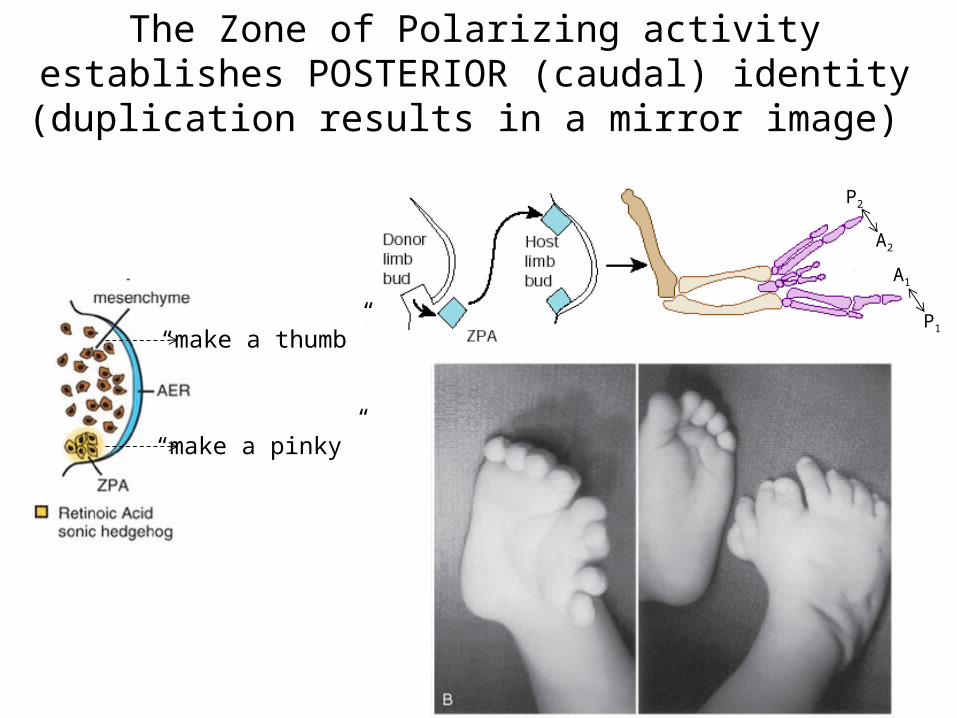
The Zone of Polarizing activity establishes POSTERIOR (caudal) identity (duplication results in a mirror image)
P2
P1
A2
A1
“make a pinky”
“make a thumb”
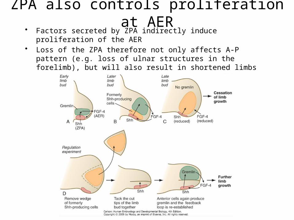
ZPA also controls proliferation at AER• Factors secreted by ZPA indirectly induce proliferation of the AER• Loss of the ZPA therefore not only affects A-P pattern (e.g. loss of ulnar
structures in the forelimb), but will also result in shortened limbs
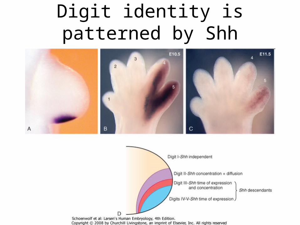
Digit identity is patterned by Shh
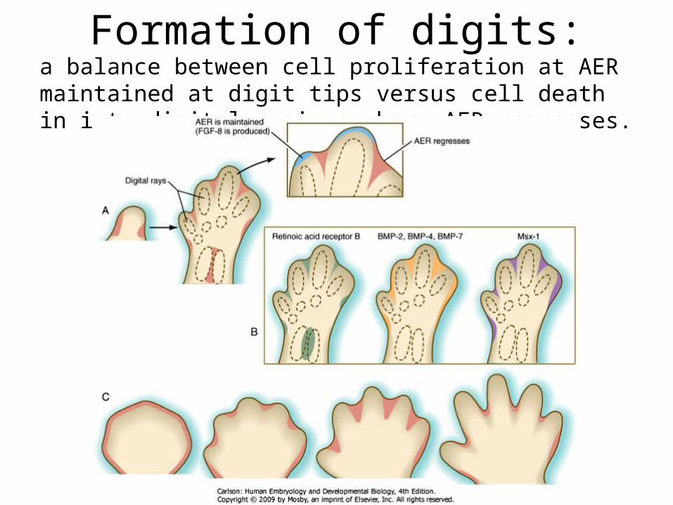
Formation of digits:a balance between cell proliferation at AER maintained at digit tips versus cell death in interdigital regions where AER regresses.
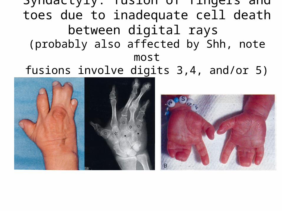
Syndactyly: fusion of fingers and toes due to inadequate cell death between digital rays
(probably also affected by Shh, note mostfusions involve digits 3,4, and/or 5)
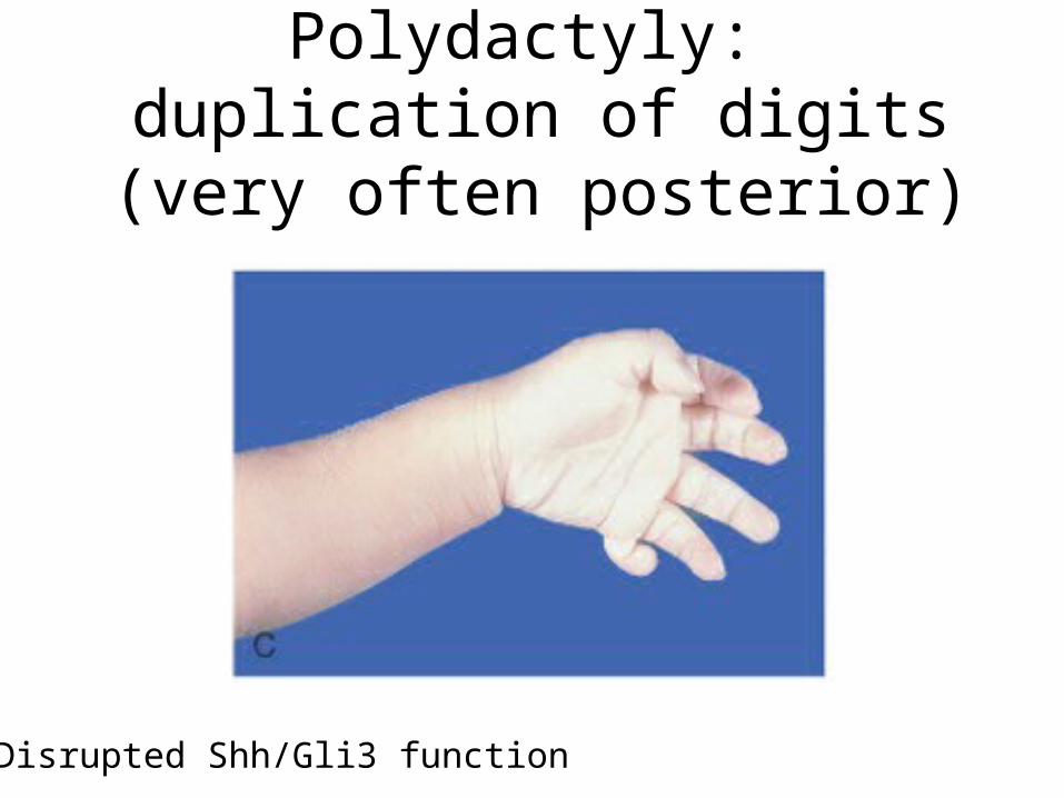
Polydactyly: duplication of digits (very often posterior)
Disrupted Shh/Gli3 function
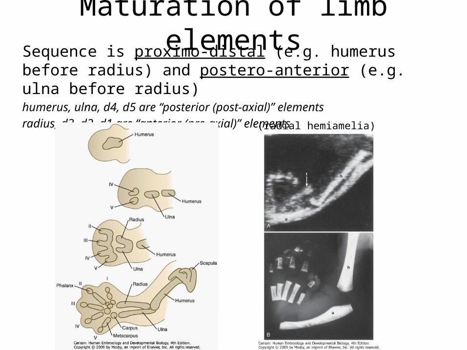
Maturation of limb elementsSequence is proximo-distal (e.g. humerus before radius) and postero-anterior (e.g. ulna before radius)humerus, ulna, d4, d5 are “posterior (post-axial)” elementsradius, d3, d2, d1 are “anterior (pre-axial)” elements
(radial hemiamelia)
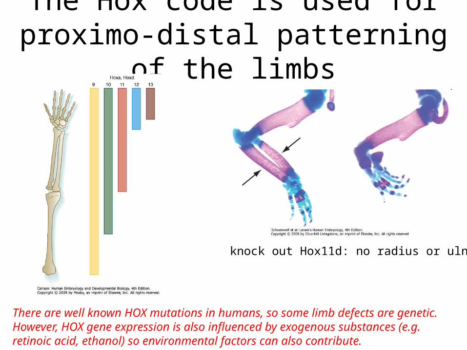
The Hox code is used for proximo-distal patterning of the limbs
knock out Hox11d: no radius or ulna
There are well known HOX mutations in humans, so some limb defects are genetic.However, HOX gene expression is also influenced by exogenous substances (e.g. retinoic acid, ethanol) so environmental factors can also contribute.
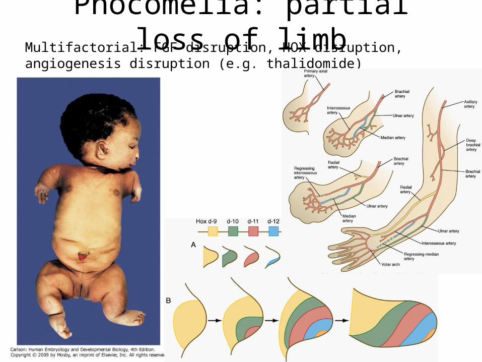
Phocomelia: partial loss of limbMultifactorial: FGF disruption, HOX disruption, angiogenesis disruption (e.g. thalidomide)
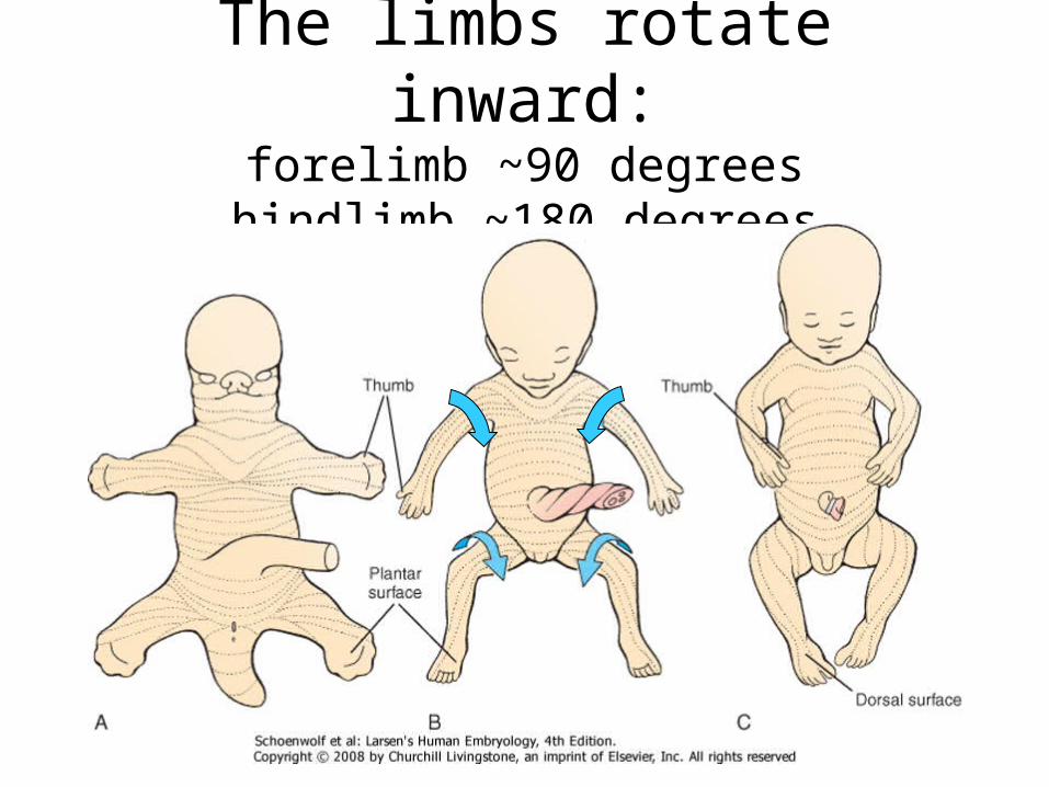
The limbs rotate inward:forelimb ~90 degrees
hindlimb ~180 degrees
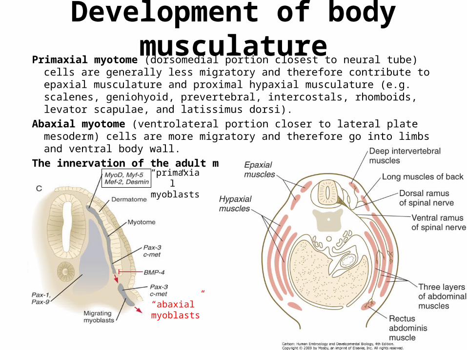
Development of body musculaturePrimaxial myotome (dorsomedial portion closest to neural tube) cells are generally less
migratory and therefore contribute to epaxial musculature and proximal hypaxial musculature (e.g. scalenes, geniohyoid, prevertebral, intercostals, rhomboids, levator scapulae, and latissimus dorsi).
Abaxial myotome (ventrolateral portion closer to lateral plate mesoderm) cells are more migratory and therefore go into limbs and ventral body wall.
The innervation of the adult muscle reflects the segment from which is was derived.
“primaxial”myoblasts
“abaxial”myoblasts
Abnormal muscle development
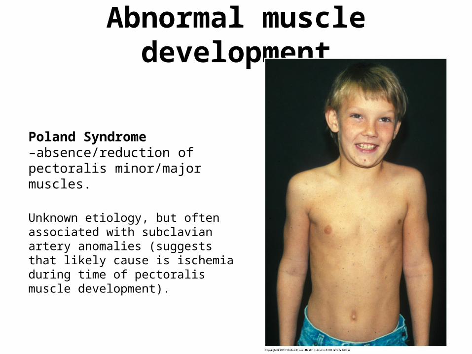
Poland Syndrome –absence/reduction of pectoralis minor/major muscles.
Unknown etiology, but often associated with subclavian artery anomalies (suggests that likely cause is ischemia during time of pectoralis muscle development).
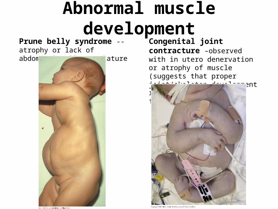
Abnormal muscle developmentPrune belly syndrome --atrophy or lack of abdominal wall musculature
Congenital joint contracture –observed with in utero denervation or atrophy of muscle (suggests that proper joint/skeleton development requires force feedback from muscles)
Slides 25-29 from Embryology02 lecture
The next five slides are from the “Basic Body Plan” lecture for your review.
click here to play the accompanying video corresponding to these slides
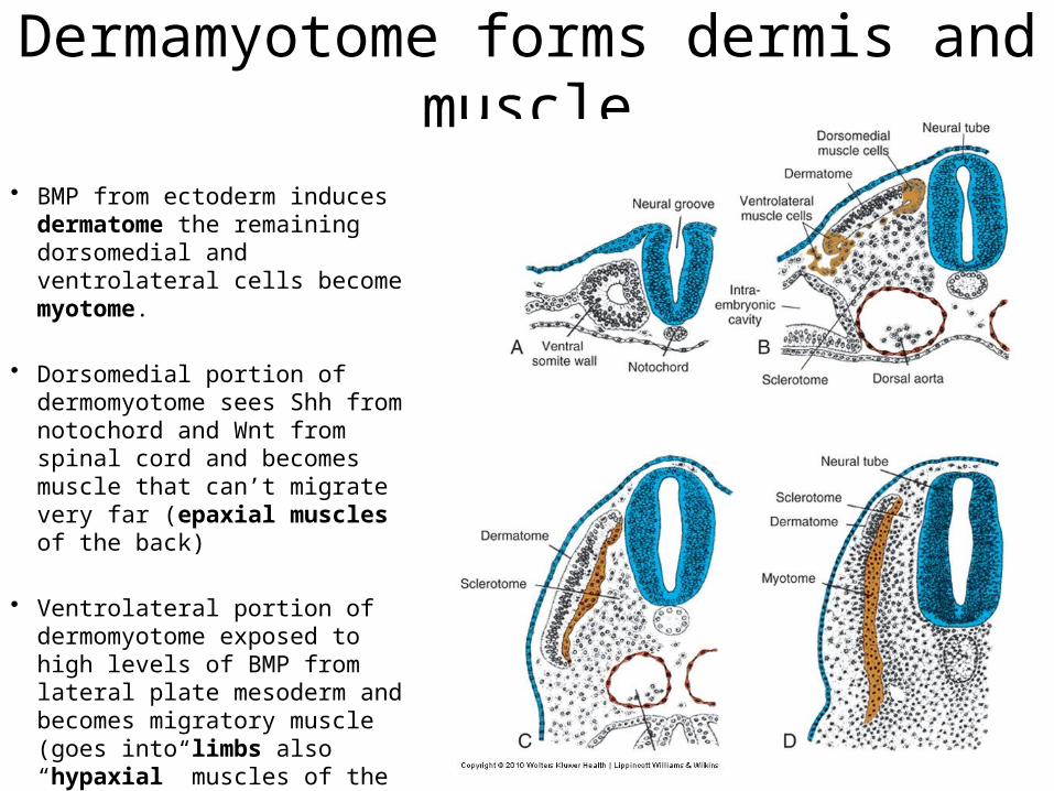
Dermamyotome forms dermis and muscle
• BMP from ectoderm induces dermatome the remaining dorsomedial and ventrolateral cells become myotome.
• Dorsomedial portion of dermomyotome sees Shh from notochord and Wnt from spinal cord and becomes muscle that can’t migrate very far (epaxial muscles of the back)
• Ventrolateral portion of dermomyotome exposed to high levels of BMP from lateral plate mesoderm and becomes migratory muscle (goes into limbs also “hypaxial” muscles of the lateral and ventral body wall, e.g. “lats” and “abs”
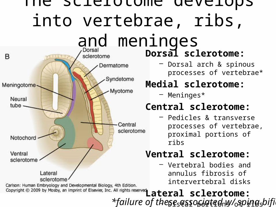
The sclerotome develops into vertebrae, ribs, and meninges
Dorsal sclerotome:– Dorsal arch & spinous processes of
vertebrae*
Medial sclerotome:– Meninges*
Central sclerotome:– Pedicles & transverse processes of
vertebrae, proximal portions of ribs
Ventral sclerotome:– Vertebral bodies and annulus
fibrosis of intervertebral disks
Lateral sclerotome:– Distal portions of ribs
*failure of these associated w/ spina bifida
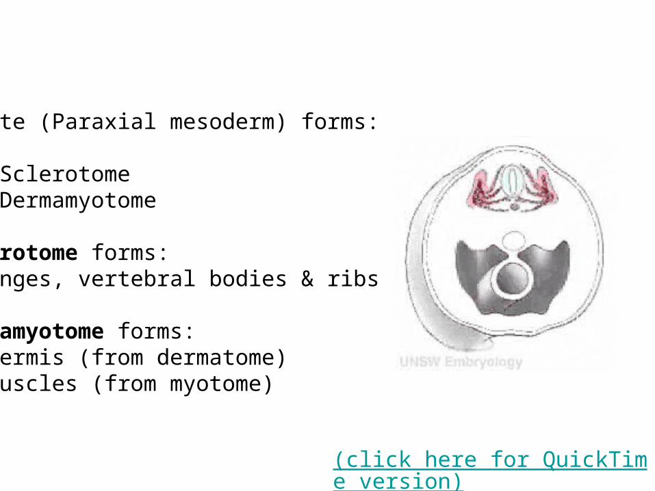
Somite (Paraxial mesoderm) forms:
1. Sclerotome2. Dermamyotome
Sclerotome forms:Meninges, vertebral bodies & ribs
Dermamyotome forms:1. Dermis (from dermatome)2. Muscles (from myotome)
(click here for QuickTime version)
Anterior and posterior portions of each sclerotome fuse to form vertebral bodies. This offsets the vertebral bodies and segmental muscles. i.e., the vertebrae (sclerotome) are out of phase with muscle (myotome) to form intervertebral joints. This allows the contracting segmental muscles to move the vertebral column laterally.
The sclerotome breaks into two parts…
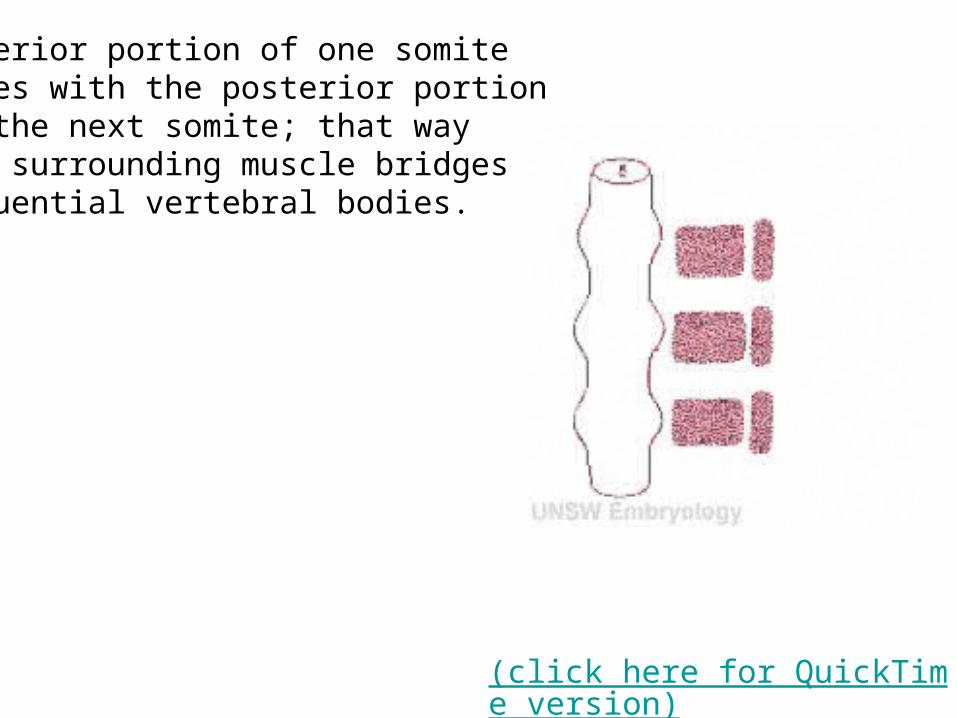
Anterior portion of one somitefuses with the posterior portionof the next somite; that wayThe surrounding muscle bridgessequential vertebral bodies.
(click here for QuickTime version)