Embed Size (px)
Citation preview

Light, Nearwork, and Visual Environment Risk Factors in Myopia
By
Amanda Aleksandra Alvarez
A dissertation submitted in partial satisfaction of the
requirements for the degree of
Doctor of Philosophy
in
Vision Science
in the
Graduate Division
of the
University of California, Berkeley
Committee in charge:
Professor Christine F. Wildsoet, Chair Professor Austin Roorda Professor Ruzena Bajcsy
Fall 2012

Light, Nearwork, and Visual Environment Risk Factors in Myopia
Copyright © 2012
by
Amanda Aleksandra Alvarez

1
Abstract
Light, Nearwork, and Visual Environment Risk Factors in Myopia
By
Amanda Aleksandra Alvarez
Doctor of Philosophy in Vision Science
University of California, Berkeley
Professor Christine F. Wildsoet, Chair
Myopia, or nearsightedness, is a form of visual impairment in which distant objects appear blurry due to excessive axial eye growth that is mismatched to the eye’s refractive power. This condition, though treatable with spectacles, contact lenses, or refractive surgery, continues to increase in prevalence, particularly in some Asian countries, where up to 80-90% of young people and students are myopic. High myopia (< -6.00 D) is associated with greater risk of glaucoma, retinal detachment, and other blinding complications.
Myopia is a complex disease with both genetic and environmental components. Rising myopia prevalence rates have mirrored lifestyle shifts that include reduced outdoor and light exposure. The directionality and impact of environmental risk factors, particularly light exposure, on myopia, continue to be poorly understood, partly due to the lack of in vivo and realtime instruments for measuring these effects. This dissertation examines the role of environmental risk factors in myopia, and introduces two new methods for quantitatively studying light and nearwork in humans.
Evidence from animal studies suggests short bursts of bright light may be sufficient to retard myopic eye growth. Recent questionnaire-based studies have found increased exposure to sunlight or outdoor environments to be correlated with reduced myopia in children. We supplemented the questionnaire approach with objectively gathered data from light sensors, and compared the accuracy of the two approaches. Maximum intensity, cumulative light exposure, frequency of intensity change, or time spent in bright light were not correlated with refractive error. Subjects overestimated time spent outdoors, and these estimates were in poor agreement with time reported by the sensor data. This is the first multi-season study to use both the questionnaire and light sensor methods coupled with local weather data to investigate light and outdoor effects in myopia.
The duration and degree of another myopia risk factor, nearwork, are typically estimated retrospectively through questionnaires that assess reading, computer use, and other visual behaviors. There are, however, no comprehensive methods of measuring working or fixation distance in realtime during natural tasks. Here we present a new approach for studying the dioptric environment in humans. A head-mounted eye tracking device was adapted to be fully mobile for the realtime measurement of eye movements, including convergence. This device was

2
validated in a small sample of young adults. We conducted exploratory analyses of possible task-related trends in fixational behavior, fixation distance, horizontal eye movements, blinks, and saccades. We found large differences in some of these metrics between reading and walking tasks; these task-dependent changes in visual behavior may underlie the nearwork effect in myopia progression. Light sensing and eye tracking are new techniques for quantifying behaviors that are thought to be involved in myopia development. Unlike questionnaires, these methods provide realtime, unbiased data at the temporal resolution that is relevant to refractive error development. Environmental pressures may be a tipping point toward pathological eye growth for genetically susceptible individuals, and further work in this vein could lead to simple behavioral interventions to curb myopia progression.

i
Table of Contents List of Figures iii List of Tables v Acknowledgements vi 1 Background 1
1.1 Introduction 1 1.2 Myopia Prevalence, Etiology, and Treatment 2 1.3 Environmental Factors in Myopia 4
1.3.1 Measures of Light and Nearwork 6 1.4 Outline of Dissertation 7 1.5 Summary 8 1.6 References 9
2 Quantifying Light Exposure 14
Abstract 14 2.1 Introduction 15 2.2 Methods and Materials 16
2.2.1 Subjects 16 2.2.2 Ocular Measurements and Questionnaire 17 2.2.3 Study Periods 18 2.2.4 Photometry 18 2.2.5 Analyses 20
2.3 Results 21 2.3.1 Light Intensity 23 2.3.2 Duration 26 2.3.3 Cumulative Light Exposure 30 2.3.4 Indoor and Outdoor Exposure 32
2.4 Discussion 37 2.5 Conclusion 41 2.6 References 43
3 Measuring the Dioptric Environment Using Eye Tracking 46
Abstract 46 3.1 Introduction 47 3.2 Technical Specifications of a Mobile Binocular Eye Tracker 50
3.2.1 Selection of Eye Tracker 53 3.3 Methods 53
3.3.1 Subjects and Ocular Measurements 54 3.3.2 Calibration Procedure 54 3.3.3 Tasks 55 3.3.4 Analyses 56 3.3.5 Eye Tracking Accuracy 59
3.4 Results 61

ii
3.4.1 The Visual Environment 61 3.4.2 Fixation Distance 63 3.4.3 Horizontal Eye Movements 66 3.4.4 Blinks and Saccades 66 3.4.5 Preliminary Refractive Error-Related Results 68
3.5 Discussion 70 3.6 Conclusion 72 3.7 References 74
4 Conclusions 78
Abstract 78 4.1 Summary 79 4.2 Light 79 4.3 Nearwork 80 4.4 Future Directions 81
4.4.1 Eye Tracker Improvements 83 4.5 References 84
5 Appendix A: Myopia Questionnaire 86

iii
List of Figures
Figure 2-1 Two subjects wearing the armband with light sensor 20 Figure 2-2a Sensor response curve, sunlight spectrum, and the eye’s photopic function 20 Figure 2-2b Light intensity measured with three devices 20 Figure 2-3a Light intensity recorded by light sensor and pyranometer during spring 22 Figure 2-3b Light intensity recorded by light sensor and pyranometer during fall 22 Figure 2-3c Light intensity recorded by light sensor and pyranometer during winter 22 Figure 2-4a Maximum daily light intensity during spring 24 Figure 2-4b Maximum daily light intensity during fall 24 Figure 2-4c Maximum daily light intensity during winter 24 Figure 2-4d Maximum daily light intensity during all seasons 24 Figure 2-5a Average daily light intensity during spring 25 Figure 2-5b Average daily light intensity during fall 25 Figure 2-5c Average daily light intensity during winter (subset) 25 Figure 2-5d Average daily light intensity during all seasons (subset) 25 Figure 2-5e Average daily light intensity during winter (complete) 25 Figure 2-5f Average daily light intensity during all seasons (complete) 25 Figure 2-6a Percentage of daily time spent outdoors during spring 27 Figure 2-6b Percentage of daily time spent outdoors during fall 27 Figure 2-6c Percentage of daily time spent outdoors during winter 27 Figure 2-6d Percentage of daily time spent outdoors during all seasons 27 Figure 2-7a Daily hours spent in bright sunlight (> 105 lux) during spring 28 Figure 2-7b Daily hours spent in bright sunlight (> 105 lux) during fall 28 Figure 2-7c Daily hours spent in bright sunlight (> 105 lux) during winter 28 Figure 2-7d Daily hours spent in bright sunlight (> 105 lux) during all seasons 28 Figure 2-8a Frequency of intensity changes during spring 29 Figure 2-8b Frequency of intensity changes during fall 29 Figure 2-8c Frequency of intensity changes during winter 29 Figure 2-8d Frequency of intensity changes during all seasons 29 Figure 2-9a Solar-normalized cumulative light exposure during spring 31 Figure 2-9b Solar-normalized cumulative light exposure during fall 31 Figure 2-9c Solar-normalized cumulative light exposure during winter 31 Figure 2-9d Solar-normalized cumulative light exposure during all seasons 31 Figure 2-10a Estimates of indoor time with sensor data means during spring 33 Figure 2-10b Estimates of outdoor time with sensor data means during spring 33 Figure 2-10c Estimates of indoor and outdoor time with sensor data means during spring 33 Figure 2-11a Estimates of indoor time with sensor data means during fall 34 Figure 2-11b Estimates of outdoor time with sensor data means during fall 34 Figure 2-11c Estimates of indoor and outdoor time with sensor data means during fall 34 Figure 2-12a Estimates of indoor time with sensor data means during winter 35 Figure 2-12b Estimates of outdoor time with sensor data means during winter 35 Figure 2-12c Estimates of indoor and outdoor time with sensor data means during winter 35 Figure 2-13 Indoor and outdoor estimates and sensor data means from all three seasons 36 Figure 2-14a Effect of changing sampling interval on hours spent in bright sunlight 38

iv
Figure 2-14b Effect of changing sampling interval on cumulative outdoor exposure 38 Figure 2-14c Effect of changing sampling interval on total cumulative light exposure 38 Figure 3-1a Native Eyelink II headgear 51 Figure 3-1b Modified binocular eye tracker headgear 51 Figure 3-2a Front view of modified binocular eye tracker 51 Figure 3-2b Hardware of the mobile binocular eye tracker 51 Figure 3-3a Subject’s head secured in the chinrest during calibration 52 Figure 3-3b Example of display setup during calibration 52 Figure 3-4 Mobile binocular eye tracker headgear and backpack worn by a subject 52 Figure 3-5a Example of near gaze captured by the eye cameras 56 Figure 3-5b Example of far gaze captured by the eye cameras 56 Figure 3-6a Scene camera images from one subject during reading 57 Figure 3-6b Scene camera images from one subject during walking 57 Figure 3-7 Method for obtaining horizontal amplitude using RMS approach 58 Figure 3-8 Effect of eye position error on fixation distance measurement 59 Figure 3-9a Post-task calibration error grouped by task 60 Figure 3-9b Post-task calibration error for each subject 60 Figure 3-10a Effect of drift correction on fixation distance during reading 60 Figure 3-10b Effect of drift correction on fixation distance during walking 60 Figure 3-11 Map of fixations in three dimensions during reading and walking 62 Figure 3-12 Average time spent at near, mid, and far gaze during reading and walking 63 Figure 3-13 Dioptric demands of fixations during reading and walking 64 Figure 3-14a Example raw traces of fixation distance (Z) during reading and walking 65 Figure 3-14b Example raw trace of fixation distance (Z) for one subject during reading 65 Figure 3-15a Average deviation from the mean for horizontal angle of fixation 65 Figure 3-15b Average deviation from the mean for Z distance 65 Figure 3-16a Example raw traces of horizontal angle of fixations during reading and walking 66 Figure 3-16b Example raw trace of horizontal angle of fixations during reading 66 Figure 3-17 Average blinks per minute during reading and walking 67 Figure 3-18 Average saccades per minute during reading and walking 67 Figure 3-19 Average saccade amplitude during reading and walking 67 Figure 3-20 Dioptric demands of fixations as a function of refractive error 68 Figure 3-21 Average blinks per minute as a function of refractive error 69 Figure 3-22 Average saccades per minute as a function of refractive error 69 Figure 3-23 Average saccade amplitude as a function of refractive error 70

v
List of Tables
Table 2-1 Refractive error and demographic characteristics of subjects in light sensor study 18 Table 2-2 Average indoor light measurements in five buildings on campus 21 Table 2-3 Mean daylight hours and integrated solar radiation during each study period 30 Table 2-4 Mean daily hours spent indoors and outdoors from both estimates and sensor data 36 Table 3-1 Comparison of eye tracker models and specifications 53 Table 3-2 Average deviations from mean for Z distance and for horizontal angle of fixation 65 Table 3-3 Range for Z distance and for horizontal angle of fixation 66

vi
Acknowledgements Thank you to the following individuals for their indispensable experimental help: Emily Liu, for screening subjects; Peter Cheng, for creating light exposure data analysis code; and Patrick Thorson of Lawrence Berkeley Lab, for providing weather data. Thank you to my classmates, especially Paul Ivanov, for doing it live and #better and always remaining positive, Nicole Putnam, for commiserating and sticking with it through the trials and tribulations, and David Hoffman, for never losing patience with me. Thank you to mom, dad, and Mel for supporting me from afar – Semper Fi! Thank you to Christine, who believed in me when no one else would. For Hany, master machinist, who has been my light in the darkness.
On sanoo: onko, onkohan? ja epäily masentaa maailman.
Ei sanoo: eikö, eiköhän! ja näemme vuorien siirtyvän.
– Lauri Viita

1
Chapter 1: Background 1.1 Introduction Ocular refractive error results from a mismatch between the eye’s axial length and the refractive power of the cornea and lens. Many animals, including humans, are born with short eyes (hyperopia, or farsightedness, in which the focal plane of the eye is behind the retina when not accommodating) that grow to a length that matches the refractive power and brings the image of distant objects to the retina, a process known as emmetropization. Hyperopia often does not require correction (at least in younger adults), because accommodation provides the extra refractive power needed to bring the image plane forward to the retina. Hyperopia is also not a condition that is associated with progression and complications, unlike myopia. Nearsightedness, or myopia, is the continued growth of the eye that pushes the retina behind the focal point, creating blur for objects viewed at distance. Since the eye cannot be made to shrink, optical or surgical corrections are employed to remove excess refractive power from the eye. Myopia is a continuum of refractive error conditions that can be progressive or even pathological, and is increasingly emerging in younger children and even adults who are past school age. Its etiology, prevalence, and treatments are discussed in Section 1.2.
Myopia is the subject of intense study genetically and clinically, but less so behaviorally. Behavioral contributions to subtle physiological phenomena are notoriously difficult to measure, especially over the course of years (the window over which one might chart the progression of myopia). The purpose of this dissertation is to gain a clearer understanding of the contributions of human behaviors and environments to myopia by quantifying these behaviors and environments using novel methods. Because human myopia appears to be more than just genetically driven (Mutti et al., 1996; Wallman, 1994), research into other risk factors is both warranted and overdue. In some respects, myopia, like diabetes or obesity, can be viewed as a lifestyle disease that arises from genetic susceptibility coupled with unhealthy environments and behaviors (Cordain et al., 2002). The healthcare costs of myopia are also not trivial. One study estimated the cost of correcting refractive error in the United States to be $12.8 billion – in 1990 (Javitt & Chiang, 1994). Uncorrected refractive error represents the largest proportion of preventable blindness cases in the world, according to the WHO (2007). Spectacles, contact lenses, or refractive surgery address the symptoms of blur, but not the causes of eye growth. Indeed, many myopes continue to progress, requiring stronger prescriptions to match their continued eye growth. When refractive error exceeds -6.00 diopters (D), the risk of developing blinding complications such as retinal detachment increases (Curtin, 1985). As in the cases of diabetes and obesity, environmental and behavioral choices may be part of the key to prevention in myopia. This dissertation focuses on two main environmental risk factors – sunlight and nearwork – but other factors such as diet and physical activity are also discussed in Section 1.3.
Emmetropization is known to be an active process, with visual input guiding eye growth. Degraded, or absent, visual stimulation, such as that produced by form deprivation, or patching or suturing of the eye, leads to elongated eyes. It is reasonable to surmise that other factors that affect retinal image quality – either optically or mechanistically, through nearwork and eyestrain

2
– or the eye’s circadian rhythm and biochemical pathways – via altered light exposure – can also be causative in the development of myopia. 1.2 Myopia Prevalence, Etiology, and Treatment Myopia rates are increasing worldwide, particularly in East Asian countries. In data reviewed by Morgan and Rose (2005), higher prevalence rates are clearly associated with urban living in developed countries, as well as schooling. For example, the prevalence of myopia in Japan grew from 39% in 1984 to 59% in 1996 (Matsumara & Harai, 1999). Increases in Taiwan (36.7% in 1983 to 60.7% in 2000) (Lin et al., 2001; Lin et al., 2004) and Hong Kong (83% in 2001, from 53% in 1991) (Lam & Goh, 1991; Lam et al., 2004) have also been reported. These data reflect myopia in adolescent 12 and 13-year-olds. By contrast, similarly aged children in India and South Africa show lower rates of myopia (4.8-10% and 4%, respectively) (Dandona et al., 1999; Dandona et al., 2002; Naidoo et al., 2003). An extreme prevalence figure of 96.5% in South Korean men was recently reported (Jung et al., 2012), and in Singapore, myopia prevalence in military conscripts was 79.3% (Wu et al., 2001). In rural China, prevalence figures of 20-35% have been reported in 13-year-olds, compared with 55% in urban areas (He et al., 2009). In the United States, the Orinda study (1953) and its follow-up (1993) reported myopia prevalence of 12.3% and 28%, respectively, in 12-year-olds (Blum et al., 1959; Zadnik, 1997). Across the American population, myopia prevalence is estimated to have increased from 25% to 41.6% in 30 years (Vitale et al., 2009). Globally, cases of high myopia (< -6.00 D) are increasing, and age of onset is decreasing. A number of gene loci associated with myopic eye growth have been identified. In addition, ethnic differences in myopia prevalence have been found, for example in Singapore (Wu et al., 2001) and the United States (Vitale et al., 2009). Having one or more myopic parents is strongly associated with developing myopia (Jones et al. 2007; Mutti et al., 2002), and this hereditary component is also manifest in emphasis on educational attainment. Education level is strongly associated with myopia level and severity in Singapore (Wu et al., 2001), and Loman et al. (2002) found a large proportion of a law student cohort to be myopic, with most progressing in their myopia over three years. Donovan et al. (2011) reported that Chinese children experienced slowed myopia progression during summer, indicating that progression data and treatment efficacy may be highly dependent on season and the balance of time spent in work/school (nearwork, low light) versus leisure (outdoors, bright light).
Genetics provides the template for eye growth, but emmetropization is an active visually guided process (Wallman & Winawer, 2004). It can thus be heavily influenced by urbanization, education, and other visual environmental factors. The role of visual input can be demonstrated most clearly in animal models that are visually manipulated to become myopic, either with negative lenses that impose hyperopic defocus on the eye, or diffusers that reduce retinal contrast and illuminance, causing form deprivation. The end result in both cases is myopic refractive error induced solely through visual environmental changes. In humans, congenital cataract provides equivalent degraded visual input that can lead to elongated eyes (Rabin et al., 1981). Myopia is now recognized as not being a malady exclusive to children or adolescents (once dubbed “school myopia”). Adults can also develop myopia, often in connection with occupational demands; some of the most well-known cases concern pilots (Hoogerheide et al. 1971) and microscopists (McBrien & Adams, 1997; Ting et al., 2004). The illusion of proximity

3
when looking through an instrument, and subsequent accommodation, have been conjectured to contribute to axial elongation in these cases. Adams and McBrien (1992) reported that 71% of the microscopists surveyed were myopic, with almost half reporting onset of myopia that coincided with commencing microscopy work. The Chinese microscopists in the sample of Ting et al. (2004) had a myopia prevalence of 87%.
There is no clear answer to why myopia develops. A number of factors, including parental myopia, education, peripheral refraction, genetics, and outdoor (light) exposure have been implicated (Pan et al., 2012). In humans, a substantial theory of myopia pathogenesis concerns the intertwined factors of accommodation, nearwork, defocus, and eye shape. Myopes are known to display a lag of accommodation, that is, insufficient accommodative response for a given accommodative target. This lag essentially results in hyperopic defocus on the retina, similar to the animal experiments described above, and is thought by some to mediate the association between nearwork and myopia progression. Extended periods of nearwork compound the exposure to this defocus, which pushes the focal plane behind the retina, inducing myopia. As the eye grows axially, it becomes more prolate (egg-shaped), with the posterior pole protruding. This extends the area of peripheral retina that receives hyperopic defocus, furthering the growth cycle. Eyes that are a priori prolate-shaped are more susceptible to defocus-induced elongation (Hoogerheide et al., 1971; Mutti et al., 2007). The discovery that eye growth is controlled by local retinal regions also helps explain how even corrected myopes can continue to progress. Myopia following ablation of the fovea in monkeys demonstrates that visual input to the periphery is sufficient to drive eye growth (Smith et al., 2007), underscoring the importance of peripheral hyperopia for myopia development. Moreover, correction with spectacles or contact lenses at the fovea leads to an equivalent situation of peripheral hyperopia. Contemporary myopia treatments are increasingly focused on selective optical stimulation of the retina to achieve peripheral myopia for eye growth inhibition. Treatments for myopia range from optical to surgical and pharmacological. Progressive addition and bifocal lenses produce small but clinically insignificant reductions in myopia progression. Forcible flattening of the cornea to reduce refractive power with orthokeratology lenses is a common and reliable alternative to spectacles that has been shown to slow myopia progression. Optically targeting specific retinal regions with multifocal contact lenses may be a promising future treatment option (Liu & Wildsoet, 2012). Many of these treatments can slow, but not completely retard, myopia progression. Permanent surgical alteration of the cornea (e.g. LASIK) can eliminate myopia and is largely successful, but it may not be an appropriate procedure for high or progressing myopes or those with thin or vulnerable corneas. At the other end of the spectrum is scleral buckle surgery to support and contain the posterior pole in cases of pathological myopia. Finally, off-label use of atropine paralyzes the ciliary muscle, with the side effects of enlarged pupils and loss of accommodation. Atropine treatment in chicks reduced axial growth and the development of myopia (McBrien et al., 1993). Atropine has also been successful in slowing myopia progression and eye elongation in children (Chua et al., 2006). The precise mechanism by which atropine retards eye growth is unknown, but increased light levels from dilated pupils may be partially responsible.

4
1.3 Environmental Factors in Myopia Visual experience drives changes in eye growth patterns (Wallman et al., 1978). As indicated in the previous Section, there is an increased understanding that behavior and visual environment can have a substantial effect on refractive error development. Since the late 19th century, myopia has been linked with education, “the companion of intellectual progress” (Smith et al., 1890), and with the excessive convergence of nearwork; elongated eyes were also thought to mirror the superior intellect of a larger brain (ibid.). “Hygiene desks” were promoted for optimal posture and reading and writing distance (Bennett, 1922). Andrews (1886) discusses light, both natural and artificial, as causal to many ocular diseases, and is enthusiastic about the adoption of incandescent lightbulbs, particularly in relation to myopia, to replace gas lighting. Dowling (1891) also favored electric light over that produced by burning gas or oil, though he counseled that studying or reading should preferably be done in sunlight, and myopes should never study at night. Despite these early insights, behavioral and environmental interventions to combat myopia have largely been non-existent. Unvalidated and potentially injurious home remedies like sun gazing and eye exercises have been promoted (e.g. Bates, 1978). The 20-20-20 rule – focusing 20 feet (6.1 meters) away for 20 seconds every 20 minutes – is a widely cited (Vertinsky & Foster, 2005) behavioral guide that appears to have no published clinical basis. The foremost treatment for myopia therefore remains optical correction, with refractive surgery gaining in popularity. A number of strands of research are converging on the conclusion that changes in visual environments and lifestyles brought about by urbanization are contributing to increases in myopia prevalence (He et al., 2009; Cordain et al., 2002). Van Rens and Arkell (1991), Morgan et al. (1975), Norn (1997), and Johnson et al. (1979) all discuss sharp increases in myopia in young Inuit who live modern lifestyles that include expanded education and changes in diet. Werner et al. (2010) specifically attribute the rise in myopia over one generation in Alaska to electrification and introduction of artificial lighting that occurred from the 1950s to the 1970s.
The role of light in eye growth remains equivocal. Nowhere is this more evident than in the debate over nightlights and myopia. The initial report (Quinn et al., 1999) found a dose-dependent association between myopia levels and nightlight exposure in early childhood; the mechanism remained unclear, but the authors suggested the absence of a daily period of darkness (i.e. a regular circadian cycle) was responsible. (They also note the limitations of collecting behavioral data via questionnaire, something that also motivates and is addressed in this dissertation.) Subsequent studies failed to replicate these results (Zadnik et al., 2000; Gwiazda et al., 2000). Stone et al. (2000) countered that, while separating genetic from environmental influences in family studies is difficult, the lack of a daily period of darkness due to nightlight use is nonetheless noteworthy as a possible accelerator or trigger for myopia development in those predisposed. Ostensible evidence for this thesis was provided by Loman et al. (2002), who found that less daily exposure to darkness was associated with myopia progression in law students.
The interest in light in relation to human myopia stems from animal research, where photoperiodicity is known to affect eye growth (Stone et al., 1995; Li et al., 2000). In mice (a nocturnal species), for example, prolonged light exposure results in myopic eye growth (Zhou et al., 2010), and in chickens, continuous exposure to bright light produces corneal flattening and hyperopia (Li et al., 1995) , with prolonged exposure leading to retinal photodamage and

5
glaucoma. In a lens paradigm, constant lighting prevented emmetropization in chicks, with stronger inhibitory effects on compensation to negative lenses, but this effect was reversible once chicks were restored to normal lighting conditions (Padmanabhan et al., 2007). Chickens undergoing myopia-inducing form deprivation had less myopic refractions when exposed to sunlight or artificial bright light for part of the day (Ashby et al., 2009). In a similar study of lens-induced myopia, exposure to bright light was found to slow, but not prevent, progression to a myopic endpoint (Ashby & Schaeffel, 2010). These experiments also showed that this effect was mediated by dopamine. A protective effect of high light levels on form deprivation myopia has also been show in monkeys (Smith et al., 2011; Smith et al., 2012).
Concurrently, human research has found apparent protective outdoor effects on myopia risk. One of the most highly cited studies (Rose et al., 2008) found that children who combined high levels of nearwork with low time spent outside had the highest odds ratios of myopia. More time spent outdoors translated into more hyperopic refractions, after adjustment for other factors. Making use of detailed and targeted questionnaires (Rose, 2008), the authors probed time use for near, mid, and far working distances both indoors and outdoors. Jones-Jordan et al. (2011) also found that children who developed myopia spent less time doing outdoor or sports activities, and that this reduction had a greater myopigenic effect than nearwork. A meta-analysis of seven studies by Sherwin et al. (2012b) found that every additional hour spent outdoors reduced the odds ratio for myopia by 2%, “a modest but significant” reduction in the risk of developing myopia or progressing.
These studies should be treated with caution, for a few reasons. First, Rose et al. (2008) and Jones-Jordan et al. (2011) both administered visual activity questionnaires to parents, not children, a shortcoming that will be discussed below. Second, little effort has been made to dissociate outdoor effects from purely light effects. Smith et al. (2012), for example, have seized on findings of Rose et al. (2008) as demonstrating a strong protective light effect, despite those authors discussing reduced accommodative demand and substitution effects (time spent outdoors is not spent indoors) in addition to light. Indeed, outdoor environments (as opposed to indoor environments) may affect myopia progression through reduced near visual stimulation, and the resultant reduced accommodative demand, increased dioptric distances and dioptric variation (Charman, 2011), smaller pupil size and associated increase in depth of focus, in addition to the availability of sunlight. That sunlight may be the most powerful factor influencing eye growth in this scenario is assumed, but not confirmed.
Outdoor effects may also be conflated with time spent doing sports or other physical activities (as occurred in Jones-Jordan et al., 2011). Rose et al. (2008) did not find an association between indoor sports and myopia, and concluded that being outdoors, rather than sports per se, was the crucial element leading to lower myopia levels. Dirani et al. (2009), who also found that greater time spent outdoors was associated with less myopia, reported that indoor sports alone was not associated with myopia, but total sports (including outdoor components) was negatively associated with myopia. Whether physical activity is a marker for outdoor activity, or has its own independent effects on myopia, has still not been firmly established, though a longitudinal study looking specifically at physical activity showed it was associated with reduced myopia progression (Jacobsen et al., 2008). A study using both a parental questionnaire and an objective physical activity measure (an accelerometer worn by the child) sought to tease apart the outdoor and sport factors. Time spent outdoors and physical activity were independently predictive of myopia onset, with the latter having a weaker effect (Guggenheim et al., 2012).

6
Dietary factors may also contribute to myopia development. Mäntyjärvi (1988) noted that intraocular changes including lens swelling appear to contribute to myopia in diabetes. The model of Cordain et al. (2002) implicates hyperinsulemia in unregulated scleral growth that leads to myopia. Edwards et al. (1996) found differences in energy, vitamin, and mineral intake in children who became myopic compared to those who did not. Dietary myopic changes may also have a link with outdoor activity, via the synthesis of vitamin D in the skin when its cholesterol precursor reacts with ultraviolet light. Mutti and Marks (2011), however, did not find a difference in blood vitamin D levels between myopes and non-myopes, nor did these subjects differ in their time spent outdoors. 1.3.1 Measures of Light and Nearwork In the context of the above factors, myopia can be considered a maladaptation of the eye to frequent near focusing distances, low light levels, and other influences. The evidence suggests environment can modulate the eye’s refractive state in both beneficial and detrimental ways, but that more data are needed to ascertain the strength and directionality of these changes. The major hurdle is having suitable objective measures of risk factors like nearwork or light exposure.
As touched on by Quinn et al. (1999) and others (Rah et al., 2002; Bryant et al., 2007), questionnaires can be insufficient study instruments. When retrospectively administered, questionnaires or surveys rely on memory for events that may have happened days, weeks or months prior. In juvenile myopia progression studies, questionnaires are often completed by proxy respondents (parents), who may be guessing or biased concerning their child’s behavior, especially in competitive academic settings. The self-reports of subjects can also be gathered by random sampling via pager or telephone, as is done in the Experience Sampling Method where subjects report the visual activity at the time of sampling, and estimate the visual distance (Rah et al., 2001; Rah et al., 2004). This method has shown good agreement with questionnaire studies (Rah et al., 2006), but still does not provide access to ground-truth or objective information about subjects’ visual activities or distances. Moreover, random sampling of activities is not applicable to the question of light levels, as there is no metric (such as arm’s length in nearwork) by which to estimate, and subjects have no intuitive idea of how to quantify the luminance levels to which they are exposed. The limitations of questionnaires for studying behavioral and environmental risk factors – including poor parent-child agreement in reporting nearwork (Rah et al., 2002), and the dependence of this agreement on the constancy of the traits being surveyed (Whiteman & Green, 1997) – motivated the methods and experiments described in this dissertation. At the time of writing, there are two objective questionnaire alternatives for gathering light exposure data. As a measure of lifetime cumulative ultraviolet light exposure, Sherwin et al. (2012a) have made use of conjunctival autofluorescence. Greater autofluorescence was associated with lower myopia in their subjects, independently of other factors, and in the associated questionnaire, myopia prevalence decreased with greater time outdoors (though the autofluorescence association was stronger). The second alternative is a light sensor. In studies conducted contemporaneously with those described in this dissertation, groups in New Zealand and Singapore have begun to use sensors to augment questionnaire data in myopia studies. In a small scale pilot study, Backhouse et al. (2011) found no correlation between refractive error and cumulative light exposure, or rate of change in light levels. These measurements were made over three weeks in winter in 13 and 14-year-old children. Dharani et al. (2012) used the same light

7
sensor and concurrent parental questionnaires to assess agreement between the two measures. They found agreement between a diary of outdoor activity and the light sensor to be poor to fair, indicating that questionnaires are far from ideal in accurately measuring outdoor or light effects. This study did not look at myopia levels or progression in the children, and only measured light exposure over a one-week period. The study of Dharani et al. (2012) is part of a larger clinical trial intervention in Singapore to counter obesity and myopia. Besides light, the FIT trial is measuring physical activity using pedometers; no results have yet been published. With objective light exposure data limited to these two studies, clinical trials of a potential light treatment for myopia are nonetheless underway. The intervention in the Guangzhou Outdoor Activity Longitudinal (GOAL) study (Xiang et al., 2011; Morgan et al., 2012) is an extra hour of daily outdoor activity for six to seven-year-old children. After one year of the three-year-study, this has resulted in statistically, but not clinically, significant reductions in myopia progression and axial elongation. With regard to the measurement of nearwork, the questionnaire approach also has limitations. A typical measure of nearwork in questionnaires is the number of books read per week (Saw et al., 2002; Dirani et al., 2009), though some studies ask for a report of a number of activities (reading, drawing, handheld gaming, etc.) that occur at a close distance (< 50 cm) (Rose et al., 2008). Myopia clearly is associated with nearwork, especially closer distances and longer durations (Ip et al., 2008). What remains unclear, as with light, is what dimension of nearwork (e.g. intensity/distance, duration, medium (books vs. electronics)) is the crucial myopigenic factor. This cannot be ascertained with questionnaire methods.
Alternative measuring techniques in this area have also emerged. A head-mounted ultrasonic device for measuring reading distance introduced by Leung et al. (2011) relies on a detectable surface in front of the subject. Myopes and non-myopes were found to have significantly different reading distances, and self-reported reading distances were not correlated with the objectively measured distances. This again underscores the poor reliability of questionnaire-based studies of environmental factors in myopia. The device of Leung et al. (2011) can only measure near working distances, as it is laboratory-based and relies on the subject engaging with a surface like a paper or screen that the device can ping to measure the distance.
Objective measurement of focusing distance requires the ability to actually follow the movements of the eyes in all environments, not just laboratories where subjects are constrained to nearwork activities. Measuring the distance from the head to the purported surface of fixation is just a proxy for recording eye movements, which are much more informative about a subject’s actual fixational behavior. To this end, Hartwig et al. (2011) used head-mounted eye tracker to study eye movements in myopes and non-myopes. This study, which was also confined to a laboratory and only assessed near working distances, found distance differences between reading and writing tasks, but not between refractive error groups. The work described in Chapter 3 of this dissertation goes beyond these efforts to measure fixation behavior at all distances. 1.4 Outline of Dissertation This dissertation concerns the roles of light and nearwork in myopia, and introduces new tools for gathering objective behavioral and environmental data about these factors.

8
Chapter 2 discusses the deployment of wearable light sensors for measuring ambient light exposure in myopic and non-myopic young adults. Light exposure patterns as a function of refractive error and season are analyzed, with the conclusion that myopia and light are not associated. Chapter 3 outlines the technical development of a new mobile binocular eye tracking device for the study of fixation distance and direction in humans via the measurement of eye movements and vergence. Experiments in both laboratory and natural settings are described, with the results suggesting large task-related differences in fixation distance and other eye movements. This type of approach may be useful in identifying visual behavior patterns associated with refractive error development. Chapter 4 summarizes these studies and the state of knowledge of the role of environmental risk factors in myopia, and discusses future work in this area. 1.5 Summary Myopia is a complex disease whose prevalence is increasing. Optical correction can address the symptom of visual blur, but not the underlying genetic, biochemical, or environmental factors that can cause continued, potentially pathological eye growth. Rising myopia prevalence rates have mirrored lifestyle shifts that include reduced outdoor and light exposure, more education, and more time spent in near focusing tasks like reading or use of computers and electronic gadgets. The potential significance of these factors has been appreciated for some time. The directionality and impact of environmental risk factors, particularly light exposure, on myopia, continue to be poorly understood, partly due to the lack of in vivo and realtime instruments for measuring these effects. Quantifying the environmental risk factors, especially light and nearwork, is one of the goals of this dissertation. A better understanding of these factors could lead to concrete treatments or behavioral interventions for myopia.

9
1.6 References
Adams, D. W., & McBrien, N. A. (1992). Prevalence of myopia and myopic progression in a population of clinical microscopists. Optometry and Vision Science 69(6), 467–473.
Andrews, J. A. (1886). The electric light as an illuminator. The effect of strong light on the eye. Transactions of the American Ophthalmological Society, 4, 228–241.
Ashby, R., Ohlendorf, A., & Schaeffel, F. (2009). The effect of ambient illuminance on the development of deprivation myopia in chicks. Investigative Ophthalmology & Visual Science, 50(11), 5348 –5354.
Ashby, R. S., & Schaeffel, F. (2010). The effect of bright light on lens compensation in chicks. Investigative Ophthalmology & Visual Science, 51(10), 5247 –5253.
Backhouse S., Ng, H., & Phillips, J. (2011) Light exposure patterns in children: A pilot study. Optometry and Vision Science, 88, 400.
Bates, W. H. (1978). The cure of imperfect sight by treatment without glasses. Health Research Books.
Bennett, H. E. (1922). Some requirements of good school seating. The Elementary School Journal, 23(3), 203–214.
Blum, H. L., Peters, H. B., & Bettman, J. W. (1959). Vision screening for elementary schools. University of California Press, Los Angeles.
Bryant, M. J., Lucove, J. C., Evenson, K. R., & Marshall, S. (2007). Measurement of television viewing in children and adolescents: A systematic review. Obesity Reviews, 8(3), 197–209.
Charman, W. N. (2011). Myopia, posture and the visual environment. Ophthalmic and Physiological Optics, 31(5), 494–501.
Chua, W.-H., Balakrishnan, V., Chan, Y.-H., Tong, L., Ling, Y., Quah, B.-L., & Tan, D. (2006). Atropine for the treatment of childhood myopia. Ophthalmology, 113(12), 2285–2291.
Cordain, L., Eaton, S. B., Brand Miller, J., Lindeberg, S., & Jensen, C. (2002). An evolutionary analysis of the aetiology and pathogenesis of juvenile-onset myopia. Acta Ophthalmologica Scandinavica, 80(2), 125–135.
Curtin, B. (1985). The myopias: Basic science and clinical management. Harper & Row, Philadelphia.
Dandona, R, Dandona, L., Naduvilath, T. J., Srinivas, M., McCarty, C. A., & Rao, G. N. (1999). Refractive errors in an urban population in Southern India: The Andhra Pradesh eye disease study. Investigative Ophthalmology & Visual Science, 40(12), 2810–2818.
Dandona, R, Dandona, L., Srinivas, M., Giridhar, P., McCarty, C. A., & Rao, G. N. (2002). Population-based assessment of refractive error in India: the Andhra Pradesh eye disease study. Clinical & Experimental Ophthalmology, 30(2), 84–93.
Dharani, R., Lee, C.-F., Theng, Z. X., Drury, V. B., Ngo, C., Sandar, M., Wong, T.-Y., et al. (2012). Comparison of measurements of time outdoors and light levels as risk factors for myopia in young Singapore children. Eye, 26(7), 911–918.
Dirani, M., Tong, L., Gazzard, G., Zhang, X., Chia, A., Young, T. L., Rose, K. A., et al. (2009). Outdoor activity and myopia in Singapore teenage children. British Journal of Ophthalmology, 93(8), 997 –1000.
Donovan, L. A., Sankaridurg, P., Kwan, J., Chen, X., Ho, A., Smith, E. L., Lazon, P., et al. (2011). The rate of myopia progression in Chinese children is slower in summer than in winter. Investigative Ophthalmology & Visual Science, 52(6), 2713.

10
Dowling, F. (1891). The prevention of myopia. The Journal of the American Medical Association, XVI(2), 42–46.
Edwards, M. H., Leung, S.S.F., & Lee, W.T.K. (1996). Do variations in normal nutrition play a role in the development of myopia? Optometry and Vision Science 73(10), 638–643.
Guggenheim, J. A., Northstone, K., McMahon, G., Ness, A. R., Deere, K., Mattocks, C., Pourcain, B. S., et al. (2012). Time outdoors and physical activity as predictors of incident myopia in childhood: a prospective cohort study. Investigative Ophthalmology & Visual Science, 53(6), 2856–2865.
Gwiazda, J., Ong, E., Held, R., & Thorn, F. (2000). Vision: Myopia and ambient night-time lighting. Nature, 404(6774), 144–144.
Hartwig, A., Gowen, E., Charman, W. N., & Radhakrishnan, H. (2011). Working distance and eye and head movements during near work in myopes and non‐myopes. Clinical and Experimental Optometry, 94(6), 536–544.
He, M., Zheng, Y., & Xiang, F. (2009). Prevalence of myopia in urban and rural children in mainland China. Optometry and Vision Science, 86(1), 40–44.
Hoogerheide, J., Rempt, F., & Hoogenboom, W. P. H. (1971). Acquired myopia in young pilots. Ophthalmologica, 163(4), 209–215.
Ip, J. M., Saw, S.-M., Rose, K. A., Morgan, I. G., Kifley, A., Wang, J. J., & Mitchell, P. (2008). Role of near work in myopia: findings in a sample of Australian school children. Investigative Ophthalmology & Visual Science, 49(7), 2903 –2910.
Jacobsen, N., Jensen, H., & Goldschmidt, E. (2008). Does the level of physical activity in university students influence development and progression of myopia? A 2-year prospective cohort study. Investigative Ophthalmology & Visual Science, 49(4), 1322–1327.
Javitt, J. C., & Chiang, Y.-P. (1994). The socioeconomic aspects of laser refractive surgery. Archives of Ophthalmology, 112(12), 1526–1530.
Johnson, G. J., Matthews, A., & Perkins, E. S. (1979). Survey of ophthalmic conditions in a Labrador community. I. Refractive errors. British Journal of Ophthalmology, 63(6), 440–448.
Jones, L. A., Sinnott, L. T., Mutti, D. O., Mitchell, G. L., Moeschberger, M. L., & Zadnik, K. (2007). Parental history of myopia, sports and outdoor activities, and future myopia. Investigative Ophthalmology & Visual Science, 48(8), 3524 –3532.
Jones-Jordan, L. A., Mitchell, G. L., Cotter, S. A., Kleinstein, R. N., Manny, R. E., Mutti, D. O., & Zadnik, K. (2011). Visual activity before and after the onset of juvenile myopia. Investigative Ophthalmology & Visual Science, 52(3), 1841–1850.
Jung, S.-K., Lee, J. H., Kakizaki, H., & Jee, D. (2012). Prevalence of myopia and its association with body stature and educational level in 19-year-old male conscripts in Seoul, South Korea. Investigative Ophthalmology & Visual Science, 53(9), 5579–5583.
Lam, C. S., & Goh, W. S. (1991). The incidence of refractive errors among school children in Hong Kong and its relationship with the optical components. Clinical and Experimental Optometry, 74(3), 97–103.
Lam, C. S. Y., Goldschmidt, E., & Edwards, M. H. (2004). Prevalence of myopia in local and international schools in Hong Kong. Optometry and Vision Science, 81(5), 317–322.
Leung, T., Flitcroft, D. I., Wallman, J., Lee, T. H., Zheng, Y., Lam, C. S., & Kee, C. (2011). A novel instrument for logging nearwork distance. Ophthalmic and Physiological Optics, 31(2), 137–144.

11
Li, T., Howland, H. C., & Troilo, D. (2000). Diurnal illumination patterns affect the development of the chick eye. Vision Research, 40(18), 2387–2393.
Li, T., Troilo, D., Glasser, A., & Howland, H. C. (1995). Constant light produces severe corneal flattening and hyperopia in chickens. Vision Research, 35(9), 1203–1209.
Lin, L. L. K., Shih, Y. F., Hsiao, C. K., & Chen, C. J. (2004). Prevalence of myopia in Taiwanese schoolchildren: 1983 to 2000. Annals of the Academy of Medicine, Singapore, 33(1), 27–33.
Lin, L. L., Shih, Y. F., Hsiao, C. K., Chen, C. J., Lee, L. A., & Hung, P. T. (2001). Epidemiologic study of the prevalence and severity of myopia among schoolchildren in Taiwan in 2000. Journal of the Formosan Medical Association, 100(10), 684–691.
Liu, Y., & Wildsoet, C. (2012). The effective add inherent in 2-zone negative lenses inhibits eye growth in myopic young chicks. Investigative Ophthalmology & Visual Science, 53(8), 5085–5093.
Loman, J., Quinn, G. E., Kamoun, L., Ying, G.-S., Maguire, M. G., Hudesman, D., & Stone, R. A. (2002). Darkness and near work: Myopia and its progression in third-year law students. Ophthalmology, 109(5), 1032–1038.
Mäntyjärvi, M. (1988). Myopia and diabetes: A review. Acta Ophthalmologica, 66(S185), 82–85.
Matsumura, H., & Hirai, H. (1999). Prevalence of myopia and refractive changes in students from 3 to 17 years of age. Survey of Ophthalmology, 44, S109–S115.
McBrien, N. A., & Adams, D. W. (1997). A longitudinal investigation of adult-onset and adult-progression of myopia in an occupational group. Refractive and biometric findings. Investigative Ophthalmology & Visual Science, 38(2), 321–333.
McBrien, N. A., Moghaddam, H. O., & Reeder, A. P. (1993). Atropine reduces experimental myopia and eye enlargement via a nonaccommodative mechanism. Investigative Ophthalmology & Visual Science, 34(1), 205–215.
Morgan, I., & Rose, K. (2005). How genetic is school myopia? Progress in Retinal and Eye Research, 24(1), 1–38.
Morgan, R. W., Speakman, J. S., & Grimshaw, S. E. (1975). Inuit myopia: An environmentally induced “epidemic”? Canadian Medical Association Journal, 112(5), 575–577.
Morgan, I. G., Xiang, F., Rose, K. A., Chen, Q., & He, M. (2012). Two year results from the Guangzhou outdoor activity longitudinal study (GOALS). ARVO Meeting Abstracts, 53(6), 2735.
Mutti, D. O., Hayes, J. R., Mitchell, G. L., Jones, L. A., Moeschberger, M. L., Cotter, S. A., Kleinstein, R. N., et al. (2007). Refractive error, axial length, and relative peripheral refractive error before and after the onset of myopia. Investigative Ophthalmology & Visual Science, 48(6), 2510–2519.
Mutti, D. O., & Marks, A. R. (2011). Blood levels of vitamin D in teens and young adults with myopia. Optometry and Vision Science, 88(3), 377–382.
Mutti, D. O., Mitchell, G. L., Moeschberger, M. L., Jones, L. A., & Zadnik, K. (2002). Parental myopia, near work, school achievement, and children’s refractive error. Investigative Ophthalmology & Visual Science, 43(12), 3633 –3640.
Mutti, D. O., Zadnik, K., & Adams, A. J. (1996). Myopia. The nature versus nurture debate goes on. Investigative Ophthalmology & Visual Science, 37(6), 952–957.

12
Naidoo, K. S., Raghunandan, A., Mashige, K. P., Govender, P., Holden, B. A., Pokharel, G. P., & Ellwein, L. B. (2003). refractive error and visual impairment in African children in South Africa. Investigative Ophthalmology & Visual Science, 44(9), 3764–3770.
Norn, M. (1997). Myopia among the Inuit population of East Greenland. Acta Ophthalmologica Scandinavica, 75(6), 723–725.
Padmanabhan, V., Shih, J., & Wildsoet, C. F. (2007). Constant light rearing disrupts compensation to imposed- but not induced-hyperopia and facilitates compensation to imposed myopia in chicks. Vision Research, 47(14), 1855–1868.
Pan, C.-W., Ramamurthy, D., & Saw, S.-M. (2012). Worldwide prevalence and risk factors for myopia. Ophthalmic and Physiological Optics, 32(1), 3–16.
Quinn, G. E., Shin, C. H., Maguire, M. G., & Stone, R. A. (1999). Myopia and ambient lighting at night. Nature, 399(6732), 113–114.
Rabin, J., Sluyters, R. C. V., & Malach, R. (1981). Emmetropization: A vision-dependent phenomenon. Investigative Ophthalmology & Visual Science, 20(4), 561–564.
Rah, M. J., Mitchell, G. L., Bullimore, M. A., Mutti, D. O., & Zadnik, K. (2001). Prospective quantification of near work using the experience sampling method. Optometry and Vision Science, 78(7), 496–502.
Rah, M. J., Mitchell, G.L., Mutti, D.O., & Zadnik, K. (2002). Levels of agreement between parents’ and children’s reports of near work. Ophthalmic Epidemiology, 9(3), 191–203.
Rah, M. J., Mitchell, G. L., & Zadnik, K. (2004). Use of the experience sampling method to measure nearwork. Optometry and Vision Science, 81(2), 82–87.
Rah, M. J., Walline, J. J., Mitchell, G. L., & Zadnik, K. (2006). Comparison of the experience sampling method and questionnaires to assess visual activities in pre-teen and adolescent children. Ophthalmic and Physiological Optics, 26(5), 483–489. doi:10.1111/j.1475-1313.2006.00372.x
Rose, K. (2008). The measurement of light intensities and effects on myopia development. Acta Ophthalmologica, 86(s243).
Rose, K. A., Morgan, I. G., Ip, J., Kifley, A., Huynh, S., Smith, W., & Mitchell, P. (2008). Outdoor activity reduces the prevalence of myopia in children. Ophthalmology, 115(8), 1279–1285.
Saw, S.-M., Chua, W.-H., Hong, C.-Y., Wu, H.-M., Chan, W.-Y., Chia, K.-S., Stone, R. A., et al. (2002). Nearwork in early-onset myopia. Investigative Ophthalmology & Visual Science, 43(2), 332–339.
Sherwin, J. C., Hewitt, A. W., Coroneo, M. T., Kearns, L. S., Griffiths, L. R., & Mackey, D. A. (2012a). The association between time spent outdoors and myopia using a novel biomarker of outdoor light exposure. Investigative Ophthalmology & Visual Science, 53(8), 4363–4370.
Sherwin, J. C., Reacher, M. H., Keogh, R. H., Khawaja, A. P., Mackey, D. A., & Foster, P. J. (2012b). The association between time spent outdoors and myopia in children and adolescents: A systematic review and meta-analysis. Ophthalmology, 119(10), 2141–2151.
Smith, E. L., Hung, L.-F., & Huang, J. (2011). Effects of high ambient lighting on the development of form-deprivation myopia in infant rhesus monkeys. ARVO Meeting Abstracts, 52(6), 3922.
Smith, E. L., Hung, L.-F., & Huang, J. (2012). Protective effects of high ambient lighting on the development of form-deprivation myopia in rhesus monkeys. Investigative Ophthalmology & Visual Science, 53(1), 421 –428.

13
Smith, E. L., Ramamirtham, R., Qiao-Grider, Y., Hung, L.-F., Huang, J., Kee, C., Coats, D., et al. (2007). Effects of foveal ablation on emmetropization and form-deprivation myopia. Investigative Ophthalmology & Visual Science, 48(9), 3914–3922.
Smith, P., Richardson Cross, F., Eales, H., Juler, H. E., Grossmann, K., Snell, S., Macnamara, C., et al. (1890). A discussion on the causes, prevention, and treatment of myopia. The British Medical Journal, 2(1552), 723–728.
Stone, R. A., Lin, T., Desai, D., & Capehart, C. (1995). Photoperiod, early post-natal eye growth, and visual deprivation. Vision Research, 35(9), 1195–1202.
Stone, R. A., Maguire, M. G., & Quinn, G. E. (2000). Vision: Reply: Myopia and ambient night-time lighting. Nature, 404(6774), 144–144.
Ting, P. W. K., Lam, C. S. Y., Edwards, M. H., & Schmid, K. L. (2004). Prevalence of myopia in a group of Hong Kong microscopists. Optometry and Vision Science 81(2), 88–93.
van Rens, G. H. M. B., & Arkell, S. M. (1991). Refractive errors and axial length among Alaskan Eskimos. Acta Ophthalmologica, 69(1), 27–32.
Vertinsky, T., & Forster, B. (2005). Prevalence of eye strain among radiologists: influence of viewing variables on symptoms. American Journal of Roentgenology, 184(2), 681–686.
Vitale, S., Sperduto, R. D., & Ferris, F. L. (2009). Increased prevalence of myopia in the United States between 1971-1972 and 1999-2004. Archives of Ophthalmology, 127(12), 1632–1639.
Wallman, J. (1994). Nature and nurture of myopia. Nature, 371(6494), 201–202. Wallman, J., Turkel, J., & Trachtman, J. (1978). Extreme myopia produced by modest change in
early visual experience. Science, 201(4362), 1249–1251. Wallman, J., & Winawer, J. (2004). Homeostasis of eye growth and the question of myopia.
Neuron, 43(4), 447–468. Werner, R. P., Mader, T. H., & Werner, S. C. (2010). Rural electrification and myopia in
residents of northwest Alaska. ARVO Meeting Abstracts, 51(5), 1693. Whiteman, D., & Green, A. (1997). Wherein lies the truth? Assessment of agreement between
parent proxy and child respondents. International Journal of Epidemiology, 26(4), 855–859. WHO (2007). Global initiative for the elimination of avoidable blindness: Action plan 2006-
2011. WHO Press, World Health Organization, Geneva. Wu, H. M., Seet, B., Yap, E. P., Saw, S. M., Lim, T. H., & Chia, K. S. (2001). Does education
explain ethnic differences in myopia prevalence? A population-based study of young adult males in Singapore. Optometry and Vision Science 78(4), 234–239.
Xiang, F., Morgan, I. G., Smith, W., Rose, K. A., & He, M. (2011). Design, methodology and one year data from a trial for preventing myopia with increased time outdoors: The Guangzhou outdoor activity longitudinal (GOAL) study. Investigative Ophthalmology & Visual Science, 52(6), 3057.
Zadnik, K. (1997). The Glenn A. Fry Award Lecture (1995). Myopia development in childhood. Optometry and Vision Science, 74, 603-608.
Zadnik, K., Jones, L. A., Irvin, B. C., Kleinstein, R. N., Manny, R. E., Shin, J. A., & Mutti, D. O. (2000). Vision: Myopia and ambient night-time lighting. Nature, 404(6774), 143–144.
Zhou, X., An, J., Wu, X., Lu, R., Huang, Q., Xie, R., Jiang, L., et al. (2010). Relative axial myopia induced by prolonged light exposure in C57BL/6 mice. Photochemistry and Photobiology, 86(1), 131–137.

14
Chapter 2: Quantifying Light Exposure
Abstract
Exposure to bright light appears to be protective against myopia in both animals (chicks, monkeys) and children. The most common study instrument in human myopia studies, the questionnaire, is qualitative and often retrospectively administered, and has been shown to produce results that are in poor agreement with more objective measures. In this study we sought to quantify light exposure using wearable sensors. Young adult myopes and non-myopes wore a light sensor continuously for two weeks during three seasons, and also completed questionnaires about their visual activities. Light data were analyzed with respect to refractive error and season, and the objective sensor data were compared with subjects’ estimates of time spent indoors and outdoors. Refractive error was not correlated with maximum light intensity, cumulative light exposure, frequency of intensity changes, or time spent in bright light. Subjects’ estimates of time spent indoors and outdoors were in poor agreement with durations reported by the sensor data. Our results also suggest light exposure should be sampled at a minimum frequency of every two minutes. Questionnaire-based studies of light exposure may thus require cautious evaluation, and the role of light in refractive error development should be investigated using diverse methods.

15
2.1 Introduction The role of light exposure in the development of myopia has been receiving increased attention. Form deprivation – essentially altered patterning and quality of light reaching the retina – can have a substantial effect on eye growth, causing axial elongation in the chicken (Wallman et al., 1978). Normal visual experience and a regular light/dark cycle are essential in many species for the eye to grow into emmetropia (Weiss & Schaeffel, 1993), yet in humans the contribution of light to eye growth and development remains a topic of research and debate. The effects of light on eye growth in animals are well-researched, though not completely unequivocal. Concurrent experiments in monkeys and chickens demonstrated that form deprivation (reduction of contrast and illuminance with diffusers) caused axial elongation (Wiesel & Raviola, 1977; Wallman et al., 1978); dark-reared monkeys did not develop myopia or longer eyes (Raviola &Wiesel, 1978). Other species react differently: tree shrews raised in normal lighting conditions develop myopia when moved to complete darkness (dark-reared shrews on the other hand do not become myopic) (Norton et al., 2006), while mice reared in constant light become highly myopic (Zhou et al., 2010). Constant light leads to hyperopia in chickens, with extended exposure resulting in retinal damage, cataract (Li et al., 1995), and glaucoma (Lauber & Oishi, 1987). While it is recognized that form deprivation is probably not the appropriate model for human myopia (Zadnik & Mutti, 1995), it does involve reductions in contrast and brightness that may be relevant to low light exposure in humans in ways that lens induction treatments are not. Studies whose experimental paradigms mimic indoor and outdoor light levels may be of more relevance to human myopia. In chickens wearing form-depriving diffusers, a short 15-minute daily dose of bright light (15,000 lux) or sunlight staved off eye elongation and myopia compared to standard indoor illumination (Ashby et al., 2009). Moreover, this effect appears to be mediated by retinal dopamine (Ashby & Schaeffel, 2010), which is released on a diurnal cycle when animals are reared with normal visual and light conditions (Weiss & Schaeffel, 1993). Most, though not all, of the monkeys wearing form-depriving diffusers remained hyperopic under bright light conditions (Smith et al., 2011; Smith et al., 2012). These protective light effects offer the possibility of a “light treatment” to counter myopia progression in young human eyes that are extensively exposed to myopigenic stimuli as occurs in reading, computer use, and other nearwork tasks. The flexibility of human behavior and the availability of numerous different light environments make determining light exposure in humans more complicated. A number of studies have measured ultraviolet exposure in humans for applications unrelated to vision. One measure of choice has been the wearable polysulfone badge that registers a cumulative effect of ultraviolet radiation. Diary recordings of outdoor exposure had fair to good correlation with UV exposure as measured by badges, but only under close experimenter supervision (Herlihy et al., 1994; Dwyer et al., 1996). This is a suitable measure when circumscribed activities and safe UV dosage are under investigation, but lacks a wide wavelength sensitivity or fine timescale or data logging capability necessary for longer-term monitoring of light exposure as it relates to vision.
Qualitative, questionnaire-based data indicate a relationship between outdoor exposure and reduced myopia risk (Rose et al., 2008; Dirani et al., 2009; Jones et al., 2007), yet the crucial dimension mediating this effect is unknown. If animal studies are to be used as a guide, transient bright light exposure (Ashby et al., 2009) or temporal modulation of light reaching the retina (Crewther et al., 2006) can offset myopigenic effects; analogous human interventions might

16
include taking outdoor breaks or short periods of light therapy. A blanket practice of spending one extra hour daily outdoors (the intervention used in the clinical trial of Xiang et al. (2011) and Morgan et al. (2012)), however, may not produce the desired anti-myopia effect, and could even be harmful if ultraviolet exposure is excessive. For an efficient and safe intervention, it is crucial to determine whether light intensity, duration of exposure, short light breaks, or season could be responsible for inhibiting myopic eye growth.
To date, only two small-scale studies have sought to quantify light exposure as it relates to myopia. Backhouse et al. (2011) measured light exposure in 12 children and found cumulative exposure and refractive error to be uncorrelated, but did not administer concurrent visual activity questionnaires. Dharani et al. (2012) compared two measures of light exposure and time outdoors over one week and found poor agreement between the two, but they did not analyze these data for refractive error-related differences.
If protective light effects are present in children, as questionnaire-based studies suggest, these differences in light exposure may persist into adulthood. Young adult university students share many of the risk factors of school myopia, and could potentially exhibit an outdoor or light exposure bias, like that found in children by Rose et al. (2008). The first purpose of this study was thus to measure light exposure in a young adult population. University students can continue to progress in their existing myopia (nine of the subjects in this study reported progressing myopia in the past year), or may be at risk of developing adult-onset myopia. The second purpose of this study was to expand the pool of light exposure data as it relates to refractive error. A central aim was to compare questionnaire-based responses (the typical measure of outdoor exposure) to objectively obtained data. This type of approach can help substantiate the relationship between light and myopia and, unlike questionnaires, can potentially clarify which aspect(s) of light exposure is beneficial with respect to eye growth. 2.2 Methods and Materials In this study we deployed wearable light sensors for measuring ambient light exposure in myopic and non-myopic young adults. These light exposure data, along with sunlight and weather data and ocular measurements, were collected during three seasons. Subjects also estimated how much time they spent indoors and outdoors. We analyzed light exposure patterns as a function of refractive error and season, and compared estimated indoor and outdoor durations with data gathered from the sensors. 2.2.1 Subjects Twenty-seven young adult university students participated in light exposure and ocular measurements. All subjects gave their informed consent to participate. This study was approved by the Committee for Protection of Human Subjects at the University of California, Berkeley and followed the tenets of the Declaration of Helsinki. The refractive errors and demographic characteristics of the subject population are listed in Table 2-1. There were four emmetropic subjects and twenty-three myopic subjects whose refractive errors covered a wide range of myopia levels.

17
Subjects were instructed to wear the light sensor all day, every day on the outside of their clothing strapped to the upper arm. They were also told to leave the sensor by their bed while sleeping. Daily text message reminders were sent to ensure compliance. 2.2.2 Ocular Measurements and Questionnaire Prior to light exposure measurements, subjects completed a vision screening and myopia and visual activity questionnaire. The screening included measurements of:
• visual acuity (computerized Snellen letter display for distance (M&S Technologies, Skokie, IL) and Bailey-Lovie near card for near)
• accommodative amplitude (push up method with a 20/40 target) • accommodative facility (using ±1.50 D flippers with a 20/40 target at 40 cm) • horizontal phorias (Von Graff prism dissociation method) • eye dominance (Miles test) • non-cycloplegic autorefraction (average of five readings; Grand Seiko WR-5100K) • axial length (average of five readings; Zeiss IOL Master)
Refractive errors are reported as right eye (OD) spherical equivalent refraction in diopters (SER = sphere + 0.5 × cylinder) throughout. No subject had anisometropia greater than 1.50 D. All subjects had normal corrected visual acuity (20/20) and age-appropriate accommodative amplitudes and facilities (see Table 2-1), and no ocular health or binocular vision anomalies. Subjects wore their habitual contact lens or spectacle corrections during the study. The questionnaire gathered data on duration of myopia, state of myopia progression, type of correction, family myopia history, and visual activities. In particular, subjects were asked to estimate the amount of time spent indoors and outdoors daily, and time spent in activities such as reading or exercise. Other questionnaire responses were used for screening for eye disease or other exclusion criteria (such as refractive surgery) and were for record-keeping only. This questionnaire was based on the WHO myopia risk factor questionnaire (Rose, 2008), which was originally intended to be completed by the parents of schoolchildren. We modified the questionnaire to be applicable to young adult students. An example is included in Appendix A.

18
Table 2-1. Refractive error and demographic characteristics of subjects in light sensor study. spring fall winter total or mean
N (female/male) 7 (4/3) 10 (7/3) 10 (6/4) 27 (17/10) Range of ages, years 18-23 18-25 19-23 20.67 ± 2 Ethnicity (% Asian/
% Caucasian/% other) 43/14/43 40/20/40 60/10/30 48/15/37
Myopes/non-myopes 5/2 10/0 8/2 23/4 % of myopes progressing 40 30 50 39.1 Mean SER of myopes, D -4.00 ± 2.72 -4.11 ± 2.33 -3.16 ± 1.38 -3.76 ± 2.09
Mean SER of non-myopes, D -0.03 ± 0.48 n/a -0.16 ± 0.22 -0.10 ± 0.31 Range of OD
refractive errors, D +0.31 to -7.75 -1.62 to -8.56 -0.01 to -4.625 +0.31 to -8.56
Mean axial length of myopes, mm 24.92 ± 0.90 25.34 ± 1.40 25.12 ± 1.04 25.17 ± 1.15
Mean axial length of non-myopes, mm 23.79 ± 0.18 n/a 23.55 ± 1.03 23.67 ± 0.62
Range of OD axial lengths, mm 23.66-26.29 23.58-27.73 22.82-26.52 22.82-27.73
Correlation of SER and axial lengths -0.99 -0.96 -0.85 -0.89
OD accommodative amplitude (D) 9.46 ± 1.00 11.24 ± 1.97 12.42 ± 1.58 11.22 ± 1.96 2.2.3 Study Periods Light sensor measurements were taken during three seasons: spring (March 30-April 13, 2011), fall (November 3-17, 2011), and winter (February 23-March 8, 2012). These periods were in the middle of the semester and did not overlap with final exams. Only the fall period coincided with the end of daylight saving time, and this was accounted for. The average amount of daylight during these periods is listed in Table 2-3. Subjects wore the light sensor continuously for 14 days during the study period. Each season, all subjects participated simultaneously, and no subjects were involved in more than one season. These three seasons were chosen to provide a diverse snapshot of the light environments of the subjects. An absence of available subjects prevented a summer data collection period. 2.2.4 Photometry The wearable light sensor used in this study was the HOBO Pendant UA-002-64 (Onset Computer Corp., Bourne, MA). It was worn on the upper arm on a custom pedestal attached to a Velcro armband, so that the sensor was pointing up as its response is cosine dependent. In its intended agricultural use, the device is designed to be mounted horizontally, with the sensor pointing skyward, as its sensitivity decreases with angle from the vertical. This is why we used a custom pedestal to maintain the sensor’s skyward orientation. The position of the armband and sensor on the arms of two subjects is shown in Figure 2-1.

19
The light sensor recorded the instantaneous ambient light intensity in lux every 10 seconds. The fastest sampling rate available on the sensor is 1 Hz, but with only 64K bytes of memory, a 10-second interval (0.1 Hz) was chosen as a compromise between good temporal resolution and being able to record continuously for the entire study period. In their studies using the same sensor, Backhouse et al. (2011) also adopted a 10-second interval, while Dharani et al. (2012) sampled every five minutes. The Nyquist sampling theorem states that faithfully capturing a data signal requires sampling at twice the frequency of the signal. In this case, the ambient light exposure signal is highly irregular and non-periodic, and so the minimum recommended sampling rate (Nyquist frequency) is difficult to determine and can be highly subject, task, and weather-dependent. If the profile of the ambient light exposure signal is not known a priori, as is the case here, sampling should be as frequent as possible. Coarse time sampling risks missing infrequent events like high intensity spikes. We further demonstrate the importance of high sampling frequency in Section 2.4.
Only data between the hours of sunrise and sunset each day were used for analysis. Sunrise and sunset times for 37.8717° N latitude and 122.272° W longitude (Berkeley, California) were calculated using the Almanac for Computers (Nautical Almanac Office, 1990) implemented in MATLAB and verified against the National Oceanic and Atmospheric Administration’s online solar calculator (NOAA, 2012).
Weather data for the study periods were obtained from the pyranometer weather station (LI200S, Campbell Scientific, Logan, UT) at Lawrence Berkeley National Laboratory (2012), adjacent to the University of California campus. These data were recorded every 15 minutes and included precipitation and solar radiation. The solar radiation data, in W/m2, were converted to lux using the conversion factor provided in Table 1 of Thimijan and Heins (1983). A photometer (IL1700, International Light Technologies, Peabody, MA) was used to measure indoor light levels in buildings on the University of California campus, as well as light levels outdoors on a typical day during the spring study period for comparison with the light sensor. This photometer was calibrated to the CIE photopic function, while the light sensor had a wider sensitivity biased to longer wavelengths. This is illustrated in Figure 2-2a, while the resulting measurement differences can be seen in Figure 2-2b, in which data from all three devices (light sensor, photometer, and pyranometer) for one day are overlaid. The data in Figure 2-2b were collected outdoors on April 17, 2011. The discrepancies seen at high lux levels were due to the aforementioned device sensitivity differences combined with instantaneous sampling at coarser intervals for the photometer and pyranometer.

20
Figure 2-2. (a) Sensor response curve (HOBO Pendant UA-002-64, Onset Computer Corp.) compared with the sunlight spectrum (American Society for Testing and Materials, 2012) and the eye’s photopic function (Judd, 1951). (b) Light intensity on a typical day during the spring study period, measured with three devices. 2.2.5 Analyses To differentiate time spent indoors from time spent outdoors, we assigned a threshold criterion of 1000 lux (identical to that used by Backhouse et al., 2011 and Dharani et al., 2012), readings above which and inclusively were labeled “outdoor exposure.” Indoor lighting is usually in the range of 100 to 1000 lux (Palmer & Grant, 2009), and this was confirmed for our study. In environments representative of those frequented by our study participants light levels never exceeded 1000 sensor lux (see Table 2-2). These measurements were made indoors at desk height with the sensor pointing skyward. For comparison, outdoor readings taken with the photometer on a cloudy day (March 2, 2011) averaged 36,418 lux, indicating that light levels outdoors are still an order of magnitude greater than indoors, even on overcast days. Linear correlation was performed to determine the strength of the relationship between the light dimensions and refractive error. As discussed in Section 2.3 (Results), all light
Figure 2-1. Two subjects wearing the armband with light sensor attached to the custom pedestal.
SENSOR FACING SKYWARD
SENSOR

21
dimensions were found to have Pearson’s r correlation statistics that were indistinguishable from zero (p > 0.05), indicating no refractive error trends. These statistics are presented on each plot.
Table 2-2. Average indoor light measurements in libraries, offices, lecture halls, and coffee shops in five buildings on the University of California campus (surveyed April 13, 2011).
Measurement location HOBO Pendant
light sensor (lux) Moffitt library 169.35
Engineering library 120.56 Optometry library 317.53
Optometry computer laboratory 613.52 Offices, 588 and 592 Minor Hall 161.46
Lecture hall, 489 Minor Hall 169.14 Yali’s cafe, Stanley Hall 369.57
Ramona’s cafe, Wurster Hall 118.37 Average 219.66
Maximum 645.8 We compared subjects’ estimates of time spent indoors and outdoors with durations reported by the sensor. Ideally the two methods would precisely overlap in their daily sampling windows. The questionnaire used here (see Appendix A) is based on the standard instrument in the field (Rose, 2008), which asks subjects about their activities during waking hours. This is a natural time window for subject self-report, but it varies from person to person and day to day, making comparisons across subjects and days difficult without an external standard. The light sensor, on the other hand, allows us to sample continuously 24 hours a day. To bring the sensor data into approximate agreement with subjects’ disparate daily windows, we only used data from the fixed window between sunrise and sunset, as mentioned in Section 2.2.4. Given that each subject’s individual awake windows are unknown, using the standard daily events of sunrise and sunset serves as the best available proxy for aligning the sensor data with subjects’ estimates. There is an error inherent to this approach, as subjects may be awake outside daylight hours. Those “unaccounted” hours, however, likely represent indoor exposure at low light levels, and so do not significantly affect cumulative or outdoor (> 1000 lux) light exposure. In their similar study Dharani et al. (2012) also employed a fixed time window to align light sensor data with subjects’ daily activities. 2.3 Results It is not known a priori which light exposure dimensions are important with respect to myopia. Patterns in light duration, intensity, cumulative light exposure, and other dimensions were investigated with respect to refractive error, and seasonal variations in light exposure were

22
studied. Subjects’ estimates of time spent indoors and outdoors were also compared to sensor data. Representative time courses of raw light sensor and solar radiation data are shown in Figure 2-3. These figures illustrate that ambient light intensity varied during and between days by orders of magnitude, and that subjects’ bright light exposure was often very short and punctate, and was not necessarily tied to the day’s weather. Most subjects’ time was spent in low light levels (data points appear coincident with the x-axis on this scale). The light sensor has a wider wavelength sensitivity range than the pyranometer, which is why its peak recorded intensities can exceed those of the solar radiation data.
Figure 2-3. Light intensity recorded by light sensor (orange) and pyranometer (black) during (a) spring, (b) fall, and (c) winter. Top rows, a cloudy day during each season. Bottom rows, a sunny day during each season. Left columns, a myopic subject. Right columns, an emmetropic/low myopic subject.

23
2.3.1 Light Intensity Daily maximum light exposure varied greatly both within and between subjects. Daily maxima ranged from values in the hundreds of lux, to around 3 × 10⁵ lux. Subjects were exposed to bright sunlight (> 105 lux) on almost all days; only 23.5% of subject-days (89 out of 378) had maxima that were below 105 lux. Most of these days (54) occurred in the darkest study period (fall), while the fewest (10 days) were in the spring study period. The daily maximum light intensities are shown in Figure 2-4 as a function of refractive error for each season. Many weekend days, especially during the fall period, were spent in indoor light levels. These seasonal differences are illustrated in Figure 2-4d. The maximum daily light exposure was not correlated with refractive error. The relative absence of data points in the region of 2-8 × 10⁴ lux indicates that exposure does follow a binary division: subjects are either in low light indoors, or high light outdoors, with few if any in-between maxima.
Average daily light exposure was typically on the order of 103 lux. The highest averages occurred during the spring, and the greatest variability was in the fall. Average daily light exposures are shown in Figure 2-5 as a function of refractive error for each season. The mean daily light exposure was 2232 lux for spring, 857 lux for fall, and 1591 lux for winter. These values are lower than daylight – during the same periods, the mean daily intensity of sunlight was 44,273 lux, 10,882 lux, and 22,563 lux – and all these values mirror the changes in day length and available sunlight across seasons (see Table 2-3). At best, subjects were experiencing average light levels comparable to an overcast day. Average light exposure tended to be lower on weekends. There was again no trend with respect to refractive error.

24
Figure 2-4. Maximum daily light intensity during (a) spring, (b) fall, (c) winter, and (d) all seasons.
r = 0.085, p =0.403 r = -0.117, p =0.169
r = 0.039, p =0.645

25
Figure 2-5. Average daily light intensity during (a) spring, (b) fall, (c) winter, and (d) all seasons. For the sake of clarity on the ordinate, (c) and (d) omit two data points at 1.236x10⁴ and 2.084x10⁴; these data are included above in (e) and (f), respectively.
r = 0.020, p =0.842 r = -0.053, p =0.538
r = -0.001, p =0.991

26
2.3.2 Duration The daily percentage of time spent outdoors (≥ 1000 lux) as a function of refractive error is shown in Figure 2-6. On most days, subjects spent less than 20% of the day outdoors. Weekend days were not marked by especially large proportions of outdoor exposure, except during the fall period (Figure 2-6b). The mean percentage of daily time spent outdoors was 12.35% in spring, 12.49% in fall, and 10.91% in winter. The number of subject-days on which less than one hour was spent outdoors were 17 for spring, 39 for fall, and 30 for winter. Outdoor exposure did not vary as a function of refractive error. Of the daily time spent outdoors, typically less than half an hour involved exposure to bright sunlight (> 105 lux). This cutoff represents the most extreme bright sunlight that subjects could be exposed to, and was chosen for a separate analysis as bright light has shown to be protective against myopia (Ashby et al., 2009; Ashby & Schaeffel, 2010; Smith et al., 2011; Smith et al., 2012). Figure 2-7 shows that subjects’ sunlight time was most restricted in the fall period. The mean daily bright light exposure was 11.20 minutes in spring, 3.19 minutes in fall, and 7.86 minutes in winter. Though these times appear very restricted, and represent only 5-10% of total outdoor time, the remainder of outdoor exposure also involved high light levels (anything from 1000 to 105 lux). Weekends again did not stand out as providing greater exposure to sunlight. The amount of time spent in bright sunlight did not vary with refractive error. As a measure of the frequency of intensity changes, or breaks taken by the subjects, the number of indoor-to-outdoor and outdoor-to-indoor transitions was calculated (i.e. how often the threshold of 1000 lux was crossed). The number of daily transitions is shown in Figure 2-8. The mean daily transitions were 63.2 for spring, 51.4 for fall, and 53.1 for winter. 50 transitions per day imply an intensity change occurred on average every 15.2 minutes in spring, 12.2 minutes in fall, and 13.5 minutes in winter. The short durations of outdoor exposure described above, however, preclude breaks taking place this frequently. Instead, these transitions across the 1000 lux criterion likely represent erroneous bright events indoors or transient dark events outdoors such as clouds or shadows. (Recall that in the indoor light measurements of Table 2-2, indoor light intensities did not approach or exceed the 1000 lux criterion).

27
Figure 2-6. Percentage of daily time spent outdoors (≥ 1000 lux) during (a) spring, (b) fall, (c) winter, and (d) all seasons.
r = -0.107, p =0.293 r = 0.102, p =0.229
r = 0.042, p =0.620

28
Figure 2-7. Daily hours spent in bright sunlight (> 105 lux) during (a) spring, (b) fall, (c) winter, and (d) all seasons.
r = 0.030, p =0.767 r = -0.051, p =0.548
r = -0.0001, p =0.999
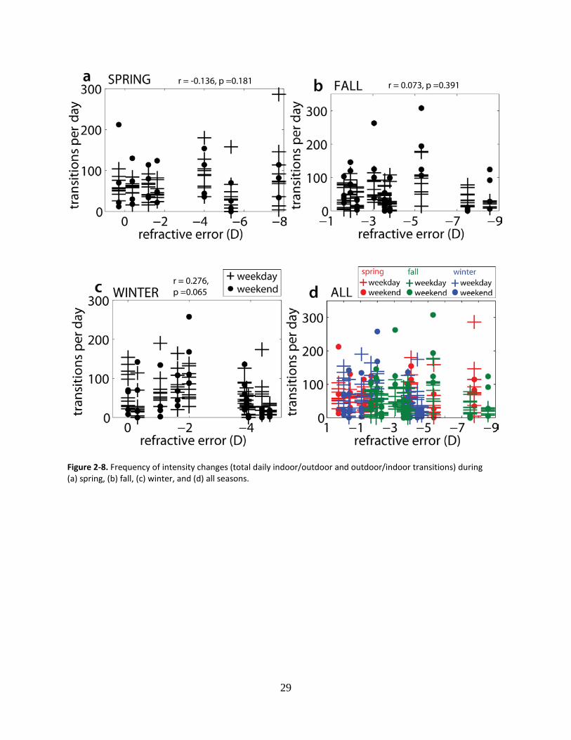
29
Figure 2-8. Frequency of intensity changes (total daily indoor/outdoor and outdoor/indoor transitions) during (a) spring, (b) fall, (c) winter, and (d) all seasons.
r = -0.136, p =0.181 r = 0.073, p =0.391
r = 0.276, p =0.065

30
2.3.3 Cumulative Light Exposure To facilitate comparison with future studies of different durations, and in different seasons, we created a weather-normalized measure of light exposure. Cumulative lux-hours were calculated by taking the integral for each subject’s intensity data over each study period. The same was done for the solar radiation data, for the same study periods (see Table 2-3). The ratio of these values is solar-normalized cumulative light exposure, as shown in Figure 2-9. Most subjects were exposed to less than 10% of the available sunlight over each 14-day study period. The mean for spring was 5.57%, for fall 6.01%, and for winter 7.31%. Seasonal or refractive error-dependent trends in light exposure were not apparent. Since solar-normalized cumulative light exposure was fairly constant, this indicates that subjects were exposed to daylight equally across seasons. Even though the absolute amount of light logged by the sensors differed across seasons, when normalized by the cumulative solar radiation, these data showed little variation. Table 2-3. Mean daylight hours (sunrise to sunset) and integrated solar radiation (lux-hours) during each study period.
spring fall winter Mean hours of daylight 12.68 10.15 11.26 Total solar lux-hours 1.30 × 10⁷ 4.70 × 10⁶ 6.94 × 10⁶

31
Figure 2-9. The percentage of available light that subjects were exposed to during (a) spring, (b) fall, (c) winter, and (d) all seasons.
r = 0.014, p =0.892 r = -0.052, p =0.545
r = 0.006, p =0.944

32
2.3.4 Indoor and Outdoor Exposure
One of the main purposes of this study was to investigate the accuracy of self-report data gathered via questionnaires, by comparing it to data obtained from the light sensors. During the pre-study screening, subjects estimated the number of daily hours spent indoors and outdoors. These estimates were evaluated against the durations of indoor and outdoor exposure reported by the light sensor, as explained in Section 2.2.5. The general trend across seasons was an overestimation of both indoor and outdoor time. The indoor and outdoor estimates, along with the means and ranges of sensor data, are shown by season in Figures 2-10 (spring), 2-11 (fall), and 2-12 (winter). Subjects’ indoor and outdoor time estimates summed to 16.14 hours for spring, 16.55 hours for fall, and 14.50 hours for winter, indicating that they were aiming for roughly 16 hours of daily awake time per day, and were accounting for approximately eight hours of sleep.
Significant differences between estimates and sensor data were found for both indoor (p<0.003) and outdoor (p<0.01) time for spring, for both indoor (p<0.01) and outdoor (p<0.01) time for fall, and for outdoor time for winter (p<0.01) (all two-tailed t-tests). There were no significant correlations between refractive error and actual time spent indoors or outdoors from the sensor data.
The means of the estimates and sensor data, grouped by season, are illustrated in Figure 2-13, and shown in tabular form in Table 2-4. There were no significant seasonal differences in indoor and outdoor estimates, or in time spent indoors or outdoors as indicated by the sensor.

33
Figure 2-10. Estimates of time spent (a) indoors and (b) outdoors, plotted with sensor data means, in daily hours for the spring study period. Subjects’ estimates (∎), the range of time spent indoors/outdoors over the experimental period from the sensor data (error bars), and sensor data means (o). (c) Combined indoor and outdoor data.

34
Figure 2-11. Estimates of time spent (a) indoors and (b) outdoors, plotted with sensor data means, in daily hours for the fall study period. Subjects’ estimates (∎), the range of time spent indoors/outdoors over the experimental period from the sensor data (error bars), and sensor data means (o). (c) Combined indoor and outdoor data.

35
Figure 2-12. Estimates of time spent (a) indoors and (b) outdoors, plotted with sensor data means, in daily hours for the winter study period. Subjects’ estimates (∎), the range of time spent indoors/outdoors over the experimental period from the sensor data (error bars), and sensor data means (o). (c) Combined indoor and outdoor data.

36
Figure 2-13. Indoor and outdoor estimates and sensor data means (hours per day) from all three seasons. Error bars are standard deviations.
Table 2-4. Mean daily hours spent indoors and outdoors from both estimate and sensor data, for all three seasons. The estimate/sensor data pairs that were found to be significantly different are highlighted in red dashed lines.
spring fall winter Outdoor estimate, mean hours 4.00 3.35 4.15 Outdoor sensor, mean hours 1.57 1.27 1.23 Indoor estimate, mean hours 12.14 13.20 10.35 Indoor sensor, mean hours 11.12 8.88 10.03
37
2.4 Discussion Few studies have been undertaken to specifically address light exposure in human myopia. The most widely cited study in the field (Rose et al., 2008) found increased outdoor exposure to be associated with lower odds of myopia. This study collected data from questionnaires administered to parents, and concluded that the protective outdoor effect was due to increased light levels, though no direct measures of light were taken. It is unclear over what period of time respondents were asked to sum and estimate visual activities, though Rose et al.’s study did distinguish between weekday and weekend activities.
Backhouse et al. (2011) surveyed the light exposure patterns in children during one season (winter) using the same light sensor employed in this dissertation (see Section 2.2.4). Similar to the results reported here, Backhouse et al. found that their subjects spent little time outside and were exposed to only 5.72% of the available sunlight over the study (cf. 7.31% during winter, and 6.30% across all seasons in the young adult sample of the present study). The subjects of Backhouse et al. received a large proportion of their light exposure while outdoors. The same was true in this study, where an average of 82% of light exposure came from time outdoors. Backhouse et al. reported a poor correlation between time spent outdoors and cumulative light from outdoor exposure. Essentially some subjects spent more time outdoors but were not exposed to bright light, and others received a large bright dose with a short outdoor duration. This does not appear to be the case in the present study, where time outdoors and cumulative light from outdoors were strongly correlated (r=0.74). Crucially, Backhouse et al. found no refractive error correlations with cumulative light exposure, or with rate of change in light levels, a result that is mirrored in the present study.
Backhouse et al. (2011) did not administer concurrent questionnaires in their study, so no comparisons between self-report and light sensor measurements were possible. This type of comparison was undertaken by Dharani et al. (2012), who found poor to fair agreement between (parentally completed) visual activity diaries and light sensor measurements over a one-week period. Moreover, no significant differences in time spent outdoors were found between myopic and non-myopic children. The authors concede that chronicling of activities through diaries (especially second-hand by parents) can lead to underreporting and errors. The flaws inherent to a questionnaire or diary method emphasize the importance of using objective measures, such as a light sensor. While the present study did not use prospective visual activity diaries, the questionnaire method employed is very similar to the gold standard used in other major studies assessing outdoor effects in myopia, including Rose et al. (2008).
There are a few crucial methodological differences between the study of Dharani et al. (2012) and the present study. First, light sensor measurements were only made every five minutes over a period of one week in the study of Dharani et al. Large changes can occur in activity level, location, and light exposure over five minutes, especially in the active life of a child. As mention in Section 2.2.4, an irregular signal such as ambient light exposure should be sampled as frequently as possible for faithful reconstruction. The sampling interval used here was 10 seconds. The difference in choice of sampling interval also highlights a potential bias toward compatibility of light data with questionnaire-based data, which is gathered on a coarse time scale (i.e. subjects are asked to divide their daily activities on an hourly basis). To prevent loss of potentially informative data such as infrequent high intensity events, a high sampling frequency should be used. To illustrate the effects of changing sampling frequency, we re-analyzed some of our light dimensions. Representative data are shown for three subjects in

38
Figure 2-14. Coarser sampling rates were compared to the original data (0.1 Hz sampling) to demonstrate a loss of information. In general, sampling less frequently than every 120 seconds (0.008 Hz) caused a deviation of ± 5% or greater from the original measurement. Hours spent in bright sunlight (Figure 2-14a), cumulative outdoor (b) and total cumulative exposure (c) all show large variation from the ±5% error bounds, especially at sampling intervals of 600, 1800, and 3600 seconds. In contrast, measurements taken at 10 to 120-second intervals are relatively stable. A sampling interval of two minutes or less thus appears preferable for faithfully capturing the ambient light exposure signal.
The second difference between the studies concerns the orientation of the light sensor. In the present study the light sensor was purposely mounted on a pedestal that aimed it skyward (see Section 2.2.4), as this is the manufacturer’s recommended installation for stationary deployment e.g. in agricultural settings. Dharani et al. (2012) had their subjects wear the sensor pinned to their shirts so that it faced outward. The cosine-dependence of the sensor’s sensitivity may cause insufficient readings in this case (and there exists the possibility that the sensor was pinned to the shirt so that it faced the body, receiving little light; with the sensor attached to the pedestal armband, this would not have occurred). In the present study, the pedestal was worn on the upper
Figure 2-14. Effect of changing sampling interval on (a) hours spent in bright sunlight, (b) cumulative outdoor exposure, and (c) total cumulative light exposure. Dashed lines indicate ± 5% of the original measurement at the 10-second sampling interval.
39
arm so the sensor would be as close as possible to the eye without undue interference to the subject. We tested the sensor’s responses in two different orientations: mounted horizontally, with the sensor facing skyward, and mounted vertically, with the sensor facing outward. Over a one-hour period of simultaneous collection outdoors in sunlight (0.1 Hz sampling), we found the vertical orientation mean was 90% lower (16,179 ± 2,512 lux) than the horizontal orientation mean (164,141 ± 10,036 lux). Additionally, sampling indoors, where the light source is non-directional (diffuse), the vertical orientation mean was again lower, by 52% (143 ± 36 lux vs. 298 ± 77 lux). The horizontal orientation has a higher response function and better resolution for low, mid, and high light levels.
The study outlined in this Chapter combines the strengths of these two previous studies, by performing both a light sensor/questionnaire comparison, and by extending the study period to three seasons (a total of 378 subject-days, vs. 252 in the study of Backhouse et al. (2011) conducted during a single season, and 819 subject-days collected over only one week by Dharani et al. (2012)). The light sensor/questionnaire comparison is vital to determining the validity of previous questionnaire-based studies that have claimed light effects in myopia progression.
The results of the present study show consistent overestimation of both time indoors and time outdoors. Dharani et al. (2012) found underestimation of time outdoors, i.e diary reports contained fewer outside hours than the light sensor reported. As the activity diaries were kept by parents, it is unsurprising that these reports and the light sensor data were significantly different for weekdays but not weekends. The mean outdoor times reported by Dharani et al. were all below two hours per day, consistent with what was found here (see Figure 2-13 and Table 2-4).
Significant differences between subjects’ indoor and outdoor estimates and durations reported by the sensor were found in all but one instance (indoor time during the winter study period). Given the comparison methods described in Section 2.2.5, however, these differences are not surprising. Subjects’ indoor and outdoor estimates are based on their individual awake windows which, being unknown to the investigator, must be roughly approximated by daylight hours. Dharani et al. (2012) also used a restricted time window in their comparison, and reported poor agreement between the two measures. The mismatch between daylight hours and subjects' daily windows results in a loss of indoor hours. The fact that the subjects' indoor estimates are greater than durations reported by the sensor is consistent with our sunset-sunrise criterion, which excluded subjects’ awake evening hours. We therefore cannot conclude that the significant differences between indoor estimates and indoor sensor durations are meaningful. Outdoor estimates, however, truly were inaccurate, because there is never daylight outside the fixed sunrise-sunset window.
This poor agreement between estimates and sensor data suggests questionnaires are an unreliable and suboptimal method for estimating outdoor activity. The questionnaire used here is based on the standard in the field (Rose, 2008). The questionnaire limits the respondent to a single integer value (“How many hours do you spend indoors/outdoors in a day?”) that captures the “typical day” without allowing for variation. Subdividing this question into finer-grained questions (“How many hours were spent reading under a tree outside?”) in the hopes of improved accuracy creates long, tedious surveys with temporal episodes that are too short for the typical person to be aware of, much less remember, with nothing approaching the resolution of 0.1 Hz, the sampling rate of the light sensor. The suggestion that ever-more detailed questionnaires can yield data of the accuracy and resolution of a sensor or app is untenable. These indirect methods should be supplemented with small, high-capacity and high-precision sensing and monitoring devices, such as the light and weather sensors used here. The current
40
questions of interest in the myopia field necessitate the use of objective measures, if clinically meaningful recommendations are to be made, particularly with regard to sun exposure, which is contraindicated otherwise for ocular and skin health.
The purpose of deploying an objective measure of light exposure was to validate and expand on questionnaire-based studies that link increased outdoor or light exposure with reduced myopia. In this study, no refractive error differences in any of the measures analyzed – maximum intensity, average intensity, percent time spent outdoors, time spent in bright sunlight, frequency of intensity changes, or cumulative light exposure – were found. The subjects in this study were young adult emmetropes and myopes, some of whom were still progressing (see Table 2-1), and who (as students) are subject to many of the same myopigenic factors as young children developing myopia. It could be argued that all the members of this small sample lead a fairly circumscribed and similar lifestyle, being young students. Our data on time spent indoors and outdoors, however, do not appear markedly different from those of Backhouse et al. (2011), whose subjects were pre-teens in New Zealand, or Dharani et al. (2012), whose subjects were children in Singapore, and reflect a trend in the developed world toward sedentary, indoor lifestyles.
Resolution of the discrepancy between questionnaire and sensor-based data requires increased data collection. The ongoing clinical study of the myopia light treatment (Xiang et al., 2011; Morgan et al., 2012) is one necessary element; thus far the GOAL study has reported small but clinically insignificant effects of the one extra daily hour of outdoor exposure in their intervention group, concluding that “greater exposures will be required to obtain clinically significant effects” (Morgan et al., 2012). Broad statements like these can and should be tempered and refined by objectively gathered data, such as those from light sensors. “Greater exposures” are not necessarily uniformly better, in myopia or vision in general, and a more analytical approach like that employed here can enhance our understanding of what in outdoor exposure is beneficial, if anything. The addition of localized weather data provides an important calibrating factor that was absent in the other two previous light sensor studies. First, data on local solar radiation allow for seasonal comparisons to be made, and can facilitate interpretation of subjects’ indoor and outdoor behavior with respect to daylight length. Dharani et al. (2012), for example, did not have all their subjects participate during the same week, but rather over a period of two months. Without taking weather data, or having the subjects participate simultaneously as we did here, the light sensor data are not comparable across weeks. Second, local solar radiation levels can act as a guide for selecting a site and season-specific indoor/outdoor threshold. The few studies in the field have converged on the 1000 lux criterion without much justification. The present study initially used a criterion of 882 lux for differentiating between indoor and outdoor exposure. This value was selected based on local solar radiation data, measurements made with three devices outdoors on a typical day during the study period (see Figure 2-2b), and indoor measurements specific to the locale of this study (Table 2-2). The initial criterion value of 882 lux was already significantly higher than any indoor measurements we recorded (see Table 2-2). Analyses of light data in the present study with criteria of 882 and 1000 lux were not found to be significantly different, so 1000 lux was adopted for the sake of consistency with the studies of Backhouse et al. (2011) and Dharani et al. (2012). The methods outlined here can improve the accuracy of data collection for epidemiological studies of myopia. This study did not find refractive error to be correlated with light exposure, in agreement with a previous study (Backhouse et al., 2011). We found

41
significant disagreement between data gathered using the existing experimental paradigm (the visual activity questionnaire) and the novel light sensor approach. While the absence of refractive error-related effects may be due to small sample size, limited time period, and cross-sectional study design, these findings serve to emphasize two points: the analyses conducted so far only cover the tip of the iceberg in terms of potential myopia-relevant factors in light exposure, which is itself only one potential outdoor effect; and second, that questionnaires, while relevant for documenting parental myopia or ocular health, may be insufficient to cover the nuances of visual activities and behavior that are now becoming pertinent to the myopia story. The effects of outdoor exposure on myopia require closer scrutiny. Some studies are already attempting to dissociate various outdoor factors, for example physical activity (Guggenheim et al., 2012) from outdoor exposure itself. Using a battery of devices to isolate the most promising therapeutic factors – sensors or dosimeters for light, accelerometers for exercise, etc. – could lead to targeted myopia interventions. Taking “light breaks” could become a rule of thumb analogous to the 20-20-20 rule for relaxing accommodation (Vertinsky & Foster, 2005). Regular interruption of myopia-inducing visual stimuli appears to be effective in reducing myopia in animals (Napper et al., 1997; Smith et al., 2002). Intensity or wavelength effects should be investigated, and could be replicated indoors with appropriate artificial lighting. To overcome the somewhat arbitrary criterion value used here (1000 lux) to differentiate indoor from outdoor, a more sophisticated sensor, or a UV filter, could be employed. Questions of wavelength effects can be addressed with multiple narrowband, (ideally photopically-calibrated) sensors. While no seasonal effects were observed in the present study, seasonal (Fulk et al., 2002) and school-year variation (Deng et al., 2010) in myopia progression have been observed, and could conceivably be latitude-dependent (Vannas et al., 2003). The interdependent contributions of wavelength, season, and the type of correction worn to myopia progression can be ascertained with a well-designed, multiple-time point study. Determining whether myopia results from low light exposure requires longitudinal monitoring of myopia progression with questionnaires and other measures.
The work presented here is a first effort to document refractive error as a function of daily light exposure, a ubiquitous environmental factor that may have cumulative effects on myopia risk. Limited light exposure may contribute to myopia progression, but other factors – weather, occupation, personality, health, to name a few – may influence whether an individual chooses to stay indoors on any given day. Demonstrating the subtle effect of light exposure on refractive error development within such an array of factors requires approaches that go beyond questionnaires in their directness and accuracy. 2.5 Conclusion Outdoor exposure, often equated with intense outdoor light levels, has been linked to lower rates of myopia and slowed myopia progression in children and animals. Human studies have relied on questionnaires, often completed by parents of myopic children, for data on time spent indoors, outdoors, in physical activity, studying, and other visual activities. The study presented in this Chapter supplemented the questionnaire approach with objectively gathered data from light sensors, and compared the accuracy of the two approaches. Duration, intensity, and other dimensions of light exposure were also studied for refractive error-related differences. Maximum

42
intensity, cumulative light exposure, frequency of intensity change, or time spent in bright light were not correlated with refractive error. Subjects’ estimates of time spent indoors and outdoors were in poor agreement with durations reported by the sensor data. Most subjects spent less than 20% of the day outdoors, were exposed to an average of only 7.42 minutes of bright sunlight per day, and received 15% or less of the total sunlight available over the study periods. These results are in broad agreement with two previous small-scale studies that used the same sensor. This is the first multi-season study to use both the questionnaire and light sensor methods coupled with local weather data to investigate light and outdoor effects in myopia. Because of the discrepancies found between questionnaire and sensor data, caution should be exercised in interpreting questionnaire-based results that indicate protective light or outdoor effects for myopia.

43
2.6 References
American Society for Testing and Materials (2012). Air mass 1.5 terrestrial reference spectra for photovoltaic performance evaluation [Data file]. Retrieved from http://rredc.nrel.gov/solar/spectra/am1.5/
Ashby, R., Ohlendorf, A., & Schaeffel, F. (2009). The effect of ambient illuminance on the development of deprivation myopia in chicks. Investigative Ophthalmology & Visual Science, 50(11), 5348 –5354.
Ashby, R. S., & Schaeffel, F. (2010). The effect of bright light on lens compensation in chicks. Investigative Ophthalmology & Visual Science, 51(10), 5247 –5253.
Backhouse S., Ng, H., & Phillips, J. (2011) Light exposure patterns in children: A pilot study. Optometry and Vision Science, 88, 400.
Crewther, S. G., Barutchu, A., Murphy, M. J., & Crewther, D. P. (2006). Low frequency temporal modulation of light promotes a myopic shift in refractive compensation to all spectacle lenses. Experimental Eye Research, 83(2), 322–328.
Deng, L., Gwiazda, J., & Thorn, F. (2010). Children’s refractions and visual activities in the school year and summer. Optometry and Vision Science, 87(6), 406-413.
Dharani, R., Lee, C.-F., Theng, Z. X., Drury, V. B., Ngo, C., Sandar, M., Wong, T.-Y., et al. (2012). Comparison of measurements of time outdoors and light levels as risk factors for myopia in young Singapore children. Eye, 26(7), 911–918.
Dirani, M., Tong, L., Gazzard, G., Zhang, X., Chia, A., Young, T. L., Rose, K. A., et al. (2009). Outdoor activity and myopia in Singapore teenage children. British Journal of Ophthalmology, 93(8), 997 –1000.
Dwyer, T., Blizzard, L., Gies, P., Ashbolt, R., & Roy, C. (1996). Assessment of habitual sun exposure in adolescents via questionnaire—a comparison with objective measurement using polysulphone badges. Melanoma Research, 6(3), 231–239.
Fulk, G.W., Cyert, L.A., & Parker, D.A. (2002). Seasonal variation in myopia progression and ocular elongation. Optometry and Vision Science, 79(1), 46–51.
Guggenheim, J. A., Northstone, K., McMahon, G., Ness, A. R., Deere, K., Mattocks, C., Pourcain, B. S., et al. (2012). Time outdoors and physical activity as predictors of incident myopia in childhood: A prospective cohort study. Investigative Ophthalmology & Visual Science, 53(6), 2856–2865.
Herlihy, E., Gies, P. H., Roy, C. R., & Jones, M. (1994). Personal dosimetry of solar UV radiation for different outdoor activities. Photochemistry and Photobiology, 60(3), 288–294.
Jones, L. A., Sinnott, L. T., Mutti, D. O., Mitchell, G. L., Moeschberger, M. L., & Zadnik, K. (2007). Parental history of myopia, sports and outdoor activities, and future myopia. Investigative Ophthalmology & Visual Science, 48(8), 3524 –3532.
Judd, D. B. (1951). Report of U.S. Secretariat Committee on colorimetry and artificial daylight. Proceedings of the Twelfth Session of the CIE, Stockholm, 1, 11. Paris: Bureau Central de la CIE. [Data retrieved from http://www.cvrl.org/lumindex.htm]
Lauber, J. K., & Oishi, T. (1987). Lid suture myopia in chicks. Investigative Ophthalmology & Visual Science, 28(11), 1851–1858.
Lawrence Berkeley National Laboratory (2012). Weather station. Retrieved from http://ehswprod.lbl.gov/weather/Home.htm
Li, T., Troilo, D., Glasser, A., & Howland, H. C. (1995). Constant light produces severe corneal flattening and hyperopia in chickens. Vision Research, 35(9), 1203–1209.

44
Morgan, I. G., Xiang, F., Rose, K. A., Chen, Q., & He, M. (2012). Two year results from the Guangzhou outdoor activity longitudinal study (GOALS). ARVO Meeting Abstracts, 53(6), 2735.
Napper, G. A., Brennan, N. A., Barrington, M., Squires, M. A., Vessey, G. A., & Vingrys, A. J. (1997). The effect of an interrupted daily period of normal visual stimulation on form deprivation myopia in chicks. Vision Research, 37(12), 1557–1564.
Nautical Almanac Office (1990). Almanac for computers. United States Naval Observatory, Washington, DC. [Retrieved from http://williams.best.vwh.net/sunrise_sunset_example.htm]
NOAA (2012). Solar calculator. Retrieved from http://www.esrl.noaa.gov/gmd/grad/solcalc/ Norton, T. T., Amedo, A. O., & Siegwart, J. T. (2006). Darkness causes myopia in visually
experienced tree shrews. Investigative Ophthalmology & Visual Science, 47(11), 4700–4707.
Palmer, J.M., & Grant, B.G. (2009) The Art of radiometry. SPIE Press, Bellingham, WA. Raviola, E., & Wiesel, T. N. (1978). Effect of dark-rearing on experimental myopia in monkeys.
Investigative Ophthalmology & Visual Science, 17(6), 485–488. Rose, K. (2008). The measurement of light intensities and effects on myopia development. Acta
Ophthalmologica, 86(s243). Rose, K. A., Morgan, I. G., Ip, J., Kifley, A., Huynh, S., Smith, W., & Mitchell, P. (2008).
Outdoor activity reduces the prevalence of myopia in children. Ophthalmology, 115(8), 1279–1285.
Smith, E. L., Hung, L.-F., & Huang, J. (2011). Effects of high ambient lighting on the development of form-deprivation myopia in infant rhesus monkeys. ARVO Meeting Abstracts, 52(6), 3922.
Smith, E. L., Hung, L.-F., & Huang, J. (2012). Protective effects of high ambient lighting on the development of form-deprivation myopia in rhesus monkeys. Investigative Ophthalmology & Visual Science, 53(1), 421 –428.
Smith, E. L., Hung, L.-F., Kee, C., & Qiao, Y. (2002). Effects of brief periods of unrestricted vision on the development of form-deprivation myopia in monkeys. Investigative Ophthalmology & Visual Science, 43(2), 291–299.
Thimijan, R.W., & Heins, R.D. (1983) Photometric, radiometric, and quantum light units of measure: a review of procedures for interconversion. HortScience, 18(6), 818–822.
Vannas, A. E., Ying, G.-S., Stone, R. A., Maguire, M. G., Jormanainen, V., & Tervo, T. (2003). Myopia and natural lighting extremes: risk factors in Finnish army conscripts. Acta Ophthalmologica Scandinavica, 81(6), 588–595.
Vertinsky, T., & Forster, B. (2005). Prevalence of eye strain among radiologists: Influence of viewing variables on symptoms. American Journal of Roentgenology, 184(2), 681–686.
Wallman, J., Turkel, J., & Trachtman, J. (1978). Extreme myopia produced by modest change in early visual experience. Science, 201(4362), 1249–1251.
Weiss, S., & Schaeffel, F. (1993). Diurnal growth rhythms in the chicken eye: Relation to myopia development and retinal dopamine levels. Journal of Comparative Physiology A: Neuroethology, Sensory, Neural, and Behavioral Physiology, 172(3), 263–270
Wiesel, T. N., & Raviola, E. (1977). Myopia and eye enlargement after neonatal lid fusion in monkeys. Nature 266, 66 – 68.

45
Xiang, F., Morgan, I. G., Smith, W., Rose, K. A., & He, M. (2011). Design, methodology and one year data from a trial for preventing myopia with increased time outdoors: the Guangzhou outdoor activity longitudinal (GOAL) study. Investigative Ophthalmology & Visual Science, 52(6), 3057.
Zadnik, K., & Mutti, D. O. (1995). How applicable are animal myopia models to human juvenile onset myopia? Vision Research, 35(9), 1283–1288.
Zhou, X., An, J., Wu, X., Lu, R., Huang, Q., Xie, R., Jiang, L., et al. (2010). Relative axial myopia induced by prolonged light exposure in C57BL/6 mice. Photochemistry and Photobiology, 86(1), 131–137.

46
Chapter 3: Measuring the Dioptric Environment Using Eye Tracking
Abstract Prolonged near working distance is a risk factor for myopia development and progression. The duration and demand of nearwork are typically estimated retrospectively through questionnaires that assess reading, computer use, and other visual behaviors. There are, however, no comprehensive methods of measuring working or fixation distance in realtime during natural tasks. Here we present a new approach to studying the dioptric environment in humans. A head-mounted eye tracking device was adapted to be fully mobile for the realtime measurement of eye movements, including convergence. This device was validated in a small sample of young adults. We conducted exploratory analyses of task-related trends in fixational behavior, fixation distance, horizontal eye movements, blinks, and saccades. We found large differences in some of these metrics between reading and walking tasks; these task-dependent differences in visual behavior may underlie the nearwork effect in myopia progression. This method opens up the possibility of new mobile experiments in natural settings, and allows for the investigation of the dioptric environment in exceptional detail.

47
3.1 Introduction The human eye has evolutionarily adapted to create a focused image on the retina when viewing distant objects. It also possesses muscular mechanisms that permit near focusing through convergence and increased accommodative refractive power, and can be aided by the postural mechanisms of the head and neck in positioning relative to visual targets. Realtime, accurate measurement of human visual behavior in and out of artificial environments has not previously been possible. This Chapter presents the first eye tracking device that is both mobile and binocular for documenting visual behavior during near and far tasks in laboratory and natural environments. This approach may be useful in identifying differences in nearwork patterns that can affect refractive error development. Myopia is considered by some to be a maladaptation to the modern, constructed environments in which humans are exposed to only near visual demands, effectively causing the eye to re-interpret the dioptric environment and elongate accordingly (Flitcroft, 2012). Urban and indoor visual environments contain many factors that have been implicated in myopia development, for example low light levels, head posture changes for near tasks, and near focusing demands that result in increased accommodation (Ip et al., 2008; Rose et al., 2008; Charman, 2011; Saw et al., 2002). Given these factors and recent experimental and theoretical emphasis on the role of peripheral hyperopic defocus in myopigenesis (Wallman & Winawer, 2004; Mutti et al., 2011), Charman (2011) proposed that large dioptric changes across the visual field in indoor environments prime emmetropization mechanisms in the periphery towards maladaptive growth. Essentially, the periphery of the retina is responsive to defocus that can guide eye growth (Smith et al., 2005), and indoor environments exacerbate hyperopic defocus in the retinal periphery. In Charman’s proposal (2011), emmetropization mechanisms make use of the eye’s inherent oblique astigmatism (Howland, 2011) to detect the symmetry of image surfaces formed on the retina. When the retina is covered by approximately symmetrical image surfaces, as occurs in viewing natural environments, myopic eye growth does not manifest. Peripheral hyperopia, such as might occur in indoor environments during near tasks, places one image surface closer to the retina, leading to ocular growth. In outdoor environments, vergences of dioptric stimuli approximate to zero across the entire visual field, so the malaptive scenario does not manifest, but in indoor environments, this consistent relationship between astigmatic image surfaces and the retina is broken. Short working distances and tilted head posture, for instance while reading or writing at a desk, exacerbate the peripheral defocus experience (Marumoto et al., 1999). The above hypothesis links indoor environments and nearwork with myopia via the altered distribution of dioptric stimuli across the visual field. Other hypotheses have implicated the prolonged contraction of the ciliary muscle during nearwork, which was thought to induce scleral stress and axial elongation. Animals with sectioned ciliary nerves, however, were found to emmetropize, thus discounting the role of accommodation (Schmid & Wildsoet, 1996; Wildsoet, 2003). Moreover, animals that cannot accommodate, such as the grey squirrel, can be made myopic with form deprivation (McBrien et al., 1993). The optical, rather than mechanistic, role of accommodation has become central again through lens studies in chick, tree shrew, and monkey (Stone et al., 2006; Norton & Siegwart, 1991; Smith et al., 2005), all of which indicate the involvement of peripheral retinal defocus in determining the direction of eye growth. Defocus produced by lags of accommodation in myopic humans is thought to play a similar role, and is the focus of optical interventions in clinical trials (Gwiazda et al., 2004; Gwiazda et al.,

48
2007; Berntsen et al., 2011). Accommodative lag can also be affected by vergence-accommodation cross-links (Norton & Gamlin, 1999; Schor, 1999). Conflicts in the neural coupling of these mechanisms, for example during stereoscopic viewing that requires accommodation to one distance but varying degrees of convergence (Hoffman et al., 2008), may adapt the cross-links towards accommodative lags that contribute to myopia. A similar conflict appears to be at play in instrument myopia (Charbonneau et al., 2010).
While the mechanism remains elusive, nearwork is nonetheless a myopia risk factor. Mutti et al. (2002) found that myopia in children was associated with more time spent studying and reading. Early onset of schooling and reading in childhood were associated with adult myopia in the study of Wong et al. (1993). Saw et al. (2002) found higher odds ratios for myopia in children who spent more time reading. Other studies, however, have not found an association between nearwork and myopia (Lu et al., 2009; Jones-Jordan et al., 2012). Cross-sectional sampling of visual activities (Lu et al., 2009) or once-yearly sampling (Jones-Jordan et al., 2012) can both be problematic data collection intervals, because they can exclude past relevant visual activities or overlook integration of visual signals over short time windows, respectively. The questionnaire approaches used in both of these studies were also subject to parental response biases as well as memory biases (Jones-Jordan et al., 2011). Frequent, direct (i.e. not by proxy) sampling of visual activities may thus be most appropriate for the investigation of the links between myopia and nearwork. More detailed study of nearwork, its duration and intensity, and its relation to other environmental factors (like the dioptric space) may require more sensitive measures than questionnaires. The current gold standard for measurement of visual behavior with respect to refractive error is the questionnaire. Nearwork is typically measured in number of books read. The diopter-hour is a nearwork measure that weights near activities like reading and computer use by dioptric demand per unit time (Saw et al., 1999; Saw et al., 2002; Mutti et al., 2002; Ip et al., 2008). There have not been any attempts to quantify nearwork in realtime both in and outside of laboratory settings. Questionnaire methods likely overlook short-term behavioral changes associated with nearwork, such as head tilt, degree of convergence, or abnormal eye movements, and often lump all outdoor activities together, as if they are equivalent in terms of light, activity, or dioptric demand. Changes in eye and head posture (Charman, 2011; Flitcroft, 2012) and convergence and extraocular muscle tension (Greene, 1980) may be important factors in myopia development. Ocular motility disturbances in high myopia (Demer & von Noorden, 1982) suggest that earlier stage progressing myopia could manifest as altered eye movements. Questionnaire methods are not sensitive to these factors, but they may be of interest in the study of myopia.
The studies discussed in this Section nearly all converge on the conclusion that the radical contraction of the dioptric environment leads to myopic ocular expansion. High density urban living (Ip et al., 2008) and intensive schooling (Saw, 2003; Jung et al., 2012) contribute to extended indoor exposure, the effects of which can be modeled or surveyed, but not easily behaviorally quantified. Fourier analyses of artificial images or photographs indicate that spatial frequency composition could be analyzed and used by the eye to guide eye growth (Hess et al., 2006; Switkes et al., 1978); this is in agreement with Charman’s hypothesis (2011). Indeed, the domain of natural scene statistics holds promise for understanding the image features that guide local retinal growth control; up to now this approach has largely been used successfully to explain attentional capture in eye movements, figure-ground segmentation, and other computer vision applications, usually with static images. As discussed further in Chapter 4, the cameras of

49
the mobile eye tracker will allow unprecedented documentation of the natural scene and extraction of depth that is directly relevant to the question of nearwork and myopia. Study of eye movement patterns and visual environment in myopia is thus warranted, but the tools for this study have been lacking. The storied history of eye movement research – covering the discovery and categorization of saccades, microsaccades, and smooth pursuit, visual search, attentional neuroscience, reading, and scene perception (Duchowski, 2002; Kowler, 2011; Tatler et al., 2010) – has largely been undertaken with stationary eye tracking devices, either video or Purkinje image-based, that restrict experiments to the laboratory environment. Stimuli in these cases are almost always artificial, and at the very least computer-displayed, requiring head stabilization and a fixed viewing distance. These methods prevent the study of potentially myopigenic visual behaviors under natural conditions. Some experimental areas do allow for eye tracking during natural behaviors. Chu et al. (2010) used the Mobile Eye (Applied Science Technologies, Bedford, MA) in their study of eye movements during presbyopic night driving. Eye movement patterns during actions in a sequence have been investigated for tasks like tea-making (Land et al., 1999) and sandwich preparation (Hayhoe, 2000; Land & Hayhoe, 2001). These studies used purpose-built equipment (camera mounted on construction helmet, etc.) that was tethered and required a specific experimental room for use; further, scene and eye video were not both recorded from the head and, at the time, had to be manually registered and analyzed. The more recent efforts of Pelz and colleagues (Pelz et al., 2000; Babcock & Pelz, 2004; Li et al., 2006) have resulted in highly portable, lightweight eye tracking solutions for the study of a variety of (mostly indoor) natural tasks. The aforementioned studies all recorded the movements of only one eye. As the discussion in this Section has emphasized, however, the ability to measure working distance is especially germane to the study of refractive error development. This necessitates the use of binocular, mobile eye tracking system. Lags of accommodation during nearwork and convergence required for reading both rely on binocular neural cross-links. Also, experimental testing of the effects of the three-dimensional scene and consequent retinal defocus on myopia (as outlined by Flitcroft, 2012) requires the type of eye tracking system that we have developed.
One recent attempt to measure working distance specifically as related to myopia did not rely on eye movement recording. Leung et al. (2011) introduced a head-mounted ultrasonic device for measuring reading distance that requires a detectable surface in front of the subject. This study found significant differences in reading distance between myopes and non-myopes, and no correlation between these objectively measured distances and subjects’ self-reported reading distances. New approaches like that of Leung et al. (2011) highlight the shortcomings of gathering data on working distance or visual behavior via questionnaire. This particular device, however, can only measure near working distance under artificial conditions, since it is laboratory-based and relies on the subject engaging with a paper or screen surface. Measuring the distance from the head to the purported surface of fixation is just a proxy for recording actual eye positions, which are necessary for determining the degree of convergence. Hartwig et al. (2011) used an eye tracker to study eye and head movements in myopes and non-myopes, but crucially did not use eye movement information to infer fixation distance. Instead, the authors chose to manually measure distance to the screen or surface with which the subject was interacting, again confining the experiments to the laboratory and near working distances. This study, which also only analyzed movements in one eye, did not find differences in working distance between refractive error groups.

50
In sum, there is an area of research spanning eye movements, natural tasks, and refractive error that has not been well investigated, but could yield insights of clinical and functional interest to myopia development. The study described in this Chapter is the first to attempt to investigate visual behavior in natural tasks, as a logical next step to the work of Hartwig et al. (2011). The following Sections describe the development of a mobile binocular eye tracker for this purpose. 3.2 Technical Specifications of a Mobile Binocular Eye Tracker A consortium of research groups made a joint grant proposal to the UC Berkeley Biology Faculty Research Fund detailing their various needs, including the ability to perform experiments in myopia, natural scene statistics, display ergonomics, and low vision. The Eyelink II was acquired from SR Research (Kanata, Ontario, Canada). Section 3.2.1 further discusses the merits of this choice compared to other alternatives. Substantial hardware and software customizations were made.1 The following lists the changes made to the Eyelink II and the final technical specifications of the mobile binocular eye tracker. • Headgear and scene cameras: The native outward-facing (scene) camera was removed, and
replaced with a stereo pair of FireWire cameras (Sony XCD-MV6) spaced 65 mm apart (see Figure 3-1). The cameras are powered by a 5V battery (Novuscell). These cameras were chosen for their low weight (37 grams). The cameras simultaneously capture black and white images (640x480 pixels) of the scene at 30 frames per second. Images are saved as uncompressed bitmaps. The stereo scene cameras are focused at infinity and tilted 10° down so their field of view coincides with that of the eye cameras. The focal length of the lenses (Kowa LM4NCL) is 3.5 mm, and the field of view is 77° horizontally and 57.7° vertically. The weight of the headgear is 420 grams. A one pound (454 g) counterweight was added to the back of the headgear for balance. The headgear (see Figure 3-1b) rested on the brow and the back of the head and was secured by headband clamps at the top and back of the head.
• Eye cameras: Two eye cameras, one per eye, were positioned 40-80 mm from the eyes. Typical gaze detection accuracy was 0.5°. Pupil position (centroid) and corneal reflection (from infrared diode) were recorded binocularly at 250 Hz. Spatial resolution with this dual detection method was 0.025°. Gaze was tracked at ± 20° in the horizontal and ± 18° in the vertical direction. The focal length of the eye cameras was 2 mm.
1 Acknowledgment: Ivana Tošić, Bill Sprague, Emily Cooper, Paul Ivanov, and Tim Blanche contributed to the software and hardware development of the eye tracker.

51
Figure 3-1. (a) Native Eyelink II headgear (diagram from SR Research). (b) Modified binocular eye tracker headgear in side view showing added stereo scene cameras.
Figure 3-2. (a) Front view of modified binocular eye tracker. (b) Most of the hardware comprising the mobile binocular eye tracker (scene camera battery not pictured). • Computing hardware: The native Eyelink system runs on two separate computers, one of
which records eye movements (“host PC”) and the other which controls the display of stimuli (“display PC”). This division, which allows for each computer’s full processing power to be devoted to just one task, was retained, with the latter computer taking up the role of recording scene camera images (the “real world” acted as the display). For mobility purposes, the host PC was transferred to a compact mini-PC case with the Eyelink PCI card. This PC runs both Windows and the Eyelink software (version 2.31) in DOS and records eye movements, stores and writes data files, and receives shutter events from the scene cameras. This PC is powered by a 19V, 130 Wh lithium battery (Novuscell). The display PC is a Lenovo X220 laptop running Windows 7 and Ubuntu Linux with an Intel core i7 2.70 GHz processor. This

52
computer records scene camera images and runs the experimental code, and interfaces with the host PC via patch Ethernet cable. The components are pictured in Figure 3-2b.
Figure 3-3. (a) Subject’s head secured in chinrest during calibration. (b) Example of display setup during calibration. Subject is at 100 cm from display.
• Display: For calibration, a 55-inch television (LG 55LW6500) displays stimuli at 1920x1080
resolution. The display is pictured in Figure 3-3b. • Calibration: A custom-machined chinrest (see Figure 3-3a) is used to position the subject’s
eyes and head at the correct height and distance relative to the display. The chinrest secures the head at the temples and has six degrees of freedom for adjustment. The cyclopean eye is positioned at a height of 83.8 cm, at distances of 50, 100, and 450 cm from the display.
• Mobility: The hardware, batteries, and cables are
packed into a backpack worn by the subject. Icepacks are added to aid in cooling the computers. The full mobile deployment is pictured in Figure 3-4.
Figure 3-4. Mobile binocular eye tracker headgear and backpack worn by a subject.

53
3.2.1 Selection of Eye Tracker The selection of the eye tracker hinged on two criteria: customizability and mobility. The diversity of experiments to be conducted with the eye tracker required modifications such as the addition of stereo scene cameras, so it was essential that the chosen system be modular rather than integrated. At the time of selection, there were no fully mobile solutions available. A mobile tracking solution from SMI has since become available, but it has a low sampling rate and cannot be customized. Virtually all eye trackers on the market are intended for stationary use with computer-displayed stimuli, or allow for only limited mobility and indoor experiments with a tether. Most vendors have not considered the requirements of experimentation in natural environments or three-dimensional gaze tracking, and so their products do not support these applications. Vendors such as SMI and Tobii offer spectacle frames with eye cameras, but these are essentially black box systems that cannot be augmented with additional cameras or other components, nor can they be calibrated in three dimensions. The spectacle frames, while lightweight, can slip and be easily displaced, while the headmount secured with clamps that we used here is much more stable.
The mobile systems developed by Pelz and colleagues (e.g. Babcock & Pelz, 2004) are also monocular. A custom solution from their spin-off Positive Science was not available in the time frame necessary for these experiments. It also was not clear whether such a system would have the necessary eye position measurement accuracy. A selection of eye trackers is compared in Table 3-1. Table 3-1. Comparison of eye tracker models and specifications.
Manufacturer SR Research Positive Science SMI Tobii Model Eyelink II Yarbus/UltraFlex Eye Tracking
Glasses Glasses Eye Tracker
Eyes Binocular Monocular Binocular Monocular Mobile? Yes (modified) Yes Yes Partial; requires
static infrared markers
Average accuracy
0.5° 1.0° 0.5° unknown
Scene cameras 2 (modified) 1 1 1 Eye sampling rate
250 Hz 30 Hz 30 Hz 30 Hz
3.3 Methods In this study subjects wore the mobile binocular eye tracker while engaging in two tasks, reading and walking. Three-dimensional gaze locations were calculated, and patterns in gaze behavior, including fixation distance, were analyzed.

54
3.3.1 Subjects and Ocular Measurements Four young adult university students participated in the eye tracking study. The mean age of the subjects was 22.25 ± 3.94 years. Right eye (OD) spherical equivalent refractive errors (SER = sphere + 0.5 × cylinder) were: +0.62 D, -0.55 D, -3.69 D, and -4.63 D. The first two subjects are approximately emmetropic, and the last two myopic. The corresponding OD axial lengths were 24.4 mm, 24.75 mm, 25.25 mm, and 26.52 mm. Both myopes were progressing. Two of the subjects were female. Subjects completed a visual screening prior to participating in the experiment. The screening included the following measurements:
• visual acuity (computerized Snellen letter display for distance (M&S Technologies, Skokie, IL) and Bailey-Lovie near card for near)
• accommodative amplitude (push up method with a 20/40 target) • accommodative facility (using ±1.50 D flippers with a 20/40 target at 40 cm) • horizontal phorias (Von Graff prism dissociation method) • eye dominance (Miles test) • nearpoint convergence (convergence nearpoint test) • fixation disparity (Saladin nearpoint balance card) • stereoacuity (Randot stereotest) • non-cycloplegic autorefraction (average of five readings; Grand Seiko WR-5100K) • axial length (average of five readings; Zeiss IOL Master)
Refractive errors are reported as right eye (OD) spherical equivalent refraction in diopters (SER = sphere + 0.5 × cylinder) throughout. No subject had anisometropia greater than 0.50 D. All subjects had normal corrected visual acuity (20/20) and age-appropriate accommodative amplitudes and facilities, and no ocular health or binocular vision anomalies. Subjects wore their habitual contact lens corrections during the study; spectacle wearers were excluded due to reflections interfering with the eye tracker.
All subjects gave their informed consent to participate. This study was approved by the Committee for Protection of Human Subjects at the University of California, Berkeley and followed the tenets of the Declaration of Helsinki. 3.3.2 Calibration Procedure For eye tracking, each subject visit started with a calibration in the research laboratory. The experimental program was first initiated on the display PC. The scene camera luminance thresholds were calibrated and matched, and the shutter speed was set manually and in the software based on task type (indoor or outdoor). The display PC (running the calibration procedure and experiment, host PC (running Eyelink and recording eye movements), batteries, ice packs for cooling, and cables were packed into the backpack. The chinrest was adjusted for the subject’s head and was centered in front of the display, initially at a distance of 100 cm (see Figure 3-3b). The subject then put on the backpack and eye tracker headgear (see Figure 3-4) with the assistance of the investigator. Eye camera positions were adjusted to the subject’s face

55
to assure pupil and corneal reflection tracking. The subject then re-seated themselves at the chinrest, wearing the backpack and eye tracker. The experimental procedure started with a nine-point calibration and validation (standard Eyelink procedure) that gauged the accuracy of the tracking. With head fixed in the chinrest, subjects were asked to fixate the stimuli on the display in succession. This was followed by the presentation of 25 targets that increased radially in eccentricity (see display in Figure 3-3b). The display was taken through a ramped set of luminance steps (seven brightness levels) to correct for pupil size artifacts (Ivanov & Blanche, in press); room lights were off, and fixation targets were presented during this step. Finally, a checkerboard stimulus was flashed to help in the registration of the stereo scene cameras’ images. This process (minus the calibration and validation) was replicated at a distance of 50 cm from the display, and at 450 cm, with screen stimuli appropriately scaled for distance. This whole procedure took approximately 30 minutes. After the final stimuli at the 450 cm distance, the subject was untethered from the display and keyboard and was ready to start the experimental task. Head position and direction were not measured during the tasks. Post-task, the experimental calibration, radial targets, and checkerboard were repeated at 100 cm. 3.3.3 Tasks Experimental tasks were chosen to cover the dioptric environment across near, mid, and far distances. The American Time Use Survey (Bureau of Labor Statistics, 2011) lists the most common daily tasks of adults. These tasks were analyzed based on frequency and grouped as near, mid, or far tasks. Out of the top 29 activities, reading and walking were selected as activities that are widespread, easy to implement, and that cover the dioptric environment, both indoors and outdoors.
The reading task consisted of reading the first chapter of Jane Austen’s Sense and Sensibility (a common choice in reading tasks, as it contains many of the most common words in English; Chung et al., 1998). The reading material was printed on paper in 12 point font and held in the air by the subject at their normal reading distance. The reading task was two minutes in duration (the same duration used by Hartwig et al. (2011) in their similar task) and took place in the research laboratory. Subjects were seated on a chair without arm rests during the task. Subjects were instructed to focus on reading at their own pace and not to speak during the task. It is evident from the data, however, that subjects did take glances up and away from the reading material during the task.
For the walking task, each subject walked an identical route outdoors around the vicinity of Evans Hall that featured stairs, ramps, and sidewalks, as well as ample opportunities for infinite gaze and naturalistic navigation. Subjects were not instructed to look at any specific targets but were left to their natural gaze. All walks took place during daylight hours. Subjects were asked not to speak during the task. Subjects were accompanied in this task by the investigator, and typically took three to eight minutes for the walk (mean=5.27 min). Differences in time were due to individual subjects’ walking speeds and other foot traffic along the route.

56
3.3.4 Analyses Experiments collected both eye tracking data and scene images, examples of which are shown in Figures 3-5 and 3-6, respectively. The scene images were used to confirm task start and end times, but were not analyzed otherwise for visual field content. Potential future uses of the stored scene images are discussed in Section 4.4. Native Eyelink EDF files were converted to ASCII format. Custom MATLAB code parsed these raw files into saccades, blink, fixations, stimulus presentations, camera shutter events, and timing information. In some cases three-dimensional gaze location and fixation distance could not be calculated due to loss of pupil or corneal reflection tracking in one or both eyes during the tasks. This occurred for one myopic subject in the walking task, and for the other myopic subject in the reading task. While four subjects participated in this study, within each task we only have complete data for three subjects. Additionally a repeat data set from one emmetropic subject was obtained for the walking task under identical conditions on the same day. The radial target locations (presented during the calibration) were calculated to obtain non-primary gaze angles and for drift correction. The raw eye coordinates were transformed from a display-referenced coordinate system (in X-Y space) to head-referenced physical metric coordinates. The origin of the coordinate system was at the subject’s cyclopean eye. The intersections of the lines of sight of the two eyes were found by fitting to the epipolar plane. Vergence, azimuth, 3D fixation points, and other information were then calculated.
Figure 3-5. Examples of (a) near and (b) far gazes captured by the eye cameras (top row). In (a) the subject is focusing on their finger held in front of the nose. In (b) the subject is looking at a wall approximately 5 m away. The pupils are false-colored blue.
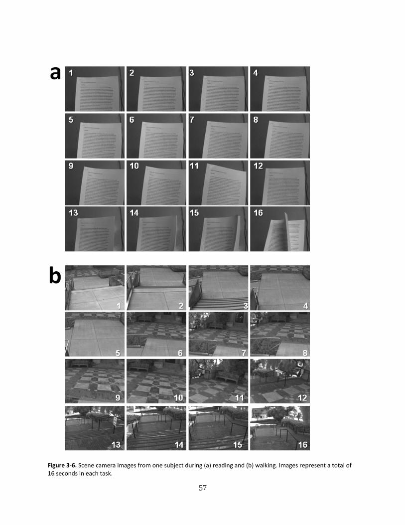
57
Figure 3-6. Scene camera images from one subject during (a) reading and (b) walking. Images represent a total of 16 seconds in each task.

58
For this study, near gaze was defined as a fixation distance in the Z dimension of <50 cm. Mid gaze was 50-400 cm, and far gaze was > 400 cm. This grouping is based on that of Rose et al. (2008), whose definition of near working distance included tasks performed at < 50 cm; their scheme also included television watching and computer use as midworking tasks, and outdoor activities (analogous to “far”), but did not specify a metric boundary. We chose 400 cm as the cutoff between mid and far because it is a distance that can be experienced indoors, e.g. during television watching, and contains a range of dioptric demands (from 2 D to 0.25 D) that cover midworking tasks. Nathan et al. (1985), for example, found the average adult television viewing distance to be 337 cm. To quantify horizontal eye movements, we identified the peaks in the sawtooth pattern that is typical of reading (see Figure 3-7) for the entire duration of the task. Through the root mean square approach, we used the deviation of these peaks from the mean to calculate the average horizontal amplitude for each subject. We also applied this method to the horizontal eye movements made during walking (see Figure 3-16a). The same root mean square approach was taken to calculate the average fluctuations in the depth dimension (Z). These results are discussed in Sections 3.4.2 and 3.4.3.
Figure 3-7. An illustration of how average horizontal amplitudes were extracted. The sawtooth pattern is one subject’s data during reading. Dashed line represents the mean. All peaks, indicated by circles, were subtracted from the mean (two examples are indicated by arrows), and the root mean square approach was applied to find the average deviation from the mean.

59
3.3.5 Eye Tracking Accuracy In this Section we attempt to quantify the performance limitations of the eye tracker. Pupil and corneal reflection tracking has an average accuracy of 0.5°, according to the Eyelink’s manufacturer. One of the main outcome measures used here, fixation distance, is based on calculating the intersection of the eyes’ lines of sight in three-dimensional space. Error in eye position measurement means that larger fixation distances have greater uncertainty attached. The uncertainty of the measurement also depends on the subject’s interpupillary distance (IPD). Figure 3-8 shows the range of possible actual fixation distances (dashed lines) given a measured fixation distance, for a 6.3 cm IPD. In this case, a measured Z distance of 340 cm has 50% uncertainty, and a Z distance of 600 cm has 100% uncertainty. A larger IPD will have a smaller uncertainty area. The effect of 0.5° of error on the geometry of the viewing situation varies by distance, with greater effects for small vergence angles at large viewing distances. While we did calibrate the eye tracker by measuring vergence at three known distances (50, 100, 450 cm), 0.5° of error in eye position measurement necessarily creates noise, especially for the measurement of mid and far viewing distances. Calculated fixation distances beyond 600 cm should therefore be treated as infinite gaze.
Figure 3-8. Effect of 0.5° of eye position error on fixation distance measurement. Solid line is identity. Dashed lines represent range of possible actual fixation distances.

60
Before each eye tracking task, subjects performed a calibration routine by fixating on displayed targets. Post-task, we re-checked the calibration at 100 cm. Using the error between the targets and fixation locations, we performed a linear drift correction, adjusting for a gradual shift over time in the coordinate system. The difference in angular error between pre and post-task calibrations is shown in Figure 3-9. As can be seen in 3-9b, these average errors were generally 0.5° or less, with the exception of one subject’s walking task.
Figure 3-9. Angular error difference between pre and post-task calibrations for (a) all subjects and (b) during reading and walking trials for each subject. Error bars are standard deviations. E=emmetropic subject, M=myopic subject. The results of the linear drift correction are shown in Figure 3-10a and b. Measured Z distance before and after correction are presented for one subject’s reading and walking tasks. The longer duration and physical nature of the walking task probably contributed to headgear displacement and increased error. For reading, this subject’s angular error was 0.002°, and for walking, 0.24°. Owing to the viewing geometry, the low error and near distances in Figure 3-10a mean very little change is seen once the drift correction is applied, while in Figure 3-10b the effect of the drift correction is substantial.
Figure 3-10. Effect of linear drift correction on measured Z fixation distance during (a) reading and (b) walking tasks. Before=before drift correction, after=after application of drift correction (final result). Note difference in ordinate scales.
61
3.4 Results The following Sections present the results of eye position measurements during reading and walking. We begin with an overview of subjects’ visual sampling strategies, followed by results of gaze distance and other eye movement parameters. 3.4.1 The Visual Environment Calculating the three-dimensional locations of all fixations during a task allows the dioptric visual experience to be mapped. This gives an overview of the direction, distance, and frequency of fixations during reading and walking: a composite of the fixational behavior during the task duration. Examples of these maps during different reading and walking trials are presented as small multiples in Figure 3-11. Near, mid, and far distances refer to the definitions in Section 3.3.4. In reading, fixations betray the stereotypical back-and-forth pattern expected in the frontal plane (first column, rows 2-3 of Figure 3-11). Viewed from the top down (second column), the pattern of fixations reveals how the subject held the reading material: at an angle that increased the distance to the left side of the paper (row 1), or more along the midline (row 2). There is a general division between the more explosive spread of points in walking tasks and the concentrated pattern of fixations seen in reading, but this is not universal. In the third row (reading), a myopic subject fixated in a pattern that resembles fixations made during walking. Walking is marked by a greater number of far fixations that tend to radiate uniformly from the origin (the cyclopean eye). In some cases there are biases to leftward gaze (rows 4 and 7). One subject (row 7) exhibits almost exclusive downward fixations. The plots also indicate how densely each subject sampled the visual scene before them. The subject in row 5, for example, extended their gaze into the distance in both the Y and X dimensions, while others (rows 3 or 7) tended to fixate along the same radial direction at different distances. Plotting the data in this way gives initial hints about the presence of large between-subjects and task-related differences in fixation behavior, and suggests subjects adopt different visual sampling strategies.

62
Figure 3-11. Map of fixations in three dimensions during trials in the reading task (top three rows) and walking task (bottom four rows), presented as small multiples. Fixations are color-coded by distance (see legend). The three dimensions are labeled and the corresponding plane is shown by the eye diagrams. XY = viewing in the frontal plane. XZ = viewing from the top down. YZ = viewing from the side. E=emmetropic subject, M=myopic subject.

63
3.4.2 Fixation Distance One of the central hypotheses of the nearwork theory of myopia progression is that (future) myopes spend more time in nearwork than those who are not or do not become myopic. Although the durations of our tasks were fairly short, one of the central aims was to use the eye tracker to measure time spent at different fixation distances. The proportion of time spent in near, mid, and far gaze (defined in Section 3.3.4) is shown in Figure 3-12. Reading was dominated by near gaze, while the two tasks did not differ on time spent in far gaze (7.62% vs. 6.58%). Walking was dominated by mid gaze. Some subjects had more unpredictable responses, spending a negligible amount of time in near gaze during reading but a large proportion of time in near gaze during walking. This may represent wandering attention during the tasks, poor gaze control, some small amount of measurement error, an aversion to blur associated with far gaze, or a combination of these factors. For some subjects fixation distance fell just outside the near cutoff (50 cm) during reading. Of interest is that the emmetropes differed in their time spent in far gaze across tasks (0% in reading vs. 8.78% in walking), while the myopic subject spent significantly more time (22.9%) in far gaze during reading.
Figure 3-12. Average time spent at near, mid, and far gaze during reading and walking tasks, all subjects.
Near < 50 cm, mid 50-400 cm, far > 400 cm.

64
Figure 3-13. The dioptric demands created by fixations
during reading and walking tasks for all subjects. Size of cluster is proportional to frequency of fixation.
Another way to visualize the fixation distance is shown in Figure 3-13, which plots the dioptric demands created by subjects’ fixations in each task. The size of each cluster is proportional to the frequency of fixation at that reciprocal distance. The bulk of the fixations created dioptric demands of 1.5-3 D during reading, and 0-1.5 D during walking. As expected, the dioptric space while walking is dominated by fixations beyond arm’s length (~ 0.67 m, or 1.5 D) and also has a larger spread. Counterintuitive far gazes during reading and near gazes during walking were largely from the myopic subjects’ data. Median Z distance was 47.6 cm during the reading task and 115.9 cm during the walking task across all subjects. Examples of raw fixation distance data over time from one subject are shown in Figure 3-14. We used the root mean square approach to find the average deviation from the mean for the Z distance (i.e. the amplitude of the peaks in the sawtooth pattern). Variation in Z distance during reading (Figure 3-14b) should be minimal, but can be perturbed by glances away from the reading material, changes in head position relative to reading material (and its angular subtense), and measurement error. It is also likely that the reading material was moved during the task because subjects were holding it in their hands. Variation in Z during walking (Figure 3-14a) is expected given the unrestricted nature of the task and the outdoor viewing environment. The Z amplitudes are shown in the top half of Table 3-2, and plotted for each subject in Figure 3-15b. The range of fixations in Z was essentially infinite (Table 3-3).

65
Figure 3-14. (a) Example raw traces of fixation distance (Z) for one emmetropic subject during reading and walking. (b) Expansion of the reading trace.
Table 3-2. Average deviations from the mean (RMS) for Z distance and for horizontal angle of fixation. E=emmetropic subject, M=myopic subject. *Average of two trials.
reading walking Z amplitude (cm) 200.96 ± 318.74 107.18 ± 115.06 E1 6.97 36.04 E2 27.09 176.03* M1 568.83 n/a M2 n/a 40.62 Horizontal amplitude (degrees) 7.52 ± 1.36 17.00 ± 9.91 E1 6.53 11.58 E2 6.96 12.29* M1 9.08 n/a M2 n/a 31.82
Figure 3-15. Average deviation from the mean (root mean square approach) for (a) horizontal angle of fixation and (b) Z distance during reading and walking trials. E=emmetropic subject, M=myopic subject.

66
Table 3-3. Range (max-min) for Z distance and for horizontal angle of fixation.
3.4.3 Horizontal Eye Movements The raw traces of the angular extents of horizontal fixations for one subject are shown in Figure 3-16. As zero represents the midline, this subject exhibited slightly more leftward horizontal fixations during walking. This may reflect the nature of the walking route, which was a loop containing only left turns. An RMS analysis of horizontal amplitudes revealed significantly smaller amplitudes during reading compared to walking. These data are presented in the bottom half of Table 3-2, and plotted for each subject in Figure 3-15a. The total horizontal range was significantly larger and more variable in walking compared to reading (Table 3-3).
Figure 3-16. (a) Example raw traces of horizontal angle of fixations for one emmetropic subject during reading and walking. (b) Expansion of the reading trace. 3.4.4 Blinks and Saccades Different visual tasks and environments call for different visual behaviors, which may also manifest in changes in the number of eye movements and blinks. The two tasks did not differ in number of blinks, though the mean number of blinks during walking was slightly higher (Figure 3-17). There was no difference in the average number of saccades made during the two tasks. The size of saccades was not significantly different across tasks, though saccades tended to be smaller during reading and the variation in amplitude was larger during walking.
reading walking Z distance range (cm) 1176.1 ± 1709.3 (= ∞) 686.03 ± 617.29 (= ∞) Horizontal angle range (degrees) 34.32 ± 12.04 84.26 ± 49.04

67
Figure 3-17. Average blinks per minute during reading and walking for all subjects.
Figure 3-18. Average saccades per minute during reading and walking for all subjects.
Figure 3-19. Average saccade amplitude during reading and walking for all subjects.

68
3.4.5 Preliminary Refractive Error-Related Results Owing to the small sample size of this study, no conclusions can be made with regard to myopia and eye movements. Some potentially interesting differences were apparent, however, and these may be of interest for future studies. These differences are presented merely as observations, since this study’s sample size does not provide sufficient statistical power. While four subjects participated in this study, within each task we only have complete data for three subjects; two different myopic subjects participated in the reading and walking tasks, respectively. The two myopic subjects tended to sample the visual environment differently, exhibiting far gazes during reading (M1) and only mid and near gazes during walking (M2) (Figure 3-20). This may be a symptom of attentional or gaze control deviations. Also, myopic subjects did not sample the visual environment as widely, but fixated along the same radial directions at different distances. The majority of near gazes during walking came from the myopic subject’s data. Subjects appear not to sample the visual environment in ways that might be expected, e.g. there are no more far gazes outdoors during walking than indoors during reading. Whether there is an aversion to far gazes, even in natural environments, is worth investigating, because it could indicate that outdoor exposure is not sufficient for disengaging from near working distances. As noted in Figure 3-15a, horizontal amplitudes are smaller in emmetropes than in myopes, both during reading and walking. Usually larger amplitudes are linked with closer working distances, because the angular subtense of reading material (for example) increases with nearer distances. Although the data with regard to fixation distance in the myopic subject was unclear in this respect, it stands to reason that the horizontal amplitude could be used to infer working distance, especially as this relationship (larger amplitude with closer distance) has been observed before (Hartwig et al., 2011).
Figure 3-20. The dioptric demands created by fixations during reading and walking tasks as a function of refractive error. Size of cluster is proportional to frequency of fixation.
E=emmetropic subject, M=myopic subject.
E2 E1 M1 M2
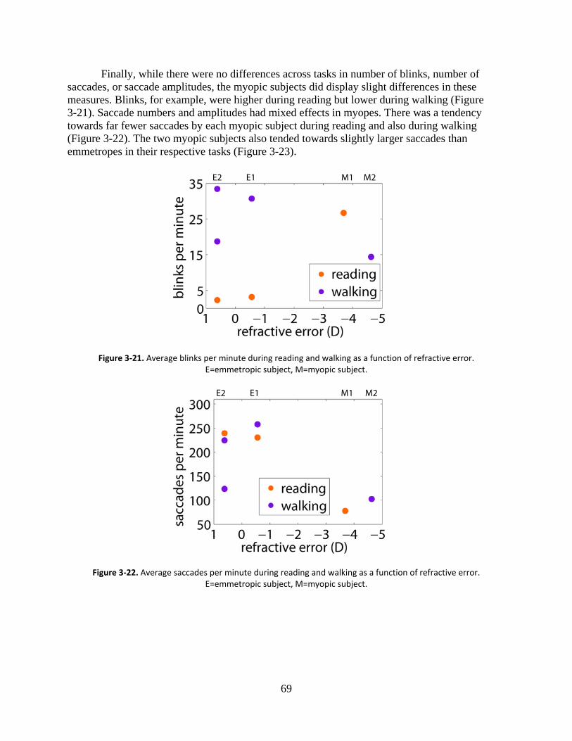
69
Finally, while there were no differences across tasks in number of blinks, number of saccades, or saccade amplitudes, the myopic subjects did display slight differences in these measures. Blinks, for example, were higher during reading but lower during walking (Figure 3-21). Saccade numbers and amplitudes had mixed effects in myopes. There was a tendency towards far fewer saccades by each myopic subject during reading and also during walking (Figure 3-22). The two myopic subjects also tended towards slightly larger saccades than emmetropes in their respective tasks (Figure 3-23).
Figure 3-21. Average blinks per minute during reading and walking as a function of refractive error. E=emmetropic subject, M=myopic subject.
Figure 3-22. Average saccades per minute during reading and walking as a function of refractive error. E=emmetropic subject, M=myopic subject.
E2 E1 M1 M2
E2 E1 M1 M2

70
Figure 3-23. Average saccade amplitude during reading and walking as a function of refractive error. Error bars are standard deviations. E=emmetropic subject, M=myopic subject.
3.5 Discussion This exploratory study of visual behavior in myopia is the first to use eye tracking during natural tasks. The results suggest there are large differences in the depth and horizontal components of eye movements, as well as in the dioptric demands and time spent at different gaze distance, during reading and walking tasks. Though collected in a small sample of subjects, these results demonstrate the feasibility of the mobile eye tracking approach for the study of visual behavior in myopia. The data yielded both anticipated and unexpected results. Subjects spent more time in near gaze during a near task (reading), for example, but did not differ in the amount of time spent in far gaze in an indoor (reading) and outdoor (walking) task. This was surprising and may be an unforeseen counterargument to the nearwork myopia hypothesis, as patients or subjects may not in fact reduce dioptrically demanding visual behaviors even when outdoors. We hypothesized that there would be a more obvious difference between time spent looking at different depths between the two tasks, and this may be based on an unfounded assumption, as natural gaze behavior in these cases has not been well studied. In their study of eye movements in reading, Hartwig et al. (2011) found larger horizontal amplitudes in myopes compared with non-myopes. These larger amplitudes were a necessary consequence of closer reading distances in myopes. Here we found that a myopic subject also had larger horizontal amplitudes while reading, though this was at odds with the fixational distances measured, which were often not in the near (< 50 cm) range. Emmetropes tended to have smaller horizontal amplitudes, and amplitudes in general were larger during walking compared with reading. In the present study, the reading distances (Z) adopted by emmetropes were smaller than those of the myopic subject. In a similar study, Hartwig et al. (2011) found a trend toward nearer reading distances in myopes compared to emmetropes, but this difference was not statistically significant. The absence of expected smaller Z distances in the present study appears to be due to the inconsistent gaze behavior of the lone myopic subject. Here we are not drawing strong
E2 E1 M1 M2

71
conclusions about these behaviors as they relate to refractive error, but are merely cataloging observations that may hint at myopia-related differences that await discovery with larger sample sizes.
This highlights a challenge in the present method, namely obtaining subjects. Finding subjects with varied refractive errors who also meet the stringent binocular vision criteria (zero or very low fixation disparity, stereoacuity < 40 arcsec, with a stable nearpoint convergence ≤ 5 cm) that enable satisfactory tracking of eye movements was not entirely straightforward. The subject pool was perhaps also restricted by the somewhat unorthodox demands of wearing headgear and a backpack while walking around in public. Fortunately, improvements in eye tracking precision and reduced hardware size and weight should mitigate these concerns in the future. This should also allow the further investigation of some potentially interesting anomalous observations, such as reduced far gaze during walking. In our measurement of fixation distance, we relied on binocular eye gaze coordinates from which three-dimensional fixation locations were calculated. This is in contrast to the manual measurement of working distance with a ruler by Hartwig et al. (2011). In that study, standard deviations were between 12 and 25% of the mean working distance measured. During reading, the standard deviations of our calculated Z values ranged from 17-52% of the mean Z, and from 29-154% of the mean Z during walking. During reading (near tasks), at least, the eye tracking method is approaching a range of precision comparable to that of manual measurement, without the intervention or bias of an investigator. Walking is an uncontrolled task for which we have no comparative accuracy information from the literature, and we expect that task to have a larger standard deviation by default. In horizontal amplitude of eye movements, the standard deviations reported by Hartwig et al. (2011) range from 18-29% of the mean amplitude. In our reading task, the standard deviations were 18% of the mean horizontal amplitude; the comparable number for walking was 58%. Using these percentages as a measure of error, our approach appears to offer commensurate precision in measuring fixation distance and horizontal eye movements. Hartwig et al. (2011) found mean book reading distance to be 37.6 cm in myopes and 41.4 cm in non-myopes; in our reading task, the mean reading distance (Z) was 46.0 cm, so again our method produces results that are in the appropriate range. No large differences were expected in blinks and saccades between the two tasks (e.g. Andrews and Coppola (1999) found very similar saccade sizes of 5-6° during reading, scene inspection, and visual search.) A normal blink rate reported in the literature is 12.55 blinks per minute (Carney & Hill, 1982); we observed 10.7 blinks/min during walking and 23.9 blinks/min during reading. The literature indicates that reading and cognitively demanding tasks in general are accompanied by fewer blinks (Bentivoglio et al., 1997; Fairclough et al., 2005). We found emmetropes had a low blink rate during reading, while the myopic subject had a higher blink rate (see Figure 3-21), but blink rate in general did not differ across tasks. This may be indicative of similar levels of attentional allocation and visual demand in the two tasks. Reading by definition requires saccades, and walking is an exploratory and navigation behavior, so naturally those tasks did not differ in number of saccades. Saccades were slightly larger during walking compared to reading, but generally did not exceed the human preference for saccades under 15° (Bahill et al., 1975). Fixational behavior varied a great deal between subjects. Even though all subjects read the same material in the same room, and took the same path with the investigator during the walking task, the visual environment and thus gaze behavior varied a great deal. Studying natural tasks increases variability that the investigator cannot control, so potentially interesting subtle

72
effects may be drowned in noise, or anomalous events may come to the fore. Increasing sample size, range of subject refractive errors, and standardizing tasks across dioptric demands should yield a set of conditions where strong myopia-related effects can be observed, in some of the measures discussed here, or potential new measures like scene defocus or entropy of the fixation point cloud. While we cannot draw robust conclusions regarding the contraction of the visual world in myopia, something about the visual behavior of myopes is different: the two subjects exhibited uncharacteristic fixation patterns during reading, and spent far more time in near gaze during walking, respectively. While these results may just be quirks of this data set, there may be noteworthy differences in eye movement control or gaze distance preferences that merit further investigation. Of course, as this Chapter presents a new experimental device, we must also entertain the hypothesis that these data are not as clean as those collected with a more mature instrument. Nonetheless, because this study is exploratory, we attempted a broad array of analyses. An eye tracker, at least in its current form, will not supplant questionnaires as a method of gathering data on working distance. It introduces a fair amount of artificiality, but provides the benefits of realtime and direct measurement of the risk factor of interest (nearwork), instead of a proxy like “books read.” If the ultimate goal is to find a correlation between a visual behavior (on a finer scale than the very general “nearwork”) and refractive error, more direct measures, like those performed in eye tracking, are necessary. This project encompassed the design, development, testing, and validation of a new method for studying visual behavior in natural tasks, with a view to determining whether those behaviors are related to task or refractive error. Automated measurement of fixation distance is possible with this head-mounted, mobile eye tracking device, and it also allows the simultaneous measurement of other eye movement patterns related to (near) working distance that may be important in myopia, such as horizontal movements. Mobile eye tracking is an addition to the toolkit for studying the visual environment in myopia that allows entirely new questions to be addressed. Possible instrument modifications and relevant future analyses are discussed in Chapter 4. 3.6 Conclusion A close visual environment and associated behaviors (nearwork) have a clear influence on the development of myopia. Head posture and eye movement patterns are known to change during reading and other near tasks, but “books read” remains the standard measure of nearwork in questionnaire-based studies. Here we described the development of an alternative technique for studying fixation distance and visual behaviors. Instead of retrospective surveys, we recorded eye movements in realtime using a mobile eye tracker that was worn by subjects during two natural tasks, reading and walking. We found large task-dependent differences in spread of fixations, fixation distance, and horizontal eye movements. While hints of some refractive error-related differences in eye movement patterns were discussed, no definitive conclusions were made in this respect due to a small sample size. Nonetheless, an entirely new way of approaching the questions of working distance and eye movements, with an application in the myopia field, was introduced and validated. This opens up the study of eye movements in

73
unprecedented environments, the very ones that are the daily settings for visual experiences that predispose towards eye growth.

74
3.7 References Andrews, T. J., & Coppola, D. M. (1999). Idiosyncratic characteristics of saccadic eye
movements when viewing different visual environments. Vision Research, 39(17), 2947–2953.
Babcock, J. S., & Pelz, J. B. (2004). Building a lightweight eyetracking headgear. Proceedings of the 2004 Symposium on Eye Tracking Research & Applications, 109-114.
Bahill, A. T., Adler, D., & Stark, L. (1975). Most naturally occurring human saccades have magnitudes of 15 degrees or less. Investigative Ophthalmology & Visual Science, 14(6), 468–469.
Bentivoglio, A. R., Bressman, S. B., Cassetta, E., Carretta, D., Tonali, P., & Albanese, A. (1997). Analysis of blink rate patterns in normal subjects. Movement Disorders, 12(6), 1028–1034.
Berntsen, D. A., Sinnott, L. T., Mutti, D. O., & Zadnik, K. (2011). Accommodative lag and juvenile-onset myopia progression in children wearing refractive correction. Vision Research, 51(9), 1039–1046.
Bureau of Labor Statistics (2011). American time use survey, Washington, DC. [Retrieved from http://www.bls.gov/tus/tables.htm]
Carney, L. G., & Hill, R. M. (1982). The nature of normal blinking patterns. Acta Ophthalmologica, 60(3), 427–433.
Charbonneau, M., Léger, A., & Claverie, B. (2010). Eyepiece focus and vergence/accommodation conflict: Implications for night-vision devices. Journal of the Society for Information Display, 18(5), 376–385.
Charman, W. N. (2011). Myopia, posture and the visual environment. Ophthalmic and Physiological Optics, 31(5), 494–501.
Chu, B. S., Wood, J. M., & Collins, M. J. (2010). The effect of presbyopic vision corrections on nighttime driving performance. Investigative Ophthalmology & Visual Science, 51(9), 4861–4866.
Chung, S. T. L., Mansfield, J. S., & Legge, G. E. (1998). Psychophysics of reading. XVIII. The effect of print size on reading speed in normal peripheral vision. Vision Research, 38(19), 2949–2962.
Demer, J. L., & von Noorden, G. K. (1982). High myopia as an unusual cause of restrictive motility disturbance. Survey of Ophthalmology, 33(4), 281–284.
Duchowski, A. T. (2002). A breadth-first survey of eye-tracking applications. Behavior Research Methods Instruments and Computers, 34(4), 455–470.
Fairclough, S. H., Venables, L., & Tattersall, A. (2005). The influence of task demand and learning on the psychophysiological response. International Journal of Psychophysiology, 56(2), 171–184.
Flitcroft, D. I. (2012). The complex interactions of retinal, optical and environmental factors in myopia aetiology. Progress in Retinal and Eye Research, 31(6), 622–660.
Greene, P. R. (1980). Mechanical considerations in myopia: Relative effects of accommodation, convergence, intraocular pressure, and the extraocular muscles. American Journal of Optometry and Physiological Optics, 57(12), 902–914.
Gwiazda, J., Hyman, L., Dong, L. M., Everett, D., Norton, T., Kurtz, D., Manny, R., et al. (2007). Factors associated with high myopia after 7 years of follow-up in the correction of myopia evaluation trial (COMET) cohort. Ophthalmic Epidemiology, 14(4), 230–237.

75
Gwiazda, J. E., Hyman, L., Norton, T. T., Hussein, M. E. M., Marsh-Tootle, W., Manny, R., Wang, Y., et al. (2004). accommodation and related risk factors associated with myopia progression and their interaction with treatment in COMET children. Investigative Ophthalmology & Visual Science, 45(7), 2143–2151.
Hartwig, A., Gowen, E., Charman, W. N., & Radhakrishnan, H. (2011). Working distance and eye and head movements during near work in myopes and non‐myopes. Clinical and Experimental Optometry, 94(6), 536–544.
Hayhoe, M. (2000). Vision using routines: A functional account of vision. Visual Cognition, 7(1-3), 43–64.
Hess, R. F., Schmid, K. L., Dumoulin, S. O., Field, D. J., & Brinkworth, D. R. (2006). What image properties regulate eye growth? Current Biology, 16(7), 687–691.
Hoffman, D. M., Girshick, A. R., Akeley, K., & Banks, M. S. (2008). Vergence–accommodation conflicts hinder visual performance and cause visual fatigue. Journal of Vision, 8(3):33, 1–30.
Howland, H. C. (2011). A possible role for peripheral astigmatism in the emmetropization of the eye. Optometry and Vision Science, 88 (3), 399.
Ip, J. M., Rose, K. A., Morgan, I. G., Burlutsky, G., & Mitchell, P. (2008). Myopia and the urban environment: Findings in a sample of 12-year-old Australian school children. Investigative Ophthalmology & Visual Science, 49(9), 3858–3863.
Ivanov, P., & Blanche, T. (in press). Improving gaze accuracy and predicting fixation onset with video based eye trackers. Journal of Vision. Retrieved from http://pirsquared.org/jov.pdf
Jones-Jordan, L. A., Mitchell, G. L., Cotter, S. A., Kleinstein, R. N., Manny, R. E., Mutti, D. O., & Zadnik, K. (2011). Visual activity before and after the onset of juvenile myopia. Investigative Ophthalmology & Visual Science, 52(3), 1841–1850.
Jones-Jordan, L. A., Sinnott, L. T., Cotter, S. A., Kleinstein, R. N., Manny, R. E., Mutti, D. O., Twelker, J. D., et al. (2012). Time outdoors, visual activity, and myopia progression in juvenile-onset myopes. Investigative Ophthalmology & Visual Science, 53(11), 7169-7175.
Jung, S.-K., Lee, J. H., Kakizaki, H., & Jee, D. (2012). Prevalence of myopia and its association with body stature and educational level in 19-year-old male conscripts in Seoul, South Korea. Investigative Ophthalmology & Visual Science, 53(9), 5579–5583.
Kowler, E. (2011). Eye movements: The past 25 years. Vision Research, 51(13), 1457–1483. Land, M. F., & Hayhoe, M. (2001). In what ways do eye movements contribute to everyday
activities? Vision Research, 41(25–26), 3559–3565. Land, M., Mennie, N., & Rusted, J. (1999). The roles of vision and eye movements in the control
of activities of daily living. Perception, 28(11), 1311–1328. Leung, T., Flitcroft, D. I., Wallman, J., Lee, T. H., Zheng, Y., Lam, C. S., & Kee, C. (2011). A
novel instrument for logging nearwork distance. Ophthalmic and Physiological Optics, 31(2), 137–144.
Li, D., Babcock, J., & Parkhurst, D. J. (2006). openEyes: A low-cost head-mounted eye-tracking solution. Proceedings of the 2006 Symposium on Eye Tracking Research & Applications, 95-100.
Lu, B., Congdon, N., Liu, X., Choi, K., Lam, D. S. C., Zhang, M., Zheng, M., et al. (2009). Associations between near work, outdoor activity, and myopia among adolescent students in rural China: The Xichang pediatric refractive error study report no. 2. Archives of Ophthalmology, 127(6), 769–775.

76
Marumoto, T., Sotoyama, M., Villanueva, M. B. G., Jonai, H., Yamada, H., Kanai, A., & Saito, S. (1999). Significant correlation between school myopia and postural parameters of students while studying. International Journal of Industrial Ergonomics, 23(1–2), 33–39.
McBrien, N. A., Moghaddam, H. O., New, R., & Williams, L. R. (1993). Experimental myopia in a diurnal mammal (Sciurus carolinensis) with no accommodative ability. The Journal of Physiology, 469(1), 427–441.
Mutti, D. O., Mitchell, G. L., Moeschberger, M. L., Jones, L. A., & Zadnik, K. (2002). Parental myopia, near work, school achievement, and children’s refractive error. Investigative Ophthalmology & Visual Science, 43(12), 3633 –3640.
Mutti, D. O., Sinnott, L. T., Mitchell, G. L., Jones-Jordan, L. A., Moeschberger, M. L., Cotter, S. A., Kleinstein, R. N., et al. (2011). Relative peripheral refractive error and the risk of onset and progression of myopia in children. Investigative Ophthalmology & Visual Science, 52(1), 199–205.
Nathan, J. G., Anderson, D. R., Field, D. E., & Collins, P. (1985). Television viewing at home: Distances and visual angles of children and adults. Human Factors, 27(4), 467–476.
Norton, T.T., & Gamlin, P.D.R. (1999). The near response, emmetropia and myopia. Ophthalmic and Physiological Optics, 19(2), 79–80.
Norton, T. T., & Siegwart, J. T. (1991). Local myopia produced by partial visual-field deprivation in tree shrew. Society for Neuroscience Abstracts, 17, 558.
Pelz, J. B., Canosa, R. L., Kucharczyk, D., Babcock, J. S., Silver, A., & Konno, D. (2000). Portable eyetracking: A study of natural eye movements. SPIE Proceedings 3959, Human Vision and Electronic Imaging V, 566-583.
Rose, K. A., Morgan, I. G., Ip, J., Kifley, A., Huynh, S., Smith, W., & Mitchell, P. (2008). Outdoor activity reduces the prevalence of myopia in children. Ophthalmology, 115(8), 1279–1285.
Saw, S. (2003). A synopsis of the prevalence rates and environmental risk factors for myopia. Clinical and Experimental Optometry, 86(5), 289–294.
Saw, S.-M., Chua, W.-H., Hong, C.-Y., Wu, H.-M., Chan, W.-Y., Chia, K.-S., Stone, R. A., et al. (2002). Nearwork in early-onset myopia. Investigative Ophthalmology & Visual Science, 43(2), 332–339.
Saw, S.-M., Nieto, F. J., Katz, J., Chew, S.-J. (1999). Distance, lighting, and parental beliefs: Understanding near work in epidemiologic studies of myopia. Optometry and Vision Science, 76(6), 355-362.
Schmid, K. L., & Wildsoet, C. F. (1996). Effects on the compensatory responses to positive and negative lenses of intermittent lens wear and ciliary nerve section in chicks. Vision Research, 36(7), 1023–1036.
Schor, C. (1999). The influence of interactions between accommodation and convergence on the lag of accommodation. Ophthalmic and Physiological Optics, 19(2), 134–150.
Smith, E. L., Kee, C., Ramamirtham, R., Qiao-Grider, Y., & Hung, L.-F. (2005). Peripheral vision can influence eye growth and refractive development in infant monkeys. Investigative Ophthalmology & Visual Science, 46(11), 3965–3972.
Stone, R. A., Pendrak, K., Sugimoto, R., Lin, T., Gill, A. S., Capehart, C., & Liu, J. (2006). Local patterns of image degradation differentially affect refraction and eye shape in chick. Current Eye Research, 31(1), 91–105.
77
Switkes, E., Mayer, M. J., & Sloan, J. A. (1978). Spatial frequency analysis of the visual environment: Anisotropy and the carpentered environment hypothesis. Vision Research, 18(10), 1393–1399.
Tatler, B. W., Wade, N. J., Kwan, H., Findlay, J. M., & Velichkovsky, B. M. (2010). Yarbus, eye movements, and vision. i-Perception, 1(1), 7–27.
Wallman, J., & Winawer, J. (2004). Homeostasis of eye growth and the question of myopia. Neuron, 43(4), 447–468.
Wildsoet, C. F. (2003). Neural pathways subserving negative lens-induced emmetropization in chicks – Insights from selective lesions of the optic nerve and ciliary nerve. Current Eye Research, 27(6) , 371-385.
Wong, L., Coggon, D., Cruddas, M., & Hwang, C. H. (1993). Education, reading, and familial tendency as risk factors for myopia in Hong Kong fishermen. Journal of Epidemiology and Community Health, 47(1), 50–53.

78
Chapter 4: Conclusions
Abstract The effects of environmental factors on refractive error development have traditionally been studied with questionnaires and surveys. Often administered retrospectively, questionnaires gauge coarse measures of factors like nearwork or outdoor exposure with proxies such as number of books read or hours spent in sports activities, often through surrogate (parental) response. This dissertation introduced two methods for finer-grained and realtime measurement of light exposure and (near) working distance. Environmental factors can have strong and lasting effects on the development and progression of refractive error. The potential for behavioral interventions with light or working distance first necessitates a clearer understanding of the impact of the duration and intensity of these factors on myopia. The light sensor and mobile eye tracking methods discussed here represent the types of tools appropriate for answering those questions.

79
4.1 Summary Eye growth and refractive error development are genetically guided processes that can be influenced by environmental factors. This is evident not just from experimental manipulations in animals, but also from human lifestyle changes that have been accompanied by a global increase in myopia prevalence. Increased myopia has been linked with reduced outdoor exposure (Rose et al., 2008), greater time spent in nearwork (Ip et al., 2008), and parental myopia (Mutti et al., 2002). While methods for studying the genetic bases of myopia have advanced, the study of environmental effects remains based in questionnaires, essentially parental or self-reports of past visual activities such as reading, computer use, or sports. “Books read” may not be an appropriate metric of nearwork in the modern world, where children and young people increasingly use smartphones, handheld gaming devices, and computers. The precision and temporal resolution of questionnaires in the study of myopigenic environmental factors is also limited, especially considering duration and intensity of light exposure or nearwork may be crucial to understanding myopia risk. This dissertation expanded the techniques available for the study of environmental factors in myopia. 4.2 Light Chapter 2 introduced a new method of studying light exposure as it relates to refractive error. As an objective supplement to traditional questionnaires, we had subjects wear light sensors over a two-week period. These sensors recorded ambient light intensity once every 10 seconds while subjects engaged in their normal activities. In our analyses of light exposure, none of the dimensions – including maximum intensity, cumulative exposure, frequency of intensity changes, or time spent in bright light – were correlated with refractive error. Moreover, durations of indoor and outdoor exposure, as measured by the sensors, differed significantly from subjects’ estimates, the conventional measure of light and outdoor exposure. Collecting these data during fall, winter, and spring periods showed that light exposure patterns did not vary significantly by season, at least at a temperate latitude in the northern hemisphere. We also demonstrated that an irregular signal such as ambient light exposure should be sampled as frequently as possible, at a minimum every two minutes. This finding, and the poor agreement between subjects’ estimates and sensor data, suggests that questionnaires may not be appropriate instruments for studying outdoor and light exposure, or they may be capturing different aspects of these factors. The timescale and sampling resolution of questionnaires at the very least are quite different from that of sensors, which can provide data specifically about light intensity, duration, and patterning that questionnaires cannot. Increased outdoor exposure has been linked with reduced risk of myopia (Rose et al., 2008; Jones-Jordan et al., 2011). Together with results showing that bright light protects against experimentally induced form deprivation myopia in chicks and monkeys (Ashby et al., 2009; Ashby & Schaeffel, 2010; Smith et al., 2011; Smith et al., 2012), these studies suggest that extra time outdoors can stave off myopia. Other studies (Jones-Jordan et al., 2012), however, failed to find associations between outdoor activity and myopia progression. In their study quantifying light exposure patterns, Backhouse et al. (2011) reported no correlation between refractive error and cumulative light exposure, a result that is mirrored in the present study. Because of these disagreements on light effects in human myopia, it seems that threshold, duration, or cumulative

80
effects may be at play, especially given animal evidence that temporal patterning of myopigenic stimuli can affect eye growth (Napper et al., 1997; Smith et al., 2002). Variation in activity and myopia progression during and out of the school year (Deng et al., 2010) has been found, so it is not implausible to hypothesize compounding light effects that are latitude, climate, and culture-dependent. These factors may explain the lack of agreement in results from diverse geographic locations, and necessitate methods that combine local geographic factors (weather, season) with subjects’ light exposure data. We created a measure of solar-normalized cumulative light exposure to facilitate comparison across season and locale. This measure is not only helpful in interpreting light exposure data, but can also guide the application of this and future studies into intervention strategies for light duration and intensity that are location and weather-specific. This study is the first to subdivide outdoor exposure into specific light dimensions such as intensity, duration, and frequency of intensity change. If protective myopia effects depend on reaching a certain threshold of outdoor or light exposure, for example, this type of analytic approach is necessary to determine the crucial dimension. While the results of the present study were negative with respect to refractive error, they demonstrated the insufficiency of the questionnaire approach for measuring light exposure. As shown in this study, the ambient light exposure patterns of subjects can be highly variable. The light effect may be subtle, and so its detection will require frequent sampling in a large subject pool. 4.3 Nearwork Chapter 3 outlined the development of a new mobile eye tracking device for the measurement of eye movements and the visual environment during natural tasks. These factors are of interest because it is conjectured that those at risk of developing myopia spend more time in near gaze, and that established myopes display eye movement changes such as reduced motility due to increased axial length. Further, the realtime study of potentially myopigenic environments and behaviors, both indoors and outdoors, has previously not been possible. The current standard measures of nearwork duration, number of books read or diopter-hours (Saw et al., 1999; Saw et al., 2002), rely on questionnaires or diaries, while attempts to make finer measurements of near working distance are still manual and laboratory-based (Hartwig et al., 2011). We sought to improve on these measures with a mobile device that does not use proxy measures of nearwork. Instead, with the eye tracker fixation distance is calculated directly from eye positions. There were large between-subjects differences in many of the measured eye movement parameters. We found that, as expected, subjects spent more time in near gaze during reading, but did not spend significantly more time in far gaze during walking compared with reading. The majority of fixations during walking, however, created dioptric demands of 1.5 D or less, while the demands during reading were 1.5 to 3 D. Horizontal fixation amplitudes were smaller during reading. Blinks, saccades, and saccade amplitudes were not significantly different between tasks. Comparing the precision of our horizontal amplitude and fixation distance results to those obtained in a similar eye tracking experiment that employed manual measurement (Hartwig et al., 2011), we concluded that the automated calculation used here produces results in the appropriate range. The mobile eye tracker is thus a valid and useful tool that does not rely on manual measurement or questionnaires to determine working distance. In addition, it allows for the measurement of working distance during natural tasks both in and out of the laboratory, covering the entire dioptric environment in a way that has not been available before. The mobile

81
eye tracker could be used to identify visual behavior patterns associated with refractive error development, and could corroborate and refine questionnaire-based assessments of nearwork.
4.4 Future Directions The future study of environmental factors in myopia, or any domain of vision, is likely to involve sensing devices and gadgets. For light exposure, the most important facet may be wavelength. The spectra of artificial light sources compared with sunlight may prove to be central to future light myopia treatments (instead of merely sending children outdoors for extra doses of damaging UV light, indoor sources could be appropriately tuned). Light measurement in myopia should incorporate sensors that are responsive in the UV and photopic ranges. Placement of these sensors on the body with respect to the eye should be considered. Indoor and outdoor pupillometry could help in determining actual retinal illuminance during different tasks, provided a photopically calibrated sensor was used. The integration of methods is what promises to really accelerate the study of environmental factors. Using pedometers to measure physical activity, sensors for light, accelerometers for head movement and posture, and an eye tracker together would make it possible to gain unprecedented detail about visual behavior. Some of these devices are already being packaged into many smartphones, reducing the need for multiple non-integrated gadgets. Coarse eye tracking could be performed through laptop and tablet built-in cameras; at the very least, the duration of use of electronic devices at near distances could be deduced this way. New proxy measures of time spent in nearwork, like apps running the background on a computer or smartphone, or sensors integrated into devices like Google Project Glass eyewear, will likely become popular for both self-monitoring and experiments. A more invasive but less cumbersome method would be to use contact lens sensors to track eye movements; though these devices are still very experimental, there’s no reason they couldn’t become as ubiquitous and easy-to-use as lenses for electroretinograms. Contact lenses have already been tested as platforms for tear glucose monitoring (Yao et al., 2011), and could integrate light-emitting diodes and biosensors (Lingley et al., 2011). Measuring environmental factors requires making observations about human behaviors and choices, and necessitates the absence of experimental interventions. This makes extracting large effects challenging, but not hopeless. For the study of nearwork, for instance, tasks should be imposed but subject-directed. The natural time course of visual activities doesn’t take place in a laboratory at a fixed appointment. The deployment of gadgets for these kinds of experiments kills two birds with one stone, allowing the investigator to continuously observe without being present, and generating objective data that doesn’t rely on memory or second-hand respondents. Some of the possible future tasks to implement with the eye tracker include navigation with and without optical correction (do myopes avoid far gaze?), daily tasks like preparing food or shopping, the use of different contact lenses (how do eye movements and peripheral awareness change under different optical conditions?), and visual behavior changes during use of electronic devices. An exciting future possibility for the mobile binocular eye tracker is the extraction of defocus experienced by subjects during natural tasks. Computer vision algorithms could be applied to the scene camera images (Figure 3-6) to obtain three-dimensional scenes. Provided a camera-to-eye coordinate system registration exists, the gaze location could then be overlaid on

82
this scene. Defocus for the scenes viewed by subjects could then be calculated, assuming zero defocus at fixation (=the fovea). This has interesting implications for theoretical predictions of the defocus patterns that are thought to contribute to myopia development, especially in near tasks and different head postures, as discussed in the next paragraph and by Flitcroft (2012).
Distance, defocus, and binocular disparity have a systematic relationship in the visual field, owing to the ubiquity of the ground plane in the natural environment. Crossed disparities and hyperopic defocus are associated with distances nearer and lower in the visual field relative to fixation, while uncrossed disparities and myopic defocus are present at far distance and in the upper visual field (Cooper et al., 2011; Previc, 1990). This smooth, systematic variation in defocus is altered indoors, and may contribute to the development of myopia. Documenting fixation distance as a proxy for defocus using the eye tracker could yield concrete results with respect to this prediction, but hints that this may be the case already exist. Lower field myopia in animals (Hodos & Erichsen, 1990; Zeng et al., 2013) and in humans (Seidemann et al., 2002) suggests that the eye makes use of the ground plane for guiding growth as it does for stereovision (e.g. humans have a vertical horopter that is height-adapted and tipped top back (Siderov et al., 1999)). The fact that the horopter adapts to become convex is not accidental: it reflects the distribution of mostly convex surfaces and shapes in the world (Cooper et al., 2011), but crucially mirrors the pattern of disparities created in near tasks, such as when reading from a computer screen or manipulating objects at near. The natural asymmetric distribution of disparities – uncrossed above and crossed below fixation – become symmetric during nearwork, with uncrossed disparities both above and below fixation. We suggest that an analogous situation arises for defocus during near tasks, because blur and disparity have analogous geometries, and represent complementary sides of the same depth cue (Held et al., 2012). Thus, at near, both upper and lower visual fields will be subject to myopic defocus. Indoors the eye is exposed to relatively more hyperopic defocus than in natural environments that normally contain both a sky and opportunities for infinite gaze. The stable and smooth gradient from near to mid and far gaze becomes a steep ramp, and a myopic defocus gradient in the upper periphery, a feature of a natural environment that includes a sky, instead becomes constant indoors. This “ceiling effect” has been observed and manipulated in laboratory animals (Zeng et al., 2013; Miles & Wallman, 1990). Thus, limited lower field or ground plane myopia in humans, combined with a ceiling effect, make for a donut-shaped distribution of defocus indoors: roughly equivalent myopic defocus in the upper and lower visual fields relative to fixation. Head posture could exacerbate this effect (eyes tend to diverge in upward gaze (Heuer et al., 1988)). This visual field symmetry of defocus could be relatively more important in susceptible individuals than lighting in predicting environmental and behavioral myopia risk. The defocus pattern could also interact with the spatial frequencies of books, computer screens, or other indoor visual targets (Diether & Wildsoet, 2005). Because the retinal periphery is thought to be active and important in influencing eye growth (Wallman & Winawer, 2004; Mutti et al., 2011), testing the effects of the three-dimensional environment on retinal defocus is a big step towards establishing viable optical and behavioral anti-myopia interventions (Flitcroft, 2012).
83
4.4.1 Eye Tracker Improvements Techniques and improvements currently under development will allow increased eye tracking accuracy. The incorporation of stereo scene camera depth maps, as outlined above, will provide a sanity check to the calculated Z distances. Drift error can be reduced through refined algorithms. Pupil and corneal reflection tracking can likely be improved by setting thresholds appropriate to experimental luminance environments. The most time-consuming experimental stage, calibration, will likely have to be expanded for improved accuracy. Ideally, the system will be re-calibrated multiple times while an experiment is underway, rather than just at the beginning and end. To compensate, the hardware and weight of the backpack can be downsized, as lighter and smaller components become available.

84
4.5 References Ashby, R., Ohlendorf, A., & Schaeffel, F. (2009). The effect of ambient illuminance on the
development of deprivation myopia in chicks. Investigative Ophthalmology & Visual Science, 50(11), 5348 –5354.
Ashby, R. S., & Schaeffel, F. (2010). The effect of bright light on lens compensation in chicks. Investigative Ophthalmology & Visual Science, 51(10), 5247 –5253.
Backhouse S., Ng, H., & Phillips, J. (2011) Light exposure patterns in children: A pilot study. Optometry and Vision Science, 88, 400.
Cooper, E. A., Burge, J., & Banks, M. S. (2011). The vertical horopter is not adaptable, but it may be adaptive. Journal of Vision, 11(3):20.
Deng, L., Gwiazda, J., & Thorn, F. (2010). Children’s refractions and visual activities in the school year and summer. Optometry and Vision Science, 87(6), 406-413.
Diether, S., & Wildsoet, C. F. (2005). Stimulus requirements for the decoding of myopic and hyperopic defocus under single and competing defocus conditions in the chicken. Investigative Ophthalmology & Visual Science, 46(7), 2242–2252.
Flitcroft, D. I. (2012). The complex interactions of retinal, optical and environmental factors in myopia aetiology. Progress in Retinal and Eye Research, 31(6), 622–660.
Hartwig, A., Gowen, E., Charman, W. N., & Radhakrishnan, H. (2011). Working distance and eye and head movements during near work in myopes and non‐myopes. Clinical and Experimental Optometry, 94(6), 536–544.
Held, R. T., Cooper, E. A., & Banks, M. S. (2012). Blur and disparity are complementary cues to depth. Current Biology, 22(5), 426–431.
Heuer, H., Dunkel-Abels, G., Brüwer, M., Kröger, H., Römer, T., & Wischmeier, E. (1988). The effects of sustained vertical gaze deviation on the resting state of the vergence system. Vision Research, 28(12), 1337–1344.
Hodos, W., & Erichsen, J. T. (1990). Lower-field myopia in birds: An adaptation that keeps the ground in focus. Vision Research, 30(5), 653–657.
Ip, J. M., Rose, K. A., Morgan, I. G., Burlutsky, G., & Mitchell, P. (2008). Myopia and the urban environment: Findings in a sample of 12-year-old Australian school children. Investigative Ophthalmology & Visual Science, 49(9), 3858–3863.
Jones-Jordan, L. A., Mitchell, G. L., Cotter, S. A., Kleinstein, R. N., Manny, R. E., Mutti, D. O., & Zadnik, K. (2011). Visual activity before and after the onset of juvenile myopia. Investigative Ophthalmology & Visual Science, 52(3), 1841–1850.
Jones-Jordan, L. A., Sinnott, L. T., Cotter, S. A., Kleinstein, R. N., Manny, R. E., Mutti, D. O., Twelker, J. D., et al. (2012). Time outdoors, visual activity, and myopia progression in juvenile-onset myopes. Investigative Ophthalmology & Visual Science, 53(11), 7169-7175.
Lingley, A. R., Ali, M., Liao, Y., Mirjalili, R., Klonner, M., Sopanen, M., & Parviz, B. A. (2011). A single-pixel wireless contact lens display. Journal of Micromechanics and Microengineering, 21(12), 125014.
Miles, F. A., & Wallman, J. (1990). Local ocular compensation for imposed local refractive error. Vision Research, 30(3), 339–349.
Mutti, D. O., Mitchell, G. L., Moeschberger, M. L., Jones, L. A., & Zadnik, K. (2002). Parental Myopia, near work, school achievement, and children’s refractive error. Investigative Ophthalmology & Visual Science, 43(12), 3633 –3640.
85
Mutti, D. O., Sinnott, L. T., Mitchell, G. L., Jones-Jordan, L. A., Moeschberger, M. L., Cotter, S. A., Kleinstein, R. N., et al. (2011). Relative peripheral refractive error and the risk of onset and progression of myopia in children. Investigative Ophthalmology & Visual Science, 52(1), 199–205.
Napper, G. A., Brennan, N. A., Barrington, M., Squires, M. A., Vessey, G. A., & Vingrys, A. J. (1997). The effect of an interrupted daily period of normal visual stimulation on form deprivation myopia in chicks. Vision Research, 37(12), 1557–1564.
Previc, F. H. (1990). Functional specialization in the lower and upper visual fields in humans: Its ecological origins and neurophysiological implications. Behavioral and Brain Sciences, 13(03), 519–542.
Rose, K. A., Morgan, I. G., Ip, J., Kifley, A., Huynh, S., Smith, W., & Mitchell, P. (2008). Outdoor activity reduces the prevalence of myopia in children. Ophthalmology, 115(8), 1279–1285.
Saw, S.-M., Chua, W.-H., Hong, C.-Y., Wu, H.-M., Chan, W.-Y., Chia, K.-S., Stone, R. A., et al. (2002). Nearwork in early-onset myopia. Investigative Ophthalmology & Visual Science, 43(2), 332–339.
Saw, S.-M., Nieto, F. J., Katz, J., Chew, S.-J. (1999). Distance, lighting, and parental beliefs: Understanding near work in epidemiologic studies of myopia. Optometry and Vision Science, 76(6), 355-362.
Seidemann, A., Schaeffel, F., Guirao, A., Lopez-Gil, N., & Artal, P. (2002). Peripheral refractive errors in myopic, emmetropic, and hyperopic young subjects. Journal of the Optical Society of America A, 19(12), 2363–2373.
Siderov, J., Harwerth, R. S., & Bedell, H. E. (1999). Stereopsis, cyclovergence and the backwards tilt of the vertical horopter. Vision Research, 39(7), 1347–1357.
Smith, E. L., Hung, L.-F., & Huang, J. (2011). Effects of high ambient lighting on the development of form-deprivation myopia in infant rhesus monkeys. ARVO Meeting Abstracts, 52(6), 3922.
Smith, E. L., Hung, L.-F., & Huang, J. (2012). Protective effects of high ambient lighting on the development of form-deprivation myopia in rhesus monkeys. Investigative Ophthalmology & Visual Science, 53(1), 421 –428.
Smith, E. L., Hung, L.-F., Kee, C., & Qiao, Y. (2002). Effects of brief periods of unrestricted vision on the development of form-deprivation myopia in monkeys. Investigative Ophthalmology & Visual Science, 43(2), 291–299.
Wallman, J., & Winawer, J. (2004). Homeostasis of eye growth and the question of myopia. Neuron, 43(4), 447–468.
Yao, H., Shum, A. J., Cowan, M., Lähdesmäki, I., & Parviz, B. A. (2011). A contact lens with embedded sensor for monitoring tear glucose level. Biosensors and Bioelectronics, 26(7), 3290–3296.
Zeng, G., Bowrey, H. E., Fang, J., Qi, Y., & McFadden, S. A. (2013). The development of eye shape and the origin of lower field myopia in the guinea pig eye. Vision Research, 76(0), 77–88.

86
Chapter 5: Appendix A Myopia Questionnaire
Screening Form The information being sought on this screening form will help us decide if you are a suitable subject, and provide other information that will help us interpret the measurements made on your eyes. All of the information will be coded for any publications that may arise from this work. In this way, your individual data will not be identifiable. This information will be destroyed if you do not participate in this study for whatever reason.
Instructions: Please complete the questionnaire by filling in the spaces or checking the most appropriate answer. If you do not know the answer to a question or if it does not apply to you, just leave it blank. This information is required to confirm your suitability as a subject and may also be used to interpret your measurements.
Subject Name:
Contact phone number:
#DOB (month/day/year):
Ethnicity:__________________________________________________________
Primary Occupation:
Major:_____________________________________________________________ Year in college:______________________________________________________

87
1. Are you short-sighted (myopic), and if so, how do you know this? � no � yes
2. At what age did you become myopic? _____ years
3. Is your myopia getting worse? � no � yes
3a. Has your prescription increased in the past year? � no � yes 4. Do you mainly wear
� spectacles � contact lenses � no correction 5. If you wear spectacles, what type do you wear?
� single vision � bifocal � multifocal (trifocal/progressives) 6. If you wear spectacles, how many years have you been wearing them? _____ years 7. If you wear spectacles, how often do you wear them?
� constantly � for distance only � other_____________________ Estimated total wearing time: ______ hours/day
8. If you wear spectacles, do you take them off when you read?
� no � yes 9. If you wear contact lenses, what type do you wear? (check all that are applicable)
� soft � hard � disposable
� single vision � toric � bifocal 10. If you wear contact lenses, how many years have you been wearing them? ______ years 11. If you wear contact lenses, how many hours per day do you wear them? ______ hours/day 12. When you take out your contact lenses, do you read with spectacles?
� no � yes � sometimes 13. Have you had refractive surgery (e.g. LASIK)?
� no � yes Date of surgery: Type of surgery: 14. Indicate all past and/or present eye conditions by writing a “p” next to present and “pa” next to past
conditions (leave blank if none of these apply to you).
� past allergic reaction to local anesthetics � glaucoma (high ocular pressure) � retinal detachment � eye infection � eye injury � ocular non-refractive surgery � strabismus (eye turned in or out) � amblyopia � eye movement disorder � other: ____________________________

88
15. Have you experienced any ocular injuries and/or conditions that have permanently affected your
vision? � no � yes Date of occurrence: Nature of problem:
16. List all prescription medications you are currently taking: __________________ 17. Are any members of your family also myopic?
� mother � father � siblings (how many: __)
� your children (how many:_____)
18. Have you ever practiced speed reading, or any eye exercises for improving vision, attention, or reading?
� no � yes: ____________________________________________
19. Do you have an eye or skin condition, or are you taking any medications, that make you light
sensitive (photophobic)? � no � yes: ________________________________ 20. Are you regularly exposed to any unusual electromagnetic sources, such as lasers, blacklights, or
magnets? � no � yes: ________________________________
21. Do you ever see double when you read? � no � yes: ________________________________ Additional comments: ________________________
I have read the screening form and agree to undertake the screening eye examination. I also understand that if I am not entered into this research, for any reason, the information I have given on these forms will be destroyed.
Signature: Date:

89
Questions about your daily activities
INSTRUCTIONS: PLEASE CONSIDER A TYPICAL DAY OVER THE PAST WEEK WHEN ANSWERING THESE QUESTIONS. 1. How many hours do you spend indoors in a day? (do not include time spent sleeping)
____________ hours
1a. While indoors, how many hours do you spend in a day:
Not at all <1 hr 1-2 hrs >2 hrs Longest continuous period
Reading from printed paper (books, etc.) __________hours
Using computers, phone, or other electronic devices __________hours
Playing sports or exercising __________hours
Are there any other indoor activities that you do for more than 2 hours in a typical day?
No
Yes:_________________________,
for ______________hours
Reading from printed paper (books, etc.) __________hours
Using computers, phone, or other electronic devices __________hours
Playing sports or exercising __________hours
Are there any other outdoor activities that you do for more than 2 hours in a typical day?
No
Yes:_________________________,
for ______________hours
2. How many hours do you spend outdoors in a day? ____________ hours
• While outdoors, do you wear sunglasses □ never, □ rarely, □ frequently, □ always
2a. While outdoors, how many hours do you spend in a day:
Not at all
<1 hr
1-2 hrs >2 hrs Longest continuous period