Embed Size (px)
Citation preview
LUND UNIVERSITY
PO Box 117221 00 Lund+46 46-222 00 00
Ligand-induced recruitment of Na+/H+-exchanger regulatory factor to the PDGF(platelet-derived growth factor) receptor regulates actin cytoskeleton reorganization byPDGF.
Demoulin, Jean-Baptiste; Seo, Jeong Kon; Ekman, Simon; Grapengiesser, Eva; Hellman, Ulf;Rönnstrand, Lars; Heldin, Carl-HenrikPublished in:Biochemical Journal
DOI:10.1042/BJ20030385
Published: 2003-01-01
Link to publication
Citation for published version (APA):Demoulin, J-B., Seo, J. K., Ekman, S., Grapengiesser, E., Hellman, U., Rönnstrand, L., & Heldin, C-H. (2003).Ligand-induced recruitment of Na+/H+-exchanger regulatory factor to the PDGF (platelet-derived growth factor)receptor regulates actin cytoskeleton reorganization by PDGF. Biochemical Journal, 376(2), 505-10. DOI:10.1042/BJ20030385
General rightsCopyright and moral rights for the publications made accessible in the public portal are retained by the authorsand/or other copyright owners and it is a condition of accessing publications that users recognise and abide by thelegal requirements associated with these rights.
• Users may download and print one copy of any publication from the public portal for the purpose of privatestudy or research. • You may not further distribute the material or use it for any profit-making activity or commercial gain • You may freely distribute the URL identifying the publication in the public portal

Take down policyIf you believe that this document breaches copyright please contact us providing details, and we will removeaccess to the work immediately and investigate your claim.
Download date: 06. Jul. 2018
Biochem. J. (2003) 376, 505–510 (Printed in Great Britain) 505
Ligand-induced recruitment of Na+/H+-exchanger regulatory factor to thePDGF (platelet-derived growth factor) receptor regulates actin cytoskeletonreorganization by PDGFJean-Baptiste DEMOULIN*1, Jeong Kon SEO*, Simon EKMAN*, Eva GRAPENGIESSER†, Ulf HELLMAN*, Lars RONNSTRAND*2
and Carl-Henrik HELDIN**Ludwig Institute for Cancer Research, Box 595, SE-75124 Uppsala, Sweden, and †Department of Medical Cell Biology, University of Uppsala, Biomedicum, Box 571,SE-75123 Uppsala, Sweden
Proteins interacting with the human PDGF (platelet-derivedgrowth factor) β-receptor were isolated using immobilized pep-tides derived from the receptor C-terminus as a bait. We identifiedtwo PDZ domain proteins, namely NHERF (Na+/H+ exchangerregulatory factor, also called EBP50) and NHERF2 (E3KARP,SIP-1, TKA-1), which have been shown previously to associatewith the murine PDGF receptor [Maudsley, Zamah, Rahman,Blitzer, Luttrell, Lefkowitz and Hall (2000) Mol. Cell. Biol. 20,8352–8363]. In porcine aortic endothelial cells and in fibroblasts,NHERF recruitment was induced by PDGF treatment, but thereceptor kinase activity was not required for the formation ofthe complex, suggesting that NHERF was not recruited in a phos-photyrosine-dependent manner. Instead, the interaction was abo-lished by mutation of the consensus C-terminal PDZ-interactingdomain of the receptor (Leu-1106 to Ala), or truncation of the last
75 amino acid residues of the receptor. Disruption of NHERFbinding to the receptor enhanced actin filament reorganization,but did not affect PDGF-induced mitogenicity and chemotaxis.Although NHERF was initially characterized as a factor requiredfor intracellular pH regulation by β2-adrenergic receptors, weobserved that it was not involved in pH regulation by PDGF.Collectively, these results suggest that the ligand-induced as-sociation of NHERF PDZ domain with the PDGF receptor tyro-sine kinase controls the extent of cytoskeleton reorganization in re-sponse to PDGF.
Key words: chemotaxis, cytoskeleton, ezrin-binding protein 50(EBP50), intracellular pH, Na+/H+ exchanger regulatory factor(NHERF), platelet-derived growth factor (PDGF).
INTRODUCTION
Platelet-derived growth factor (PDGF) isoforms play key rolesin embryonic development by acting on cells of mesenchymalorigin, such as mesangial cells, fibroblasts and vascular-smooth-muscle cells [1]. They induce reorganization of the cytoskeleton,increase in intracellular pH and calcium concentration, massivechanges in gene expression and, ultimately, cell migration andproliferation [2]. Four different PDGF chains assemble into fiveisoforms, namely PDGF-AA, -BB, -AB, -CC and -DD, which bindwith different affinities to two structurally similar PDGF recep-tor (PDGFR) tyrosine kinases, PDGFRα and PDGFRβ [2–4].Signalling by PDGFRs involves the following steps: (i) acti-vation of the receptor tyrosine kinase activity on ligand-induced dimerization; (ii) phosphorylation of the receptor onmultiple tyrosine residues; and (iii) docking of signalling proteinscontaining SH2 domains that interact with phosphorylated tyro-sines and surrounding amino acid residues [2,5]. Well-charac-terized signal transduction proteins that are recruited to thePDGFRs include PI3K (phosphoinositide 3-kinase), PLCγ , Src,Grb2 (growth-factor-receptor-bound protein 2) and SHP-2 (SH2domain-containing phosphatase 2), which are responsible foractivation of downstream kinases such as Akt/protein kinase B,protein kinase C and ERK (extracellular-signal-regulated kinase).
Recently, the murine PDGFR was shown to recruit NHERF(Na+/H+-exchanger regulatory factor), also called ezrin-bindingprotein 50 (EBP50), which lacks SH2 domains [6]. In cells over-
Abbreviations used: BCECF, 2′,7′-bis(carboxyethyl)-5(6)-carboxyfluorescein; CCD, charge-coupled-device; ERK, extracellular-signal-regulated kinase;HEK-293T cells, human embryonic kidney 293T cells; NHE, Na+/H+ exchanger; NHERF, NHE regulatory factor; PAE, porcine aortic endothelial (cells);PDGF, platelet-derived growth factor; PDGFR, PDGF receptor; PI3K, phosphoinositide 3-kinase; PY99, anti-phosphotyrosine; WGA, wheatgerm agglutinin.
1 To whom correspondence should be addressed (e-mail [email protected]).2 Present address: Department of Experimental Clinical Chemistry, Lund University, Malmo University Hospital, Malmo, Sweden.
expressing both the receptor and NHERF, this interaction wasconstitutive and depended on the binding of the N-terminal PDZdomain of NHERF to the C-terminus of the PDGFR. NHERFtargets proteins for apical localization in epithelial cells, mediatesintracellular pH regulation by the β2-adrenergic receptor andregulates signalling in response to parathyroid hormone [7–10].Maudsley et al. [6] suggested that NHERF binding enhanced thePDGFR tyrosine kinase activity, thereby potentiating the cellulareffects of PDGF.
In the present study, we show that in PAE (porcine aorticendothelial) cells expressing normal levels of PDGFRs, NHERFrecruitment was induced by PDGF stimulation, independent of thetyrosine phosphorylation of the receptor. A thorough investigationof the possible involvement of NHERF in the cellular activities ofPDGF revealed a role of NHERF in PDGF-induced reorganizationof the cytoskeleton.
EXPERIMENTAL
Cell culture and reagents
PAE cells were cultured in Ham’s F-12 medium supplementedwith 10 % foetal calf serum. HEK-293T (human embryonickidney 293T) cells and AG01518 fibroblasts were cultured inDulbecco’s modified Eagle’s medium with 10 % foetal calf serum.Anti-phospho-Akt (Ser-473) and anti-phospho-ERK antibodies
c© 2003 Biochemical Society
506 J.-B. Demoulin and others
were purchased from Cell Signaling (Beverly, MA, U.S.A.). Anti-phosphotyrosine (PY99), anti-PDGFRβ (958) and anti-Akt1/2were obtained from Santa Cruz Biotechnologies (Santa Cruz, CA,U.S.A.). Anti-NHERF antibodies were raised against a syn-thetic peptide corresponding to amino acid residues 342–355 ofhuman NHERF (QMDWSKKNELFSNL) and purified as des-cribed in [11], using the same peptide coupled with Sulfolinkbeads (Pierce, Rockford, IL, U.S.A.). Anti-ERK2 antiserum EEThas been described previously [11]. Peptides, synthesized onan Applied Biosystems 433A instrument using Fmoc chemistry,were purified by preparative reversed-phase chromatography.
Mutagenesis and transfection
Site-directed mutagenesis was performed on a cDNA encodingthe full-length PDGFRβ inserted into the pcDNA3 cloningvector (Invitrogen, Carlsbad, CA, U.S.A.), using QuickChange(Stratagene, La Jolla, CA, U.S.A.). The mutations were con-firmed by DNA sequencing. PAE and HEK-293 cells weretransfected using LIPOFECTAMINETM Plus as recommended bythe manufacturer (Invitrogen). PAE clones were selected for G418resistance, and tested for homogeneous PDGFRβ expression byflow cytometry using a monoclonal antibody directed against theextracellular part of the receptor. At least two independent clonesof each type were used in each experiment.
Immunoprecipitation and Western-blot analysis
Subconfluent HEK-293T or PAE cells cultured in 10 cm disheswere washed, starved overnight in a medium containing 0.1 %BSA and then stimulated with PDGF-BB (50 ng/ml) for 10 min,unless otherwise stated. Cells were washed in ice-cold PBS,lysed and processed for immunoprecipitation with anti-NHERFantibodies and Protein-A–Sepharose. One-fifth of the lysatewas incubated with WGA (wheatgerm agglutinin)–Sepharose(Amersham Biosciences). After boiling in Laemmli reducingbuffer, samples were separated by SDS/PAGE, transferred on toan Immobilon membrane, blocked with fat-free 5 % (w/v) drymilk in PBS for 1 h and probed with PY99 (Santa Cruz Bio-technologies), anti-PDGFRβ or anti-NHERF antibodies (1 µg/ml). Blots were visualized by chemiluminescence using aFUJI LAS2000 cooled CCD (charge-coupled-device) camera.AG01518 cells were processed similarly, except that lysate fromtwo 15 cm dishes was used for each immunoprecipitation. Theprotein content of each sample was measured using BCA (bi-cinchoninic acid assay; Pierce).
Intracellular pH measurements
PAE cells were allowed to attach to coverslips for a few hours inculture medium containing 10 % foetal calf serum, washed andstarved overnight in a medium supplemented with 0.1 % BSA.Cells were washed with Hanks buffer (2.5 mM Hepes, pH 7.4/140 mM NaCl/5 mM KCl/2 mM CaCl2/1 mM Na2HPO4/25 mMglucose/0.05 % BSA) and loaded with 2 µM BCECF [2′,7′-bis(carboxyethyl)-5(6)-carboxyfluorescein] acetoxymethylester(Molecular Probes, Eugene, OR, U.S.A.) and 0.1 % pluronic acidfor 30 min in Hanks buffer [12]. Coverslips with the BCECF-loaded cells were rinsed, and used as exchangeable bottomsof an open superfusion chamber thermostatically regulated at37 ◦C. Cannulas fixed to this chamber were connected to a per-istaltic pump allowing steady superfusion of a 2.5 mm Hanksbuffer layer at the rate of 1 ml/min. Intracellular pH was measuredwith a dual-wavelength microfluorometric system (Deltascan,
Photon Technology International, Princeton, NJ, U.S.A.) [13].The excitation light was alternatively directed to two monochro-mators by a chopper mirror spinning at 50 Hz. The mono-chromator outputs were connected via a bifurcated optical fibreto the epifluorescence attachment of an inverted microscope(Nikon Diaphot) equipped with a ×100 objective. The pH-dependent fluorescence of 3–5 cells was recorded at 530 nm witha photomultiplier using a 25 nm half-bandwidth interference filter.The background-subtracted signals, obtained by excitation at 440and 490 nm, were recorded at 2 Hz using FeliX software (PhotonTechnology International). PDGF-BB (20 ng/ml) was added tothe superfusion medium 5 min after stabilization of the baseline,and fluorescence was followed for another 30 min. Finally, wemonitored cell fluorescence in the presence of K+-rich calibrationbuffers (125 mM KCl/1 mM CaCl2/1 mM MgCl2/25 mM Mops,pH 6.80–7.40) containing 10 µg/ml nigericin (Sigma). This cali-bration was used to convert corrected 490 nm/440 nm excitationratios to intracellular pH values [12].
Chemotaxis, mitogenicity and actin reorganization
PAE cells were washed, starved overnight as described above,trypsinized quickly and washed in the presence of 1 % Trasylol(Bayer, Leverkussen, Germany) to inactivate trypsin. Chemotaxiswas measured using ChemoTx microplates as recommended bythe manufacturer (Neuro Probes, Gaithersburg, MD, U.S.A.).Polycarbonate filters (8 µm pore size) were coated with purifiedcollagen (100 µg/ml; Cohesion, Palo Alto, CA, U.S.A.) or humanfibronectin (50 µg/ml; Becton Dickinson, Bedford, MA, U.S.A.)on both sides. Cells were resuspended at 2 × 10⁶ cells/ml in PhenolRed-free medium, containing 0.1 % BSA, deposited in 30 µldrops on the upper side of the filter and allowed to migrate towardsPDGF-BB (1–100 ng/ml) for 4 h at 37 ◦C. The filter was removedfrom the chamber, washed with PBS to remove cells that did notmigrate and stained with a Giemsa solution. The number of cellsthat have migrated through the filter was estimated using a cooledCCD camera (Fuji) and the AIDA software. Similar results wereobtained using a modified Boyden chamber (results not shown).Mitogenicity and actin reorganization were measured as describedpreviously [14].
RESULTS
NHERF interacts with PDGFRβ in a ligand-dependent manner
Using an immobilized peptide corresponding to the last 13 aminoacid residues of the human PDGFRβ as a bait, we isolated twoproteins, NHERF (also called EBP50) and NHERF2 that wereidentified by MS and Western-blot analysis. NHERF contains twoPDZ domains that have been shown to interact with the DSFL C-terminus in murine PDGFRs in vitro and in transfected cells [6].To study further the interaction between NHERF and PDGFRβ,we performed immunoprecipitations of NHERF from PAE cellsexpressing PDGFRβ at physiological level. In unstimulatedcells, the association was weak and not consistent, but it wasdramatically enhanced by treatment with PDGF-BB (Figure 1A).It reached a maximum 20 min after ligand addition. At that timepoint, most of the PDGFRβ had been internalized and a significantpart degraded, as shown from the amount of receptor isolatedwith WGA–Sepharose, which binds glycosylated proteins, suchas mature PDGFRβ, with high affinity. Similar results wereobtained in PAE cells transfected with PDGFRα (not shown).Next, we immunoprecipitated NHERF from AG01518 normalhuman foreskin fibroblasts, which naturally express PDGFRβ.
c© 2003 Biochemical Society
Na+/H+-exchanger regulatory factor regulates actin reorganization by PDGF 507
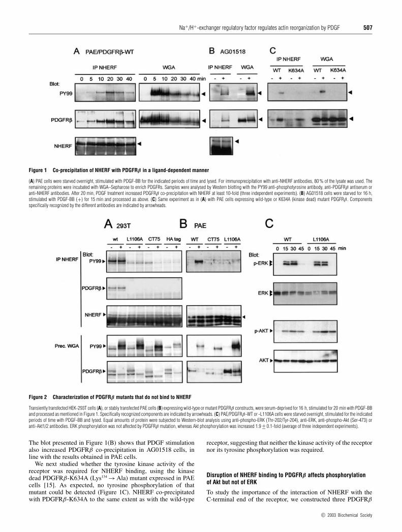
Figure 1 Co-precipitation of NHERF with PDGFRβ in a ligand-dependent manner
(A) PAE cells were starved overnight, stimulated with PDGF-BB for the indicated periods of time and lysed. For immunoprecipitation with anti-NHERF antibodies, 80 % of the lysate was used. Theremaining proteins were incubated with WGA–Sepharose to enrich PDGFRs. Samples were analysed by Western blotting with the PY99 anti-phosphotyrosine antibody, anti-PDGFRβ antiserum oranti-NHERF antibodies. After 20 min, PDGF treatment increased PDGFRβ co-precipitation with NHERF at least 10-fold (three independent experiments). (B) AG01518 cells were starved for 16 h,stimulated with PDGF-BB (+) for 15 min and processed as above. (C) Same experiment as in (A) with PAE cells expressing wild-type or K634A (kinase dead) mutant PDGFRβ . Componentsspecifically recognized by the different antibodies are indicated by arrowheads.
Figure 2 Characterization of PDGFRβ mutants that do not bind to NHERF
Transiently transfected HEK-293T cells (A), or stably transfected PAE cells (B) expressing wild-type or mutant PDGFRβ constructs, were serum-deprived for 16 h, stimulated for 20 min with PDGF-BBand processed as mentioned in Figure 1. Specifically recognized components are indicated by arrowheads. (C) PAE/PDGFRβ-WT or -L1106A cells were starved overnight, stimulated for the indicatedperiods of time with PDGF-BB and lysed. Equal amounts of protein were subjected to Western-blot analysis using anti-phospho-ERK (Thr-202/Tyr-204), anti-ERK, anti-phospho-Akt (Ser-473) oranti-Akt1/2 antibodies. ERK phosphorylation was not affected by PDGFRβ mutation, whereas Akt phosphorylation was increased 1.9 +− 0.1-fold (average of three independent experiments).
The blot presented in Figure 1(B) shows that PDGF stimulationalso increased PDGFRβ co-precipitation in AG01518 cells, inline with the results obtained in PAE cells.
We next studied whether the tyrosine kinase activity of thereceptor was required for NHERF binding, using the kinasedead PDGFRβ-K634A (Lys134 → Ala) mutant expressed in PAEcells [15]. As expected, no tyrosine phosphorylation of thatmutant could be detected (Figure 1C). NHERF co-precipitatedwith PDGFRβ-K634A to the same extent as with the wild-type
receptor, suggesting that neither the kinase activity of the receptornor its tyrosine phosphorylation was required.
Disruption of NHERF binding to PDGFRβ affects phosphorylationof Akt but not of ERK
To study the importance of the interaction of NHERF with theC-terminal end of the receptor, we constructed three PDGFRβ
c© 2003 Biochemical Society
508 J.-B. Demoulin and others
mutants. First, the last amino acid residue, a leucine, whichplays an essential role in the NHERF-binding motif, was changedto alanine (L1106A) [6,8,16]. We also deleted the last 75 aminoacids (CT75), creating a DPKP C-terminal end which is unlikelyto bind to NHERF [8]. This mutant retains all the phosphorylatedtyrosines of the receptor. Finally, a haemagglutinin tag(GYPYDVPDYA) was added to the C-terminal end after the-EDSFL sequence. These mutant receptors were first transientlytransfected in HEK-293T cells. As shown in Figure 2(A), allthree modifications abolished binding of endogenous NHERF.Similarly, neither the L1106A mutant nor the CT75 mutantinteracted with NHERF in stably transfected PAE cells (Fig-ure 2B). Taken together, these results confirmed that NHERFinteracts with the C-terminal end of the receptor.
Interestingly, we found that the association was constitutivein transfected HEK-293 cells (Figure 2A), consistent with obser-vations by Maudsley et al. [6]. In their report, they suggestedthat in cells overexpressing both PDGFRβ and NHERF, NHERFbinding to the receptor enhanced the receptor activity and ERKactivation. However, we found that mutants that were unableto bind to endogenous NHERF were phosphorylated to a verysimilar level compared with wild-type receptor, both in HEK-293 and in PAE cells (Figure 2C). In line with this result, ERKphosphorylation was not affected by the L1106A mutation. Incontrast, Akt phosphorylation was reproducibly increased approx.2-fold (Figure 2C).
NHERF is involved in PDGF-induced cytoskeletal rearrangement,but not mitogenicity or chemotaxis
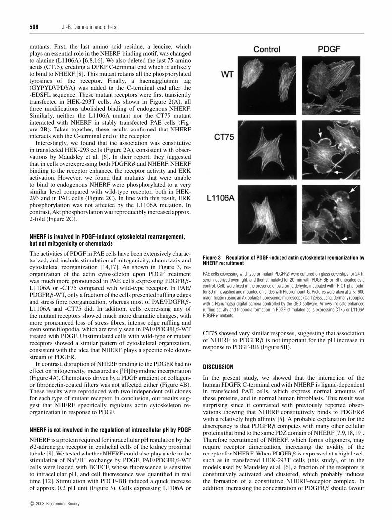
The activities of PDGF in PAE cells have been extensively charac-terized, and include stimulation of mitogenicity, chemotaxis andcytoskeletal reorganization [14,17]. As shown in Figure 3, re-organization of the actin cytoskeleton upon PDGF treatmentwas much more pronounced in PAE cells expressing PDGFRβ-L1106A or -CT75 compared with wild-type receptor. In PAE/PDGFRβ-WT, only a fraction of the cells presented ruffling edgesand stress fibre reorganization, whereas most of PAE/PDGFRβ-L1106A and -CT75 did. In addition, cells expressing any ofthe mutant receptors showed much more dramatic changes, withmore pronounced loss of stress fibres, intense edge ruffling andeven some filopodia, which are rarely seen in PAE/PDGFRβ-WTtreated with PDGF. Unstimulated cells with wild-type or mutantreceptors showed a similar pattern of cytoskeletal organization,consistent with the idea that NHERF plays a specific role down-stream of PDGFR.
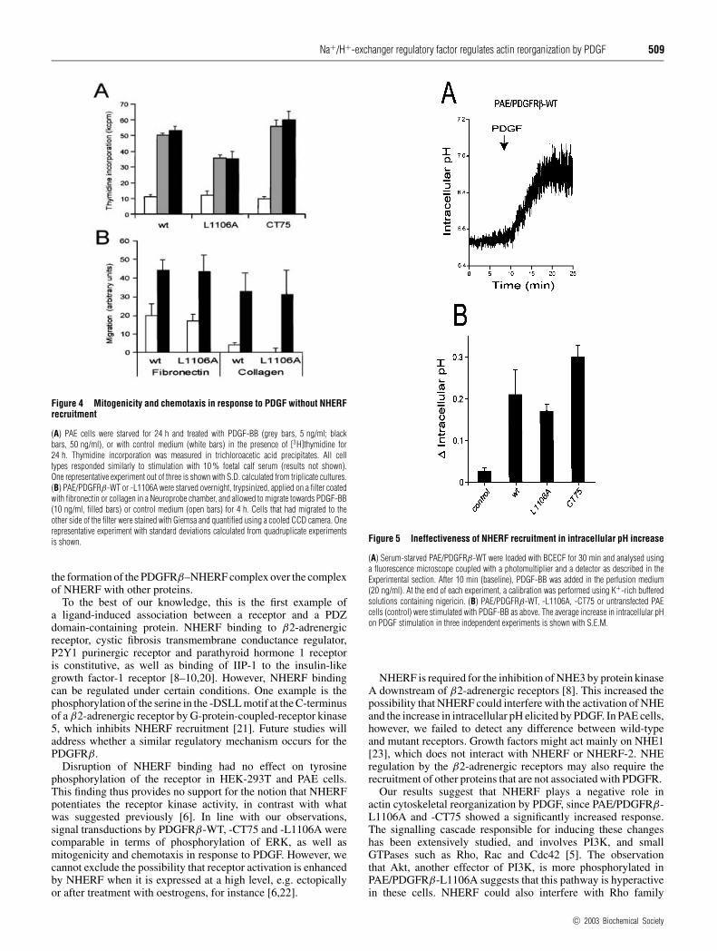
In contrast, disruption of NHERF binding to the PDGFR had noeffect on mitogenicity, measured as [3H]thymidine incorporation(Figure 4A). Chemotaxis driven by a PDGF gradient on collagen-or fibronectin-coated filters was not affected either (Figure 4B).These results were reproduced with two independent cell clonesfor each type of mutant receptor. In conclusion, our results sug-gest that NHERF specifically regulates actin cytoskeleton re-organization in response to PDGF.
NHERF is not involved in the regulation of intracellular pH by PDGF
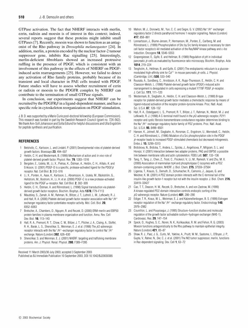
NHERF is a protein required for intracellular pH regulation by theβ2-adrenergic receptor in epithelial cells of the kidney proximaltubule [8]. We tested whether NHERF could also play a role in thestimulation of Na+/H+ exchange by PDGF. PAE/PDGFRβ-WTcells were loaded with BCECF, whose fluorescence is sensitiveto intracellular pH, and cell fluorescence was quantified in realtime [12]. Stimulation with PDGF-BB induced a quick increaseof approx. 0.2 pH unit (Figure 5). Cells expressing L1106A or
Figure 3 Regulation of PDGF-induced actin cytoskeletal reorganization byNHERF recruitment
PAE cells expressing wild-type or mutant PDGFRβ were cultured on glass coverslips for 24 h,serum-deprived overnight, and then stimulated for 20 min with PDGF-BB or left untreated as acontrol. Cells were fixed in the presence of paraformaldehyde, incubated with TRICT-phalloidinfor 30 min, washed and mounted on slides with Fluoromount-G. Pictures were taken at a × 600magnification using an Axioplan2 fluorescence microscope (Carl Zeiss, Jena, Germany) coupledwith a Hamamatsu digital camera controlled by the QED software. Arrows indicate enhancedruffling activity and filopodia formation in PDGF-stimulated cells expressing CT75 or L1106APDGFRβ mutants.
CT75 showed very similar responses, suggesting that associationof NHERF to PDGFRβ is not important for the pH increase inresponse to PDGF-BB (Figure 5B).
DISCUSSION
In the present study, we showed that the interaction of thehuman PDGFR C-terminal end with NHERF is ligand-dependentin transfected PAE cells, which express normal amounts ofthese proteins, and in normal human fibroblasts. This result wassurprising since it contrasted with previously reported obser-vations showing that NHERF constitutively binds to PDGFRβwith a relatively high affinity [6]. A probable explanation for thediscrepancy is that PDGFRβ competes with many other cellularproteins that bind to the same PDZ domain of NHERF [7,9,18,19].Therefore recruitment of NHERF, which forms oligomers, mayrequire receptor dimerization, increasing the avidity of thereceptor for NHERF. When PDGFRβ is expressed at a high level,such as in transfected HEK-293T cells (this study), or in themodels used by Maudsley et al. [6], a fraction of the receptors isconstitutively activated and clustered, which probably inducesthe formation of a constitutive NHERF–receptor complex. Inaddition, increasing the concentration of PDGFRβ should favour
c© 2003 Biochemical Society
Na+/H+-exchanger regulatory factor regulates actin reorganization by PDGF 509
Figure 4 Mitogenicity and chemotaxis in response to PDGF without NHERFrecruitment
(A) PAE cells were starved for 24 h and treated with PDGF-BB (grey bars, 5 ng/ml; blackbars, 50 ng/ml), or with control medium (white bars) in the presence of [3H]thymidine for24 h. Thymidine incorporation was measured in trichloroacetic acid precipitates. All celltypes responded similarly to stimulation with 10 % foetal calf serum (results not shown).One representative experiment out of three is shown with S.D. calculated from triplicate cultures.(B) PAE/PDGFRβ-WT or -L1106A were starved overnight, trypsinized, applied on a filter coatedwith fibronectin or collagen in a Neuroprobe chamber, and allowed to migrate towards PDGF-BB(10 ng/ml, filled bars) or control medium (open bars) for 4 h. Cells that had migrated to theother side of the filter were stained with Giemsa and quantified using a cooled CCD camera. Onerepresentative experiment with standard deviations calculated from quadruplicate experimentsis shown.
the formation of the PDGFRβ–NHERF complex over the complexof NHERF with other proteins.
To the best of our knowledge, this is the first example ofa ligand-induced association between a receptor and a PDZdomain-containing protein. NHERF binding to β2-adrenergicreceptor, cystic fibrosis transmembrane conductance regulator,P2Y1 purinergic receptor and parathyroid hormone 1 receptoris constitutive, as well as binding of IIP-1 to the insulin-likegrowth factor-1 receptor [8–10,20]. However, NHERF bindingcan be regulated under certain conditions. One example is thephosphorylation of the serine in the -DSLL motif at the C-terminusof a β2-adrenergic receptor by G-protein-coupled-receptor kinase5, which inhibits NHERF recruitment [21]. Future studies willaddress whether a similar regulatory mechanism occurs for thePDGFRβ.
Disruption of NHERF binding had no effect on tyrosinephosphorylation of the receptor in HEK-293T and PAE cells.This finding thus provides no support for the notion that NHERFpotentiates the receptor kinase activity, in contrast with whatwas suggested previously [6]. In line with our observations,signal transductions by PDGFRβ-WT, -CT75 and -L1106A werecomparable in terms of phosphorylation of ERK, as well asmitogenicity and chemotaxis in response to PDGF. However, wecannot exclude the possibility that receptor activation is enhancedby NHERF when it is expressed at a high level, e.g. ectopicallyor after treatment with oestrogens, for instance [6,22].
Figure 5 Ineffectiveness of NHERF recruitment in intracellular pH increase
(A) Serum-starved PAE/PDGFRβ-WT were loaded with BCECF for 30 min and analysed usinga fluorescence microscope coupled with a photomultiplier and a detector as described in theExperimental section. After 10 min (baseline), PDGF-BB was added in the perfusion medium(20 ng/ml). At the end of each experiment, a calibration was performed using K+-rich bufferedsolutions containing nigericin. (B) PAE/PDGFRβ-WT, -L1106A, -CT75 or untransfected PAEcells (control) were stimulated with PDGF-BB as above. The average increase in intracellular pHon PDGF stimulation in three independent experiments is shown with S.E.M.
NHERF is required for the inhibition of NHE3 by protein kinaseA downstream of β2-adrenergic receptors [8]. This increased thepossibility that NHERF could interfere with the activation of NHEand the increase in intracellular pH elicited by PDGF. In PAE cells,however, we failed to detect any difference between wild-typeand mutant receptors. Growth factors might act mainly on NHE1[23], which does not interact with NHERF or NHERF-2. NHEregulation by the β2-adrenergic receptors may also require therecruitment of other proteins that are not associated with PDGFR.
Our results suggest that NHERF plays a negative role inactin cytoskeletal reorganization by PDGF, since PAE/PDGFRβ-L1106A and -CT75 showed a significantly increased response.The signalling cascade responsible for inducing these changeshas been extensively studied, and involves PI3K, and smallGTPases such as Rho, Rac and Cdc42 [5]. The observationthat Akt, another effector of PI3K, is more phosphorylated inPAE/PDGFRβ-L1106A suggests that this pathway is hyperactivein these cells. NHERF could also interfere with Rho family
c© 2003 Biochemical Society
510 J.-B. Demoulin and others
GTPase activation. The fact that NHERF interacts with merlin,ezrin, radixin and moesin is of interest in this context; indeed,several reports suggest that these proteins might inhibit smallGTPases [7]. Recently, moesin was shown to function as an antag-onist of the Rho pathway in Drosophila melanogaster [24]. Inaddition, merlin, a protein encoded by the nuclear factor-2 tumoursuppressor gene, inhibits Rac signalling [25]. Interestingly,merlin-deficient fibroblasts showed an increased protrusiveruffling in the presence of PDGF, which is consistent with aninvolvement of this pathway in the effects of NHERF on PDGF-induced actin rearrangements [25]. However, we failed to detectany activation of Rho family proteins, probably because of itstransient and local character in PAE cells treated with PDGF.Future studies will have to assess whether recruitment of ezrinor radixin or moesin to the PDGFR complex by NHERF cancontribute to the termination of small GTPase signals.
In conclusion, our experiments suggest that NHERF can berecruited by the PDGFRβ in a ligand-dependent manner, and has aspecific role in cytoskeleton reorganization on PDGF stimulation.
J. B. D. was supported by a Marie Curie post-doctoral fellowship (European Commission).This research was funded in part by the Swedish Research Council (grant no. 72X-562).We thank Ann-Sofi Johansson and Sofia Edlund for helpful discussions and Ulla Engstromfor peptide synthesis and purification.
REFERENCES
1 Betsholtz, C., Karlsson, L. and Lindahl, P. (2001) Developmental roles of platelet-derivedgrowth factors. Bioessays 23, 494–507
2 Heldin, C. H. and Westermark, B. (1999) Mechanism of action and in vivo role ofplatelet-derived growth factor. Physiol. Rev. 79, 1283–1316
3 Bergsten, E., Uutela, M., Li, X., Pietras, K., Ostman, A., Heldin, C. H., Alitalo, K. andEriksson, U. (2001) PDGF-D is a specific, protease-activated ligand for the PDGFβreceptor. Nat. Cell Biol. 3, 512–516
4 Li, X., Ponten, A., Aase, K., Karlsson, L., Abramsson, A., Uutela, M., Backstrom, G.,Hellstrom, M., Bostrom, H., Li, H. et al. (2000) PDGF-C is a new protease-activatedligand for the PDGF α-receptor. Nat. Cell Biol. 2, 302–309
5 Heldin, C. H., Ostman, A. and Ronnstrand, L. (1998) Signal transduction via platelet-derived growth factor receptors. Biochim. Biophys. Acta 1378, F79–F113
6 Maudsley, S., Zamah, A. M., Rahman, N., Blitzer, J. T., Luttrell, L. M., Lefkowitz, R. J.and Hall, R. A. (2000) Platelet-derived growth factor receptor association with Na+/H+
exchanger regulatory factor potentiates receptor activity. Mol. Cell. Biol. 20,8352–8363
7 Bretscher, A., Chambers, D., Nguyen, R. and Reczek, D. (2000) ERM-merlin and EBP50protein families in plasma membrane organization and function. Annu. Rev. CellDev. Biol. 16, 113–143
8 Hall, R. A., Premont, R. T., Chow, C. W., Blitzer, J. T., Pitcher, J. A., Claing, A., Stoffel,R. H., Barak, L. S., Shenolikar, S., Weinman, E. J. et al. (1998) The β2-adrenergicreceptor interacts with the Na+/H+-exchanger regulatory factor to control Na+/H+
exchange. Nature (London) 392, 626–6309 Shenolikar, S. and Weinman, E. J. (2001) NHERF: targeting and trafficking membrane
proteins. Am. J. Physiol. Renal. Physiol. 280, F389–F395
10 Mahon, M. J., Donowitz, M., Yun, C. C. and Segre, G. V. (2002) Na+/H+ exchangerregulatory factor 2 directs parathyroid hormone 1 receptor signalling. Nature (London)417, 858–861
11 Lennartsson, J., Blume-Jensen, P., Hermanson, M., Ponten, E., Carlberg, M. andRonnstrand, L. (1999) Phosphorylation of Shc by Src family kinases is necessary for stemcell factor receptor/c-kit mediated activation of the Ras/MAP kinase pathway and c-fosinduction. Oncogene 18, 5546–5553
12 Grapengiesser, E., Gylfe, E. and Hellman, B. (1989) Regulation of pH in individualpancreatic β-cells as evaluated by fluorescence ratio microscopy. Biochim. Biophys. Acta1014, 219–224
13 Tengholm, A., Hellman, B. and Gylfe, E. (2001) The endoplasmic reticulum is a glucose-modulated high-affinity sink for Ca2+ in mouse pancreatic β-cells. J. Physiol.(Cambridge, U.K.) 530, 533–540
14 Ruusala, A., Sundberg, C., Arvidsson, A. K., Rupp-Thuresson, E., Heldin, C. H. andClaesson-Welsh, L. (1998) Platelet-derived growth factor (PDGF)-induced actinrearrangement is deregulated in cells expressing a mutant Y778F PDGF β-receptor.J. Cell Sci. 111, 111–120
15 Westermark, B., Siegbahn, A., Heldin, C. H. and Claesson-Welsh, L. (1990) B-typereceptor for platelet-derived growth factor mediates a chemotactic response by means ofligand-induced activation of the receptor protein-tyrosine kinase. Proc. Natl. Acad.Sci. U.S.A. 87, 128–132
16 Hall, R. A., Ostedgaard, L. S., Premont, R. T., Blitzer, J. T., Rahman, N., Welsh, M. J. andLefkowitz, R. J. (1998) A C-terminal motif found in the β2-adrenergic receptor, P2Y1receptor and cystic fibrosis transmembrane conductance regulator determines binding tothe Na+/H+ exchanger regulatory factor family of PDZ proteins. Proc. Natl. Acad.Sci. U.S.A. 95, 8496–8501
17 Hansen, K., Johnell, M., Siegbahn, A., Rorsman, C., Engstrom, U., Wernstedt, C., Heldin,C. H. and Ronnstrand, L. (1996) Mutation of a Src phosphorylation site in the PDGFβ-receptor leads to increased PDGF-stimulated chemotaxis but decreased mitogenesis.Embo J. 15, 5299–5313
18 Brdickova, N., Brdicka, T., Andera, L., Spicka, J., Angelisova, P., Milgram, S. L. andHorejsi, V. (2001) Interaction between two adapter proteins, PAG and EBP50: a possiblelink between membrane rafts and actin cytoskeleton. FEBS Lett. 507, 133–136
19 Tang, Y., Tang, J., Chen, Z., Trost, C., Flockerzi, V., Li, M., Ramesh, V. and Zhu, M. X.(2000) Association of mammalian trp4 and phospholipase C isozymes with a PDZdomain-containing protein, NHERF. J. Biol. Chem. 275, 37559–37564
20 Ligensa, T., Krauss, S., Demuth, D., Schumacher, R., Camonis, J., Jaques, G. andWeidner, K. M. (2001) A PDZ domain protein interacts with the C-terminal tail of theinsulin-like growth factor-1 receptor but not with the insulin receptor. J. Biol. Chem. 276,33419–33427
21 Cao, T. T., Deacon, H. W., Reczek, D., Bretscher, A. and von Zastrow, M. (1999)A kinase-regulated PDZ-domain interaction controls endocytic sorting of theβ2-adrenergic receptor. Nature (London) 401, 286–290
22 Ediger, T. R., Kraus, W. L., Weinman, E. J. and Katzenellenbogen, B. S. (1999) Estrogenreceptor regulation of the Na+/H+ exchange regulatory factor. Endocrinology 140,2976–2982
23 Counillon, L. and Pouyssegur, J. (1995) Structure–function studies and molecularregulation of the growth factor activatable sodium–hydrogen exchanger (NHE-1).Cardiovasc. Res. 29, 147–154
24 Speck, O., Hughes, S. C., Noren, N. K., Kulikauskas, R. M. and Fehon, R. G. (2003)Moesin functions antagonistically to the Rho pathway to maintain epithelial integrity.Nature (London) 421, 83–87
25 Shaw, R. J., Paez, J. G., Curto, M., Yaktine, A., Pruitt, W. M., Saotome, I., O’Bryan, J. P.,Gupta, V., Ratner, N., Der, C. J. et al. (2001) The Nf2 tumor suppressor, merlin, functionsin Rac-dependent signaling. Dev. Cell 1, 63–72
Received 11 March 2003/30 July 2003; accepted 3 September 2003Published as BJ Immediate Publication 10 September 2003, DOI 10.1042/BJ20030385
c© 2003 Biochemical Society