Embed Size (px)
Citation preview
Ligand Discrimination in Myoglobin from Linear-Scaling DFT+UDaniel J. Cole,† David D. O’Regan,‡ and Mike C. Payne*,†
†Theory of Condensed Matter Group, Cavendish Laboratory, University of Cambridge, J. J. Thomson Avenue, Cambridge CB3 0HE,U.K.‡Theory and Simulation of Materials, Ecole Polytechnique Federale de Lausanne, MXC 341, Station 12, CH-1015 Lausanne,Switzerland
*S Supporting Information
ABSTRACT: Myoglobin modulates the binding of diatomic molecules to its heme group viahydrogen-bonding and steric interactions with neighboring residues, and is an important benchmarkfor computational studies of biomolecules. We have performed calculations on the heme bindingsite and a significant proportion of the protein environment (more than 1000 atoms) using linear-scaling density functional theory and the DFT+U method to correct for self-interaction errorsassociated with localized 3d states. We confirm both the hydrogen-bonding nature of thediscrimination effect (3.6 kcal/mol) and assumptions that the relative strain energy stored in theprotein is low (less than 1 kcal/mol). Our calculations significantly widen the scope for tacklingproblems in drug design and enzymology, especially in cases where electron localization, allostery,or long-ranged polarization influence ligand binding and reaction.
SECTION: Biophysical Chemistry and Biomolecules
Myoglobin (Mb) is a small, globular protein that isresponsible for storing oxygen in muscle tissues. Mb
contains a single heme group, which is packed within apredominantly α-helical secondary structure and is coordinatedby a histidine residue (known as the proximal histidine) as thefifth ligand of the heme’s central iron ion. The 3d electrons ofthe ferrous heme iron ion (Fe(II)) are energetically well-aligned with π* acceptor orbitals in CO and O2 and, as such,are capable of strongly binding these gaseous molecules. TheMb protein famously reduces the heme group’s naturalpreference for CO binding: the binding energy of CO, relativeto O2, is reduced approximately 1000-fold (or ∼4 kcal/mol) inthe protein environment.1 The influence of the protein istraditionally split into two effects,2 mediated by two distalprotein residues H64 and V68 (Figure 1). First, the π* acceptororbitals on O2 are lower in energy than on CO, resulting ingreater charge transfer from the Fe 3d orbitals and, hence, astronger electrostatic interaction with the neighboring H64.Second, the symmetry of unoccupied CO π* acceptor orbitalsresults in a linear lowest energy binding conformation withheme: the Fe−C−O bond angle is close to 180°, while Fe−O−O is closer to 120°. It has been argued that steric interactionsinvolving the H64 and V68 residues of Mb reduce the affinity ofCO relative to O2, which can be more easily incorporated intothe binding cavity in its lowest energy bent conformation.3
The nature of the protein effect has been well-studied andmakes Mb an important benchmark for biocatalysis studies andstructure−function relationships. Density functional theory(DFT),4 quantum mechanics/molecular mechanics (QM/MM),5 and Poisson−Boltzmann6 studies indicate that thefirst, hydrogen-bonding factor is the dominant one indetermining preference for O2 binding in Mb. Site-directedmutagenesis of H64 to various hydrophobic residues implies
that it contributes around 3.7 kcal/mol to ligand discrim-ination.7 In conjunction with DFT studies showing that thestrain energy stored in the CO ligand in more recent crystalstructures is quite low (<1 kcal/mol),2 a consistent picture ofligand discrimination via hydrogen-bonding to the distalhistidine emerges. However, as with many similar computa-tional simulations of ligand binding and reactions in proteins,the protein environment of the heme group beyond a smallnumber of residues is generally neglected or is included in aQM/MM description. This leaves two important questionsunanswered. First, how does long-range polarization by theprotein environment affect charge transfer to the O2 ligand andits electrostatic interactions with H64? Second, how muchstrain energy is stored in the protein itself, and does this factoraffect its ability to discriminate sterically between ligands?Here, we address these questions by means of linear-scaling
DFT as implemented in the ONETEP8 code, which combinesbasis set accuracy equivalent to that of plane-wave DFTmethods, with a computational cost that scales linearly with thenumber of atoms in the system. In combination with a DFTenergy functional augmented by damped London terms capableof describing van der Waals interaction in weakly boundsystems (DFT+D),9 this method allows for an accurate, fullyQM description of systems of thousands of atoms,10 includingentire proteins.11,12 However, conventional local or semilocalapproximate exchange-correlation functionals for DFT oftenfail to describe the physics of strongly localized orbitals,particularly those of 3d symmetry, due to self-interaction errors
Received: April 5, 2012Accepted: May 10, 2012Published: May 10, 2012
Letter
pubs.acs.org/JPCL
© 2012 American Chemical Society 1448 dx.doi.org/10.1021/jz3004188 | J. Phys. Chem. Lett. 2012, 3, 1448−1452
associated with the lack, or underestimation, of the exactfunctional’s derivative discontinuity with respect to particlenumber.13 Typical symptoms of self-interaction error mayinclude, for example, underestimated localization and relatedlocal moments, spurious selection of low-spin states, under-estimated single-particle and optical excitation energies, andqualitatively incorrect metal−ligand binding. The latterproblem, in particular, inhibits the reliable application of suchfunctionals to the study of the transition metal binding sitescentral to the function of a range of proteins. Sinceconventional, inexpensive functionals often perform acceptablywell for describing lighter elements, and hybrid functionalscomprising a fraction of exact exchange are excessivelyexpensive for large systems, we favor the DFT+Hubbard U(DFT+U, also known as LDA+U) approach for correcting thefunctional locally, thus canceling the self-interaction errorsdirectly in the 3d subspaces where they are most grave. TheDFT+U method is most well-known in solid-state physics,14,15
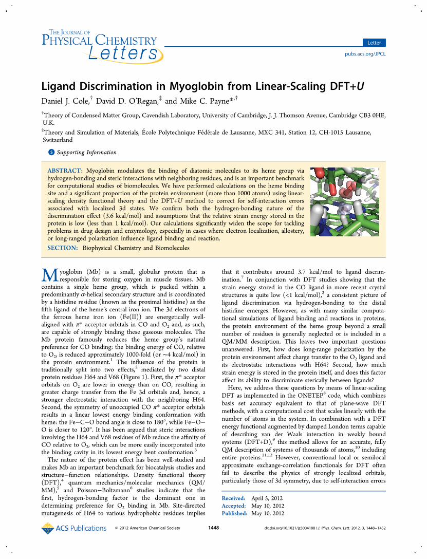
but has been demonstrated to be particularly efficient andeffective for correcting the self-interaction error in transitionmetal chemistry16−18 and is used with increasing frequency forbiological systems. The implementation of the DFT+U methodin the linear-scaling framework, with full optimization of boththe local orbitals describing the Kohn−Sham states and thestates for correction with DFT+U, has been recentlydemonstrated19−21 and it is with this implementation that weperform simulations of a realistic model of the Mb hemeprotein (1007 atoms) in complex with two ligands, CO and O2,at a fully QM level.In Table 1, we compare the structural properties of the three
computational models shown in Figure 1 extracted from both
Mb−CO and Mb−O2 X-ray crystal structures22 and optimized
using DFT, with a Hubbard parameter of U = 0 eV. The largestsystems require the optimization of 113 atoms within an 894atom protein environment (see Computational Methodsection). The modeled geometries are, in general, in goodagreement with each other and with the experimentalstructures, and our Mb-O2 geometries compare favorably withoptimized DFT/MM structures employing a range of differentexchange-correlation functionals.23 The Fe−C−O bond angledecreases by ∼5° on the addition of the neighboring proteinresidues, H64 and V68, while the O2 molecule retains a bentconformation of ∼120°. The root-mean-square deviation(rmsd) of the computed atomic positions of the hemestructures from those in the experimental structures are lowand most of the rmsd increase in the 53-residue system is dueto the propionate side chains. Residue H64 is observed toadopt multiple conformations in crystal structures of Mb-O2.
22,24 We have found the more distant conformation (O−NH64 distance of 2.97 Å) to be the more energetically favorableconformation in the 3-residue Mb-O2 simulation (by 1.9 kcal/mol) and results reported in this Letter are specifically for thisconformation. Following complete relaxation in the 53-residuesystem, the O−NH64 distance is intermediate between the tworeported experimental values.22
The strength of the hydrogen bond between the distalhistidine H64 and the two ligands is expected to dependstrongly on the charge transfer from the heme group to theligand. Table 2 reveals that the magnitude of the natural bondorbital charge population25 on CO is less than 0.1 e in allmodels studied. In contrast, there is substantial charge transferto the O2 ligand, which increases with system size and reaches
Figure 1. Computational models used in the current study. Iron and oxygen ligand atoms are represented by orange and red spheres, respectively.Distal residues H64 and V68, identified as important in determining ligand binding, are labeled, as well as the proximal histidine H93. In the 53-residue system, the full Mb molecule is shown in gray, while the system used in our calculations is colored.
Table 1. Structural Data for Computational Models, with an Increasing Number of Protein Residues, Together withExperimental Structuresa
Mb−CO Mb−O2
1 3 53 1A6G 1 3 53 1A6M
Fe−X 1.79 1.78 1.77 1.82 1.92 1.87 1.86 1.81X−O 1.16 1.16 1.16 1.09 1.24 1.26 1.27 1.24X−NH64 − 3.47 3.46 3.42 − 3.09 3.01 3.08/3.02O−NH64 − 3.25 3.33 3.16 − 2.87 2.78 2.97/2.67Fe−NH93 2.08 2.09 2.10 2.06 2.14 2.08 2.06 2.06Fe−Nheme 2.03 2.04 2.03 1.99 2.03 2.03 2.03 2.01Fe−X−O 175.0° 170.2° 170.3° 171.1° 120.9° 120.3° 120.4° 122.5°rmsd 0.07 0.07 0.11 − 0.07 0.06 0.10 −
aX = C,O for Mb-CO and Mb-O2 respectively. Histidine residues are labeled in Figure 1. The rmsd of the computed atomic positions from those inthe experimental structures is measured for non-hydrogen atoms of the heme group and ligand. Two alternative ligand-to-NH64 distances are quotedfor the 1A6M crystal structure, corresponding to two equally occupied conformations of the distal histidine. Distances are measured in Å.
The Journal of Physical Chemistry Letters Letter
dx.doi.org/10.1021/jz3004188 | J. Phys. Chem. Lett. 2012, 3, 1448−14521449
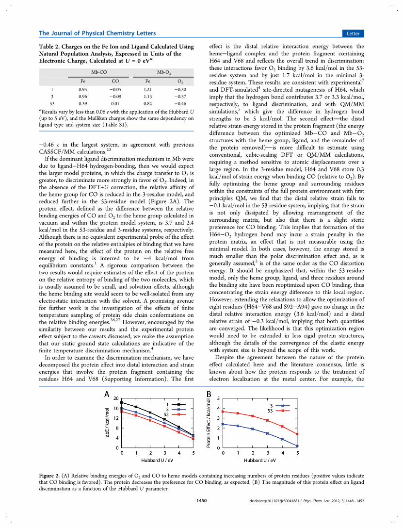
−0.46 e in the largest system, in agreement with previousCASSCF/MM calculations.23
If the dominant ligand discrimination mechanism in Mb weredue to ligand−H64 hydrogen-bonding, then we would expectthe larger model proteins, in which the charge transfer to O2 isgreater, to discriminate more strongly in favor of O2. Indeed, inthe absence of the DFT+U correction, the relative affinity ofthe heme group for CO is reduced in the 3-residue model, andreduced further in the 53-residue model (Figure 2A). Theprotein effect, defined as the difference between the relativebinding energies of CO and O2 to the heme group calculated invacuum and within the protein model system, is 3.7 and 2.4kcal/mol in the 53-residue and 3-residue systems, respectively.Although there is no equivalent experimental probe of the effectof the protein on the relative enthalpies of binding that we havemeasured here, the effect of the protein on the relative freeenergy of binding is inferred to be ∼4 kcal/mol fromequilibrium constants.1 A rigorous comparison between thetwo results would require estimates of the effect of the proteinon the relative entropy of binding of the two molecules, whichis usually assumed to be small, and solvation effects, althoughthe heme binding site would seem to be well-isolated from anyelectrostatic interaction with the solvent. A promising avenuefor further work is the investigation of the effects of finitetemperature sampling of protein side chain conformations onthe relative binding energies.26,27 However, encouraged by thesimilarity between our results and the experimental proteineffect subject to the caveats discussed, we make the assumptionthat our static ground state calculations are indicative of thefinite temperature discrimination mechanism.4
In order to examine the discrimination mechanism, we havedecomposed the protein effect into distal interaction and strainenergies that involve the protein fragment containing theresidues H64 and V68 (Supporting Information). The first
effect is the distal relative interaction energy between theheme−ligand complex and the protein fragment containingH64 and V68 and reflects the overall trend in discrimination:these interactions favor O2 binding by 3.6 kcal/mol in the 53-residue system and by just 1.7 kcal/mol in the minimal 3-residue system. These results are consistent with experimental7
and DFT-simulated4 site-directed mutagenesis of H64, whichimply that the hydrogen bond contributes 3.7 or 3.3 kcal/mol,respectively, to ligand discrimination, and with QM/MMsimulations,5 which give the difference in hydrogen bondstrengths to be 5 kcal/mol. The second effectthe distalrelative strain energy stored in the protein fragment (the energydifference between the optimized Mb−CO and Mb−O2structures with the heme group, ligand, and the remainder ofthe protein removed)is more difficult to estimate usingconventional, cubic-scaling DFT or QM/MM calculations,requiring a method sensitive to atomic displacements over alarge region. In the 3-residue model, H64 and V68 store 0.3kcal/mol of strain energy when binding CO (relative to O2). Byfully optimizing the heme group and surrounding residueswithin the constraints of the full protein environment with firstprinciples QM, we find that the distal relative strain falls to−0.1 kcal/mol in the 53-residue system, implying that the strainis not only dissipated by allowing rearrangement of thesurrounding matrix, but also that there is a slight stericpreference for CO binding. This implies that formation of theH64−O2 hydrogen bond may incur a strain penalty in theprotein matrix, an effect that is not measurable using theminimal model. In both cases, however, the energy stored ismuch smaller than the polar discrimination effect and, as isgenerally assumed,2 is of the same order as the CO distortionenergy. It should be emphasized that, within the 53-residuemodel, only the heme group, ligand, and three residues aroundthe binding site have been reoptimized upon CO binding, thusconcentrating the strain energy difference to this local region.However, extending the relaxations to allow the optimization ofeight residues (H64−V68 and S92−A94) gave no change in thedistal relative interaction energy (3.6 kcal/mol) and a distalrelative strain of −0.3 kcal/mol, implying that both quantitiesare converged. The likelihood is that this optimization regionwould need to be extended in less rigid protein structures,although the details of the convergence of the elastic energywith system size is beyond the scope of this work.Despite the agreement between the nature of the protein
effect calculated here and the literature consensus, little isknown about how the protein responds to the treatment ofelectron localization at the metal center. For example, the
Table 2. Charges on the Fe Ion and Ligand Calculated UsingNatural Population Analysis, Expressed in Units of theElectronic Charge, Calculated at U = 0 eVa
Mb-CO Mb-O2
Fe CO Fe O2
1 0.95 −0.05 1.21 −0.303 0.96 −0.09 1.13 −0.3753 0.39 0.01 0.82 −0.46
aResults vary by less than 0.06 e with the application of the Hubbard U(up to 5 eV), and the Mulliken charges show the same dependency onligand type and system size (Table S1).
Figure 2. (A) Relative binding energies of O2 and CO to heme models containing increasing numbers of protein residues (positive values indicatethat CO binding is favored). The protein decreases the preference for CO binding, as expected. (B) The magnitude of this protein effect on liganddiscrimination as a function of the Hubbard U parameter.
The Journal of Physical Chemistry Letters Letter
dx.doi.org/10.1021/jz3004188 | J. Phys. Chem. Lett. 2012, 3, 1448−14521450
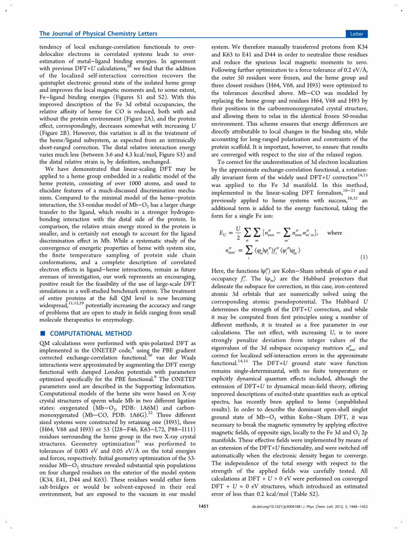
tendency of local exchange-correlation functionals to over-delocalize electrons in correlated systems leads to over-estimation of metal−ligand binding energies. In agreementwith previous DFT+U calculations,28 we find that the additionof the localized self-interaction correction recovers thequintuplet electronic ground state of the isolated heme groupand improves the local magnetic moments and, to some extent,Fe−ligand binding energies (Figures S1 and S2). With thisimproved description of the Fe 3d orbital occupancies, therelative affinity of heme for CO is reduced, both with andwithout the protein environment (Figure 2A), and the proteineffect, correspondingly, decreases somewhat with increasing U(Figure 2B). However, this variation is all in the treatment ofthe heme/ligand subsystem, as expected from an intrinsicallyshort-ranged correction. The distal relative interaction energyvaries much less (between 3.6 and 4.3 kcal/mol, Figure S3) andthe distal relative strain is, by definition, unchanged.We have demonstrated that linear-scaling DFT may be
applied to a heme group embedded in a realistic model of theheme protein, consisting of over 1000 atoms, and used toelucidate features of a much-discussed discrimination mecha-nism. Compared to the minimal model of the heme−proteininteraction, the 53-residue model of Mb−O2 has a larger chargetransfer to the ligand, which results in a stronger hydrogen-bonding interaction with the distal side of the protein. Incomparison, the relative strain energy stored in the protein issmaller, and is certainly not enough to account for the liganddiscrimination effect in Mb. While a systematic study of theconvergence of energetic properties of heme with system size,the finite temperature sampling of protein side chainconformations, and a complete description of correlatedelectron effects in ligand−heme interactions, remain as futureavenues of investigation, our work represents an encouraging,positive result for the feasibility of the use of large-scale DFTsimulations in a well-studied benchmark system. The treatmentof entire proteins at the full QM level is now becomingwidespread,11,12,29 potentially increasing the accuracy and rangeof problems that are open to study in fields ranging from smallmolecule therapeutics to enzymology.
■ COMPUTATIONAL METHODQM calculations were performed with spin-polarized DFT asimplemented in the ONETEP code,8 using the PBE gradientcorrected exchange-correlation functional.30 van der Waalsinteractions were approximated by augmenting the DFT energyfunctional with damped London potentials with parametersoptimized specifically for the PBE functional.9 The ONETEPparameters used are described in the Supporting Information.Computational models of the heme site were based on X-raycrystal structures of sperm whale Mb in two different ligationstates: oxygenated (Mb−O2, PDB: 1A6M) and carbon-monoxygenated (Mb−CO, PDB: 1A6G).22 Three differentsized systems were constructed by retaining one (H93), three(H64, V68 and H93) or 53 (I28−F46, K63−L72, P88−I111)residues surrounding the heme group in the two X-ray crystalstructures. Geometry optimization31 was performed totolerances of 0.003 eV and 0.05 eV/Å on the total energiesand forces, respectively. Initial geometry optimization of the 53-residue Mb−O2 structure revealed substantial spin populationson four charged residues on the exterior of the model system(K34, E41, D44 and K63). These residues would either formsalt-bridges or would be solvent-exposed in their realenvironment, but are exposed to the vacuum in our model
system. We therefore manually transferred protons from K34and K63 to E41 and D44 in order to neutralize these residuesand reduce the spurious local magnetic moments to zero.Following further optimization to a force tolerance of 0.2 eV/Å,the outer 50 residues were frozen, and the heme group andthree closest residues (H64, V68, and H93) were optimized tothe tolerances described above. Mb−CO was modeled byreplacing the heme group and residues H64, V68 and H93 bytheir positions in the carbonmonoxygenated crystal structure,and allowing them to relax in the identical frozen 50-residueenvironment. This scheme ensures that energy differences aredirectly attributable to local changes in the binding site, whileaccounting for long-ranged polarization and constraints of theprotein scaffold. It is important, however, to ensure that resultsare converged with respect to the size of the relaxed region.To correct for the underestimation of 3d electron localization
by the approximate exchange-correlation functional, a rotation-ally invariant form of the widely used DFT+U correction14,15
was applied to the Fe 3d manifold. In this method,implemented in the linear-scaling DFT formalism,19−21 andpreviously applied to heme systems with success,28,32 anadditional term is added to the energy functional, taking theform for a single Fe ion:
∑ ∑ ∑
∑ φ ψ ψ φ
= −
= ⟨ | ⟩ ⟨ | ⟩σ
σ σ σ
σ σ σ σ′
′ ′
′ ′
EU
n n n
n f
2[ ], where
mmm
mmm m m
mmi
m i i i m
U
(1)
Here, the functions |ψiσ⟩ are Kohn−Sham orbitals of spin σ and
occupancy f iσ. The |φm⟩ are the Hubbard projectors that
delineate the subspace for correction, in this case, iron-centeredatomic 3d orbitals that are numerically solved using thecorresponding atomic pseudopotential. The Hubbard Udetermines the strength of the DFT+U correction, and whileit may be computed from first principles using a number ofdifferent methods, it is treated as a free parameter in ourcalculations. The net effect, with increasing U, is to morestrongly penalize deviation from integer values of theeigenvalues of the 3d subspace occupancy matrices nmm′
σ andcorrect for localized self-interaction errors in the approximatefunctional.14,15 The DFT+U ground state wave functionremains single-determinantal, with no finite temperature orexplicitly dynamical quantum effects included, although theextension of DFT+U to dynamical mean-field theory, offeringimproved descriptions of excited-state quantities such as opticalspectra, has recently been applied to heme (unpublishedresults). In order to describe the dominant open-shell singletground state of Mb−O2 within Kohn−Sham DFT, it wasnecessary to break the magnetic symmetry by applying effectivemagnetic fields, of opposite sign, locally to the Fe 3d and O2 2pmanifolds. These effective fields were implemented by means ofan extension of the DFT+U functionality, and were switched offautomatically when the electronic density began to converge.The independence of the total energy with respect to thestrength of the applied fields was carefully tested. Allcalculations at DFT + U > 0 eV were performed on convergedDFT + U = 0 eV structures, which introduced an estimatederror of less than 0.2 kcal/mol (Table S2).
The Journal of Physical Chemistry Letters Letter
dx.doi.org/10.1021/jz3004188 | J. Phys. Chem. Lett. 2012, 3, 1448−14521451
■ ASSOCIATED CONTENT
*S Supporting InformationSupporting methods, calculation of distal interaction and strainenergies, Mulliken charge analysis, energetics of FeP(Im),variation of local magnetic moments and distal interactionenergy with U, and tests of our geometry optimization protocolat nonzero Hubbard U. This material is available free of chargevia the Internet at http://pubs.acs.org/.
■ AUTHOR INFORMATION
Corresponding Author*E-mail: [email protected].
NotesThe authors declare no competing financial interest.
■ ACKNOWLEDGMENTS
We are grateful to Nicholas Hine for helpful discussions.Computational resources were provided by the CambridgeHPC Service, funded by EPSRC Grant EP/F032773/1. D.J.C.,D.D.O’R. and M.C.P. acknowledge support from the EPSRC.
■ REFERENCES(1) Olson, J. S.; Phillips, G. N., Jr. Myoglobin Discriminates BetweenO2, NO, and CO by Electrostatic Interactions with the Bound Ligand.J. Biol. Inorg. Chem. 1997, 2, 544−522.(2) Spiro, T. G.; Kozlowski, P. M. Is the CO Adduct of MyoglobinBent, and Does It Matter? Acc. Chem. Res. 2001, 34, 137−144.(3) Collman, J. P.; Brauman, J. I.; Halbert, T. R.; Suslick, K. S. Natureof O2 and CO Binding to Metalloporphyrins and Heme Proteins. Proc.Natl. Acad. Sci. U.S.A. 1976, 73, 3333−3337.(4) De Angelis, F.; Jarzecki, A. A.; Car, R.; Spiro, T. G. QuantumChemical Evaluation of Protein Control over Heme Ligation: CO/O2Discrimination in Myoglobin. J. Phys. Chem. B 2005, 109, 3065−3070.(5) Sigfridsson, E.; Ryde, U. Theoretical Study of the Discriminationbetween O2 and CO by Myoglobin. J. Inorg. Biochem. 2002, 91, 101−115.(6) Phillips, G. N., Jr.; Teodoro, M. L.; Li, T.; Smith, B.; Olson, J. S.Bound CO is a Molecular Probe of Electrostatic Potential in the DistalPocket of Myoglobin. J. Phys. Chem. B 1999, 103, 8817−8829.(7) Springer, B. A.; Sligar, S. G.; Olson, J. S.; Phillips, G. N., Jr.Mechanisms of Ligand Recognition in Myoglobin. Chem. Rev. 1994,94, 699−714.(8) Skylaris, C. K.; Haynes, P. D.; Mostofi, A. A.; Payne, M. C.Introducing ONETEP: Linear-Scaling Density Functional Simulationson Parallel Computers. J. Chem. Phys. 2005, 122, 084119.(9) Hill, Q.; Skylaris, C. K. Including Dispersion Interactions in theONETEP Program for Linear-Scaling Density Functional TheoryCalculations. Proc. R. Soc. A 2009, 465, 669−683.(10) Hine, N. D. M.; Haynes, P. D.; Mostofi, A. A.; Skylaris, C. K.;Payne, M. C. Linear-Scaling Density-Functional Theory with Tens ofThousands of Atoms: Expanding the Scope and Scale of Calculationswith ONETEP. Comput. Phys. Commun. 2009, 180, 1041−1053.(11) Cole, D. J.; Skylaris, C. K.; Rajendra, E.; Venkitaraman, A. R.;Payne, M. C. Protein−Protein Interactions from Linear-Scaling First-Principles Quantum-Mechanical Calculations. Europhys. Lett. 2010, 91,37004.(12) Cole, D. J.; Rajendra, E.; Roberts-Thomson, M.; Hardwick, B.;McKenzie, G. J.; Payne, M. C.; Venkitaraman, A. R.; Skylaris, C. K.Interrogation of the Protein−Protein Interactions between HumanBRCA2 BRC Repeats and RAD51 Reveals Atomistic Determinants ofAffinity. PLoS Comput. Biol. 2011, 7, e1002096.(13) Cohen, A. J.; Mori-Sanchez, P.; Yang, W. Insights into CurrentLimitations of Density Functional Theory. Science 2008, 321, 792−794.
(14) Cococcioni, M.; de Gironcoli, S. Linear Response Approach tothe Calculation of the Effective Interaction Parameters in the LDA+UMethod. Phys. Rev. B 2005, 71, 035105.(15) Pickett, W. E.; Erwin, S. C.; Ethridge, E. C. Reformulation of theLDA+U Method for a Local-Orbital Basis. Phys. Rev. B 1998, 58,1201−1209.(16) Kulik, H. J.; Cococcioni, M.; Scherlis, D. A.; Marzari, N.Density-Functional Theory in Transition-Metal Chemistry: A Self-Consistent Hubbard U Approach. Phys. Rev. Lett. 2006, 97, 103001.(17) Kulik, H. J.; Marzari, N. A Self-Consistent Hubbard U Density-Functional Theory Approach to the Addition−Elimination Reactionsof Hydrocarbons on Bare FeO+. J. Chem. Phys. 2008, 129, 134314.(18) Kulik, H. J.; Marzari, N. Transition-Metal Dioxides: A Case forthe Intersite Term in Hubbard-Model Functionals. J. Chem. Phys.2011, 134, 094103.(19) O’Regan, D. D.; Hine, N. D. M.; Payne, M. C.; Mostofi, A. A.Projector Self-Consistent DFT+U using Non-Orthogonal GeneralizedWannier Functions. Phys. Rev. B 2010, 82, 081102(R).(20) O’Regan, D. D.; Payne, M. C.; Mostofi, A. A. SubspaceRepresentations in Ab Initio Methods for Strongly Correlated Systems.Phys. Rev. B 2011, 83, 245124.(21) O’Regan, D. D.; Hine, N. D. M.; Payne, M. C.; Mostofi, A. A.Linear-Scaling DFT+U with Full Local Orbital Optimization. Phys.Rev. B 2012, 85, 085107.(22) Vojtechovsky, J.; Chu, K.; Berendzen, J.; Sweet, R. M.;Schlichting, I. Crystal Structures of Myoglobin-Ligand Complexes atNear-Atomic Resolution. Biophys. J. 1999, 77, 2153−2174.(23) Chen, H.; Ikeda-Saito, M.; Shaik, S. Nature of the Fe-O2Bonding in Oxy-Myoglobin: Effect of the Protein. J. Am. Chem. Soc.2008, 130, 14778−14790.(24) Unno, M.; Chen, H.; Kusama, S.; Shaik, S.; Ikeda-Saito, M.Structural Characterization of the Fleeting Ferric Peroxo Species inMyoglobin: Experiment and Theory. J. Am. Chem. Soc. 2008, 129,13394−13395.(25) Reed, A. E.; Weinstock, R. B.; Weinhold, F. Natural PopulationAnalysis. J. Chem. Phys. 1985, 83, 735−746.(26) Alcantara, R. E.; Xu, C.; Spiro, T. G.; Guallar, V. A QuantumChemical Picture of Hemoglobin Affinity. Proc. Natl. Acad. Sci. U.S.A.2007, 104, 18451−18455.(27) Strickland, N.; Mulholland, A. J.; Harvey, J. N. The Fe−COBond Energy in Myoglobin: A QM/MM Study of the Effect ofTertiary Structure. Biophys. J. 2006, 90, L27−L29.(28) Scherlis, D. A.; Cococcioni, M.; Sit, P.; Marzari, N. Simulation ofHeme Using DFT+U: A Step toward Accurate Spin-State Energetics. J.Phys. Chem. B 2007, 111, 7384−7391.(29) Ufimtsev, I. S.; Luehr, N.; Martinez, T. J. Charge Transfer andPolarization in Solvated Proteins from Ab Initio Molecular Dynamics.J. Phys. Chem. Lett. 2011, 2, 1789−1793.(30) Perdew, J. P.; Burke, K.; Ernzerhof, M. Generalized GradientApproximation Made Simple. Phys. Rev. Lett. 1996, 77, 3865−3868.(31) Hine, N. D. M.; Robinson, M.; Haynes, P. D.; Skylaris, C. K.;Payne, M. C.; Mostofi, A. A. Accurate Ionic Forces and GeometryOptimisation in Linear Scaling Density-Functional Theory with LocalOrbitals. Phys. Rev. B 2011, 83, 195102.(32) Sena, A. M. P.; Brazdova, V.; Bowler, D. R. Density FunctionalTheory Study of the Iron-Based Porphyrin Haem(b) on the Si(111):HSurface. Phys. Rev. B 2009, 79, 245404.
The Journal of Physical Chemistry Letters Letter
dx.doi.org/10.1021/jz3004188 | J. Phys. Chem. Lett. 2012, 3, 1448−14521452