Embed Size (px)
Citation preview

Resveratrol delays age-related deterioration and mimicstranscriptional aspects of dietary restriction without extendinglifespan
Kevin J. Pearson1,*, Joseph A. Baur2,*, Kaitlyn N. Lewis1, Leonid Peshkin3, Nathan L.Price1, Nazar Labinskyy4, William R. Swindell5, Davida Kamara1, Robin K. Minor1, EvelynPerez1, Hamish A. Jamieson6, Yongqing Zhang7, Stephen R. Dunn8, Kumar Sharma9, NancyPleshko10, Laura A. Woollett11, Anna Csiszar4, Yuji Ikeno12, David Le Couteur6, Peter J.Elliott13, Kevin G. Becker7, Placido Navas14, Donald K. Ingram15, Norman S. Wolf16, ZoltanUngvari4, David A. Sinclair2,†, and Rafael de Cabo1,†
1 Laboratory of Experimental Gerontology, Harvard Medical School, 77 Avenue Louis Pasteur, Boston MA,02115, USA, Ph. 617 432 3931, Fax: 617 432 1313
2 Department of Pathology and Paul F. Glenn Laboratories for the Biological Mechanisms of Aging, HarvardMedical School, 77 Avenue Louis Pasteur, Boston MA, 02115, USA, Ph. 617 432 3931, Fax: 617 432 1313
3 Department of Systems Biology and Center for Bio-Medical Informatics, Harvard Medical School, 77Avenue Louis Pasteur, Boston MA, 02115, USA, Ph. 617 432 3931, Fax: 617 432 1313
4 Department of Physiology, New York Medical College, Valhalla, NY 10595, USA
5 Department of Pathology, University of Michigan, 109 Zina Pitcher Place, Ann Arbor, MI 48103, USA
6 Centre for Education and Research on Ageing, and the ANZAC Research Institute University of Sydney,Concord NSW 2139, Australia
7 Gene Expression and Genomics Unit, National Institute on Aging, National Institutes of Health, 5600 NathanShock Drive, Baltimore, Maryland, 21224, USA, Ph: 410 558 8510; Fax: 410 558 8302
8 Cancer Genomics, Nucleic Acid/Microarray Facility, Kimmel Cancer Center, Thomas Jefferson University,233 South 10th Street, Suite 1009 BLSB, Philadelphia, PA 19107, USA
9 Translational Research in Kidney Disease, 9500 Gilman Drive, MC 0711, UCSD, La Jolla, CA 92014-0711,USA
10 Hospital for Special Surgery, 535 E. 70th St., New York, NY 10021; current address, Exponent, 3401Market Street, Suite 300, Philadelphia, PA 19104, USA
11 Department of Pathology and Laboratory Medicine, Genome Research Center, University of CincinnatiMedical Center, 2180 East Galbraith Road, Cincinnati, OH 45237, USA
12 Barshop Institute for Longevity and Aging Studies and Department of Pathology, University of TexasHealth Science Center at San Antonio, and Research Service, Audie Murphy VA Hospital (STVHCS).15355Lambda Drive, San Antonio, TX 78245-3207, USA
13 Sirtris Pharmaceuticals Inc, 200 Technology Square, Cambridge, MA 02139, USA
† Correspondence and requests for materials should be addressed to R.deC. ([email protected]) or D.S.([email protected]).*These authors contributed equally.
NIH Public AccessAuthor ManuscriptCell Metab. Author manuscript; available in PMC 2009 August 1.
Published in final edited form as:Cell Metab. 2008 August ; 8(2): 157–168. doi:10.1016/j.cmet.2008.06.011.
NIH
-PA Author Manuscript
NIH
-PA Author Manuscript
NIH
-PA Author Manuscript
14 Centro Andaluz de Biología del Desarrollo, and Centro de Investigación Biomédica en Red: EnfermedadesRaras, Instituto de Salud Carlos III, Universidad Pablo de Olavide-CSIC, 41013 Sevilla, Spain
15 Nutritional Neuroscience and Aging Laboratory, Pennington Biomedical Research Center, LouisianaState University System, 6400 Perkins Road, Baton Rouge, LA 70808, USA
16 Department of Pathology, University of Washington, Seattle, WA 98195-7470, USA
SUMMARYA small molecule that safely mimics the ability of dietary restriction (DR) to delay age-relateddiseases in laboratory animals is greatly sought after. We and others have shown that resveratrolmimics effects of DR in lower organisms. In mice, we find that resveratrol induces gene expressionpatterns in multiple tissues that parallel those induced by DR and every-other-day feeding. Moreover,resveratrol-fed elderly mice show a marked reduction in signs of aging including reducedalbuminuria, decreased inflammation and apoptosis in the vascular endothelium, increased aorticelasticity, greater motor coordination, reduced cataract formation, and preserved bone mineraldensity. However, mice fed a standard diet did not live longer when treated with resveratrol beginningat 12 months of age. Our findings indicate that resveratrol treatment has a range of beneficial effectsin mice but does not increase the longevity of ad libitum-fed animals when started mid-life.
INTRODUCTIONIn developed countries, much of the population now survives to the point where chronic age-associated diseases such as cardiovascular disease, cancer, diabetes, sarcopenia, osteoporosis,stroke, and kidney disease are major determinants of morbidity and mortality (Crews, 2005).Numerous studies have shown that dietary restriction (DR) alleviates many of these conditionsin mammals. Reduction of caloric intake to 30–50% below ad libitum levels, or every-other-day feeding (EOD) of a nutritious diet, can delay the onset of age-related diseases, improvestress resistance, and decelerate functional decline (Barger et al., 2003; Goodrick et al.,1982; McCay et al., 1935). Although DR has beneficial effects in humans (Heilbronn et al.,2006), such a diet is unlikely to be widely adopted, and would pose a significant risk to thefrail, critically ill, or the elderly. As such, we have focused on the development of “DR mimetic”compounds that provide some of the benefits of DR without a reduction in caloric intake(Ingram et al., 2004). Strategies that have been proposed, include inhibition of glycolysis (2-deoxyglucose) (Lane et al., 1998), enhancing insulin action (glucophage/metformin) (Dhahbiet al., 2005), and small molecule activators of SIRT1 (e.g. 3,5,4’-trihydroxystilbene/resveratrol) (Howitz et al., 2003).
Sirtuins are a family of NAD+-dependent deacetylases and ADP-ribosyltransferases that arehomologous to the Saccharomyces cerevisiae Sir2 protein. Extra copies of SIR2 or its homologsextend lifespan in yeast (Kaeberlein et al., 1999), worms (Tissenbaum and Guarente, 2001),and flies (Rogina and Helfand, 2004) and are proposed to underlie some of the physiologicaleffects of DR in simple organisms (Anderson et al., 2003; Lin et al., 2000) and in mammals(Boily et al., 2008; Bordone et al., 2007; Chen et al., 2005). To study sirtuins in mammals, weemployed resveratrol, a small polyphenol identified in an in vitro screen for SIRT1 activatorsthat can extend the lifespan of S. cerevisiae (Howitz et al., 2003; Jarolim et al., 2004),Caenorhabditis elegans (Viswanathan et al., 2005; Wood et al., 2004), Drosophilamelanogaster (Bauer et al., 2004; Wood et al., 2004) and the vertebrate fish Nothobranchiusfurzeri (Valenzano et al., 2006), although one group has failed to detect a significant effect inworms or flies (Bass et al., 2007). In the first three species, lifespan extension is dependent onSIR2, and this has not yet been tested for N. furzeri. In obese mice, resveratrol improves anumber of health parameters including glucose homeostasis, endurance, and survival (Baur et
Pearson et al. Page 2
Cell Metab. Author manuscript; available in PMC 2009 August 1.
NIH
-PA Author Manuscript
NIH
-PA Author Manuscript
NIH
-PA Author Manuscript
al., 2006; Lagouge et al., 2006; Sun et al., 2007), at least partly due to the increased activity ofSIRT1 and AMPK (Baur et al., 2006; Lagouge et al., 2006).
Here, we test the hypothesis that resveratrol imparts health benefits by inducing DR physiology.At one year of age, C57BL/6NIA mice were placed on a standard control diet (SD) or DR byevery-other-day feeding (EOD) with or without resveratrol. We present evidence that long-term resveratrol treatment slows age-related degeneration and functional decline and mimicsthe gene expression patterns induced by DR. We discuss the potential implications of thesefindings for human health.
RESULTSWe previously reported that resveratrol improves the health and survival of obese mice fed ahigh calorie diet (Baur et al., 2006). This raised two key questions: Can resveratrol improvethe health of non-obese mice and if so, is this due to an ability to mimic the effects of DR? Toanswer these questions, we examined the effects of resveratrol on mice fed a standard diet (SD)ad libitum, subjected to every-other-day feeding (EOD), or fed a high calorie diet (HC) adlibitum. Initially, each dietary group was divided into no resveratrol (negative control; SD,EOD, or HC), low resveratrol (100 mg/kg of food, SDLR, EODLR, or HCLR), or resveratrol(400 mg/kg of food, SDR, EODR, or HCR). Later, additional groups of mice were given ahigher dose of resveratrol along with the standard or HC diets (2400 mg/kg of food, SDHR orHCHR). The HC plus resveratrol (HCR) group was the subject of a previous report, and thatnomenclature is preserved herein (Baur et al., 2006).
Resveratrol mimics transcriptional effects of DROne of the most comprehensive ways to assess global changes in physiology is to comparetranscriptional changes across major organs and tissues (Spindler, 2006). To test the hypothesisthat resveratrol is a mimetic of DR, we compared the transcriptional profiles of resveratrol andEOD feeding in liver, skeletal muscle, adipose, and heart. Z ratios were calculated for eachgene as described previously (Cheadle et al., 2003), and false discovery rates were estimatedusing Rankprod (Hong et al., 2006). A subset of the expression changes was verified by RT-PCR (Figure S1). Most transcriptional changes induced by resveratrol were subtle (fold change< 1.5) and tissue-specific. The ten largest changes induced by resveratrol treatment in eachtissue are listed in Table S1. In liver Cyp7A1, a rate-limiting enzyme in the conversion ofcholesterol to bile acids (Russell and Setchell, 1992), was upregulated, while glucose-6-phosphatase, a rate limiting enzyme in the production of glucose (Trinh et al., 1998), wasrepressed. In skeletal muscle and heart, numerous transcripts involved in contractility werealtered, while in white adipose tissue (WAT) the most prominently affected transcripts werebeta defensins, antimicrobial peptides involved in innate and adaptive immunity (Bowdish etal., 2006). The two changes that were consistent across all four tissues based on the Rankprodanalysis were reductions in the expression of the anion exchanger Slc4a1, and the interferon-inducible transcript Ifi27/Isg12, which could indicate suppression of inflammatory responses.Microarray data on the effects of SIRT1 overexpression in these tissues are not available,making it currently difficult to assess whether SIRT1 is a mediator of these effects. No globalcorrelation with previous studies of SIRT1 overexpression in cultured NIH3T3 or beta-cells(Moynihan et al., 2005; Revollo et al., 2004) was observed (data not shown).
To test the hypothesis that resveratrol treatment induces global transcriptional changes thatresemble EOD feeding, we performed principal component analysis (PCA) on the microarraydata from each tissue. Each principal component (PC) can be roughly considered to representa set of correlated changes in gene expression, and to be independent of every other PC. PCsare ranked based on the contribution each makes to the total variability between samples. Whenthe entire data set is used to generate principal components, the effects of age and diet
Pearson et al. Page 3
Cell Metab. Author manuscript; available in PMC 2009 August 1.
NIH
-PA Author Manuscript
NIH
-PA Author Manuscript
NIH
-PA Author Manuscript
predominate (data not shown), however an effect of resveratrol treatment can also be observedthat parallels the changes induced by EOD feeding. If the SD, SDR, and EOD groups are usedfor PCA (eliminating the influence of age and HC diet), the effects of resveratrol and EODfeeding correlate in the first PC (i.e. the set of correlated changes making the greatestcontribution to variability between groups) in liver, muscle, and adipose. Restricting theanalysis to differentially expressed genes strengthens the correlation considerably (see panels1A (liver vs. muscle) and 1B (adipose vs. heart)).
To comprehensively measure the variability between groups, we calculated pairwise distancesin high-dimensional space. The position of each data point (microarray) is specified by theexpression levels of many different genes, each represented mathematically as a distance in anindependent spatial dimension; a straight line connecting any two points can always be drawn,and its length is a reflection of the similarity between the two microarrays in question. Suchcalculations were performed for each pair of microarrays in each tissue, and the results arepresented in Figure 1C, with deep red representing a high degree of similarity, and whiterepresenting divergent samples. These calculations also allow a statistical evaluation of thehypothesis that resveratrol treatment shifted the overall pattern of gene expression toward thatinduced by EOD feeding. This conclusion could be reached based on spatial distances for liver,muscle, and heart. The statistical power in adipose tissue was limited by the number of samples,although the same trend was apparent. Thus the transcriptional effects of resveratrol and EODfeeding show significant overlap in multiple tissues.
Although EOD feeding and conventional caloric restriction (reducing daily energy intake by~40%, “CR”) share key features, including extending mean and maximum lifespan, preventingage-related disease, and improving insulin sensitivity, they have not been shown to workthrough a common mechanism. Therefore, we were interested in comparing our transcriptionalprofiles of EOD and resveratrol treatment to previously reported effects of CR. Differences inage, strain, gender, duration of treatment, array platform, and tissue compound this analysis.Nonetheless, by testing the overlap based on differential expression signatures (Swindell,2008), we detected significant associations between either EOD feeding or resveratroltreatment and previous studies of caloric restriction (Figure 1D). In the case of liver, the effectsof EOD and resveratrol treatment overlapped significantly with those of CR for 5 of 7 publishedstudies.
We next used parametric analysis of gene-set enrichment (PAGE) to highlight specificfunctional pathways within the microarray data. Gene sets were obtained from the MolecularSignatures Database (MSigDB, C2 collection) and analyzed as described previously(Subramanian et al., 2005). Pathways that were significantly altered by EOD feeding orresveratrol treatment are presented in Figure 2A. The effects of the two treatments werecorrelated (by direction of change) in 82% (liver), 76% (muscle), 96% (adipose), and 64%(heart) of the affected pathways, supporting the idea that resveratrol can mimic many effectsof DR in vivo. Among the notable changes were an increase in mitochondrial gene expressionin liver and muscle (Figure 2B) and a decrease in apoptosis across the four tissues (Figure 2C).Full names and Z scores for the gene sets in Figures 2 and S2 are presented in Table S5.
We also sought to identify changes in gene expression patterns that occur during normal agingand to test whether resveratrol treatment or DR resulted in more youthful gene expressionpatterns in the elderly mice. Based on a comparison between SD fed mice at 18 and 27 monthsof age, gene expression patterns induced by either resveratrol treatment or EOD feeding inliver resembled patterns from mice nine months younger (Figure 2D), while in muscle this wasonly true for resveratrol treatment. In adipose both EOD feeding and resveratrol enhancedchanges that occurred with aging, while in heart, neither treatment had a significant effect.
Pearson et al. Page 4
Cell Metab. Author manuscript; available in PMC 2009 August 1.
NIH
-PA Author Manuscript
NIH
-PA Author Manuscript
NIH
-PA Author Manuscript
These results indicate that both resveratrol and EOD feeding can slow the transcriptionalchanges that occur with aging in some but not all tissues.
We previously showed that resveratrol opposes the majority of the transcriptional changes inliver induced by a high calorie (HC) diet. Using newly isolated RNA, a different array platform,and the current version of the MSigDB pathways (1687 pathways vs. 522 analyzed previously),we have confirmed this result and extended our analysis to three additional tissues: skeletalmuscle, adipose, and heart (Figure S2). These results from additional tissues support theconclusion that resveratrol treatment induces gene expression profiles that resemble mice ona lower calorie diet.
Resveratrol delays functional declineOsteoporosis is a major age-associated disease in humans (Gass and Dawson-Hughes, 2006).Resveratrol increases the osteogenic response of osteoblasts (Su et al., 2007) and bone densityin ovariectomized rats (Liu et al., 2005), but the effect of resveratrol on age-induced bone lossin normal mice has not previously been tested. Following their natural deaths, femurs wereremoved from mice (ages 30–33 months) and analyzed by micro-computed tomography (microCT) and mechanical measurements. In the distal femur, resveratrol significantly improved thetissue mineral density (TMD) in SDLR and SDR compared to SD control bones (Figure 3A),and tended to increase trabecular thickness (P = 0.13). Cortical TMD (Figure 3B) trendedhigher in resveratrol-treated groups (P = 0.14), and the effect was statistically significant whenthe two doses were pooled. In addition, resveratrol significantly increased the bone volume tototal volume ratio over the entire femur in the SD fed mice (Figure 3C). Bone strength wasdetermined as the load that is endured by a bone prior to failing in the 3 point bend test.Resveratrol caused a trend towards increased maximum load (P = 0.18, Figure 3D), thatapproached statistical significance when the two doses were pooled (p = 0.058). Overall,resveratrol improved the structure and strength of the femurs tested, suggesting that it mayimprove bone health.
The development of age-related cataracts involves mis-migration of lens epithelial cells andthe accumulation of reactive oxygen species (Wolf et al., 2005). A pathologist trained incataract assessment in aging mice, and blinded to the groups, rated lens opacity in live micefrom 0 to 4 by half steps of 0.5, with 4 representing the complete lens opacity of a maturecataract. Consistent with previous studies (Wolf et al., 2000), the extent of cataract formationsignificantly increased with age in ad libitum-fed mice (Figure 3E). Strikingly, this increasewas attenuated by resveratrol treatment, which was more effective than EOD at 30 months ofage.
Decreased locomotor function resulting in the loss of balance and coordination occurs withincreasing age in humans and rodents. To test the effect of resveratrol on locomoter function,we measured the time to fall from an accelerating rotarod every 3 months. Rotarod is a taskthat contains a learning component (Welsh et al., 2005), so it is normal to observe improvedperformance over time. The SDR group, however, showed a pronounced and statisticallysignificant improvement at 21 and 24 months (Figure 3F), indicating that resveratrol improvesbalance and motor coordination in aged animals.
Improved vascular functionIncreased albuminuria is a marker of vascular dysfunction in mice and a clinical marker ofoverall increased cardiovascular risk in humans (Guzik and Harrison, 2007; Scalia et al.,2007). Urine albumin/creatinine ratios were assessed in the SD, HC, HCLR and HCR groupsat 21 and 26 months of age. The HC mice, but not the HCLR or HCR mice, had significantly
Pearson et al. Page 5
Cell Metab. Author manuscript; available in PMC 2009 August 1.
NIH
-PA Author Manuscript
NIH
-PA Author Manuscript
NIH
-PA Author Manuscript
increased albumin/creatinine ratios compared to SD controls at both time points (Figure S3A),indicating that resveratrol affords protection against vascular or kidney dysfunction.
Because the original cohort had reached an advanced age and had few members remaining, weassessed vascular function in an additional cohort of mice placed on a diet containing 2400mg/kg resveratrol at 12 months of age. Total plasma cholesterol was significantly reduced in22-month-old non-obese mice (SDHR) following 10 months of resveratrol treatment (FigureS3B), while plasma triglycerides showed a slight trend toward a decrease (Figure S3C).Fractionating pooled plasma samples revealed that resveratrol reduced the amount ofcholesterol carried in all lipoprotein fractions (Figure S3D). One potential explanation for thedecrease in circulating cholesterol is diversion to bile acid synthesis via Cyp7A1, which washighly upregulated in the livers of resveratrol-treated animals (Table S1). However, changesin bile acid pool sizes were not detected (data not shown).
Aortic dysfunction and stiffening occur with increased age in humans (Lakatta and Levy,2003; Vaitkevicius et al., 1993), and it has been suggested that resveratrol might be protectiveagainst these effects (Labinskyy et al., 2006). Therefore, we investigated the effects ofresveratrol (2400 mg/kg/food) on vascular function in mice fed a SD or HC diet. Aortas of 3(SD only) or 18-month old animals were dissected and tested for responsiveness to theendothelium-dependent vasodilator acetylcholine (ACh). Both age-related and obesity-relatedfunctional decline were prevented by resveratrol treatment (Figure 3G). The responsivenessof aortas from SDHR mice was significantly better than that of age-matched controls, andcomparable to that of younger (SD 3m) controls. The HC diet significantly worsenedresponsiveness, and the HCHR mice were protected such that there was no difference betweenHCHR and age-matched SD controls. The loss of ACh-induced relaxation with aging andobesity was most likely due to increased superoxide production, since pre-incubation of theHC control vessels with superoxide dismutase restored function (Figure 3G).
To directly measure levels of oxidative stress aortic rings were incubated with dihydroethidine,which reacts with superoxide to generate the fluorescent molecule ethidium bromide (EB).Oxidative stress, as measured by EB fluorescence, increased with age, and was attenuated byresveratrol-treatment in aortas from SD-fed mice (Figure S6P). Separately, mean EBfluorescence intensities were quantified in cross sections of aortas from the SD 3m, SD 18m,HC and HCHR mice (Figure S6Q). HC diet increased, and resveratrol attenuated, oxidativestress, to the point where HCHR aortas were not different from those of age-matched SDcontrols. These experiments are summarized in Figure 3H.
Since nitric oxide (NO) is a key mediator of ACh-induced vasorelaxation that is sensitive tooxidative stress, we measured endothelial nitric oxide synthase (eNOS) expression. Theresveratrol-treated mice displayed increased levels of eNOS mRNA, suggesting an enhancedcapacity for NO production (Figures S6C and S6D), which could potentially help offsetinactivation by superoxide. NADPH oxidase is the primary source of O2.- in vascular tissueand gp91phox, its catalytic subunit, is up-regulated during aging in endothelial cells (Csiszar etal., 2007b). Expression of gp91phox was up-regulated in response to aging and HC diet, and inboth cases, the effects were reversed by resveratrol treatment (Figure S6A and S6B). Consistentwith these data, vascular NADPH oxidase activity was increased in mice fed a HC diet, andthis was prevented by resveratrol treatment (Figure S6O). Thus, resveratrol may enhancevasorelaxation by both increasing NO and decreasing O2.- production.
Apoptotic death of endothelial cells is thought to contribute to vascular pathophysiology inaging and is attenuated by DR (Csiszar et al., 2004). Since our PAGE analysis suggested thatresveratrol can suppress apoptosis (Figure 2C), we performed TUNEL staining of aortas fromSD 3m, SD 18m, SDHR 18 m, HC 18 m and HCHR 18m mice (Figures 3I and 3J). Apoptotic
Pearson et al. Page 6
Cell Metab. Author manuscript; available in PMC 2009 August 1.
NIH
-PA Author Manuscript
NIH
-PA Author Manuscript
NIH
-PA Author Manuscript
index (TUNEL positive nuclei as a percentage of total propidium iodide stained endothelialcell nuclei) was increased by aging and further increased by the HC diet, but these increasesdid not occur in the presence of resveratrol (Figure 3K).
Inflammatory processes contribute to endothelial dysfunction in aging (Csiszar et al., 2007a)and obesity (Roberts et al., 2006) and are suppressed by DR. In agreement with in vitro studiessuggesting that resveratrol can reduce the production of inflammatory cytokines andchemokines through inhibition of NF-κB (Mayo et al., 2003), we found that age- and diet-related increases in the expression of the inflammatory markers TNFα, IL-6, IL-1β, ICAM-1,and iNOS were attenuated by resveratrol treatment (Figures S6E- S6N). Consistent with theseobservations, we found that a number of pathways containing NF-κB responsive genes weresuppressed in multiple tissues during the PAGE analysis (see Table S5).
SurvivalWe have reported that resveratrol treatment increased the survival of mice fed a high calorie(HC) diet to 114 weeks of age (Baur et al., 2006). Here, we provide the complete Kaplan-Meiersurvival analysis (Figure 4B–4D) and maximum lifespan (final 20% surviving) for the HCgroups, as well as mice fed a standard diet or placed on an EOD feeding regimen (Figure 4E).In the context of the HC diet, resveratrol increased remaining lifespan of 1 year old mice byan average of 26% for the HCLR group (P = 0.005) and 25% for HCR (P = 0.001) to the pointwhere survival was not significantly different from that of non-obese SD controls. In the lowerdose (HCLR) group, maximum lifespan was also increased, while this effect did not reachsignificance for the higher dose (HCR) (Figure 4E). The major factor contributing to lifespanextension in the resveratrol-treated HC groups was a reduction in the number of deathsattributed to cardiopulmonary distress (specifically, fatty changes in the liver combined withsevere congestion and edema in the lungs, Table S3).
Interestingly, the increases in longevity could be completely uncoupled from changes in bodyweight. While the HCR group had a very slight decrease in body weight that was not statisticallysignificant, the HCLR group displayed a significant increase in body weight, despiteconsuming a similar amount of food (Figures 4A, S4 and S5). Thus, the beneficial effects ofresveratrol on health and lifespan are not dependent on weight loss.
In the context of the standard diet, resveratrol did not increase overall survival or maximumlifespan (Figures 4B and 4E). Importantly, the SD control group had a lifespan similar to thatof a much larger cohort of C57BL/6NIA mice (Turturro et al., 1999). EOD feeding produceda trend towards increased longevity compared to the SD control group, but the effect did notreach statistical significance. Our results are consistent with the previous observation that theeffect of EOD on longevity is diminished in older C57BL/6 mice (Goodrick et al., 1990), whichis also true of DR by 40% restriction (Weindruch and Walford, 1982). Notably, EOD feedingin combination with the lower dose of resveratrol did extend both mean and maximal lifespanby 15% compared to SD controls (Figure 4C and 4E). We have also tested the effect of a higherdose of resveratrol beginning at 12 months of age (SDHR) on lifespan, and again found thatlongevity was not significantly affected (Figure 4F).
HistopathologyBlinded post-mortem histopathology for disease or pre-disease states was performed onvisceral organs including the heart, kidneys, liver, spleen, lungs, and pancreas (Table S3).Resveratrol treatment did not significantly alter the distribution of pathologies in SD groups.This included neoplasias, despite the potency of resveratrol against implanted or chemicallyinduced tumors, recently reviewed elsewhere (Baur and Sinclair, 2006). This may be relatedto the fact that the vast majority of these cases were lymphomas, a tumor type for which the
Pearson et al. Page 7
Cell Metab. Author manuscript; available in PMC 2009 August 1.
NIH
-PA Author Manuscript
NIH
-PA Author Manuscript
NIH
-PA Author Manuscript
efficacy of resveratrol has not been thoroughly assessed, and that is thought to be triggeredmainly by endogenous retroviruses in mice (Kaplan, 1967;Risser et al., 1983).
DISCUSSIONHere we present a long-term evaluation of resveratrol as a DR mimetic in mice. In agreementwith a concurrent study (Barger et al., 2008), we show that resveratrol induces changes in thetranscriptional profiles of key metabolic tissues that closely resemble those induced by DR. Inliver and muscle, these changes can also be correlated to the gene expression patterns inyounger animals, while in adipose tissue, the trend is reversed. Overall health was improvedunder all dietary conditions, as reflected by the reduction of osteoporosis, cataracts, vasculardysfunction, and declines in motor coordination; however, longevity was increased only in thecontext of a HC diet, as reported previously (Baur et al., 2006).
The effects of DR on longevity are diminished when the regimen is initiated at increasing ages(Goodrick et al., 1990; Weindruch and Walford, 1982), although many of the characteristictranscriptional changes can be induced rapidly regardless of age (Cao et al., 2001). Indeed, ourEOD feeding regimen was initiated at 12 months of age, and while average lifespan wasincreased, the effect did not reach statistical significance, except in combination withresveratrol (EODLR vs. SD). Thus, it is possible that the age of our animals at the start of theexperiment might have diminished the potential effects of resveratrol on lifespan in non-obesemice. To address this question we are following up with lifespan studies in which resveratroltreatment is begun at weaning. It may also be that resveratrol slows the general age-relateddecline, but does not impact the specific causes of death in these mice. Indeed, resveratrol didnot suppress lymphoma, a major cause of mortality in C57BL/6 mice.
There is increasing evidence that the anti-aging action of DR, at least in part, stems from theattenuation of the age-associated increase in oxidative stress (Sohal and Weindruch, 1996). Inparticular, DR attenuates oxidative stress in the aortas of aged rodents and this may contributeto its vasoprotective effects (Guo et al., 2002). Many of the changes we observed in resveratrol-treated animals involved a generalized reduction in oxidative stress and inflammation,consistent with known effects of DR. This was particularly apparent in the aorta, where adecrease in superoxide production was detected directly (Figure 3H), and a variety oftranscripts related to inflammatory processes were repressed (Figure S6E–S6N). Thesechanges likely had direct functional consequences, since ACh-induced responsiveness wasrestored by resveratrol treatment (Figure 3G). In addition, the reduction in cataract formationin resveratrol-treated animals suggests that oxidative stress was reduced in the eye (Figure 3E).One clear difference between EOD and resveratrol was that EOD strongly upregulatedglutathione metabolism whereas resveratrol had no effect. Thus, there may be differences inmechanisms by which EOD and resveratrol reduce oxidative stress. Another difference wasthat while both increased expression of ribosomal proteins in liver, heart, and adipose, onlyEOD had this effect in skeletal muscle. Since increased protein synthesis in skeletal musclehas previously been implicated as a major effect of DR (Lee et al., 1999), this result highlightsa potentially important difference in the resveratrol-treated animals.
Whether it is possible to find a DR mimetic that is also safe for long-term consumption is ofconsiderable debate in the field. Throughout the experiment, mice were subjected to behavioraltests and examined for signs of distress or disease. Postmortem histopathological assessmentswere performed on mice from all three dietary regimens (SD, EOD, and HC), with or withoutresveratrol, and no obvious detrimental change was seen. Although we did not find evidencefor detrimental effects of long-term resveratrol treatment at modest doses, we cannot rule outthe possibility that resveratrol exerted harmful effects that limited its ability to extend lifespan.In a small pilot study using 7.5 times our highest dose (18,000 mg/kg resveratrol in the food),
Pearson et al. Page 8
Cell Metab. Author manuscript; available in PMC 2009 August 1.
NIH
-PA Author Manuscript
NIH
-PA Author Manuscript
NIH
-PA Author Manuscript
5 out of 6 mice died within 3–4 months, consistent with an earlier study of extremely highdoses in rats (Crowell et al., 2004).
In conclusion, long-term resveratrol treatment of mice can mimic transcriptional changesinduced by dietary restriction, and allow them to live healthier, more vigorous lives. In additionto improving insulin sensitivity and increasing survival in HC mice, we show that resveratrolimproves cardiovascular function, bone density, and motor coordination, and delays cataracts,even in non-obese rodents. Together these findings confirm the feasibility of finding an orallyavailable DR mimetic. Since cardiovascular disease is a major cause of age-related morbidityand mortality in humans but not mice, it is possible that DR mimetics such as resveratrol couldhave a greater impact on humans. However, resveratrol does not seem to mimic all of thesalutary effects of DR in that its introduction into the diet of normal one year old mice did notincrease longevity.
EXPERIMENTAL PROCEDURESAnimals and diets
Male C57BL/6NIA were purchased from the National Institute on Aging Aged Rodent Colony(Harlan Sprague-Dawley, Indianapolis, IN). Beginning at one year of age, mice were fed astandard AIN-93G diet (SD and EOD) or AIN-93G modified to provide 60% of calories fromfat (HC) plus 0, 0.01%, or 0.04% resveratrol. Average daily doses over the course of the study(mg/kg/day) were: 7.9 ± 0.2 (SDLR), 30.9 ± 0.6 (SDR), 7.6 ± 0.2 (EODLR), 30.4 ± 0.6 (EODR),5.4 ± 0.2 (HCLR), 24.2 ± 0.8 (HCR), 204 ± 4 (SDHR), and 167 ± 16 (HCHR, ages 12–18months). Additional details are provided in the supplemental material.
Micro CT and bone strengthFemurs were removed following natural deaths between the ages of 30–33 months and theirarchitecture was analyzed on an EVS micro CT MS-8 system (GE Healthcare, London, ON).Mechanical behavior of the femurs was measured on an ELF 3200 test instrument (Bose Corp,Eden Prairie, MN). Additional details are provided in the supplemental material.
Cataracts assessmentAge-related lens opacity was scored in all living mice by an experienced pathologist using aslit lamp, who was blinded as to the experimental groups.
RotarodTime to fall from an accelerating rotarod was measured for all survivors from a pre-designatedsubset of each group; n = 15 (SD), 11 (SDLR), and 16 (SDR). The mice were tested at 15, 18,21, and 24 months of age. At each time-point the mice were given a habituation trial at a constantspeed of 4 rpm. The following day each mouse was given three trials, separated by 30 minuterest periods, during which the rotarod accelerated from 4 to 40 rpm over a period of five min.
Vessel isolation and functional studiesEndothelial function was assessed as described previously (Csiszar et al., 2007a; Csiszar et al.,2007b). In brief, aortas (n = 6 per group) were cut into ring segments 1.5 mm in length andmounted in myographs chambers (Danish Myo Technology A/S, Inc., Denmark) formeasurement of isometric tension. The vessels were superfused with Krebs buffer solution(118 mM NaCl, 4.7 mM KCl, 1.5 mM CaCl2, 25 mM NaHCO3, 1.1 mM MgSO4, 1.2 mMKH2PO4, and 5.6 mM glucose; at 37°C; gassed with 95% air and 5% CO2). After anequilibration period of 1 hour during which an optimal passive tension was applied to the rings(as determined from the vascular length-tension relationship), they were pre-contracted with
Pearson et al. Page 9
Cell Metab. Author manuscript; available in PMC 2009 August 1.
NIH
-PA Author Manuscript
NIH
-PA Author Manuscript
NIH
-PA Author Manuscript
10−6 M phenylephrine and relaxation in response to acetylcholine (ACh) over the range of10−9 to 10−6 M was measured. In some experiments rings were pre-incubated for 30 min with200 U/mL superoxide dismutase to test the involvement of oxidative stress.
Apoptosis assaysTUNEL assays were performed on aorta sections using the Apop Tag Fluorescein in SituApoptosis Detection Kit (Chemicon/Millipore, Billerica, MA), according to manufacturer’sinstructions, as previously described (Csiszar et al., 2007b). Endothelial cells were identifiedby their anatomical location (Ungvari et al., 2007).
MicroarraysRNA was extracted from liver, muscle, adipose and heart at 18 or 27 months of age andhybridized to Illumina Mouseref 8 whole genome microarrays (version 1.1). Raw data weresubjected to Z normalization as described previously (Cheadle et al., 2003) and are availableat http://www.ncbi.nlm.nih.gov/geo, accession number GSE11845. Gene set enrichment wastested using the PAGE method as previously described (Kim and Volsky, 2005) and pathwaydefinitions from MSigDB (Subramanian et al., 2005). Pathways involving mitochondria andapoptosis (Figure 2B and 2C) were selected based on the names and descriptions provided byMSigDB while blinded to the data. Differentially expressed genes were converted intodifferential expression signatures and used to evaluate overlap with previously reported effectsof 10–44% CR as described previously (Swindell, 2008). Characteristics of the animals in 10–44% restriction studies are presented in Table S2. Additional details, including calculation ofprincipal components and pairwise distances, are provided in the supplemental material.
Supplementary MaterialRefer to Web version on PubMed Central for supplementary material.
AcknowledgementsDavid A. Sinclair declares he is a consultant to Sirtris, a GSK sirtuin company, and a board member/shareholder ofGenocea Biosciences, a vaccine company. Peter J. Elliott declares he is a full-time employee and shareholder of GSK-Sirtris. We would like to thank Dawn Phillips for animal care, Hank Rasnow for purchasing the mice, William Woodfor microarray assistance, Katie Burke for help with the plasma lipid and lipoprotein cholesterol concentrations, PatrickLoerch for helpful suggestions and advice, and Mark Beasley and David Allison for assistance with Cox RegressionModeling. We would like to thank Steve Sollott, Alexei Sharov, and Dan Longo for critical reading of the manuscript.This work utilized the facilities of the HSS Musculoskeletal Repair and Regeneration Core Center (NIH AR46121)and was supported by the Intramural Research Program of the National Institute on Aging, National Institutes ofHealth. D.A.S. is an Ellison Medical Foundation Senior Scholar. This work was supported by grants from the AmericanHeart Association (0425834T to J.A.B. and 0435140N to A.C.) and from the NIH (RO1GM068072, AG19972, andAG19719 to D.A.S.), (HL077256 to Z.U.), (HD034089 to L.W), (2RO1 EY011733 to N.S.W.), Spanish grant(BFU2005-03017 to P.N.), and by the generous support of Mr. Paul F. Glenn and The Paul F. Glenn Laboratories forthe Biological Mechanisms of Aging.
ReferencesAnderson RM, Bitterman KJ, Wood JG, Medvedik O, Sinclair DA. Nicotinamide and PNC1 govern
lifespan extension by calorie restriction in Saccharomyces cerevisiae. Nature 2003;423:181–185.[PubMed: 12736687]
Barger JL, Kayo T, Vann JM, Arias EB, Wang J, Hacker TA, Wang Y, Raederstorff D, Morrow JD,Leeuwenburgh C, Allison DB, Saupe KW, Cartee GD, Weindruch R, Prolla TA. A low dose of dietaryresveratrol partially mimics caloric restriction and retards aging parameters in mice. PLoS ONE2008;3:e2264. [PubMed: 18523577]
Barger JL, Walford RL, Weindruch R. The retardation of aging by caloric restriction: its significance inthe transgenic era. Exp Gerontol 2003;38:1343–1351. [PubMed: 14698815]
Pearson et al. Page 10
Cell Metab. Author manuscript; available in PMC 2009 August 1.
NIH
-PA Author Manuscript
NIH
-PA Author Manuscript
NIH
-PA Author Manuscript
Bass TM, Weinkove D, Houthoofd K, Gems D, Partridge L. Effects of resveratrol on lifespan inDrosophila melanogaster and Caenorhabditis elegans. Mech Ageing Dev 2007;128:546–552.[PubMed: 17875315]
Bauer JH, Goupil S, Garber GB, Helfand SL. An accelerated assay for the identification of lifespan-extending interventions in Drosophila melanogaster. Proc Natl Acad Sci U S A 2004;101:12980–12985. [PubMed: 15328413]
Baur JA, Pearson KJ, Price NL, Jamieson HA, Lerin C, Kalra A, Prabhu VV, Allard JS, Lopez-Lluch G,Lewis K, Pistell PJ, Poosala S, Becker KG, Boss O, Gwinn D, Wang M, Ramaswamy S, Fishbein KW,Spencer RG, Lakatta EG, Le Couteur D, Shaw RJ, Navas P, Puigserver P, Ingram DK, de Cabo R,Sinclair DA. Resveratrol improves health and survival of mice on a high-calorie diet. Nature2006;444:337–342. [PubMed: 17086191]
Baur JA, Sinclair DA. Therapeutic potential of resveratrol: the in vivo evidence. Nature reviews2006;5:493–506.
Benjamini Y, Hochberg Y. Controlling the false discovery rate: a powerful and practical approach tomultiple testing. J Roy Stat Soc B 1995;57:289–300.
Boily G, Seifert EL, Bevilacqua L, He XH, Sabourin G, Estey C, Moffat C, Crawford S, Saliba S, JardineK, Xuan J, Evans M, Harper ME, McBurney MW. SirT1 regulates energy metabolism and responseto caloric restriction in mice. PLoS ONE 2008;3:e1759. [PubMed: 18335035]
Bordone L, Cohen D, Robinson A, Motta MC, van Veen E, Czopik A, Steele AD, Crowe H, Marmor S,Luo J, Gu W, Guarente L. SIRT1 transgenic mice show phenotypes resembling calorie restriction.Aging cell 2007;6:759–767. [PubMed: 17877786]
Bowdish DM, Davidson DJ, Hancock RE. Immunomodulatory properties of defensins and cathelicidins.Curr Top Microbiol Immunol 2006;306:27–66. [PubMed: 16909917]
Cao SX, Dhahbi JM, Mote PL, Spindler SR. Genomic profiling of short- and long-term caloric restrictioneffects in the liver of aging mice. Proc Natl Acad Sci U S A 2001;98:10630–10635. [PubMed:11535822]
Cheadle C, Vawter MP, Freed WJ, Becker KG. Analysis of microarray data using Z score transformation.J Mol Diagn 2003;5:73–81. [PubMed: 12707371]
Chen D, Steele AD, Lindquist S, Guarente L. Increase in activity during calorie restriction requires Sirt1.Science 2005;310:1641. [PubMed: 16339438]
Corton JC, Apte U, Anderson SP, Limaye P, Yoon L, Latendresse J, Dunn C, Everitt JI, Voss KA,Swanson C, Kimbrough C, Wong JS, Gill SS, Chandraratna RA, Kwak MK, Kensler TW, StulnigTM, Steffensen KR, Gustafsson JA, Mehendale HM. Mimetics of caloric restriction include agonistsof lipid-activated nuclear receptors. J Biol Chem 2004;279:46204–46212. [PubMed: 15302862]
Crews DE. Artificial environments and an aging population: designing for age-related functional losses.Journal of physiological anthropology and applied human science 2005;24:103–109. [PubMed:15684554]
Crowell JA, Korytko PJ, Morrissey RL, Booth TD, Levine BS. Resveratrol-associated renal toxicity.Toxicol Sci 2004;82:614–619. [PubMed: 15329443]
Csiszar A, Labinskyy N, Smith K, Rivera A, Orosz Z, Ungvari Z. Vasculoprotective effects of anti-tumornecrosis factor-alpha treatment in aging. The American journal of pathology 2007a;170:388–398.[PubMed: 17200210]
Csiszar A, Labinskyy N, Zhao X, Hu F, Serpillon S, Huang Z, Ballabh P, Levy RJ, Hintze TH, WolinMS, Austad SN, Podlutsky A, Ungvari Z. Vascular superoxide and hydrogen peroxide productionand oxidative stress resistance in two closely related rodent species with disparate longevity. Agingcell 2007b;6:783–797. [PubMed: 17925005]
Csiszar A, Ungvari Z, Koller A, Edwards JG, Kaley G. Proinflammatory phenotype of coronary arteriespromotes endothelial apoptosis in aging. Physiological genomics 2004;17:21–30. [PubMed:15020720]
Dhahbi JM, Mote PL, Fahy GM, Spindler SR. Identification of potential caloric restriction mimetics bymicroarray profiling. Physiological genomics 2005;23:343–350. [PubMed: 16189280]
Dhahbi JM, Tsuchiya T, Kim HJ, Mote PL, Spindler SR. Gene expression and physiologic responses ofthe heart to the initiation and withdrawal of caloric restriction. The journals of gerontology2006;61:218–231. [PubMed: 16567370]
Pearson et al. Page 11
Cell Metab. Author manuscript; available in PMC 2009 August 1.
NIH
-PA Author Manuscript
NIH
-PA Author Manuscript
NIH
-PA Author Manuscript
Edwards MG, Anderson RM, Yuan M, Kendziorski CM, Weindruch R, Prolla TA. Gene expressionprofiling of aging reveals activation of a p53-mediated transcriptional program. BMC genomics2007;8:80. [PubMed: 17381838]
Fu C, Hickey M, Morrison M, McCarter R, Han ES. Tissue specific and non-specific changes in geneexpression by aging and by early stage CR. Mech Ageing Dev 2006;127:905–916. [PubMed:17092546]
Gass M, Dawson-Hughes B. Preventing osteoporosis-related fractures: an overview. The Americanjournal of medicine 2006;119:S3–S11. [PubMed: 16563939]
Goodrick CL, Ingram DK, Reynolds MA, Freeman JR, Cider N. Effects of intermittent feeding uponbody weight and lifespan in inbred mice: interaction of genotype and age. Mech Ageing Dev1990;55:69–87. [PubMed: 2402168]
Goodrick CL, Ingram DK, Reynolds MA, Freeman JR, Cider NL. Effects of intermittent feeding upongrowth and life span in rats. Gerontology 1982;28:233–241. [PubMed: 7117847]
Guo Z, Mitchell-Raymundo F, Yang H, Ikeno Y, Nelson J, Diaz V, Richardson A, Reddick R. Dietaryrestriction reduces atherosclerosis and oxidative stress in the aorta of apolipoprotein E-deficient mice.Mech Ageing Dev 2002;123:1121–1131. [PubMed: 12044962]
Guzik TJ, Harrison DG. Endothelial NF-kappaB as a mediator of kidney damage: the missing linkbetween systemic vascular and renal disease? Circulation research 2007;101:227–229. [PubMed:17673681]
Heilbronn LK, de Jonge L, Frisard MI, DeLany JP, Larson-Meyer DE, Rood J, Nguyen T, Martin CK,Volaufova J, Most MM, Greenway FL, Smith SR, Deutsch WA, Williamson DA, Ravussin E. Effectof 6-month calorie restriction on biomarkers of longevity, metabolic adaptation, and oxidative stressin overweight individuals: a randomized controlled trial. Jama 2006;295:1539–1548. [PubMed:16595757]
Higami Y, Pugh TD, Page GP, Allison DB, Prolla TA, Weindruch R. Adipose tissue energy metabolism:altered gene expression profile of mice subjected to long-term caloric restriction. Faseb J2004;18:415–417. [PubMed: 14688200]
Hong F, Breitling R, McEntee CW, Wittner BS, Nemhauser JL, Chory J. RankProd: a bioconductorpackage for detecting differentially expressed genes in meta-analysis. Bioinformatics (Oxford,England) 2006;22:2825–2827.
Howitz KT, Bitterman KJ, Cohen HY, Lamming DW, Lavu S, Wood JG, Zipkin RE, Chung P,Kisielewski A, Zhang LL, Scherer B, Sinclair DA. Small molecule activators of sirtuins extendSaccharomyces cerevisiae lifespan. Nature 2003;425:191–196. [PubMed: 12939617]
Ingram DK, Anson RM, de Cabo R, Mamczarz J, Zhu M, Mattison J, Lane MA, Roth GS. Developmentof calorie restriction mimetics as a prolongevity strategy. Ann N Y Acad Sci 2004;1019:412–423.[PubMed: 15247056]
Jarolim S, Millen J, Heeren G, Laun P, Goldfarb DS, Breitenbach M. A novel assay for replicative lifespanin Saccharomyces cerevisiae. FEMS yeast research 2004;5:169–177. [PubMed: 15489200]
Kaeberlein M, McVey M, Guarente L. The SIR2/3/4 complex and SIR2 alone promote longevity inSaccharomyces cerevisiae by two different mechanisms. Genes Dev 1999;13:2570–2580. [PubMed:10521401]
Kaplan HS. On the natural history of the murine leukemias: presidential address. Cancer Res1967;27:1325–1340. [PubMed: 4860293]
Kim SY, Volsky DJ. PAGE: parametric analysis of gene set enrichment. BMC bioinformatics 2005;6:144.[PubMed: 15941488]
Labinskyy N, Csiszar A, Veress G, Stef G, Pacher P, Oroszi G, Wu J, Ungvari Z. Vascular dysfunctionin aging: potential effects of resveratrol, an anti-inflammatory phytoestrogen. Current medicinalchemistry 2006;13:989–996. [PubMed: 16611080]
Lagouge M, Argmann C, Gerhart-Hines Z, Meziane H, Lerin C, Daussin F, Messadeq N, Milne J, LambertP, Elliott P, Geny B, Laakso M, Puigserver P, Auwerx J. Resveratrol improves mitochondrial functionand protects against metabolic disease by activating SIRT1 and PGC-1alpha. Cell 2006;127:1109–1122. [PubMed: 17112576]
Pearson et al. Page 12
Cell Metab. Author manuscript; available in PMC 2009 August 1.
NIH
-PA Author Manuscript
NIH
-PA Author Manuscript
NIH
-PA Author Manuscript
Lakatta EG, Levy D. Arterial and cardiac aging: major shareholders in cardiovascular disease enterprises:Part I: aging arteries: a “set up” for vascular disease. Circulation 2003;107:139–146. [PubMed:12515756]
Lane MA, Ingram DK, Roth GS. 2-Deoxy-D-glucose feeding in rats mimics physiological effects ofcalorie restriction. J Anti Aging Med 1998;1:327–337.
Lee CK, Allison DB, Brand J, Weindruch R, Prolla TA. Transcriptional profiles associated with agingand middle age-onset caloric restriction in mouse hearts. Proc Natl Acad Sci U S A 2002;99:14988–14993. [PubMed: 12419851]
Lee CK, Klopp RG, Weindruch R, Prolla TA. Gene expression profile of aging and its retardation bycaloric restriction. Science 1999;285:1390–1393. [PubMed: 10464095]
Lin SJ, Defossez PA, Guarente L. Requirement of NAD and SIR2 for life-span extension by calorierestriction in Saccharomyces cerevisiae. Science 2000;289:2126–2128. [PubMed: 11000115]
Liu ZP, Li WX, Yu B, Huang J, Sun J, Huo JS, Liu CX. Effects of trans-resveratrol from Polygonumcuspidatum on bone loss using the ovariectomized rat model. Journal of medicinal food 2005;8:14–19. [PubMed: 15857203]
Mayo MW, Denlinger CE, Broad RM, Yeung F, Reilly ET, Shi Y, Jones DR. Ineffectiveness of histonedeacetylase inhibitors to induce apoptosis involves the transcriptional activation of NF-kappa Bthrough the Akt pathway. J Biol Chem 2003;278:18980–18989. [PubMed: 12649266]
McCay CM, Crowell MF, Maynard LA. The effect of retarded growth upon the length of life span andupon the ultimate body size. The Journal of nutrition 1935;10:63–79.
Moynihan KA, Grimm AA, Plueger MM, Bernal-Mizrachi E, Ford E, Cras-Meneur C, Permutt MA, ImaiS. Increased dosage of mammalian Sir2 in pancreatic beta cells enhances glucose-stimulated insulinsecretion in mice. Cell Metab 2005;2:105–117. [PubMed: 16098828]
Revollo JR, Grimm AA, Imai S. The NAD biosynthesis pathway mediated by nicotinamidephosphoribosyltransferase regulates Sir2 activity in mammalian cells. J Biol Chem 2004;279:50754–50763. [PubMed: 15381699]
Risser R, Horowitz JM, McCubrey J. Endogenous mouse leukemia viruses. Annual review of genetics1983;17:85–121.
Roberts CK, Barnard RJ, Sindhu RK, Jurczak M, Ehdaie A, Vaziri ND. Oxidative stress and dysregulationof NAD(P)H oxidase and antioxidant enzymes in diet-induced metabolic syndrome. Metabolism:clinical and experimental 2006;55:928–934. [PubMed: 16784966]
Rogina B, Helfand SL. Sir2 mediates longevity in the fly through a pathway related to calorie restriction.Proc Natl Acad Sci U S A 2004;101:15998–16003. [PubMed: 15520384]
Russell DW, Setchell KD. Bile acid biosynthesis. Biochemistry 1992;31:4737–4749. [PubMed: 1591235]Scalia R, Gong Y, Berzins B, Zhao LJ, Sharma K. Hyperglycemia is a major determinant of albumin
permeability in diabetic microcirculation: the role of mucalpain. Diabetes 2007;56:1842–1849.[PubMed: 17446533]
Selman C, Kerrison ND, Cooray A, Piper MD, Lingard SJ, Barton RH, Schuster EF, Blanc E, Gems D,Nicholson JK, Thornton JM, Partridge L, Withers DJ. Coordinated multitissue transcriptional andplasma metabonomic profiles following acute caloric restriction in mice. Physiological genomics2006;27:187–200. [PubMed: 16882887]
Sohal RS, Weindruch R. Oxidative stress, caloric restriction, and aging. Science 1996;273:59–63.[PubMed: 8658196]
Spindler SR. Use of microarray biomarkers to identify longevity therapeutics. Aging cell 2006;5:39–50.[PubMed: 16441842]
Su JL, Yang CY, Zhao M, Kuo ML, Yen ML. Forkhead proteins are critical for bone morphogeneticprotein-2 regulation and anti-tumor activity of resveratrol. J Biol Chem 2007;282:19385–19398.[PubMed: 17513867]
Subramanian A, Tamayo P, Mootha VK, Mukherjee S, Ebert BL, Gillette MA, Paulovich A, PomeroySL, Golub TR, Lander ES, Mesirov JP. Gene set enrichment analysis: a knowledge-based approachfor interpreting genome-wide expression profiles. Proc Natl Acad Sci U S A 2005;102:15545–15550.[PubMed: 16199517]
Sun C, Zhang F, Ge X, Yan T, Chen X, Shi X, Zhai Q. SIRT1 improves insulin sensitivity under insulin-resistant conditions by repressing PTP1B. Cell Metab 2007;6:307–319. [PubMed: 17908559]
Pearson et al. Page 13
Cell Metab. Author manuscript; available in PMC 2009 August 1.
NIH
-PA Author Manuscript
NIH
-PA Author Manuscript
NIH
-PA Author Manuscript
Swindell WR. Comparative analysis of microarray data identifies common responses to caloric restrictionamong mouse tissues. Mech Ageing Dev 2008;129:138–153. [PubMed: 18155270]
Tissenbaum HA, Guarente L. Increased dosage of a sir-2 gene extends lifespan in Caenorhabditis elegans.Nature 2001;410:227–230. [PubMed: 11242085]
Trinh KY, O’Doherty RM, Anderson P, Lange AJ, Newgard CB. Perturbation of fuel homeostasis causedby overexpression of the glucose-6-phosphatase catalytic subunit in liver of normal rats. J Biol Chem1998;273:31615–31620. [PubMed: 9813078]
Tsuchiya T, Dhahbi JM, Cui X, Mote PL, Bartke A, Spindler SR. Additive regulation of hepatic geneexpression by dwarfism and caloric restriction. Physiological genomics 2004;17:307–315. [PubMed:15039484]
Turturro A, Witt WW, Lewis S, Hass BS, Lipman RD, Hart RW. Growth curves and survivalcharacteristics of the animals used in the Biomarkers of Aging Program. The journals of gerontology1999;54:B492–501. [PubMed: 10619312]
Ungvari Z, Orosz Z, Rivera A, Labinskyy N, Xiangmin Z, Olson S, Podlutsky A, Csiszar A. Resveratrolincreases vascular oxidative stress resistance. American journal of physiology 2007;292:H2417–2424. [PubMed: 17220179]
Vaitkevicius PV, Fleg JL, Engel JH, O’Connor FC, Wright JG, Lakatta LE, Yin FC, Lakatta EG. Effectsof age and aerobic capacity on arterial stiffness in healthy adults. Circulation 1993;88:1456–1462.[PubMed: 8403292]
Valenzano DR, Terzibasi E, Genade T, Cattaneo A, Domenici L, Cellerino A. Resveratrol prolongslifespan and retards the onset of age-related markers in a short-lived vertebrate. Curr Biol2006;16:296–300. [PubMed: 16461283]
Viswanathan M, Kim SK, Berdichevsky A, Guarente L. A role for SIR-2.1 regulation of ER stressresponse genes in determining C. elegans life span. Developmental cell 2005;9:605–615. [PubMed:16256736]
Weindruch R, Walford RL. Dietary restriction in mice beginning at 1 year of age: effect on life-span andspontaneous cancer incidence. Science 1982;215:1415–1418. [PubMed: 7063854]
Welsh JP, Yamaguchi H, Zeng XH, Kojo M, Nakada Y, Takagi A, Sugimori M, Llinas RR. Normal motorlearning during pharmacological prevention of Purkinje cell long-term depression. Proc Natl AcadSci U S A 2005;102:17166–17171. [PubMed: 16278298]
Wolf N, Penn P, Pendergrass W, Van Remmen H, Bartke A, Rabinovitch P, Martin GM. Age-relatedcataract progression in five mouse models for anti-oxidant protection or hormonal influence.Experimental eye research 2005;81:276–285. [PubMed: 16129095]
Wolf NS, Li Y, Pendergrass W, Schmeider C, Turturro A. Normal mouse and rat strains as models forage-related cataract and the effect of caloric restriction on its development. Experimental eye research2000;70:683–692. [PubMed: 10870527]
Wood JG, Rogina B, Lavu S, Howitz K, Helfand SL, Tatar M, Sinclair D. Sirtuin activators mimic caloricrestriction and delay ageing in metazoans. Nature 2004;430:686–689. [PubMed: 15254550]
Pearson et al. Page 14
Cell Metab. Author manuscript; available in PMC 2009 August 1.
NIH
-PA Author Manuscript
NIH
-PA Author Manuscript
NIH
-PA Author Manuscript
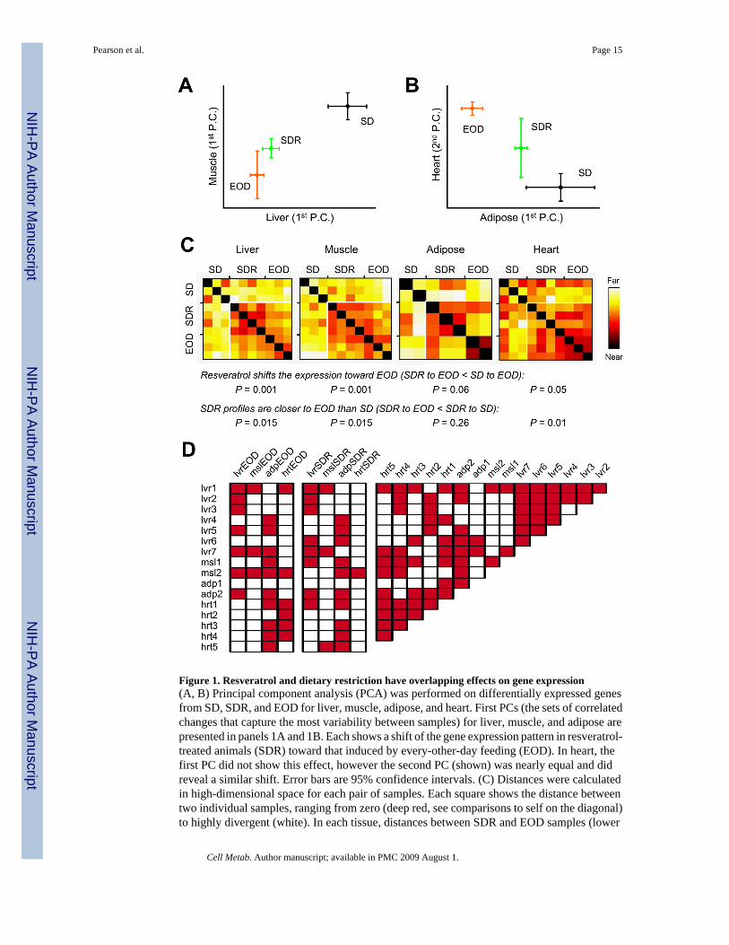
Figure 1. Resveratrol and dietary restriction have overlapping effects on gene expression(A, B) Principal component analysis (PCA) was performed on differentially expressed genesfrom SD, SDR, and EOD for liver, muscle, adipose, and heart. First PCs (the sets of correlatedchanges that capture the most variability between samples) for liver, muscle, and adipose arepresented in panels 1A and 1B. Each shows a shift of the gene expression pattern in resveratrol-treated animals (SDR) toward that induced by every-other-day feeding (EOD). In heart, thefirst PC did not show this effect, however the second PC (shown) was nearly equal and didreveal a similar shift. Error bars are 95% confidence intervals. (C) Distances were calculatedin high-dimensional space for each pair of samples. Each square shows the distance betweentwo individual samples, ranging from zero (deep red, see comparisons to self on the diagonal)to highly divergent (white). In each tissue, distances between SDR and EOD samples (lower
Pearson et al. Page 15
Cell Metab. Author manuscript; available in PMC 2009 August 1.
NIH
-PA Author Manuscript
NIH
-PA Author Manuscript
NIH
-PA Author Manuscript
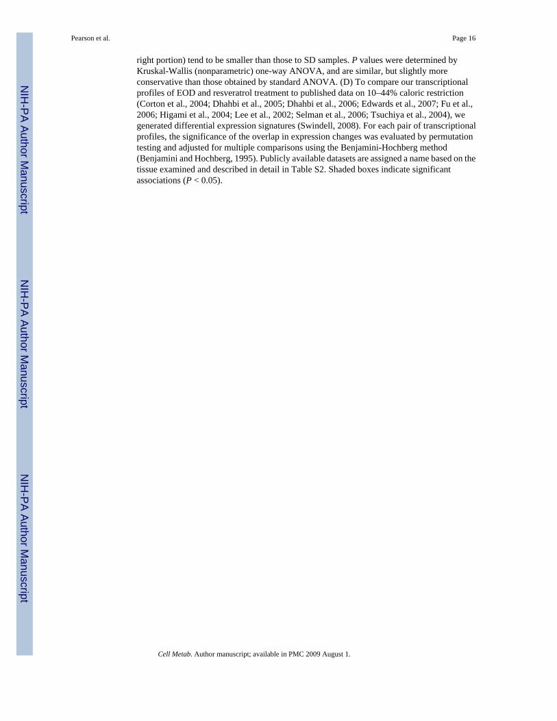
right portion) tend to be smaller than those to SD samples. P values were determined byKruskal-Wallis (nonparametric) one-way ANOVA, and are similar, but slightly moreconservative than those obtained by standard ANOVA. (D) To compare our transcriptionalprofiles of EOD and resveratrol treatment to published data on 10–44% caloric restriction(Corton et al., 2004; Dhahbi et al., 2005; Dhahbi et al., 2006; Edwards et al., 2007; Fu et al.,2006; Higami et al., 2004; Lee et al., 2002; Selman et al., 2006; Tsuchiya et al., 2004), wegenerated differential expression signatures (Swindell, 2008). For each pair of transcriptionalprofiles, the significance of the overlap in expression changes was evaluated by permutationtesting and adjusted for multiple comparisons using the Benjamini-Hochberg method(Benjamini and Hochberg, 1995). Publicly available datasets are assigned a name based on thetissue examined and described in detail in Table S2. Shaded boxes indicate significantassociations (P < 0.05).
Pearson et al. Page 16
Cell Metab. Author manuscript; available in PMC 2009 August 1.
NIH
-PA Author Manuscript
NIH
-PA Author Manuscript
NIH
-PA Author Manuscript
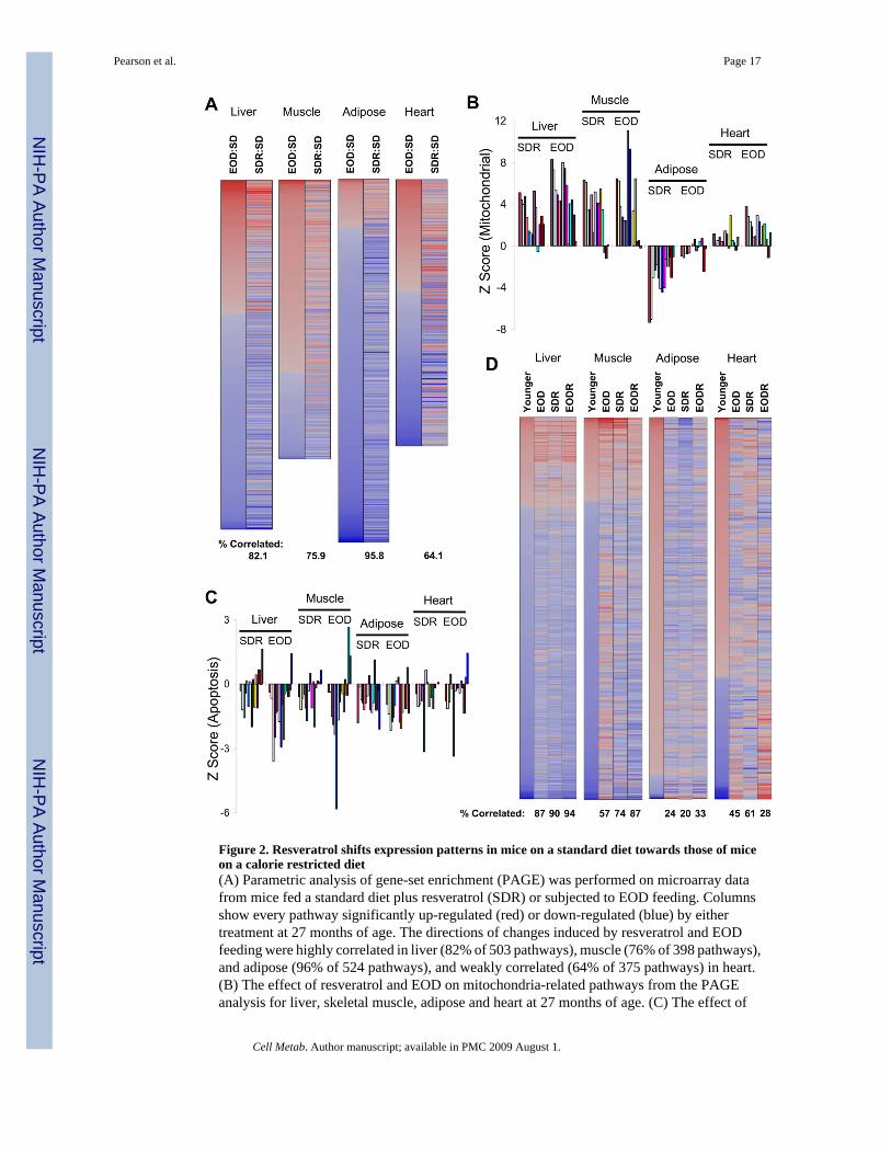
Figure 2. Resveratrol shifts expression patterns in mice on a standard diet towards those of miceon a calorie restricted diet(A) Parametric analysis of gene-set enrichment (PAGE) was performed on microarray datafrom mice fed a standard diet plus resveratrol (SDR) or subjected to EOD feeding. Columnsshow every pathway significantly up-regulated (red) or down-regulated (blue) by eithertreatment at 27 months of age. The directions of changes induced by resveratrol and EODfeeding were highly correlated in liver (82% of 503 pathways), muscle (76% of 398 pathways),and adipose (96% of 524 pathways), and weakly correlated (64% of 375 pathways) in heart.(B) The effect of resveratrol and EOD on mitochondria-related pathways from the PAGEanalysis for liver, skeletal muscle, adipose and heart at 27 months of age. (C) The effect of
Pearson et al. Page 17
Cell Metab. Author manuscript; available in PMC 2009 August 1.
NIH
-PA Author Manuscript
NIH
-PA Author Manuscript
NIH
-PA Author Manuscript
resveratrol and EOD feeding on apoptosis-related pathways from the PAGE analysis for liver,skeletal muscle, adipose, and heart at 27 months of age. Full names and Z scores for each ofthe pathways represented in panels B and C are presented in the supplemental material. (D)PAGE analysis was reexamined to identify changes in transcriptional patterns between control(SD) mice at 18 and 27 months of age for liver, muscle, adipose and heart. For each of thepathways found to be significantly different in the 18-month-old (“younger”) mice comparedto 27-month-old mice (359, 270, 365, and 256 pathways in liver, muscle, adipose, and heart,respectively), the corresponding Z scores are also presented for EOD, SDR, and EODR miceat 27 months of age (compared to the same 27-month-old controls).
Pearson et al. Page 18
Cell Metab. Author manuscript; available in PMC 2009 August 1.
NIH
-PA Author Manuscript
NIH
-PA Author Manuscript
NIH
-PA Author Manuscript
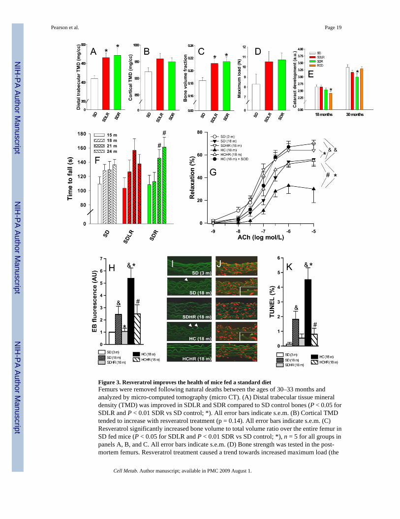
Figure 3. Resveratrol improves the health of mice fed a standard dietFemurs were removed following natural deaths between the ages of 30–33 months andanalyzed by micro-computed tomography (micro CT). (A) Distal trabecular tissue mineraldensity (TMD) was improved in SDLR and SDR compared to SD control bones (P < 0.05 forSDLR and P < 0.01 SDR vs SD control; *). All error bars indicate s.e.m. (B) Cortical TMDtended to increase with resveratrol treatment (p = 0.14). All error bars indicate s.e.m. (C)Resveratrol significantly increased bone volume to total volume ratio over the entire femur inSD fed mice (P < 0.05 for SDLR and P < 0.01 SDR vs SD control; *), n = 5 for all groups inpanels A, B, and C. All error bars indicate s.e.m. (D) Bone strength was tested in the post-mortem femurs. Resveratrol treatment caused a trend towards increased maximum load (the
Pearson et al. Page 19
Cell Metab. Author manuscript; available in PMC 2009 August 1.
NIH
-PA Author Manuscript
NIH
-PA Author Manuscript
NIH
-PA Author Manuscript

load that is endured by a bone prior to it failing in the three-point bending to failure test) (p =0.18), n = 5 for SD and SDLR and n = 4 for SDR. All error bars indicate s.e.m. (E) Resveratroltreatment delayed the onset of age-related cataracts. Lens opacity was scored in living miceon a scale from 0 to 4 by half steps of 0.5, with 4 representing the complete lens opacity of amature cataract. Age-related cataract development was significantly decreased at 30 monthsof age in SDR mice compared to the SD control, whereas EOD feeding caused an earlyprotective effect that was lost (P < 0.05; *). All error bars indicate s.e.m. (F) Time to fall froman accelerating rotarod was measured every 3 months for all survivors from a pre-designatedsubset of each group; n = 15 (SD), 11 (SDLR), and 16 (SDR). The SDR group improvedsignificantly at 21 and 24 months vs. 15 months (P < 0.05; #), showing increased motorcoordination over time. All error bars indicate s.e.m. (G) Acetylcholine-induced relaxation inaortic ring preparations. Both age-related (SD 18m vs SD 3m) and obesity-related (HC 18mvs SD 18m) declines in endothelial function were prevented by resveratrol treatment. Pre-incubation with SOD restored ACh-induced relaxation in the HC 18m rings to youthful levels,n = 6 for each group. (&P < 0.05 vs. SD 3m, *P < 0.05 vs SD 18m and #P < 0.05 vs. HC 18m).All error bars indicate s.e.m. (H) Quantification of total nuclear ethidium bromide fluorescenceas a marker of increased oxidative stress, n = 6 aortas for each group. This panel combines datafrom two experiments shown separately in Fig. S6P and S6Q. Both age-related (SD 18m vsSD 3m) and obesity-related (HC 18m vs SD 18m) increases in oxidative stress were preventedby resveratrol treatment. (&P < 0.05 vs. SD 3m, *P < 0.05 vs. SD 18m and #P < 0.05 vs. HC18m). All error bars indicate s.e.m. (I, J) Representative TUNEL staining of aortas from miceof the indicated ages and diets. Nuclei from apoptotic endothelial cells (intense green) in aortasof SD 18m and HC 18m mice are highlighted in insets. Autofluorescence of elastic laminae(faint green) and nuclear counterstaining (propidium iodide, red) are shown for orientationpurposes. (K) Apoptotic index (% of TUNEL positive endothelial cell nuclei), was increasedin the aortas of obese mice, and this change was prevented by resveratrol treatment, n = 6 aortasfor each group, 10 to 15 images per aorta were analyzed. (&P < 0.05 vs. SD 3m, *P < 0.05 vs.SD 18m and #P < 0.05 vs. HC 18m). All error bars indicate s.e.m.
Pearson et al. Page 20
Cell Metab. Author manuscript; available in PMC 2009 August 1.
NIH
-PA Author Manuscript
NIH
-PA Author Manuscript
NIH
-PA Author Manuscript
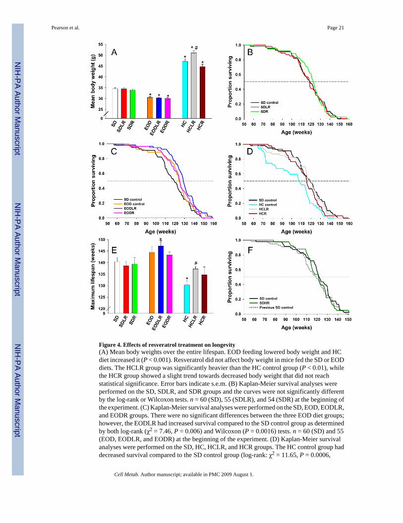
Figure 4. Effects of resveratrol treatment on longevity(A) Mean body weights over the entire lifespan. EOD feeding lowered body weight and HCdiet increased it (P < 0.001). Resveratrol did not affect body weight in mice fed the SD or EODdiets. The HCLR group was significantly heavier than the HC control group (P < 0.01), whilethe HCR group showed a slight trend towards decreased body weight that did not reachstatistical significance. Error bars indicate s.e.m. (B) Kaplan-Meier survival analyses wereperformed on the SD, SDLR, and SDR groups and the curves were not significantly differentby the log-rank or Wilcoxon tests. n = 60 (SD), 55 (SDLR), and 54 (SDR) at the beginning ofthe experiment. (C) Kaplan-Meier survival analyses were performed on the SD, EOD, EODLR,and EODR groups. There were no significant differences between the three EOD diet groups;however, the EODLR had increased survival compared to the SD control group as determinedby both log-rank (χ2 = 7.46, P = 0.006) and Wilcoxon (P = 0.0016) tests. n = 60 (SD) and 55(EOD, EODLR, and EODR) at the beginning of the experiment. (D) Kaplan-Meier survivalanalyses were performed on the SD, HC, HCLR, and HCR groups. The HC control group haddecreased survival compared to the SD control group (log-rank: χ2 = 11.65, P = 0.0006,
Pearson et al. Page 21
Cell Metab. Author manuscript; available in PMC 2009 August 1.
NIH
-PA Author Manuscript
NIH
-PA Author Manuscript
NIH
-PA Author Manuscript
Wilcoxon: P = 0.0003), whereas, survival in the HCLR and HCR groups did not differsignificantly from that of SD controls. When compared to HC controls, survival wassignificantly increased in both the HCLR group (log-rank: χ2 = 8.31, P = 0.004, Wilcoxon:P = 0.005) and the HCR group (log-rank: χ2 = 4.83, P = 0.03, Wilcoxon: P = 0.001). n = 60(SD) and 55 (HC, HCLR, and HCR) at the beginning of the experiment. (E) Maximum lifespanwas calculated as the mean of the final 20% of mice in each group as determined by Kaplan-Meier analysis. Compared to the SD control group, the maximum lifespan was significantlyincreased in the EODLR (P = 0.03) and significantly decreased in the HC control (P = 0.003)groups. In addition, HCLR had significantly increased maximum lifespan compared to the HCcontrol group (P = 0.04), and there was a trend towards increased lifespan in HCR micecompared to HC controls. *,P < 0.05 vs SD control; #, P < 0.05 vs HC control. Error barsindicate s.e.m. (F) Kaplan-Meier survival analyses were performed on the SD and SDHRgroups, and the curves were not significantly different. The earlier SD survival curve (brokenline) is shown for reference. n = 48 (SD, SDHR), and 60 (previous SD control) at the beginningof the experiment.
Pearson et al. Page 22
Cell Metab. Author manuscript; available in PMC 2009 August 1.
NIH
-PA Author Manuscript
NIH
-PA Author Manuscript
NIH
-PA Author Manuscript