Embed Size (px)
Citation preview
Life Science Journal 2016;13(1s) http://www.lifesciencesite.com
93
Aluminum –Induced Oxidative Stress and Hepato -Renal Impairment in Male Albino Rats: Possible Protective trial with Naringenin
Tamer S. Imam1, Hesham A. Khalifa2, Mohamed M.A.Hussein3 and Haytham. A. Ali3
1Department of Forensic Medicine & Toxicology, Faculty of Vet, Medicine, Zagazig University, Zagazig, Egypt. 2Department of Pharmacology, Faculty of Vet, Medicine, Zagazig University, Zagazig, Egypt.
3Department of Biochemistry, Faculty of Vet, Medicine, Zagazig University, Zagazig Egypt. [email protected]
Abstract: Aluminum is widely distributed in the environment and present in various food additives, manufactured foods and therapeutic treatment. Also, it is added to drinking water during purification purposes, thus increasing human and animal exposure risk to this metal. A body of evidence has accumulated implicating the free radical generation with subsequent oxidative stress in the biochemical and molecular mechanisms of aluminum toxicity. Naringenin (NAR) is a naturally occurring plant bioflavonoid, which has been reported to have awide range of pharmacological properties and free radicals scavenging activities. Since liver and kidney considered critical organs for Al toxicity, so this study was conducted to elucidate the effectiveness of naringenin in alleviating Al-induced liver and kidney toxicity in rats. For this, forty male albino rats were allocated into four groups. Group I (control) received normal saline. Group II was administered NAR at a dose of 50 mg/kg b.wt. Group III was orally treated with AlCl3 at a dose of 34 mg/kg b.wt. Group IV was simultaneously treated with AlCl3 and NAR together at the same doses. All treatments were performed daily via oral route and maintained for 70 days. Hepatic and renal dysfunction was evaluated by investigating serum biomarkers. Liver and kidney GPx activity, GSH level and MDA level as indicator of lipid peroxidation were assayed. Histopathological examination of liver and kidney was conducted. Aluminum treatment resulted in a significant increase in serum AST, ALT, ALP activities, billirubin concentration, urea and creatinine level with decreased serum total protein and albumin. AlCl3 significantly inhibited the GPx activity, reduced GSH level and increased MDA level in liver and kidney. Histopathological examination revealed deformities in hepatic and renal tissues due to aluminum exposure which augment the aforementioned results. Co- administration of NAR along with Al significantly restore the serum biomarkers to their near-normal levels and have the ability to overcome Al –induced oxidative stress, manifested by significant reduction in hepatic and renal MDA level, with enhanced cellular antioxidant defense particulary GPx and GSH and preserved normal hepatic and renal histological architecture. From these results, it could be concluded that NAR can mop up Al-induce toxicity, suggesting that the hepatoprotective and nephroprotective potential of NAR in aluminum toxicity may be owed to its antioxidant and metal chelating properties, which may be useful in achieving optimum effects in Al-induced hepatic and renal damage. [Tamer S. Imam, Hesham A. Khalifa, Mohamed M.A.Hussein and Haytham. A. Ali. Aluminum –Induced Oxidative Stress and Hepato -Renal Impairment in Male Albino Rats: Possible Protective trial with Naringenin. Life Sci J 2016;13(1s):93-104]. ISSN 1097-8135 (print); ISSN 2372-613X (online) http://www.lifesciencesite.com. 10. doi:10.7537/marslsj1301s1610. Keywords Aluminum, naringenin, antioxidants activities, liver, kidney, rats. 1. Introduction
Aluminum is the third most abundant metal, comprising about (8%) of the earth’s crust, is found in combination with oxygen, silicon, fluorine and other elements in soil, rocks, clays and gems and has a significant toxic potential for humans (Verstraeten et al., 2008). Aluminum is widely distributed in the environment and extensively used in daily life, which was enhanced by the belief that it is non-toxic and is quickly excreted from the body in urine. However, this element has a negative impact on human health (Kumar and Gill, 2009). Aluminum is a constituent of cooking utensils especially in developing countries and medicines such as antacids, phosphate binders, buffered aspirins, vaccines, antiperspirants and
allergen injection (Exley,1998) and this has allowed its easy access into the body (Yokel, 2000). The primary sources of aluminum are corn, yellow cheese, herbs, spices, salts, tea, cosmetics, cookware, aluminum ware and containers. Also it is widely used in food additives, deodorants and toothpaste (Abbasali et al., 2005). Aluminum gets access to the body via the gastrointestinal and respiratory tracts and accumulates in many tissues, such as kidney, liver, heart, blood, bone and brain (Al-Kahtani, 2010). Environmental pollution with the different aluminum containing compounds, especially those in industrial waste water, and from particulate matter distributed by cement –producing factories containing high amount of aluminum expose the populations residing in the

Life Science Journal 2016;13(1s) http://www.lifesciencesite.com
94
vicinity to higher than normal level of aluminum (Shehla et al., 2001).
The toxic effect of aluminum have been suggested to be mediated by reactive oxygen species generation resulting in the oxidative deterioration of cellular lipids, proteins and DNA and also induces changes in the activities of tissue antioxidant enzymes (Farina et al., 2005; Mailloux et al., 2011), altered gene expression and apoptosis (Stohs et al., 2000). The induced oxidative stress by aluminum and its salts is responsible for hepatotoxicity (Mailloux et al., 2011), nephrotoxicity (Farina et al., 2005), cardiac toxicity (Nan et al., 2009), reproductive toxicity (Al-Hashem, 2009) and also neurodegenerative disease and Alzheimer like neurofibrillary tangle formation (Nehru and Anand, 2005). Aluminum ions alter the properties and structure of cellular membranes, inhibit many enzymes like alkaline phosphatase, acetylocholinestrase, and adenylcyclase (Exley and Birchall, 1992; Ward et al., 2001). Additionally, antagonistic interactions between Al ions and other elements such as magnesium, calcium, silicon, iron, phosphorus, zinc, and copper were observed in biological systems (Ward et al., 2001). The excessive mitochondrial reactive oxygen species generation triggers hepatocyte apoptosis and depletes the endogenous antioxidant enzymes through activation of the caspases cascade, such as caspase-3, -8, and 9. Therefore, the external supply of antioxidants is important to suppress caspase activation and for the defense against the deleterious effects of oxidative stress (Schulze-Bergkamen et al., 2006; Ozben, 2007). Although several chelating agents and antagonists are establishedto reduce the metal toxicity, some of them are burned with undesirable side effects. Due to the intrinsic limitations and variability of efficacy of heavy metal chelating agents, metal intoxication therapy is looking for the development of new therapeutic agents with different actions especially from phytochemicals. Flavonoids are one of the most numerous and widespread group of naturally occurring antioxidants that can inhibit lipid oxidation in a biological membrane. They are found invegetables, fruits, nuts, seeds, leaves and barks of plants (Middleton et al., 2000). They usually contain one or more aromatic hydroxyl groups in their moiety which is responsible for the antioxidant properties of the flavonoids (Van Acker et al., 2000). Naringenin(4, 5, 7-trihydroxy flavonone) is a bioflavonoid plant found in tomato, cherries, grapefruit, citrus fruits, and coca. Naringenin (NAR) have already been pharmacologically evaluated as anticancer (So et al., 1997), a potential antioxidant (Santos et al., 1999), antiatherogenic (Lee et al., 2001), hepatoprotective (Lee et al., 2004), nephroprotective (Badary et al., 2005) and anti-
mutagenic (Choi et al., 1994) activities. The number of hydroxyl substitutions of NAR can donate hydrogen to ROS, allowing acquisition of stable structure, thus enabling scavenging of these free Radicals (Shen et al., 2004; Heo et al., 2004). Naringenin may modulate cytochrome P450-dependent monooxygenase, the primary enzyme involved in the metabolism of many xenobiotics (Ueng et al., 1999). Furthermore, Van Acker et al. (2000) reported that aglycone of naringenin, naringin can assume the role of alpha-tocopherol as achain-breaking antioxidant in liver microsomal membrane. To our knowledge the role of naringenin against Al-induced deteriorations in hepatic and renal tissues has not so far been studied. Therefore, taking the above into account, the present study was carried out to determine hepatotoxicity and nephrotoxicity of AlCl3 in adult male albino rats and to evaluate the antioxidant, hepatoprotective and nephroprotective potential of naringenin against the possible -induced hepatic and renal dysfunction caused by AlCl3. 2. Material and Methods I-Chemicals and Reagents
Aluminum chloride (AlCl3) was purchased from Al Gomhoria Chemical Company, Egypt. Naringenin (NAR) was purchased from Sigma Chemical Company, St. Louis, Missouri, USA. All other chemicals used in this study were of analytical grade. II-Animals
Forty male albino rats, weighing at the beginning of the experiment (220±20)gm., were purchased from laboratory animal farm (Helwan, Cairo, Egypt) and housed (Animal House, Faculty of Veterinary Medicine, Zagazig University, Egypt) in standard cages in groups of ten animals per cage under controlled conditions (temperature 25±0.5°C, a 12:12 light/dark cycle), and relative humidity (50–60%) and were given a standard diet and water ad libitum. Animal handling and treatment procedures were conducted according to the Guidelines for the Care and Use of laboratory animals of the National Inistitute of Health(NIH) and approved by a research ethics committee of the Faculty of Veterinary Medicine at Zagazig University. Animals were acclimatized under control condition at the laboratory for two weeks before use. III-Experimental Design and Sampling protocol
Rats were randomly divided into four main groups (ten/each group):
Group I (control group) received normal saline. Group II (NAR-treated group) was gavaged with NAR at dose of 50 mg /kg/ b.wt (Mershibo et al., 2013). Group III (AlCl3-treated group) was orally administered AlCl3 at a dose of 34 mg /kg b.wt (Newairy et al., 2009). Group IV (AlCl3+ NAR-
Life Science Journal 2016;13(1s) http://www.lifesciencesite.com
95
treated group) was simultaneously treated with AlCl3
(34mg/ kg b.wt) and NAR (50 mg /kg b.wt). Rats were orally administered their respective doses daily for 70 days.
At the end of the experimental period, rats of each group were scarified by decapitation; blood was collected directly into plain tubes without anticoagulants then centrifuged at 3000 rpm for 10 min to obtain serum, stored at -20°C in clean capped tubes until conducting the biochemical analysis of liver and kidney functions. Livers and kidneys were dissected rapidly, a part of these tissues were separately homogenized in 5ml cold 20 mM HEPES buffer, pH 7.2, containing 1mM EDTA, 210 mM mannitol, and 70 mM sucrose per gram tissue. Homogenates were centrifuged at 10.000 x g for15 minutes at 4 °C and the resultant supernatant was removed and stored at -80°C until used for antioxidant enzyme activities and lipid peroxidation (MDA) assays. The other part of liver and kidney tissues were promptly washed with normal saline and processed for histopathological studies. IV-Biochemical investigation 1-Estimation of serum biochemical parameters
Serum ALT and AST were determined using Spectrum Kits, Egyptian Company for Biotechnology, Cairo, Egypt (REF: 265 002 for ALT and 261 002 for
AST) following the steps recommended by kits according to (Breuer, 1996). Alkaline phosphatase (ALP) was measured calorimetrically (Belfield and Golberg, 1971). Serum total proteins and albumin were determined according to (Doumas et al., 1981) and (Drupt, 1974) respectively, while total bilirubin was determined calorimetrically according to (Schmidt and Eisenburg, 1975). Serum urea and creatinine were determined according to (Patton and Crouch, 1977) and (Bonsens and Taussky, 1984) respectively. 2-Antioxidants and lipid peroxidation assays in tissue homogenate
Reduced glutathione (GSH) level was determined in the liver and kidney tissue homogenates using a kit supplied by Cayman (Cat. No. 703002, Cayman, USA) according to (Ellman, 1959). Glutathione peroxidase (GPx) activity was measured by a kit (Cat. No. NWK-GPX01) obtained from Northwest Life Science Specialties (NWLSSTM) according to (Lawrence and Burk, 1976). Malondialdehyde (MDA) level was analyzed by estimation of the produced thiobarbituric acid reactive substances (TBARS) by the method of (Buege and Aust, 1978) using a TBARS assay kit (Cat. No. 10009055, Cayman, USA).
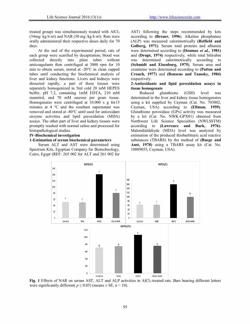
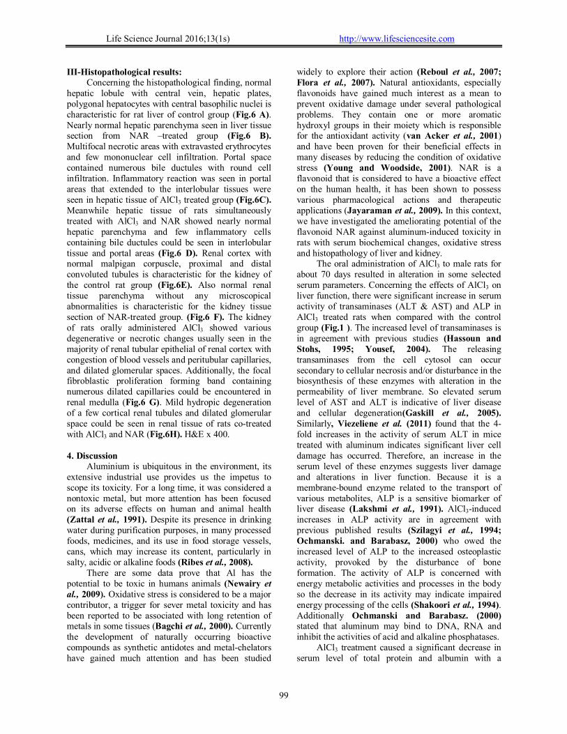
Fig. 1 Effects of NAR on serum AST, ALT and ALP activities in AlCl3-treated rats. Bars bearing different letters were significantly different( p ≤ 0.05) (means ± SE, n = 10).
Life Science Journal 2016;13(1s) http://www.lifesciencesite.com
96
V-Histopathological Examination Small pieces of liver and kidney tissues were
collected and fixed at 10% buffered neutral formalin solution, dehydrated in graded ethanol (70-100%), cleared in xylene and embedded in paraffin. Five micron thick paraffin sections were prepared and routinely stained with hematoxylin and eosin (HE) dyes (Wilson and Gamble, 2008) and then examined microscopically. VI-Statistical analysis
The results were expressed as the mean ± standard error (SE). Statistical analysis was performed using oneway analysis of variance (ANOVA) followed by the post hoc Duncan’s test for comparison between different experimental groups. This was carried out using IBM SPSS Statistics computer software (version 21). p-values <0.05 were considered statistically significant. Results I- Effects of Al and co-treatment with NAR on serum liver and kidney functions (serum biochemistry)
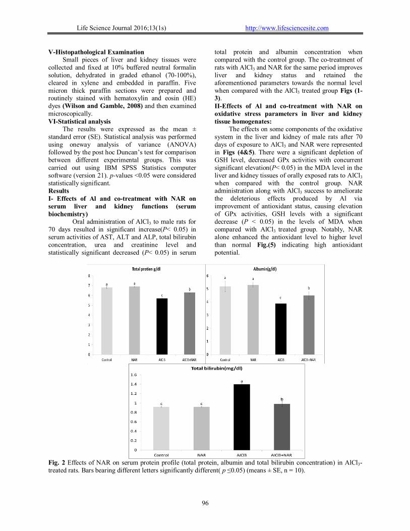
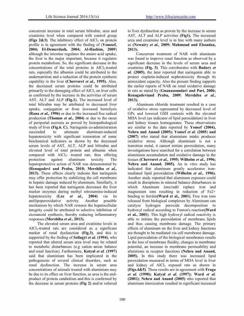
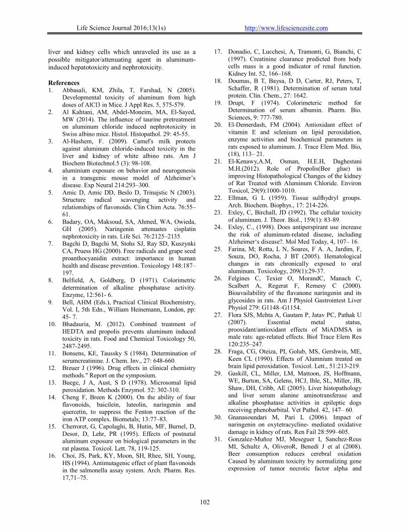
Oral administration of AlCl3 to male rats for 70 days resulted in significant increase(P˂ 0.05) in serum activities of AST, ALT and ALP, total bilirubin concentration, urea and creatinine level and statistically significant decreased (P˂ 0.05) in serum
total protein and albumin concentration when compared with the control group. The co-treatment of rats with AlCl3 and NAR for the same period improves liver and kidney status and retained the aforementioned parameters towards the normal level when compared with the AlCl3 treated group Figs (1-3). II-Effects of Al and co-treatment with NAR on oxidative stress parameters in liver and kidney tissue homogenates:
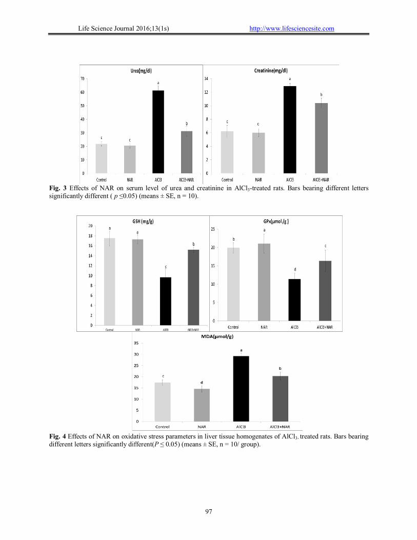
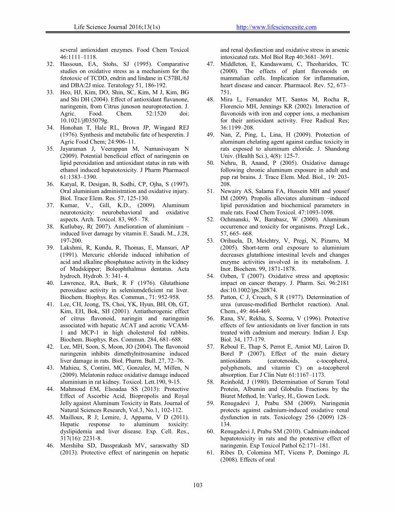
The effects on some components of the oxidative system in the liver and kidney of male rats after 70 days of exposure to AlCl3 and NAR were represented in Figs (4&5). There were a significant depletion of GSH level, decreased GPx activities with concurrent significant elevation(P˂ 0.05) in the MDA level in the liver and kidney tissues of orally exposed rats to AlCl3 when compared with the control group. NAR administration along with AlCl3 success to ameliorate the deleterious effects produced by Al via improvement of antioxidant status, causing elevation of GPx activities, GSH levels with a significant decrease (P ˂ 0.05) in the levels of MDA when compared with AlCl3 treated group. Notably, NAR alone enhanced the antioxidant level to higher level than normal Fig.(5) indicating high antioxidant potential.
Fig. 2 Effects of NAR on serum protein profile (total protein, albumin and total bilirubin concentration) in AlCl3-treated rats. Bars bearing different letters significantly different( p ≤0.05) (means ± SE, n = 10).
Life Science Journal 2016;13(1s) http://www.lifesciencesite.com
97
Fig. 3 Effects of NAR on serum level of urea and creatinine in AlCl3-treated rats. Bars bearing different letters significantly different ( p ≤0.05) (means ± SE, n = 10).
Fig. 4 Effects of NAR on oxidative stress parameters in liver tissue homogenates of AlCl3- treated rats. Bars bearing different letters significantly different(P ≤ 0.05) (means ± SE, n = 10/ group).
Life Science Journal 2016;13(1s) http://www.lifesciencesite.com
98
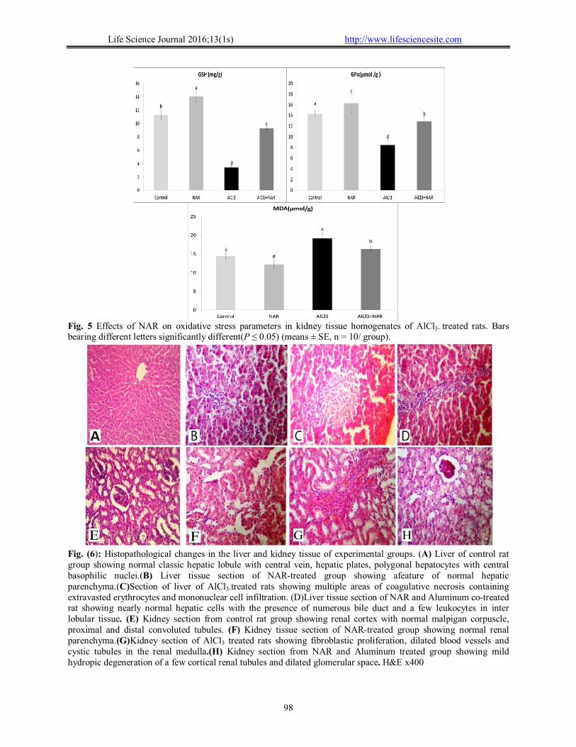
Fig. 5 Effects of NAR on oxidative stress parameters in kidney tissue homogenates of AlCl3- treated rats. Bars bearing different letters significantly different(P ≤ 0.05) (means ± SE, n = 10/ group).
Fig. (6): Histopathological changes in the liver and kidney tissue of experimental groups. (A) Liver of control rat group showing normal classic hepatic lobule with central vein, hepatic plates, polygonal hepatocytes with central basophilic nuclei.(B) Liver tissue section of NAR-treated group showing afeature of normal hepatic parenchyma.(C)Section of liver of AlCl3-treated rats showing multiple areas of coagulative necrosis containing extravasted erythrocytes and mononuclear cell infiltration. (D)Liver tissue section of NAR and Aluminum co-treated rat showing nearly normal hepatic cells with the presence of numerous bile duct and a few leukocytes in inter lobular tissue. (E) Kidney section from control rat group showing renal cortex with normal malpigan corpuscle, proximal and distal convoluted tubules. (F) Kidney tissue section of NAR-treated group showing normal renal parenchyma.(G)Kidney section of AlCl3 treated rats showing fibroblastic proliferation, dilated blood vessels and cystic tubules in the renal medulla.(H) Kidney section from NAR and Aluminum treated group showing mild hydropic degeneration of a few cortical renal tubules and dilated glomerular space. H&E x400
Life Science Journal 2016;13(1s) http://www.lifesciencesite.com
99
III-Histopathological results: Concerning the histopathological finding, normal
hepatic lobule with central vein, hepatic plates, polygonal hepatocytes with central basophilic nuclei is characteristic for rat liver of control group (Fig.6 A). Nearly normal hepatic parenchyma seen in liver tissue section from NAR –treated group (Fig.6 B). Multifocal necrotic areas with extravasted erythrocytes and few mononuclear cell infiltration. Portal space contained numerous bile ductules with round cell infiltration. Inflammatory reaction was seen in portal areas that extended to the interlobular tissues were seen in hepatic tissue of AlCl3 treated group (Fig.6C). Meanwhile hepatic tissue of rats simultaneously treated with AlCl3 and NAR showed nearly normal hepatic parenchyma and few inflammatory cells containing bile ductules could be seen in interlobular tissue and portal areas (Fig.6 D). Renal cortex with normal malpigan corpuscle, proximal and distal convoluted tubules is characteristic for the kidney of the control rat group (Fig.6E). Also normal renal tissue parenchyma without any microscopical abnormalities is characteristic for the kidney tissue section of NAR-treated group. (Fig.6 F). The kidney of rats orally administered AlCl3 showed various degenerative or necrotic changes usually seen in the majority of renal tubular epithelial of renal cortex with congestion of blood vessels and peritubular capillaries, and dilated glomerular spaces. Additionally, the focal fibroblastic proliferation forming band containing numerous dilated capillaries could be encountered in renal medulla (Fig.6 G). Mild hydropic degeneration of a few cortical renal tubules and dilated glomerular space could be seen in renal tissue of rats co-treated with AlCl3 and NAR (Fig.6H). H&E x 400.
4. Discussion
Aluminium is ubiquitous in the environment, its extensive industrial use provides us the impetus to scope its toxicity. For a long time, it was considered a nontoxic metal, but more attention has been focused on its adverse effects on human and animal health (Zattal et al., 1991). Despite its presence in drinking water during purification purposes, in many processed foods, medicines, and its use in food storage vessels, cans, which may increase its content, particularly in salty, acidic or alkaline foods (Ribes et al., 2008).
There are some data prove that Al has the potential to be toxic in humans animals (Newairy et al., 2009). Oxidative stress is considered to be a major contributor, a trigger for sever metal toxicity and has been reported to be associated with long retention of metals in some tissues (Bagchi et al., 2000). Currently the development of naturally occurring bioactive compounds as synthetic antidotes and metal-chelators have gained much attention and has been studied
widely to explore their action (Reboul et al., 2007; Flora et al., 2007). Natural antioxidants, especially flavonoids have gained much interest as a mean to prevent oxidative damage under several pathological problems. They contain one or more aromatic hydroxyl groups in their moiety which is responsible for the antioxidant activity (van Acker et al., 2001) and have been proven for their beneficial effects in many diseases by reducing the condition of oxidative stress (Young and Woodside, 2001). NAR is a flavonoid that is considered to have a bioactive effect on the human health, it has been shown to possess various pharmacological actions and therapeutic applications (Jayaraman et al., 2009). In this context, we have investigated the ameliorating potential of the flavonoid NAR against aluminum-induced toxicity in rats with serum biochemical changes, oxidative stress and histopathology of liver and kidney.
The oral administration of AlCl3 to male rats for about 70 days resulted in alteration in some selected serum parameters. Concerning the effects of AlCl3 on liver function, there were significant increase in serum activity of transaminases (ALT & AST) and ALP in AlCl3 treated rats when compared with the control group (Fig.1 ). The increased level of transaminases is in agreement with previous studies (Hassoun and Stohs, 1995; Yousef, 2004). The releasing transaminases from the cell cytosol can occur secondary to cellular necrosis and/or disturbance in the biosynthesis of these enzymes with alteration in the permeability of liver membrane. So elevated serum level of AST and ALT is indicative of liver disease and cellular degeneration(Gaskill et al., 2005). Similarly, Viezeliene et al. (2011) found that the 4-fold increases in the activity of serum ALT in mice treated with aluminum indicates significant liver cell damage has occurred. Therefore, an increase in the serum level of these enzymes suggests liver damage and alterations in liver function. Because it is a membrane-bound enzyme related to the transport of various metabolites, ALP is a sensitive biomarker of liver disease (Lakshmi et al., 1991). AlCl3-induced increases in ALP activity are in agreement with previous published results (Szilagyi et al., 1994; Ochmanski. and Barabasz, 2000) who owed the increased level of ALP to the increased osteoplastic activity, provoked by the disturbance of bone formation. The activity of ALP is concerned with energy metabolic activities and processes in the body so the decrease in its activity may indicate impaired energy processing of the cells (Shakoori et al., 1994). Additionally Ochmanski and Barabasz. (2000) stated that aluminum may bind to DNA, RNA and inhibit the activities of acid and alkaline phosphatases.
AlCl3 treatment caused a significant decrease in serum level of total protein and albumin with a
Life Science Journal 2016;13(1s) http://www.lifesciencesite.com
100
concurrent increase in total serum bilirubin, urea and creatinine level when compared with control group (Figs 2&3). The inhibitory effect of AlCl3 on protein profile is in agreement with the finding of (Youssef, 2004; El-Demerdash, 2004; Al-Hashim, 2009) although the intestine regulates the amino acid uptake, the liver is the major important, because it regulates protein metabolism. So, the significant decrease in the concentrations of the total proteins in AlCl3-treated rats, espicially the albumin could be attributed to the undernutrition and a reduction of the protein synthesis capability in the liver (Cherroret et al., 1995). Also, the decreased serum proteins could be attributed primarily to the damaging effect of AlCl3 on liver cells as confirmed by the increase in the activities of serum AST, ALT and ALP (Fig.1). The increased level of total bilirubin may be attributed to decreased liver uptake, conjugation or from increased hemolysis (Rana et al., 1996) or due to the increased free radical production (Thomas et al., 2004) or due to the onset of periportal necrosis as proved by histopathological study of liver (Fig.6. C). Naringenin co-administration succeeded to attenuate aluminum-induced hepatotoxicity with significant restoration of some biochemical indicators as shown by the decreased serum levels of AST, ALT, ALP and bilirubin and elevated level of total protein and albumin when compared with AlCl3- treated rats, thus offering protection against aluminum toxicity. The hepatoprotective action of NAR was demonstrated by (Renugadevi and Prabu, 2010; Mershiba et al., 2013). These effects clearly indicate that naringenin may offer protection by stabilizing the cell membrane in hepatic damage induced by aluminum. Simillarly, it has been reported that naringenin decreases the liver marker enzymes during methyl nitrosamine-induced hepatotoxicity (Lee et al., 2004) via its antilipoperoxidative activity. Another possible mechanism by which NAR restores the hepatocellular integrity could be attributed to selective inhibition of eicosanoid synthesis, thereby reducing inflammatory responses (Mershiba et al., 2013).
The elevated serum urea and creatinine levels in AlCl3-treated rats are considered as a significant marker of renal dysfunction (Fig.3), and this is supported by the finding of Szilagyi et al. (1994), who reported that altered serum urea level may be related to metabolic disturbances (e.g cation–anion balance and renal function). Furthermore, Katyal et al. (1997) said that aluminium has been implicated in the pathogenesis of several clinical disorders, such as renal dysfunction. The increase in serum urea concentrations of animals treated with aluminium may be due to its effect on liver function, as urea is the end-product of protein catabolism and this is confirmed by the decrease in serum proteins (Fig 2) and/or referred
to liver dysfunction as proven by the increase in serum AST, ALT and ALP activities (Fig1). The increased urea and creatinine level is in line with many authors as (Newairy et al., 2009; Mahmoud and Elsoadaa, 2013).
Concurrent treatment of NAR with aluminum was found to improve renal function as observed by a significant decrease in the levels of serum urea and creatinine (Fig. 3). This corroborates with Badary et al. (2005), the later reported that naringenin able to protect cisplatin-induced nephrotoxicity through its antioxidant capacity. Also the present finding supports the earlier reports of NAR on renal oxidative damage in rats as stated by (Gnanasoundari and Pari, 2006; Renugadeviand Prabu, 2009; Mershiba et al., 2013).
Aluminum chloride treatment resulted in a case of oxidative stress represented by decreased level of GPx and lowered GSH contents with the elevated MDA level (an indicator of lipid peroxidation) in liver and kidney tissues homogenates. These observations are similar to the data reported by Yousef (2004), Nehru and Anand (2005); Yousef et al. (2005) and (2007) who stated that aluminium intake produces oxidative stress. Although aluminium is not a transition metal, it cannot initiate peroxidation, many investigations have searched for a correlation between aluminium accumulation and oxidative damage in the tissues (Cherroret et al., 1995; Wilhelm et al., 1996; Nehru and Anand. 2005). An in vitro study has indicated that aluminum greatly accelerates iron-mediated lipid peroxidation (Wilhelm et al., 1996). Another study reported that aluminum exposure could result in disruptions in mineral balance disturbances in which Aluminum ions/radii replace iron and magnesium ions resulting in reduction of Fe2+ binding to ferritin(Ward et al., 2001). Free iron ions released from biological complexes by Aluminum can catalyze hydrogen peroxide decomposition to hydroxyl radical according to Fenton's reaction(Ward et al., 2001). This high hydroxyl radical reactivity is able to initiate the peroxidation of membrane lipids and thus causing membrane damage. The primary effects of aluminum on the liver and kidney functions are thought to be mediated via cell membrane damage. Lipid peroxidation of the biological membranes results in the loss of membrane fluidity, changes in membrane potential, an increase in membrane permeability and alterations in receptor functions (Nehru and Anand, 2005). In this study there was increased lipid peroxidation measured in terms of MDA level in liver and kidney of AlCl3 exposed rats as shown in (Figs.4&5). These results are in agreement with Fraga et al. (1990); Katyal et al. (1997); Ward et al. (2001); Nehru and Anand (2005) who reported that aluminum intoxication resulted in significant increased

Life Science Journal 2016;13(1s) http://www.lifesciencesite.com
101
lipid peroxidation markers. The increased lipid peroxidation is due to an inhibition or changing the activity of non-enzymatic and enzymatic components of the oxidative system. Also Orihuela et al. (2005) reported that a high dose of aluminum is able to induce free radicals and so decrease the GSH level. Aluminum decreases the GSH synthesis probable by decreasing the activity of glutathione synthesis, thus leading to decreased GSH level. On the other hand, it has been demonstrated that aluminum can inhibit NADPH-generating enzymes such as NADP-isocitrate dehydrogenase and glucose 6-phosphate dehydrogenase. Since, NADPH is shown to be a main factor for the GSH regeneration, the decreased GSH level could be also ascribed to insufficient supply of NADPH (Newairy et al., 2009). The decreased GPx activities in liver and kidney are in accordance with Nehru and Anand, (2005); Newairy et al.(2009). The higher intracellular aluminum concentration reduced protein synthesis of antioxidant enzymes and subsequently reduced their activities (Nehru and Anand, 2005).
Simultaneous administrations of NAR with AlCl3 replenish the antioxidant enzyme activities near to normal seen as increased GPx activities, GSH level and diminish MDA level as a marker of LPO in liver and kidney when compared with AlCl3-treated rats. This might be due to the ability of naringenin to reduce the accumulation of free radical generation during Al-induced lipid peroxidation. Mira et al. (2002) reported that naringenin protects cells from ROS mediated cell death through its antioxidant nature. Administration of NAR can chelate the ferrous iron and decrease the formation of hydroxyl radical via inhibition of iron-dependent Fenton reaction (Cheng and Breen, 2000). It is well documented that NAR effectively quenches free radicals because of their 4-hydroxyl group in the ring possessing electron donating properties, also being on radical target which thereby protect the membrane from free radical attack and thus protect the membrane and inhibit the lipid peroxidation (Amic et al., 2003). The lipophilic nature of naringenin is able to facilitate its adherence to lipid bilayer which might decrease the formation of free radicals and protects the cell membrane (Honohan et al., 1976). Naringenin able to inhibit the radical generation, could further reduce the oxidative threat caused by aluminum, which could mitigate the consumption of endogenous enzymatic and non-enzymatic antioxidants and increased their levels and markedly reduces the hepatic and renal LPO. The biological mechanism may be associated with their metal-binding abilities which urge on the antioxidant action of NAR in the liver and kidney tissue. Additionally, Felgines et al. (2000) reported that the
phenolic acid metabolites of NAR were involved in the antioxidant activity.
Our aforementioned results were corroborated by histopathological studies of liver and kidney. The histopathological observations of the liver and kidney tissues revealed many deformities due to aluminum treatment. The observed histological alterations in hepatic tissues of aluminum treatment include multifocal necrotic areas with extravasted erythrocytes and leukocytes infiltration (Fig.6C). Various degenerative or necrotic changes seen in the majority of renal tubular epithelial of renal cortex with congestion of blood vessels and peritubular capillaries, and dilated glomerular spaces were seen in examining kidney tissue (Fig.6G). These results are concordant with other reports (El-Kenawy et al., 2012; Bhaduria, 2012; Mahmoud and Elsoadaa, 2013; Shirvastava, 2013; Al Kahtani et al., 2014). These histopathological changes may be attributed to the Al accumulation in the liver and kidneys, which promote degeneration of hepatic and renal tubular cells(El-Kenawy et al., 2012). Additionally, Al- induced hepatotoxicity (Kutlubay, 2007) and nephrotoxicity (Mahieu et al., 2009) is mediated by free radical generation and subsequent lipid peroxidation that can cause cytotoxicity and hepatocellular damage. Administration of NAR reduced the histological alterations provoked by aluminum quite appreciable. It can be attributed to the NAR antiradical/ antioxidant efficacy, which significantly decreased the oxidative stress leading to the reduction of histopathological alterations and restoration of normal physiological state of an organism. Previous results have suggested that NAR reduced the histological alterations caused by cadmium-induced nephrotoxicity Renugadevi and Prabu (2009), hepatotoxicity Renugadevi and Prabu (2010) and oxytetracycline induced- acute proximal tubular necrosis Gnanasoundari and Pari (2006).
Taken together, our findings indicate that the administration of naringenin in aluminum intoxicated rats counteracted the oxidative hepatic and renal dysfunction attributed by aluminum. Treatment with naringenin appreciably reduced the abnormal changes induced by aluminum and restored the serum biomarkers, increased antioxidant enzyme activities and decreased level of MDA towards near normal. Hepatoprotective and nephroprotective nature of naringenin against aluminum was further supported by the improvement in the histopathological changes occasioned by aluminum in liver and kidney.
Conclusion
In view of the present study, it can be concluded that naringenin played a role of an antioxidant, which includes free radical scavenging and metal-chelating property and thereby improved the detrimental state of
Life Science Journal 2016;13(1s) http://www.lifesciencesite.com
102
liver and kidney cells which unraveled its use as a possible mitigator/attenuating agent in aluminum- induced hepatotoxicity and nephrotoxicity.
References 1. Abbasali, KM, Zhila, T, Farshad, N (2005).
Developmental toxicity of aluminum from high doses of AlCl3 in Mice. J Appl Res. 5, 575-579.
2. Al Kahtani, AM, Abdel-Moneim, MA, El-Sayed, MW (2014). The influence of taurine pretreatment on aluminum chloride induced nephrotoxicity in Swiss albino mice. Histol. Histopathol. 29: 45-55.
3. Al-Hashem, F. (2009). Camel's milk protects against aluminum chloride-induced toxicity in the liver and kidney of white albino rats. Am J Biochem Biotechnol.5 (3): 98-108.
4. aluminium exposure on behavior and neurogenesis in a transgenic mouse model of Alzheimer’s disease. Exp Neural 214:293–300.
5. Amic D, Amic DD, Beslo D, Trinajstic N (2003). Structure radical scavenging activity and relationships of flavonoids. Clin Chim Acta. 76:55–61.
6. Badary, OA, Maksoud, SA, Ahmed, WA, Owieda, GH (2005). Naringenin attenuates cisplatin nephrotoxicity in rats. Life Sci. 76:2125–2135.
7. Bagchi D, Bagchi M, Stohs SJ, Ray SD, Kuszynki CA, Pruess HG (2000). Free radicals and grape seed proanthocyanidin extract: importance in human health and disease prevention. Toxicology 148:187–197.
8. Belfield, A, Goldberg, D (1971). Colorimetric determination of alkaline phosphatase activity. Enzyme, 12:561- 6.
9. Bell, AHM (Eds.), Practical Clinical Biochemistry, Vol. I, 5th Edn., William Heinemann, London, pp: 45- 7.
10. Bhadauria, M. (2012). Combined treatment of HEDTA and propolis prevents aluminum induced toxicity in rats. Food and Chemical Toxicology 50, 2487-2495.
11. Bonsens, KE, Taussky S (1984). Determination of serumcreatinine. J. Chem. Inv., 27: 648-660.
12. Breuer J (1996). Drug effects in clinical chemistry methods." Report on the symposium.
13. Buege, J A, Aust, S D (1978). Microsomal lipid peroxidation. Methods Enzymol. 52: 302-310.
14. Cheng F, Breen K (2000). On the ability of four flavonoids, baicilein, luteolin, naringenin and quercetin, to suppress the Fenton reaction of the iron ATP complex. Biometals; 13:77–83.
15. Cherroret, G, Capolaghi, B, Hutin, MF, Burnel, D, Desor, D, Lehr, PR (1995). Effects of postnatal aluminum exposure on biological parameters in the rat plasma. Toxicol. Lett. 78, 119-125.
16. Choi, JS, Park, KY, Moon, SH, Rhee, SH, Young, HS (1994). Antimutagenic effect of plant flavonoids in the salmonella assay system. Arch. Pharm. Res. 17,71–75.
17. Donadio, C, Lucchesi, A, Tramonti, G, Bianchi, C (1997). Creatinine clearance predicted from body cells mass is a good indicator of renal function. Kidney Int. 52, 166–168.
18. Doumas, B T, Baysa, D D, Carter, RJ, Peters, T, Schaffer, R (1981). Determination of serum total protein. Clin. Chem., 27: 1642.
19. Drupt, F (1974). Colorimeteric method for Determination of serum albumin. Pharm. Bio. Sciences, 9: 777-780.
20. El-Demerdash, FM (2004). Antioxidant effect of vitamin E and selenium on lipid peroxidation, enzyme activities and biochemical parameters in rats exposed to aluminum. J. Trace Elem Med. Bio, (18), 113– 21.
21. El-Kenawy,A.M, Osman, H.E.H, Daghestani M.H.(2012). Role of Propolis(Bee glue) in improving Histopathological Changes of the kidney of Rat Treated with Aluminum Chloride. Environ Toxicol, 29(9):1000-1010.
22. Ellman, G L (1959). Tissue sulfhydryl groups. Arch. Biochem. Biophys., 17: 214-226.
23. Exley, C, Birchall, JD (1992). The cellular toxicity of aluminum. J. Theor. Biol., 159(1): 83-89.
24. Exley, C., (1998). Does antiperspirant use increase the risk of aluminum-related disease, including Alzheimer‘s disease?. Mol Med Today, 4, 107– 16.
25. Farina, M; Rotta, L N, Soares, F A. A, Jardim, F, Souza, DO, Rocha, J BT (2005). Hematological changes in rats chronically exposed to oral aluminum. Toxicology, 209(1):29-37.
26. Felgines C, Texier O, MorandC, Manach C, Scalbert A, Regerat F, Remesy C (2000). Bioavailability of the flavanone naringenin and its glycosides in rats. Am J Physiol Gastrointest Liver Physiol 279: G1148–G1154.
27. Flora SJS, Mehta A, Gautam P, Jatav PC, Pathak U (2007). Essential metal status, prooxidant/antioxidant effects of MiADMSA in male rats: age-related effects. Biol Trace Elem Res 120:235–247.
28. Fraga, CG, Oteiza, PI, Golub, MS, Gershwin, ME, Keen CL (1990). Effects of Alumnium treated on brain lipid peroxidation. Toxicol. Lett., 51:213-219.
29. Gaskill, CL, Miller, LM, Mattoon, JS, Hoffmann, WE, Burton, SA, Gelens, HCJ, Ihle, SL, Miller, JB, Shaw, DH, Cribb, AE (2005). Liver histopathology and liver serum alanine aminotransferase and alkaline phosphatase activities in epileptic dogs receiving phenobarbital. Vet Pathol. 42, 147– 60.
30. Gnanasoundari M, Pari L (2006). Impact of naringenin on oxytetracycline- mediated oxidative damage in kidney of rats. Ren Fail 28:599–605.
31. Gonzalez-Muñoz MJ, Meseguer I, Sanchez-Reus MI, Schultz A, OliveroR, Benedí J et al (2008). Beer consumption reduces cerebral oxidation Caused by aluminum toxicity by normalizing gene expression of tumor necrotic factor alpha and
Life Science Journal 2016;13(1s) http://www.lifesciencesite.com
103
several antioxidant enzymes. Food Chem Toxicol 46:1111–1118.
32. Hassoun, EA, Stohs, SJ (1995). Comparative studies on oxidative stress as a mechanism for the fetotoxic of TCDD, endrin and lindane in C57BL/6J and DBA/2J mice. Teratology 51, 186-192.
33. Heo, HJ, Kim, DO, Shin, SC, Kim, M J, Kim, BG and Shi DH (2004). Effect of antioxidant flavanone, naringenin, from Citrus junoson neuroprotection. J. Agric. Food. Chem. 52:1520 doi: 10.1021/jf035079g.
34. Honohan T, Hale RL, Brown JP, Wingard REJ (1976). Synthesis and metabolic fate of hesperetin. J Agric Food Chem; 24:906–11.
35. Jayaraman J, Veerappan M, Namasivayam N (2009). Potential beneficial effect of naringenin on lipid peroxidation and antioxidant status in rats with ethanol induced hepatotoxicity. J Pharm Pharmacol 61:1383–1390.
36. Katyal, R, Desigan, B, Sodhi, CP, Ojha, S (1997). Oral aluminium administration and oxidative injury. Biol. Trace Elem. Res. 57, 125-130.
37. Kumar, V., Gill, K.D., (2009). Aluminum neurotoxicity: neurobehavioral and oxidative aspects. Arch. Toxicol. 83, 965– 78.
38. Kutlubay, R( 2007). Amelioration of aluminium –induced liver damage by vitamin E. Saudi. M., J.28, 197-200.
39. Lakshmi, R, Kundu, R, Thomas, E, Mansuri, AP (1991). Mercuric chloride induced inhibation of acid and alkaline phosphatase activity in the kidney of Mudskipper; Boleophthalmus dentatus. Acta hydroch. Hydrob. 3: 341- 4.
40. Lawrence, RA, Burk, R F (1976). Glutathione peroxidase activity in seleniumdeficient rat liver. Biochem. Biophys. Res. Commun., 71: 952-958.
41. Lee, CH, Jeong, TS, Choi, YK, Hyun, BH, Oh, GT, Kim, EH, Bok, SH (2001). Antiatherogenic effect of citrus flavonoid, naringin and naringenin associated with hepatic ACAT and acrotic VCAM-1 and MCP-1 in high cholesterol fed rabbits. Biochem. Biophys. Res. Commun. 284, 681–688.
42. Lee, MH, Soon, S, Moon, JO (2004). The flavonoid naringenin inhibits dimethylnitrosamine induced liver damage in rats. Biol. Pharm. Bull. 27, 72–76.
43. Mahieu, S, Contini, MC, Gonzalez, M, Millen, N (2009). Melatonin reduce oxidative damage induced aluminium in rat kidney. Toxicol. Lett.190, 9-15.
44. Mahmoud EM, Elsoadaa SS (2013): Protective Effect of Ascorbic Acid, Biopropolis and Royal Jelly against Aluminum Toxicity in Rats. Journal of Natural Sciences Research, Vol.3, No.1, 102-112.
45. Mailloux, R J; Lemire, J, Appama, V D (2011). Hepatic response to aluminum toxicity: dyslipidemia and liver disease. Exp. Cell. Res., 317(16): 2231-8.
46. Mershiba SD, Dassprakash MV, saraswathy SD (2013). Protective effect of naringenin on hepatic
and renal dysfunction and oxidative stress in arsenic intoxicated rats. Mol Biol Rep 40:3681–3691.
47. Middleton, E, Kandaswami, C, Theoharides, TC (2000). The effects of plant flavonoids on mammalian cells. Implication for inflammation, heart disease and cancer. Pharmacol. Rev. 52, 673–751.
48. Mira L, Fernandez MT, Santos M, Rocha R, Florencio MH, Jennings KR (2002). Interaction of flavonoids with iron and copper ions, a mechanism for their antioxidant activity. Free Radical Res; 36:1199–208.
49. Nan, Z, Ping, L, Lina, H (2009). Protection of aluminum chelating agent against cardiac toxicity in rats exposed to aluminum chloride. J. Shandong Univ. (Health Sci.), 4(8): 125-7.
50. Nehru, B, Anand, P (2005). Oxidative damage following chronic aluminum exposure in adult and pup rat brains. J. Trace Elem. Med. Biol., 19: 203-208.
51. Newairy AS, Salama FA, Hussein MH and yousef IM (2009). Propolis alleviates aluminum –induced lipid peroxidation and biochemical parameters in male rats. Food Chem Toxicol. 47:1093-1098.
52. Ochmanski, W, Barabasz, W (2000). Aluminum occurrence and toxicity for organisms. Przegl Lek., 57, 665- 668.
53. Orihuela, D, Meichtry, V, Pregi, N, Pizarro, M (2005). Short-term oral exposure to aluminium decreases glutathione intestinal levels and changes enzyme activities involved in its metabolism. J. Inor. Biochem. 99, 1871-1878.
54. Ozben, T (2007). Oxidative stress and apoptosis: impact on cancer therapy. J. Pharm. Sci. 96:2181 doi:10.1002/jps.20874.
55. Patton, C J, Crouch, S R (1977). Determination of urea (urease-modified Berthelot reaction). Anal. Chem., 49: 464-469.
56. Rana, SV, Rekha, S, Seema, V (1996). Protective effects of few antioxidants on liver function in rats treated with cadmium and mercury. Indian J. Exp. Biol. 34, 177-179.
57. Reboul E, Thap S, Perrot E, Amiot MJ, Lairon D, Borel P (2007). Effect of the main dietary antioxidants (carotenoids, c-tocopherol, polyphenols, and vitamin C) on a-tocopherol absorption. Eur J Clin Nutr 61:1167–1173.
58. Reinhold, J (1980). Determination of Serum Total Protein, Albumin and Globulin Fractions by the Biuret Method, In: Varley, H., Gowen Lock.
59. Renugadevi J, Prabu SM (2009). Naringenin protects against cadmium-induced oxidative renal dysfunction in rats. Toxicology 256 (2009) 128–134.
60. Renugadevi J, Prabu SM (2010). Cadmium-induced hepatotoxicity in rats and the protective effect of naringenin. Exp Toxicol Pathol 62:171–181.
61. Ribes D, Colomina MT, Vicens P, Domingo JL (2008). Effects of oral
Life Science Journal 2016;13(1s) http://www.lifesciencesite.com
104
62. Santos, KF, Oliveria, TT, Nagem, TJ, Pinto, AS, Oliveria, MG (1999). Hypolipidemic effects of naringenin, rutin, nicotinic acid and their association. Pharmacol. Res. 40, 493–496.
63. Schmidt, M, Eisenburg, J (1975). Serum bilirubin determination in new born infants. A new micro method for the determination of serum of plasma bilirubin in new born infants. Fortschr. Med., 93:1461-1466.
64. Schulze-Bergkamen H, Schuchmann, M, Fleischer, B, Galle, PR(2006). The role of apoptosis versus oncotic necrosis in liver injury: facts or faith? J. Hepatol. 44:984. doi: 10.1016/j. jhep. 2006.02.004.
65. Shakoori, AR, Butt U, Riffat, R, Aziz F (1994). Hematological and biochemical effects of danitol administered for two months on the blood and liver of rabbits. Zeitschrift Fuer Angewandte Zoologie, 80:165-180.
66. Shehla, KF, Prabhavathi, PA, Pamavathi, P, Reddy, PP (2001). Analysis of chromosomal aberrations in men occupationally exposed to cement dust. Mutat. Res., 490:179-186.
67. Shen, S C. Ko, CH, S W, Tseng, SH, Tsai, Chen. Y C (2004). Structurally related antitumor effects of flavanones in vitro and in vivo: involvement of caspase 3 activation, p21 gene expression, and reactive oxygen species production. Toxicol. Appl. Pharmacol. 197:84. doi:10.1016/j.taap.2004.02.002.
68. Shrivastava, S (2013). Amelioration of aluminum induced toxicity by Allium Sativum. Sci. Res. Essays, 8 (4): 168-177.
69. So, FV, Guthrie, N, Chambers, AF, Carroll, KK (1997). Inhibition of proliferation of estrogen receptor-positive MCF-7 human. Cancer Lett. 112, 127–133.
70. Stohs SJ, Bagchi D, Hassoun E, Bagchi M(2000). Oxidative mechanisms in the toxicity of chromium and cadmium ions. J Environ Pathol Toxicol Oncol; 20:77–88.
71. Szilagyi, M., Bokori, J, Fekete, S, Vetesi, F, Albert, M, Kadar, I (1994). Effects of long-term aluminum exposure on certain serum constituents in broiler chickens. Eur J. Clin Chem Clin Biochem. 32 (6), 485- 6.
72. Thomas, W, Sedlak, MD, Solomon, H, Snyder, MD (2004). Bilirubin benefits: Cellular protection by a
biliverdin reductase antioxidant cycle. Pediatrics 113, 1776-1782.
73. Ueng, YF, Chang, YL, Oda, Y, Park, SS, Liao, JF, Lin, MF, Chen, CF (1999). In vitro and in vivo effects of naringin on cytochrome P450-dependent monooxygenase in mouse liver. Life Sci. 65, 2591–2602.
74. Van Acker FA, Hulshof JW, Haenen GR, Menge WM, van der Vijgh WJ, Bast A (2001). New synthetic flavonoids as potent protectors against doxorubicin-induced cardiotoxicity. Free Radic Biol Med 31:31–37.
75. Van Acker FAA, Schouten, O, Haenen GR, VanDervijgh WJF, Bast, A (2000). Flavonoids can replace a-tocopherol as an antioxidant. FEBS Lett; 473:145–8.
76. Verstraeten, SV, Aimo, L, Oteiza, PI (2008). Aluminium and lead: molecular mechanisms of brain toxicity. Arch. Toxicol. 82, 789–802.
77. Viezeliene, D, Jansenb, E, Rodoviciusc, H, Kasauskas, A, Ivanova, L, (2011). Protective effect of selenium on aluminum-induced oxidative stress in mouse liver in vivo. Environ. Toxicol. Pharmacol. 31(2), 302– 6.
78. Ward RJ, Zhang, Y, Crichton, R R (2001). Aluminum toxicity and iron homeostasis. J. Inorg. Biochem. 87 (1-2):9-14.
79. Wilhelm, M, Jaeger, DE, Schull-Cablitz, H, Hafner, D, Idel, H (1996). Hepatic clearance and retention of aluminium: studies in the isolated perfused rat liver. Toxicol. Lett. 89, 257-263.
80. Wilson, I. and Gamble, M. (2008): The Hematoxylins and Eosins. In: Theory and Practice of Histological Techniques, Bancroft, J. D. and M. Gamble (Eds.), Elsevier Health Sciences, London, UK. ISBN-10: 0443102791, pp: 125- 138.
81. Xie, CX, Yokel, RA (1996). Aluminum facilitation of iron mediated lipid peroxidation is dependent on substrate, pH and aluminum and iron concentrations. Arch. Biochem. Biophys. 327, 222-226.
82. Xu F, Fok TF, Yung E, Yin JA, To KF (2000). Antioxidant enzyme activities and antioxidant enzyme gene expression in hyperoxiainduced lung injury in premature rat. HK J Paediatr 5:3–7.
83. Yokel, RA (2000). The toxicology of aluminum in the brain: a review. Neurotoxicology 21(5), 13–828.
12/25/2016