Embed Size (px)
Citation preview
Life Science Journal 2015;12(2) http://www.lifesciencesite.com
14
Altered chicken cecal microbial communities affect Salmonella colonization
Yeshi Yin1, †, Fang Lei2, 6, †, Dan Fang3, Liying Zhu1, Hongwei David Yu4, Charlie Xiang5, Baoli Zhu2, Jian Wu3, *, Xin Wang1, *
1. State Key Laboratory of Breeding Base for Zhejiang Sustainable Pest and Disease Control, Institute of
PlantProtection and Microbiology, Zhejiang Academy of Agricultural Sciences, Hangzhou, 310021, China 2. CAS Key Laboratory of Pathogenic Microbiology and Immunology, Institute of Microbiology, Chinese Academy
of Sciences, Beijing 100101, China 3. Department of Basic Veterinary Medicine, College of Veterinary Medicine, Huazhong Agricultural University,
Wuhan 430070, China 4. Departments of Biochemistry and Microbiology, Joan C. Edwards School of Medicine at Marshall University,
Huntington, 25755, USA 5. State Key Laboratory for Diagnosis and Treatment of Infectious Diseases, The First Affiliated Hospital, Zhejiang
University, Hangzhou 310003, China 6Current Address: Tianjin Institute of Industrial Biotechnology, Chinese Academy of Sciences, Tianjin, 300308,
China † Co-authors: These authors contributed equally to this article.
[email protected] Abstract: Altering the diets of chicken and other poultry shows promise as a mean for controlling and preventing Salmonellosis. However, it is not known whether alteration of the gut microbiota from changes in diet in turn changes the colonization of Salmonella. The current study describes an investigation of the effects of medium composition on chicken cecal microbiota and subsequent colonization of Salmonella in vitro. Both batch fermentation and chemostat systems were used to compare the effects on bacterial community by different culture media, which were either rich in starch and peptone (Veal Infusion medium, VI) or rich in simple sugars and hydrolyzed peptide (Viande-Levure medium, VL). Polymerase Chain Reaction-Denaturing Gradient Gel Electrophoresis (PCR-DGGE) and quantitive real-time PCR (qPCR) were used to ascertain that the growth of chicken cecal microbiota in VL medium resulted in a Bacteroides-enriched community that was closer to the initial cecal inoculum. However, VI medium produced a microbiota represented mainly by Lactobacillus and Bifidobacterium. A time-dependent reduction of Salmonella was observed only in VI microbiota and the decrease was correlated with a marked increase in the proportion of Lactobacillus and Bifidobacterium relative to other species. Supernatant from lactic-acid bacteria (LAB) isolates from VI microbiota showed lactic acid-independent bacteriocidal activity on Salmonella. Animal challenge experiment also showed that LAB isolates have the function of decrease S. pullorum colonization in vivo. In summary, our results suggest that in vitro enriched microbiota with probiotic compositions can reduce colonization by Salmonella. [Yin Y, Lei F, Fang D, et al. Altered chicken cecal microbial communities affect Salmonella colonization. Life Sci J 2015;12(2):14-25]. (ISSN:1097-8135). http://www.lifesciencesite.com. 3 Keywords: Chemostat, Chicken cecal microbiota, Salmonella, Lactic-acid bacteria, PCR-DGGE, Real-time PCR 1. Introduction
Salmonella, like other zoonotic pathogens, such as Campylobacter and Escherichia coli O157:H7, has been recognized as a major cause of outbreaks of food-borne disease in humans [1, 2]. Ten million cases of Salmonellosis occur annually from 2000-2006 in the United States resulting in 19,586 hospitalizations and 378 deaths [3]. Food animals, including the carcasses of chickens and turkeys are generally considered the major reservoirs of human Salmonella infection. In humans, Salmonella infection causes acute gastroenteritis, but in chickens, intestinal infection by Salmonella occurs at early age and becomes a chronic, asymptomatic infection [4]. Due to the over-utilization of antibiotics in animal feed as
growth promoters, the incidence of multi-drug resistant Salmonella has increased [5-7]. For this reason, an alternative regime for the control of Salmonella colonization in commercial poultry farms is in great demand [8].
Like those of all vertebrates, the large intestines of chickens are inhabited by an abundant and diverse community of bacteria and the cecum is the primary site of colonization of commensal bacteria, as well as of Salmonella. It has been established that the quick development of balanced microbial communities in ceca can protect young birds against Salmonella colonization [9]. Certain products of competitive exclusion have been developed and used as one of alternative means for
Life Science Journal 2015;12(2) http://www.lifesciencesite.com
15
preventing pathogenic bacteria colonization in Europe and United States. Meanwhile, probiotic microorganisms, such as lactobacilli have been introduced as feed supplements [10]. The mechanism by which these techniques prevent Salmonella infection remains unclear, but leading hypotheses include competition for receptor sites, stimulation of the host immune system, and production of active antimicrobial substances [11, 12].
Previous studies have established a relationship between dietary nutritional status and Salmonella colonization in the chicken ceca. For example, incorporation of 5% to 10% lactose in the diet is able to significantly decrease the numbers of Salmonella in the chicken ceca by increasing levels of bacteriostatic acetic and propionic acids [13, 14]. Moreover, the addition of indigestible oligosaccharides, including oligofructose from chicory roots and arabinoxylooligosaccharides, to the subjects’ diet has also shown a positive trend in the reduction of the Salmonella population [15, 16]. Teirlynck et al. reported that a maize-soybean-based diet, as opposed to the standard wheat-soybean-based diet, can significantly affect Salmonella colonization in broilers. This implies that the type of cereal in the feed can affect Salmonella colonization through alteration of the physiological condition of the cecal microbial community [17]. Understanding the nutritional and physiological conditions involved in the maintenance of the chicken cecal microbial ecosystem will help us to design cost-effective and user-friendly methods for controlling Salmonella.
In vitro gut modeling systems, also known as chemostat systems, are considered as a useful in vitro system for the study of colonic metabolism and the interactions between colonic bacteria and other factors [18]. Under operation parameters set to simulate the physiological conditions in different regions of the gut, the major groups of colonic bacteria can be maintained at levels numerically similar to those observed in vivo [19, 20]. Chemostat systems have been used successfully in the production of competitive exclusion products for use in chickens [21-24]. However, the impact of media compositions on the structures of microbial communities has not been studied in details. One of the advantages in the application of chemostat systems is to analyze the nutritional requirements of certain microbial community for maintaining its homeostasis without interference from the host physiological background. Therefore, the chemostat systems could be used as an effective tool to study the contribution of non-host-associated factors in the manipulation of Salmonella population in the gut microbial community.
In this study, we compared the effects of different growth media on microbiota community in
chemostats and then investigated the impact of bacterial composition on Salmonella colonization. Furthermore two lactic-acid bacteria (LAB) strains were isoloated from cecal microbiota grown in chemostat with veal infusion medium and their supernatants showed bacteriocidal activity on Salmonella. Results showed that probiotic microbiota enriched in chemostat can reduce Salmonella colonization in vitro, and the animal challenge experiments showed that isolated LAB strains have the function of inhibit Salmonella colonization in vivo. 2. Material and Methods Bacterial strains
Salmonella enterica serovar Pullorum (CVCC 519) were obtained from the China Veterinary Culture Collection and Salmonella enterica serovar Typhimurium (CGMCC 1.1174) were obtained from the China General Microbiological Culture Collection Center. Other bacterial strains used in current experiment included Staphylococcus aureus (ATCC6358P), Micrococcus luteus (ATCC10240), Bacillus cereus (CGMCC 1.1846), Salmonella enterica serovar Paratyphi B (CMCC (B)50094), Escherichia coli (daily used in our laborotary). Stocks of bacterial strains were maintained frozen at -70°C until use. All bacteria stock cultures were grown in LB broth for 14 to 18 hours and cell pellets were collected by centrifugation. The cell pellets were washed twice with 0.1 mol L-1 sodium phosphate buffer (PBS, pH 6.8) and then resuspended in PBS. The cell numbers were counted using a Neubauer counting chamber (Yancheng Hengtai Glass Instrument Factory, Yancheng, China) and then adjusted to the desired concentrations for further experiments. Batch culture fermentation
Batch culture fermentation was performed in serum bottles with two growth media, veal infusion medium (VI) and Viande-Levure medium (VL). The modified VI medium consisted of the following ingredients (g L-1): starch, 8.0; tryptone, 3.0; peptone, 3.0; yeast extract, 4.5; bile salt no. 3, 0.4; L-cysteine hydrochloride, 0.8; haemin, 0.05; NaCl, 4.5; KCl, 2.5; MgCl2·6H2O, 0.45; CaCl2·6H2O, 0.2; KH2PO4, 0.4; and Tween 80, 1.0; 2 ml of trace elements solution was also added [25]. Modified VL medium contained the following (g L-1): beef extract, 2.4; yeast extract, 5.0; glucose, 2.5; tryptose, 10.0; L-cysteine hydrochloride, 0.6; and NaCl, 5.0 [26]. Both media were adjusted to pH 6.2 before autoclaving.
Five adult chickens were anesthetized using ether and killed by cervical dislocation. The cecal contents of each chicken were immediately squeezed into plastic bags and mixed with anaerobic PBS to make 10% (w/v) slurries. Ten milliliters of each slurry was inoculated into serum bottle containing 90 ml of
Life Science Journal 2015;12(2) http://www.lifesciencesite.com
16
sterilized growth medium and then incubated at 37°C in anaerobic chamber (gas phase: 10% CO2, 5% H2, and 85% N2) (miniMACS Anaerobic Workstation, Don Whitley Scientific, UK). After sampling at 12 hours, 1×10⁷ CFU of S. pullorum was added and samples were allowed to incubate for an additional six and twelve hours for enumeration of S. pullorum and analysis of volatile fatty acids (VFA). The effects of VI and VL media on growth of S. pullorum in the absence of chicken cecal microbiota were included as controls (CTR).
The Salmonella population in fermentation samples was enumerated on DHL Agar (Microbial Reagent, Hangzhou, China). In brief, all samples were homogenized in a sufficient volume of PBS to give a 1 to 10 dilution factor and then plated onto DHL agar in triplicates at 37°C for 18 hours. Only black colonies were counted as Salmonella [27]. Chemostat systems
Parallel single stage chemostat systems (working volume: 330 ml) containing VI and VL media were set up as described previously [25, 26, 28]. Temperature (37°C) and pH (6.2) was automatically controlled. The systems were operated anaerobically by continuous sparging with O2-free N2 gas. Cecal contents of healthy chicken were suspended in anaerobic PBS to make 10% slurry as inocula. Thirty milliliters of slurry were inoculated into each chemostat. After equilibrating overnight, medium was pumped into the working vessels continuously. Both chemostats were operated at a dilution rate of D=0.04 ml h-1, based on the parameters used in the production of commercial competitive exclusion products [28]. The system was equilibrated for seven turn-overs to reach a steady state. Fermentation samples (15 ml) from each system were collected daily from day 7 to day 10. A sample of S. pullorum numbering 5×10⁷
CFU was inoculated next into both chemostats on day 10, and samples (15 ml) were collected daily for a further 7 days for enumeration Salmonella population..To further confirm the impact of bacterial community composition on Salmonella growth, the chemostat experiment was repeated with the cecal inocula from a second chicken under the same operational conditions except using S. typhimurium as the test strain. DNA extraction and PCR-DGGE
DNA was extracted from chicken cecal microbiota and chemostat samples (before and after S. pullorum inoculation) using the bead-beating method described previously [29]. The concentration of DNA was determined by NanoDrop ND-2000 (NanoDrop Technologies, U.S.) and confirmed by 1.0% agarose gel electrophoresis. All DNA was stored at -20°C for further analysis.
Changes in the bacterial communities in chemostat samples cultured with VI and VL medium were analyzed by PCR-DGGE profiling. PCR amplification, DGGE electrophoresis of PCR amplicons, and visualization of DNA bands were conducted as described previously [29]. Primers 341F and 534R, with GC clips against the V3 region of the 16S rRNA genes (positions 339 to 539 in the E. coli gene), were used to analyze the total bacterial population [30, 31]. DGGE was performed with a DCode universal mutation detection system (Bio-Rad, Hercules, CA, U.S.) and 200-ng PCR products were electrophoresed in 8% (wt/vol) polyacrylamide gels containing a linear 30% to 60% denaturant gradient (100% denaturant corresponds to 7 mol L-1 urea and 40% deionized formamide). Electrophoresis was conducted at a constant voltage of 200 V at 60°C for 4 hours. Gels were stained three times with 7 ml 1×SYBR® Green I nucleic acid gel stain (Sigma, St. Louis, MO, U.S.) for 15 minutes, washed with deionized water, and viewed by UV transillumination using Quantity One software (Version 4.6.1, BIO-RAD Laboratories Inc, U.S.). The similarities between the DGGE profiles were analyzed using Quantity One with a match tolerance of 4% based on the UPGMA method. Principal Component Analysis (PCA) plots were generated by Matlab software (Version 7.0; MathWorks, Natick, U.S.) after digital processing with Quantity One software. Enumeration of the major bacterial populations by quantitative real-time PCR (qPCR)
The major bacterial groups in the chicken cecal content and chemostat samples were assessed by qPCR using 7500 Fast Real-Time PCR system (Applied Biosystems, Carlsbad, U.S.). Three fermentation samples were obtained from chemostats at steady state on days 7, 9, and 10 (B7, B9, B10) and three more samples that had been inoculated with S. pullorum on days 3, 5, and 7 (A3, A5, A7) were analyzed. All primers used are listed in Table 1. The reaction mixture (20 μl) comprised 0.5 μmol L-1 of each primer, 1×SYBR Green Real-time PCR Master Mix (Toyobo, Japan), and 20ng template DNA. The amplification program consisted of one cycle of 95°C for 1 minute for initial denaturation followed by 40 cycles of 95°C for 15 seconds, primer-specific annealing temperature for 25 seconds, and then 72°C for 30 seconds. To determine the specificity of PCR reactions, melt curve analysis was carried out after amplification by slowly heating the PCR mixtures from 60°C to 95°C with fluorescence collection at 0.5 °C intervals and a hold of 10 seconds at each decrement. Standard curves were made from known concentrations of plasmid DNA containing the corresponding amplicon for each set of primers.
Life Science Journal 2015;12(2) http://www.lifesciencesite.com
17
Table 1. Primers used for real-time PCR assay
Target bacterial group Primer name
Primer sequence (5-3) Amplicon size (bp)
Annealing temp (°C)
Reference
Total bacteria Bac1114F CGGCAACGAGCGCAACCC
161 66 [32] Bac1275R CCATTGTAGCACGTGTGTAGCC
Bacteroides group Bfra-F ATAGCCTTTCGAAAGRAAGAT
495 50 [33] Bfra-R CCAGTATCAACTGCAATTTTA
Clostridium coccoides Eubacterium group
CcocF CGGTACCTGACTAAGAAGC 429 55 [34]
CcocR AGTTTYATTCTTGCGAACG Clostridium leptum subgroup
ClepF GCACAAGCAGTGGAGT 239 62 [33]
ClepR CTTCCTCCGTTTTGTCAA
Enterobacteriaceae Eco1457F CATTGACGTTACCCGCAGAAGAAGC
195 63 [35] Eco1652R CTCTACGAGACTCAAGCTTGC
Bifidobacterium genus
Bif164F GGGTGGTAATGCCGGATG 437 59 [35]
Bif601R TAAGCCATGGACTTTCACACC
Lactobacillus group LactoF TGGAAACAGRTGCTAATACCG
233 62 [36] LactoR GTCCATTGTGGAAGATTCCC
Isolation of LAB strains and determination of their lethality to S. pullorum
LAB strains were isolated from the chemostat systems innoculated with S. pullorum using selective medium deMan Rogosa Sharpe (MRS) agar (Hangzhou Microbial Reagent Co., LTD, Hangzhou, China) and grown at 37°C for 24 hours under the anaerobic conditions (gas phase: 10% CO2, 5% H2, and 85% N2) achieved using an anaerobic hood (miniMACS Anaerobic Workstation, Don Whitley Scientific, UK). Two candidate strains were identified based on the 16S rRNA sequence, as described previously [37]. After sequencing by automated ABI 377 sequencer (Sangon Biotech Co., Ltd, Shanghai, China), the isolates were classified to species by comparing with GenBank using the Blastn algorithm (http://blast.ncbi.nlm.nih.gov/).
Two LAB isolates were grown in 10 ml of MRS broth for 24 hours at 37°C. LAB cell-free culture supernatants (CFCS) were obtained by centrifugation at 10,000×g for 10 minutes. They were then passed through a sterile 0.22 μm MillexGS filter (Millipore, Molsheim, France). All the testing pathogen bacteria were grown overnight for 18h in LB medium. The cultures were centrifuged at 5000×g for 10 minutes at 4°C. After discarding the supernatant, pellets were washed twice with PBS and resuspended in LB medium or Dulbecco’s modified Eagle’s minimum essential medium (DMEM; Invitrogen, U.S.) to give a final volume containing 4×10⁷ CFU ml-
1 bacteria. For S. pullorum, the total killing activity of the
LAB cultures and CFCS on the pathogenic bacteria were determined in the presence of LB broth. Meanwhile lactate-independent killing activity was measured in the presence of DMEM [38]. As described previously, the killing activities were assessed by incubating 500 μl of S. pullorum (2×10⁷
CFU) with 500 μl of LAB culture or CFCS for 4 hours
at 37°C. Because the LAB isolates were grown in MRS, the effects of MRS and MRS containing lactic acid were also tested. 500 μl of MRS medium or MRS contained 60 mmol L-1 lactic acid (MRS-LA) were mixed with 500 μl of S. pullorum cell suspension (2×10⁷ CFU) and incubated for 4 hours at 37°C. The aliquots were then removed, serially diluted, and plated on DHL agar to determine bacterial colony counts.
For other bacteria including S. aureus, M. luteus, B. cereus, S. pullorum, S. tyhpimurium, S. paratyphi B, E. coli, killing activities were assessed by incubating 500 μl of testing bacterial suspension (2×10⁷ CFU) in LB medium with 500 μl of LAB CFCS for 4 hours at 37°C. Then the aliquots were removed, serially diluted, and plated on LB agar to determine bacterial colony counts. VFA analysis
Quantitative analysis of VFA including acetate, propionate, butyrate, valerate, and isovalate was performed using a gas chromatograph (Agilent 6820GE, Agilent Technologies, U.S.) fitted with a flame ionization detector. For VFA analysis, the batch fermentation samples were centrifuged at 13,000×g for 10 minutes at 4°C. The supernatants were acidified with 50 mM H2SO4 and then extracted with diethyl ether to isolate the VFA derivatives [39]. VFA was detected by injecting samples into glass column (20 m×0.30 mm) containing a porous aromatic polymer (Chromosorb 101, 80–100 mesh) (Nade Scientific, Hangzhou, China). The temperatures of the flame ionization detector, injection port, and column were 225°C, 225°C, and 200°C, respectively. N2 was used as the carrier gas. The concentrations of VFA were determined according to a standard calibration curve. Animal experiment
Eighty newly hatched chicks (commercial line, Hubbard layer) used in this experiment were obtained from a local chicken farm (Zhejiang Zhenda Broilers
Life Science Journal 2015;12(2) http://www.lifesciencesite.com
18
Co., Ltd, Hangzhou, China) and fed a maize-and-soybean-based diet formulated according to the Feeding Standard of Chickens in China [40]. The birds were raised under commercial growth conditions, and completed with normal vaccinations. In order to determine whether these isolated LAB strains had any ability to inhibit the growth of S. pullorum in vivo, these chicks were randomly divided into four groups and each group was kept in a separate cage. Group LAB 206 was treated by intragastric administration 0.2 ml 2×10⁸ CFU ml-1 isolator L206 from day 1 to day 7; Group LAB mixture was treated by intragastric administration 0.2 ml 2×10⁸ CFU ml-1 mixture of isolators L206 and L207 from day 1 to day 7; Group Tetracycline was treated with adding 0.1g/L tetracycline in drink water from day 1 to day 7; and Group Control do not have any special treatment. The challenge of S. pullorum was performed at day 5 with 0.2 ml 2×10⁸ CFU ml-1 S. pullorum. At the end of this study (day 22), the number of S. pullorum (log10) and the average daily weight gain (ADG) were calculated for evaluating the effects of different treatments on chicken growth. Six chicks from each group were randomly selected for weighting and slaughter, the cecal contents were collected for evaluation the number of S. pullorum.
All the animals used in the current experiment were handled in strict compliance with the current regulations and guidelines concerning the use of laboratory animals in China, and our experimental procedure was approved by the Laboratory Animal Care and Usage Committee of Zhejiang Academy of Agricultural Sciences. Statistical analysis
Data were analyzed using SPSS 13.0 software (SPSS Inc., Chicago, IL). Logarithms of CFU were used for enumeration of Salmonella and other
pathogen bacteria, then means ± standard deviation (SD) were calculated. Statistical significance of qPCR data was assessed by student’s t test and the statistical significances of VFA data and inhibitory effect on pathogen strains were assessed by ANOVA followed by the post hoc Tukey’s tests. The differences were considered significant at P values <0.05. 3. Results Effects of growth media on the viability of S. pullorum in batch fermentation
Batch culture fermentation was first used to investigate the effects of growth media (VI and VL) on the growth of S. pullorum in the presence and absence of chicken cecal microbiota. As shown in Table 2, no inhibitive effect was observed on the growth of S. pullorum after 6 hours of incubation in either type of growth medium in the absence of chicken cecal microbiota (CTR), and a slight increase in the number of S. pullorum was observed after 12 hours of incubation, indicating that the two media were both able to sustain the growth of S. pullorum. In contrast, slight inhibitive effects on the growth of S. pullorum were observed in both VI and VL batch fermentations in the presence of chicken cecal microbiota. The log10 of S. pullorum populations decreased from 7.29 to 7.21 and from 7.44 to 7.35 in VI and VL media, respectively (Table 2).
The amount of VFA produced through fermentation by cecal microbiota was determined by gas chromatography and was shown in Table 3. In general, more VFAs were produced in VI than in VL, and adding S. pullorum to the batch fermentors resulted in a slight increase of the VFA yield in both growth media. The concentrations of acetate and propionate in VI were higher than those in VL, but isovalerate was only detected in VL medium.
Table 2. Effects of the culture media with or without inoculation of chicken cecal microbiota on the growth of S. pullorum in batch fermentation*
Media types
Samples collection time† Number of S. pullorum(CFU ml-1, log10)
Cecal microbiota CTR‡ VI 18h 7.29±0.07ab 7.89±0.05c VI 24h 7.21±0.06b 8.35±0.06ab VL 18h 7.44±0.10a 7.96±0.08c VL 24h 7.35±0.05a 8.46±0.06a
*Data are means and SD obtained from duplicate batch fermentors inoculated with cecal slurries of five individual chickens (n=5). †The samples in batch fermentors were challenged with 1×10⁷ CFU S. pullorum at 12 h, then samples were collected at 18 h and 24 h for enumeration the numbers of S. pullorum. The viable counts of S. pullorum were determined by plating dilutions onto DHL Agar in triplicates. ‡ CTR groups represent the media without chicken cecal microbiota inocula. a,b,c Different superscripts within a column indicate significant differences with the P values <0.05.
Life Science Journal 2015;12(2) http://www.lifesciencesite.com
19
Table 3. Volatile fatty acids (VFA) production of chicken cecal microbiota with media VI and VL before and after Salmonella challenge in batch fermentation* Media types Samples colletion time Acetate Propionate Butyrate Isovalerate Total VI 12h† 19.56±4.41a 28.05±2.77a 3.70±0.97ab ND‡ 51.31±5.50ab VI 18h 20.66±1.72a 28.90±4.86a 4.17±0.25ab ND 54.55±6.14a VI 24h 21.48±3.07a 28.48±0.88a 4.44±0.40a ND 53.58±2.21a VL 12h† 16.53±1.76 b 19.87±1.42b 2.65±0.54b 1.12±0.20b 40.64±3.94c VL 18h 16.18±2.10 b 20.63±1.55b 3.05±0.75b 2.78±1.32a 47.34±8.83bc VL 24h 18.14±3.34 b 22.03±3.31ab 4.61±1.77a 2.56±0.75a 48.26±7.14ab Effects of medium composition on microbiota community in chemostats A
B
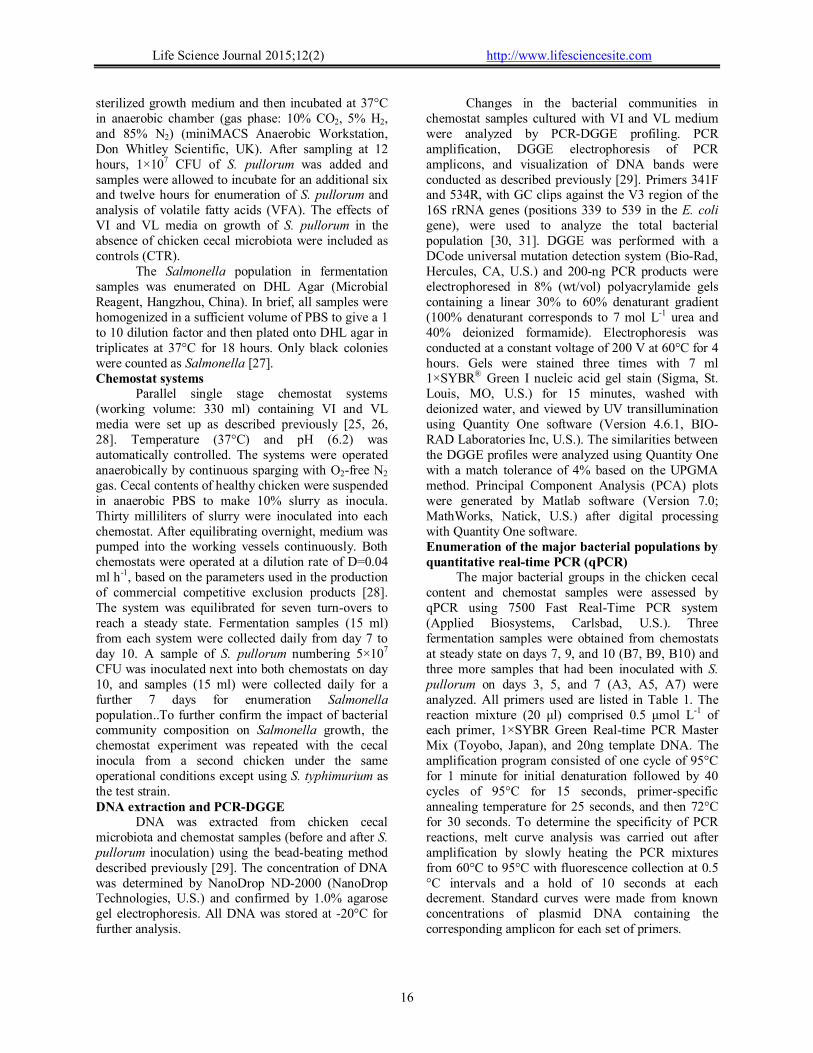
Figure 1. The effects of different culture media on the chicken cecal microbiota community in chemostat. A: Clustering tree based on UPGMA correlations of the DGGE profiles in chicken cecal content (duplicate samples), chemostat samples collected from VL medium and VI medium. B7 and B9 represent chemostat samples collected on days 7 and 9. A1, A3, A5, and A7 represent chemostat samples collected after Salmonella challenge on days 1, 3, 5, and 7. B: PCA plots of microbial communities according to the DGGE profiles in chicken cecal contents (CC, □), chemostat samples collected from VL medium (VL,○) and VI medium (VI, *).
To investigate the effects of VI and VL
medium on the compositions of the microbiota,
parallel chemostat systems with two growth media were set up and inoculated with cecal microbiota derived from the same chicken. The microbiota community structures of the chicken cecal content and fermentation samples were analyzed by PCR-DGGE profiles and qPCR. As shown in Figure 1A, stabilized PCR-DGGE profiles on B7 and B9 indicated that the microbiota community in both chemostats had reached an equilibrium stage. UPGMA analysis of PCR-DGGE profiles showed that the bacterial community generated from the samples of same chemostat were grouped together but separated by the different types of growth media (Figure 1A). In addition, significant separation between VI and VL media was observed by PCA analysis (Figure 1B). PCA plots indicated that the structure of the bacterial community generated from VL medium was much closer to the chicken cecal inoculum than to the one generated from VI medium, implying that the VL medium was more suitable for simulation of chicken cecal microbiota in vitro.
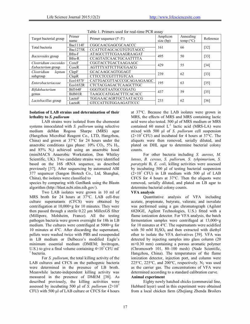
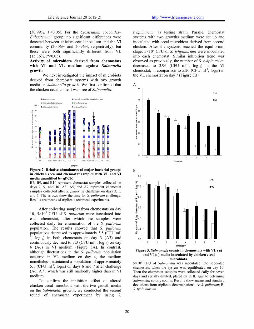
The major bacterial groups in the chemostat samples at steady state (B7, B9, B10), including the Bacteriodes group, Bifidobacterium genus, Lactubacillus group, Enterobacteriaceae, Clostridium coccoides-Eubacterium group, and Clostridium leptum group, were assessed by qPCR with specific primers, as shown in Table 1 The total bacterial populations in both chemostats reached over 1011 (16S rDNA copy numbers ml-1). The propotions of major bacterial groups are expressed as the percentage of total bacteria. In general, the bacterial community structures in chemostats containing either VL or VI medium were different from the one containing cecal inoculum (Figure 2). For example, the Clostridium leptum group accounted for 9.75% of total bacteria in the cecal inoculum but for only 0.127% in VL and 0.173% in VI (both P<0.05). Similar differences were observed in Enterobacteriaceae, which was 5.04% in the cecal inoculum and 1.59% in VL medium and 0.05% in the VI medium (P<0.05). In contrast, Lactobacillus was more plentiful in both chemostats (P<0.05) but more profound differences were associated with the VI medium. For the Bacteroides group, the numbers were significantly lower in the VI medium than in cecal inoculum (17.19% and 8.28%, respectively, P<0.05) but higher in VL medium
Life Science Journal 2015;12(2) http://www.lifesciencesite.com
20
(30.99%, P<0.05). For the Clostridium coccoides–Eubacterium group, no significant differences were detected between chicken cecal inoculum and the VI community (20.06% and 20.96%, respectively), but these were both significantly different from VL (15.36%, P<0.05). Activity of microbiota derived from chemostats with VI and VL medium against Salmonella growth
We next investigated the impact of microbiota derived from chemostat systems with two growth media on Salmonella growth. We first confirmed that the chicken cecal content was free of Salmonella.
Figure 2. Relative abundances of major bacterial groups in chicken ceca and chemostat samples with VL and VI media quantified by qPCR. B7, B9, and B10 represent chemostat samples collected on days 7, 9, and 10. A3, A5, and A7 represent chemostat samples collected after S. pullorum challenge on days 3, 5, and 7. The arrows show the time for S. pullorum challenge. Results are means of triplicate technical experiments.
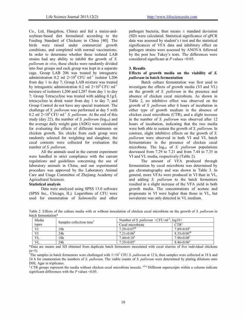
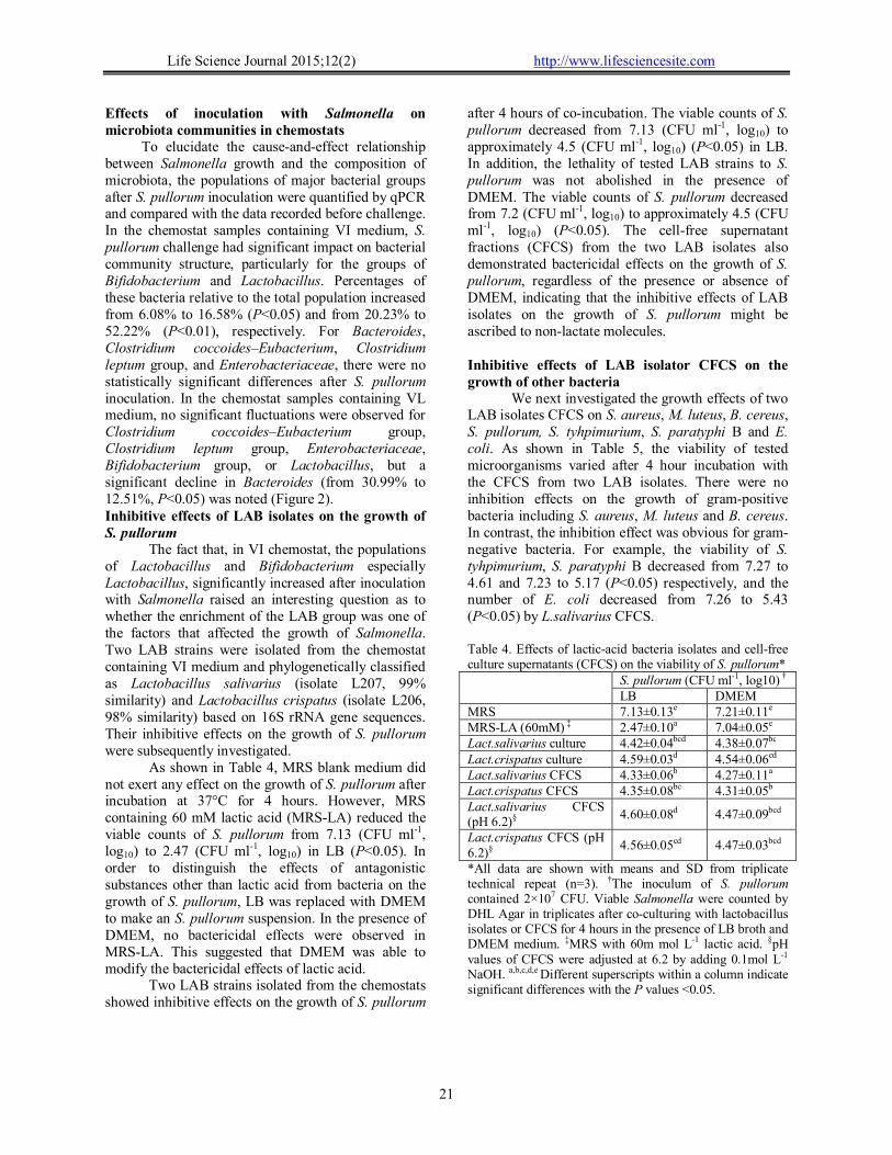
After collecting samples from chemostats on day 10, 5×10⁷ CFU of S. pullorum were inoculated into each chemostat, after which the samples were collected daily for enumeration of the S. pullorum population. The results showed that S. pullorum populations decreased to approximately 5.5 (CFU ml-
1, log10) in both chemostats on day 3 (A3) and continuously declined to 3.3 (CFU ml-1, log10) on day 6 (A6) in VI medium (Figure 3A). In contrast, although fluctuations in the S. pullorum population occurred in VL medium on day 4, the medium nonetheless maintained a population of approximately 5.1 (CFU ml-1, log10) on days 6 and 7 after challenge (A6, A7), which was still markedly higher than in VI medium.
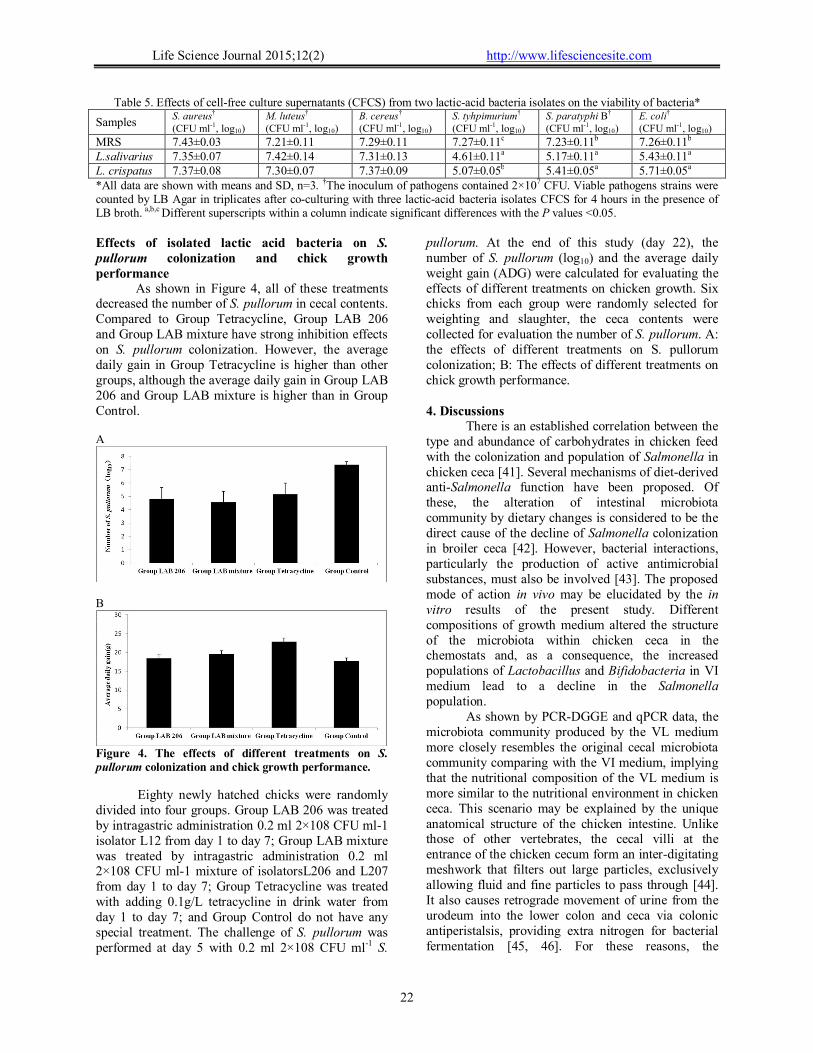
To confirm the inhibition effect of altered chicken cecal microbiota with the two growth media on the Salmonella growth, we conducted the second round of chemostat experiment by using S.
tyhpimurium as testing strain. Parallel chemostat systems with two growths medium were set up and inoculated with cecal microbiota derived from second chicken. After the systems reached the equilibrium stage, 5×10⁷ CFU of S. tyhpimurium were inoculated into each chemostat. Similar inhibition trend was observed as previously, the number of S. tyhpimurium decreased to 3.96 (CFU ml-1, log10) in the VI chemostat, in comparison to 5.20 (CFU ml-1, log10) in the VL chemostat on day 7 (Figure 3B). A
2.5
3.5
4.5
5.5
6.5
7.5
8.5
0 1 2 3 4 5 6 7
Nu
mb
er
of S.pullorum
(CF
U·m
l-1,
log
10
)
VI
VL
Time (day)
B
2.5
3.5
4.5
5.5
6.5
7.5
8.5
0 1 2 3 4 5 6 7
Nu
mb
er
of
S.t
yp
him
uri
um
(C
FU
ml-1
, lo
g1
0)
Time (day)
VI
VL
Figure 3. Salmonella counts in chemostats with VL (■) and VI (□) media inoculated by chicken cecal
microbiota. 5×10⁷ CFU of Salmonella was inoculated into seperated chemostats when the system was equilibrated on day 10. Then the chemostat samples were collected daily for seven days and serially diluted, plated on DHL agar to determine Salmonella colony counts. Results show means and standard deviations from triplicate determinations. A: S. pullorum; B: S. typhimurium.
0
10
20
30
40
50
60
70
80
90
100
CC VIB7 VIB9 VIB10 VIA3 VIA5 VIA7 VLB7 VLB9 VLB10 VLA3 VLA5 VLA7
Bacteroides group Clostridium coccoides–Eubacterium group
Clostridium leptum subgroup Enterobacteriaceae
Bifidobacterium genus Lactobacillus group
Pe
rce
nta
ge
of m
ajo
r ba
cte
ria
l gro
up
s (%
)
samples
Life Science Journal 2015;12(2) http://www.lifesciencesite.com
21
Effects of inoculation with Salmonella on microbiota communities in chemostats
To elucidate the cause-and-effect relationship between Salmonella growth and the composition of microbiota, the populations of major bacterial groups after S. pullorum inoculation were quantified by qPCR and compared with the data recorded before challenge. In the chemostat samples containing VI medium, S. pullorum challenge had significant impact on bacterial community structure, particularly for the groups of Bifidobacterium and Lactobacillus. Percentages of these bacteria relative to the total population increased from 6.08% to 16.58% (P<0.05) and from 20.23% to 52.22% (P<0.01), respectively. For Bacteroides, Clostridium coccoides–Eubacterium, Clostridium leptum group, and Enterobacteriaceae, there were no statistically significant differences after S. pullorum inoculation. In the chemostat samples containing VL medium, no significant fluctuations were observed for Clostridium coccoides–Eubacterium group, Clostridium leptum group, Enterobacteriaceae, Bifidobacterium group, or Lactobacillus, but a significant decline in Bacteroides (from 30.99% to 12.51%, P<0.05) was noted (Figure 2). Inhibitive effects of LAB isolates on the growth of S. pullorum
The fact that, in VI chemostat, the populations of Lactobacillus and Bifidobacterium especially Lactobacillus, significantly increased after inoculation with Salmonella raised an interesting question as to whether the enrichment of the LAB group was one of the factors that affected the growth of Salmonella. Two LAB strains were isolated from the chemostat containing VI medium and phylogenetically classified as Lactobacillus salivarius (isolate L207, 99% similarity) and Lactobacillus crispatus (isolate L206, 98% similarity) based on 16S rRNA gene sequences. Their inhibitive effects on the growth of S. pullorum were subsequently investigated.
As shown in Table 4, MRS blank medium did not exert any effect on the growth of S. pullorum after incubation at 37°C for 4 hours. However, MRS containing 60 mM lactic acid (MRS-LA) reduced the viable counts of S. pullorum from 7.13 (CFU ml-1, log10) to 2.47 (CFU ml-1, log10) in LB (P<0.05). In order to distinguish the effects of antagonistic substances other than lactic acid from bacteria on the growth of S. pullorum, LB was replaced with DMEM to make an S. pullorum suspension. In the presence of DMEM, no bactericidal effects were observed in MRS-LA. This suggested that DMEM was able to modify the bactericidal effects of lactic acid.
Two LAB strains isolated from the chemostats showed inhibitive effects on the growth of S. pullorum
after 4 hours of co-incubation. The viable counts of S. pullorum decreased from 7.13 (CFU ml-1, log10) to approximately 4.5 (CFU ml-1, log10) (P<0.05) in LB. In addition, the lethality of tested LAB strains to S. pullorum was not abolished in the presence of DMEM. The viable counts of S. pullorum decreased from 7.2 (CFU ml-1, log10) to approximately 4.5 (CFU ml-1, log10) (P<0.05). The cell-free supernatant fractions (CFCS) from the two LAB isolates also demonstrated bactericidal effects on the growth of S. pullorum, regardless of the presence or absence of DMEM, indicating that the inhibitive effects of LAB isolates on the growth of S. pullorum might be ascribed to non-lactate molecules.
Inhibitive effects of LAB isolator CFCS on the growth of other bacteria
We next investigated the growth effects of two LAB isolates CFCS on S. aureus, M. luteus, B. cereus, S. pullorum, S. tyhpimurium, S. paratyphi B and E. coli. As shown in Table 5, the viability of tested microorganisms varied after 4 hour incubation with the CFCS from two LAB isolates. There were no inhibition effects on the growth of gram-positive bacteria including S. aureus, M. luteus and B. cereus. In contrast, the inhibition effect was obvious for gram-negative bacteria. For example, the viability of S. tyhpimurium, S. paratyphi B decreased from 7.27 to 4.61 and 7.23 to 5.17 (P<0.05) respectively, and the number of E. coli decreased from 7.26 to 5.43 (P<0.05) by L.salivarius CFCS. Table 4. Effects of lactic-acid bacteria isolates and cell-free culture supernatants (CFCS) on the viability of S. pullorum*
S. pullorum (CFU ml-1, log10) † LB DMEM
MRS 7.13±0.13e 7.21±0.11e MRS-LA (60mM) ‡ 2.47±0.10a 7.04±0.05e Lact.salivarius culture 4.42±0.04bcd 4.38±0.07bc Lact.crispatus culture 4.59±0.03d 4.54±0.06cd Lact.salivarius CFCS 4.33±0.06b 4.27±0.11a Lact.crispatus CFCS 4.35±0.08bc 4.31±0.05b Lact.salivarius CFCS (pH 6.2)§
4.60±0.08d 4.47±0.09bcd
Lact.crispatus CFCS (pH 6.2)§
4.56±0.05cd 4.47±0.03bcd
*All data are shown with means and SD from triplicate technical repeat (n=3). †The inoculum of S. pullorum contained 2×10⁷ CFU. Viable Salmonella were counted by DHL Agar in triplicates after co-culturing with lactobacillus isolates or CFCS for 4 hours in the presence of LB broth and DMEM medium. ‡MRS with 60m mol L-1 lactic acid. §pH values of CFCS were adjusted at 6.2 by adding 0.1mol L-1 NaOH. a,b,c,d,e Different superscripts within a column indicate significant differences with the P values <0.05.
Life Science Journal 2015;12(2) http://www.lifesciencesite.com
22
Table 5. Effects of cell-free culture supernatants (CFCS) from two lactic-acid bacteria isolates on the viability of bacteria*
Samples S. aureus† (CFU ml-1, log10)
M. luteus†
(CFU ml-1, log10) B. cereus†
(CFU ml-1, log10) S. tyhpimurium†
(CFU ml-1, log10) S. paratyphi B†
(CFU ml-1, log10) E. coli†
(CFU ml-1, log10)
MRS 7.43±0.03 7.21±0.11 7.29±0.11 7.27±0.11c 7.23±0.11b 7.26±0.11b L.salivarius 7.35±0.07 7.42±0.14 7.31±0.13 4.61±0.11a 5.17±0.11a 5.43±0.11a L. crispatus 7.37±0.08 7.30±0.07 7.37±0.09 5.07±0.05b 5.41±0.05a 5.71±0.05a *All data are shown with means and SD, n=3. †The inoculum of pathogens contained 2×10⁷ CFU. Viable pathogens strains were counted by LB Agar in triplicates after co-culturing with three lactic-acid bacteria isolates CFCS for 4 hours in the presence of LB broth. a,b,c Different superscripts within a column indicate significant differences with the P values <0.05. Effects of isolated lactic acid bacteria on S. pullorum colonization and chick growth performance
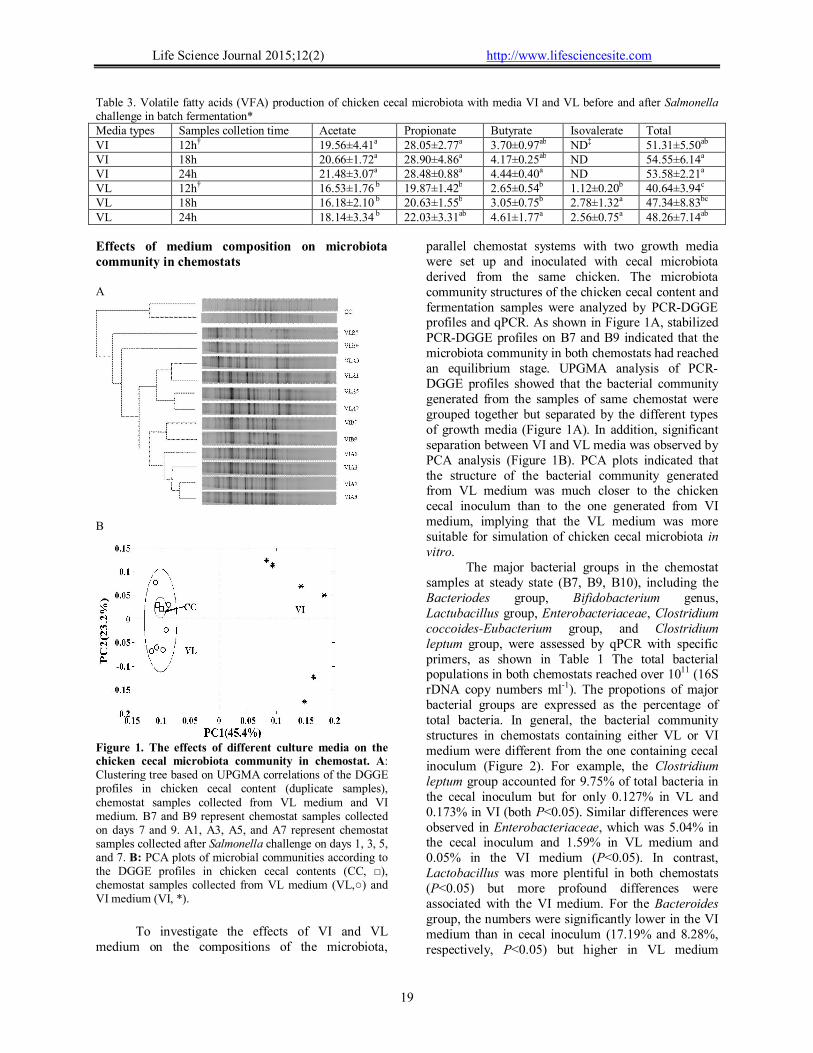
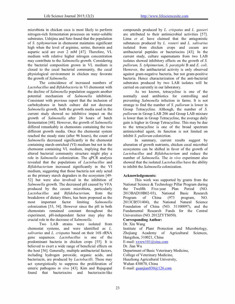
As shown in Figure 4, all of these treatments decreased the number of S. pullorum in cecal contents. Compared to Group Tetracycline, Group LAB 206 and Group LAB mixture have strong inhibition effects on S. pullorum colonization. However, the average daily gain in Group Tetracycline is higher than other groups, although the average daily gain in Group LAB 206 and Group LAB mixture is higher than in Group Control. A
B
Figure 4. The effects of different treatments on S. pullorum colonization and chick growth performance.
Eighty newly hatched chicks were randomly divided into four groups. Group LAB 206 was treated by intragastric administration 0.2 ml 2×10⁸ CFU ml-1 isolator L12 from day 1 to day 7; Group LAB mixture was treated by intragastric administration 0.2 ml 2×10⁸ CFU ml-1 mixture of isolatorsL206 and L207 from day 1 to day 7; Group Tetracycline was treated with adding 0.1g/L tetracycline in drink water from day 1 to day 7; and Group Control do not have any special treatment. The challenge of S. pullorum was performed at day 5 with 0.2 ml 2×10⁸ CFU ml-1 S.
pullorum. At the end of this study (day 22), the number of S. pullorum (log10) and the average daily weight gain (ADG) were calculated for evaluating the effects of different treatments on chicken growth. Six chicks from each group were randomly selected for weighting and slaughter, the ceca contents were collected for evaluation the number of S. pullorum. A: the effects of different treatments on S. pullorum colonization; B: The effects of different treatments on chick growth performance. 4. Discussions
There is an established correlation between the type and abundance of carbohydrates in chicken feed with the colonization and population of Salmonella in chicken ceca [41]. Several mechanisms of diet-derived anti-Salmonella function have been proposed. Of these, the alteration of intestinal microbiota community by dietary changes is considered to be the direct cause of the decline of Salmonella colonization in broiler ceca [42]. However, bacterial interactions, particularly the production of active antimicrobial substances, must also be involved [43]. The proposed mode of action in vivo may be elucidated by the in vitro results of the present study. Different compositions of growth medium altered the structure of the microbiota within chicken ceca in the chemostats and, as a consequence, the increased populations of Lactobacillus and Bifidobacteria in VI medium lead to a decline in the Salmonella population.
As shown by PCR-DGGE and qPCR data, the microbiota community produced by the VL medium more closely resembles the original cecal microbiota community comparing with the VI medium, implying that the nutritional composition of the VL medium is more similar to the nutritional environment in chicken ceca. This scenario may be explained by the unique anatomical structure of the chicken intestine. Unlike those of other vertebrates, the cecal villi at the entrance of the chicken cecum form an inter-digitating meshwork that filters out large particles, exclusively allowing fluid and fine particles to pass through [44]. It also causes retrograde movement of urine from the urodeum into the lower colon and ceca via colonic antiperistalsis, providing extra nitrogen for bacterial fermentation [45, 46]. For these reasons, the
Life Science Journal 2015;12(2) http://www.lifesciencesite.com
23
microbiota in chicken ceca is most likely to perform nitrogen-rich fermentation processes on water-soluble substrates. Ushijina and Seto found that the population of S. typhymurium in chemostat maintains significant high when the level of arginine, serine, theronin and aspartic acid are over 2 mM [47]. Therefore, VL medium with relative higher nitrogen concentration may contribute to the Salmonella growth. Considering the bacterial composition grown in VL medium is closed to the cecal bacterial community, the cecal physiological environment in chicken may favorate the growth of Salmonella.
The coincidence of increased numbers of Lactobacillus and Bifidobacteria in VI chemostat with the decline of Salmonella population suggests another potential mechanism of Salmonella inhibition. Consistent with previous report that the inclusion of carbohydrates in batch culture did not decrease Salmonella growth, both the growth media used in the current study showed no inhibitive impact on the growth of Salmonella after 24 hours of batch fermentation [48]. However, the growth of Salmonella differed remarkably in chemostats containing the two different growth media. Once the chemostat system reached the steady state (after 96 hours), the count of Salmonella decreased significantly in the chemostat containing starch-enriched (VI) medium but not in the chemostat containing VL medium, implying that the altered bacterial community structure might play a role in Salmonella colonization. The qPCR analysis revealed that the populations of Lactobacillus and Bifidobacterium increased significantly in the VI medium, suggesting that those bacteria not only acted as the primary starch degraders in the ecosystem [49-52] but were also involved in the inhibition of Salmonella growth. The decreased pH caused by VFA produced by the cecum microbiota, particularly Lactobacillus and Bifidobacterium, from the breakdown of dietary fibers, has been proposed as the most important factor limiting Salmonella colonization [53, 54]. However since the pH in both chemostats remained constant throughout the experiment, pH-independent factor may play the crucial role in the decrease of Salmonella.
Two LAB strains were isolated from chemostat systems, and were identified as L. salivarius and L. crispatus based on their 16S rRNA gene sequences. Lactobacillus is one of the predominant bacteria in chicken crops [55]. It is believed to exert a wide range of beneficial effects on the host [56]. Generally, multiple antibacterial factors, including hydrogen peroxide, organic acids, and bacteriacin, are produced by Lactobacilli. These may act synergistically to suppress the proliferation of enteric pathogens in vivo [43]. Kim and Rajagopal found that bacteriocins and bacteriocin-like
compounds produced by L. crispatus and L. gasseri are attributed to their antimicrobial activities [57]. Lima et al. have claimed that the antagonistic substances produced by L. reuteri and L. salivarius isolated from chicken crops and cecum are antibacterial peptides or bacteriocins [43]. In the current study, culture supernatants from two LAB isolates showed inhibitory effects on the growth of S. pullorum, S. tyhpimurium, S. paratyphi B and E. coli. However, the antibacterial activity is only observed against gram-negative bacteria, but not gram-positive bacteria. Hence characterization of the anti-bacterial substrates produced by two LAB isolates will be carried on currently in our laboratory.
As we known, tetracycline is one of the normally used antibiotics for controlling and preventing Salmonella infection in farms. It is not strange to find the number of S. pullorum is lower in Group Tetracycline. Although, the number of S. pullorum in Group LAB 206 and Group LAB mixture is lower than in Group Tetracycline, the average daily gain is higher in Group Tetracycline. This may be due to the tetracycline is one of the broad spectrum antimicrobial agent, its function is not limited on inhibit S. pullorum coloniztion.
In summary, current results suggest by alteration of growth nutrients, chicken cecal microbial ecosystems can be shifted in favor of the growth of Lactobacillus and Bifidobacterium and reduce the number of Salmonella. The in vivo experiment also showed that the isolated Lactobacillus have the ability to inhibit the Salmonella colonization. Acknowledgements:
This work was supported by grants from the National Science & Technology Pillar Program during the Twelfth Five-year Plan Period (NO. 2013BAD10B02-03), National Basic Research Program of China (973 program, NO. 2013CB531404), the National Natural Science Foundation of China (NO. 31100097), and the Fundamental Research Funds for the Central Universities (NO. 2012ZYTS050). Corresponding Author: Dr. Xin Wang Institute of Plant Protection and Microbiology, Zhejiang Academy of Agricultural Sciences, Hangzhou, 310021, China E-mail: [email protected] Dr. Jian Wu Department of Basic Veterinary Medicine, College of Veterinary Medicine, Huazhong Agricultural University, Wuhan 430070, China E-mail: [email protected]
Life Science Journal 2015;12(2) http://www.lifesciencesite.com
24
References 1 Allos BM, Moore MR, Griffin PM, et al. Surveillance
for sporadic foodborne disease in the 21st century: the FoodNet perspective. Clin Infect Dis 2004;38 Suppl 3:S115-120.
2 Thorns CJ. Bacterial food-borne zoonoses. Rev Sci Tech 2000;19(1):226-239.
3 Scallan E, Hoekstra RM, Angulo FJ, et al. Foodborne illness acquired in the United States--major pathogens. Emerg Infect Dis 2011;17(1):7-15.
4 Marin C, Lainez M. Salmonella detection in feces during broiler rearing and after live transport to the slaughterhouse. Poultry science 2009;88(9):1999-2005.
5 Chiu LH, Chiu CH, Horn YM, et al. Characterization of 13 multi-drug resistant Salmonella serovars from different broiler chickens associated with those of human isolates. BMC Microbiol 2010;10:86.
6 Luangtongkum T, Jeon B, Han J, et al. Antibiotic resistance in Campylobacter: emergence, transmission and persistence. Future Microbiol 2009;4(2):189-200.
7 Rayamajhi N, Jung BY, Cha SB, et al. Antibiotic resistance patterns and detection of blaDHA-1 in Salmonella species isolates from chicken farms in South Korea. Appl Environ Microbiol 2010;76(14):4760-4764.
8 Zhao S, White DG, Friedman SL, et al. Antimicrobial resistance in Salmonella enterica serovar Heidelberg isolates from retail meats, including poultry, from 2002 to 2006. Appl Environ Microbiol 2008;74(21):6656-6662.
9 Nurmi E, Rantala M. New aspects of Salmonella infection in broiler production. Nature 1973;241(5386):210-211.
10 Chambers JR, Lu X. Probiotics and Maternal Vaccination for Salmonella Control in Broiler Chickens. J APPL POULT RES 2002;11(3):320-327.
11 Barnes E, Impey C, Cooper D. Manipulation of the crop and intestinal flora of the newly hatched chick. Am J Clin Nutr 1980;33(11):2426-2433.
12 Nisbet DJ, Corrier DE, Ricke SC, et al. Cecal Propionic Acid as a Biological Indicator of the Early Establishment of a Microbial Ecosystem Inhibitory toSalmonellain Chicks. Anaerobe 1996;2(6):345-350.
13 McReynolds JL, Byrd JA, Genovese KJ, et al. Dietary lactose and its effect on the disease condition of necrotic enteritis. Poult Sci 2007;86(8):1656-1661.
14 Nisbet DJ, Corrier DE, DeLoach JR. Effect of mixed cecal microflora maintained in continuous culture and of dietary lactose on Salmonella typhimurium colonization in broiler chicks. Avian diseases 1993;37(2):528-535.
15 Chambers JR, Spencer JL, Modler HW. The influence of complex carbohydrates on Salmonella typhimurium colonization, pH, and density of broiler ceca. Poultry science 1997;76(3):445-451.
16 Eeckhaut V, Van Immerseel F, Dewulf J, et al. Arabinoxylooligosaccharides from wheat bran inhibit Salmonella colonization in broiler chickens. Poult Sci 2008;87(11):2329-2334.
17 Teirlynck E, Haesebrouck F, Pasmans F, et al. The cereal type in feed influences Salmonella Enteritidis
colonization in broilers. Poult Sci 2009;88(10):2108-2112.
18 Macfarlane GT, Macfarlane S. Models for intestinal fermentation: association between food components, delivery systems, bioavailability and functional interactions in the gut. Curr Opin Biotechnol 2007;18(2):156-162.
19 Allison C, McFarlan C, MacFarlane GT. Studies on mixed populations of human intestinal bacteria grown in single-stage and multistage continuous culture systems. Appl Environ Microbiol 1989;55(3):672-678.
20 Van den Abbeele P, Grootaert C, Marzorati M, et al. Microbial community development in a dynamic gut model is reproducible, colon region specific, and selective for Bacteroidetes and Clostridium cluster IX. Appl Environ Microbiol 2010;76(15):5237-5246.
21 Pivnick H, Nurmi E: The Nurmi concept and its role in the control of Salmonellae in poultry in: Developments in Food Microbiology -1. Barking, Essex England: Applied Science Publishers Ltd 1982.
22 Schneitz C. Competitive exclusion in poultry--30 years of research. Food Control 2005;16(8):657-667.
23 Nisbet D. Defined competitive exclusion cultures in the prevention of enteropathogen colonisation in poultry and swine. Antonie Van Leeuwenhoek 2002;81(1-4):481-486.
24 Revolledo L, Ferreira CS, Ferreira AJ. Prevention of Salmonella Typhimurium colonization and organ invasion by combination treatment in broiler chicks. Poultry science 2009;88(4):734-743.
25 Smith EA, Macfarlane GT. Enumeration of amino acid fermenting bacteria in the human large intestine: effects of pH and starch on peptide metabolism and dissimilation of amino acids. FEMS Microbiol Ecol 1998;25(4):355-368.
26 Genovese KJ, Anderson RC, Harvey RB, et al. Competitive exclusion of Salmonella from the gut of neonatal and weaned pigs. Journal of food protection 2003;66(8):1353-1359.
27 Midorikawa Y, Newton PN, Nakamura S, et al. A phenomenon useful for the detection of Salmonella implementing a device from citrus extracts. Trop Med Health 2009;37:115-120.
28 Nisbet DJ, Corrier DE, Stanker LH: Competitive exclusion culture for swine. In. USA; 1999: 6.
29 Li M, Wang B, Zhang M, et al. Symbiotic gut microbes modulate human metabolic phenotypes. Proc Natl Acad Sci U S A 2008;105(6):2117-2122.
30 Muyzer G, de Waal EC, Uitterlinden AG. Profiling of complex microbial populations by denaturing gradient gel electrophoresis analysis of polymerase chain reaction-amplified genes coding for 16S rRNA. Appl Environ Microbiol 1993;59(3):695-700.
31 Holben WE, Feris KP, Kettunen A, et al. GC fractionation enhances microbial community diversity assessment and detection of minority populations of bacteria by denaturing gradient gel electrophoresis. Appl Environ Microbiol 2004;70(4):2263-2270.
32 Denman SE, McSweeney CS. Development of a real-time PCR assay for monitoring anaerobic fungal and cellulolytic bacterial populations within the rumen. FEMS Microbiol Ecol 2006;58(3):572-582.
Life Science Journal 2015;12(2) http://www.lifesciencesite.com
25
33 Matsuki T, Watanabe K, Fujimoto J, et al. Development of 16S rRNA-gene-targeted group-specific primers for the detection and identification of predominant bacteria in human feces. Appl Environ Microbiol 2002;68(11):5445-5451.
34 Rinttila T, Kassinen A, Malinen E, et al. Development of an extensive set of 16S rDNA-targeted primers for quantification of pathogenic and indigenous bacteria in faecal samples by real-time PCR. J Appl Microbiol 2004;97(6):1166-1177.
35 Bartosch S, Fite A, Macfarlane GT, et al. Characterization of bacterial communities in feces from healthy elderly volunteers and hospitalized elderly patients by using real-time PCR and effects of antibiotic treatment on the fecal microbiota. Appl Environ Microbiol 2004;70(6):3575-3581.
36 Frank JA, Reich CI, Sharma S, et al. Critical evaluation of two primers commonly used for amplification of bacterial 16S rRNA genes. Appl Environ Microbiol 2008;74(8):2461-2470.
37 Pavlova SI, Kilic AO, Kilic SS, et al. Genetic diversity of vaginal lactobacilli from women in different countries based on 16S rRNA gene sequences. J Appl Microbiol 2002;92(3):451-459.
38 Fayol-Messaoudi D, Coconnier-Polter MH, Moal VL, et al. The Lactobacillus plantarum strain ACA-DC287 isolated from a Greek cheese demonstrates antagonistic activity in vitro and in vivo against Salmonella enterica serovar Typhimurium. J Appl Microbiol 2007;103(3):657-665.
39 Macfarlane GT, Gibson GR, Cummings JH. Comparison of fermentation reactions in different regions of the human colon. J Appl Bacteriol 1992;72(1):57-64.
40 Wang ML, Suo X, Gu JH, et al. Influence of grape seed proanthocyanidin extract in broiler chickens: effect on chicken coccidiosis and antioxidant status. Poult Sci 2008;87(11):2273-2280.
41 McHan F, Shotts EB, Brown J. Effect of feeding selected carbohydrates on the in vivo attachment of Salmonella typhimurium in chick ceca. Avian diseases 1991;35(2):328-331.
42 Fernandez F, Hinton M, Van Gils B. Dietary mannan-oligosaccharides and their effect on chicken caecal microflora in relation to Salmonella Enteritidis colonization. Avian Pathol 2002;31(1):49-58.
43 Lima ET, Andreatti Filho RL, Okamoto AS, et al. Evaluation in vitro of the antagonistic substances produced by Lactobacillus spp. isolated from chickens. Can J Vet Res 2007;71(2):103-107.
44 Duke GE: Alimentary canal: anatomy, regulation of feeding, and motility. New York: Springer-Verlag; 1986.
45 Duke GE. Relationship of cecal and colonic motility to diet, habitat, and cecal anatomy in several avian species. J Exp Zool 1989;252(S3):38-47.
46 Lai HC, Duke GE. Colonic motility in domestic Turkeys. Dig Dis Sci 1978;23(8):673-681.
47 Ushijima T, Seto A. Selected faecal bacteria and nutrients essential for antagonism of Salmonella typhimurium in anaerobic continuous flow cultures. J Med Microbiol 1991;35(2):111-117.
48 Martin-Pelaez S, Gibson GR, Martin-Orue SM, et al. In vitro fermentation of carbohydrates by porcine faecal inocula and their influence on Salmonella Typhimurium growth in batch culture systems. FEMS Microbiol Ecol 2008;66(3):608-619.
49 Wang X, Brown IL, Evans AJ, et al. The protective effects of high amylose maize (amylomaize) starch granules on the survival of Bifidobacterium spp. in the mouse intestinal tract. J Appl Microbiol 1999;87(5):631-639.
50 Siew-Wai L, Zi-Ni T, Karim AA, et al. Fermentation of Metroxylon sagu resistant starch type III by Lactobacillus sp. and Bifidobacterium bifidum. J Agric Food Chem 2010;58(4):2274-2278.
51 Iyer C, Phillips M, Kailasapathy K. Release studies of Lactobacillus casei strain Shirota from chitosan-coated alginate-starch microcapsules in ex vivo porcine gastrointestinal contents. Lett Appl Microbiol 2005;41(6):493-497.
52 Giraud E, Champailler A, Raimbault M. Degradation of Raw Starch by a Wild Amylolytic Strain of Lactobacillus plantarum. Appl Environ Microbiol 1994;60(12):4319-4323.
53 Dunkley KD, McReynolds JL, Hume ME, et al. Molting in Salmonella Enteritidis-challenged laying hens fed alfalfa crumbles. II. Fermentation and microbial ecology response. Poultry science 2007;86(10):2101-2109.
54 Nisbet DJ, Corrier DE, Ricke SC, et al. Cecal propionic acid as a biological indicator of the early establishment of a microbial ecosystem inhibitory to Salmonella in chicks. Anaerobe 1996;2:345-350.
55 Smith HW. Observations on the flora of the alimentary tract of animals and factors affecting its composition. J Pathol Bacteriol 1965;89(1):95-122.
56 Brooker BE, Fuller R. Adhesion of lactobacilli to the chicken crop epithelium. J Ultrastruct Res 1975;52(1):21-31.
57 Kim J-W, Rajagopal SN. Antibacterial Activities of Lactobacillus crispatus ATCC 33820 and Lactobacillus gasseri ATCC 33323. J Microbiol 2001;39(2):146-148.
1/27/2015