Embed Size (px)
Citation preview
Wound Practice and Research 196
Eric J Lew DPM University of Arizona — Southern Arizona Limb Salvage Alliance, Tucson, Arizona, USA
Nicholas A Giovinco DPM University of Arizona — Southern Arizona Limb Salvage Alliance, Tucson, Arizona, USA
David G Armstrong DPM, MD, PhD University of Arizona — Southern Arizona Limb Salvage Alliance, Tucson, Arizona, USA
Fontaine closely evaluate these clinical parameters. However, they fail to adequately categorise the extent of tissue loss and the severity of infection, if present2,3.
It was intended and noted in Bell’s 1982 publication that diabetic patients be excluded or defined in a separate category due to a varied clinical picture of neuropathy, ischaemia and sepsis1. However, in modern practice, patients who present with a threatened lower extremity carry a broader spectrum of contributing factors. Ischaemia and PAD is one such factor, but neuropathy and foot deformity associated with uncontrolled diabetes are other compounding factors. It is now known that diabetic foot ulcers carry neuropathic, ischaemic, and neuroischaemic aetiologies4. In fact, there has been a rising incidence of PAD in patients with diabetes. Recent revascularisation trials have shown the prevalence of diabetes in patients undergoing revascularisation for limb salvage to be as high as 50% to 80%5-8.
One of the most widely known wound classification systems intended for diabetic feet was published by Wagner9. While it has continued to maintain use in wound descriptions including osteomyelitis and location of gangrene, this system does not provide a means to quantify vascular influence over the lower extremity wound. It also fails to address soft tissue infection nor does it differentiate gangrene due to ischaemia or infection. Similarly, the University of Texas (UT) wound classification system, the first validated diabetic foot wound classification system, did describe depth, infection, and ischaemia. However, it only dichotomised infection and ischaemia as “present”
ABSTRACTPeripheral arterial disease and associated critical limb ischaemia is a well-respected predictor of wound morbidity and tissue necrosis. With respect to diabetes, patients who present with a threatened lower extremity may carry a broader spectrum of contributing factors including neuroischaemic and infectious aetiologies. The authors of the Society for Vascular Surgery (SVS) Lower Extremity Threatened Limb Classification have refocused an approach to the patient with a threatened limb and not only address ischaemia, but also have taken into account the key factors of tissue loss and the presence and extent of infection. Case examples are presented for practical application of this system which may aid in characterising and stratifying the clinical state and risk of amputation in patients whose disease burden has given way for a limb-at-risk. With this new classification system and risk assessment tool, meaningful clinical trials may be developed to improve current operative and non-operative treatment options for the threatened limb.
Clinical application of the Society for Vascular Surgery (SVS) Lower Extremity Threatened Limb Classification system: risk stratification based on Wound, Ischaemia, and foot Infection (WIfI)Lew EJ, Giovinco NA & Armstrong DG
INTRODUCTIONPeripheral arterial disease (PAD) and associated critical limb ischaemia (CLI) is a long-known and well-respected predictor of wound morbidity and tissue necrosis. An earlier publication first described CLI based on certain parameters: pressure <40 mmHg in the presence of rest pain and <60 mmHg in the presence of tissue necrosis1. Existing CLI classifications such as Rutherford and
Lew EJ et al. Clinical application of the Society for Vascular Surgery (SVS) Lower Extremity Threatened Limb Classification system
Volume 22 Number 4 – November 2014197
or “absent”10. The Infectious Disease Society of America (IDSA) does provide a system for stratifying amputation risk pertaining to pedal infection, but does not address wound extent or ischaemia11. Therefore, the authors of the Society for Vascular Surgery (SVS) Lower Extremity Threatened Limb Classification have refocused an approach to the patient with a threatened limb that not only addresses ischaemia, but also has taken into account the key factors of wound depth and presence and extent of infection12. The SVS is a not-for-profit organisation consisting of primarily vascular surgeons and other specialists who are dedicated to the prevention and cure of vascular disease. Based in the United States, its stated purpose is to advance excellence and innovation in vascular health through education, advocacy, research, and public awareness.
The purpose of this article is to provide a concise summary and description of the new SVS Wound, Ischaemia, and foot Infection (WIfI) classification for the threatened limb. Case examples are also presented for practical application of this system. It is the expressed purpose of the authors of this article that foot and ankle specialists use this system to aid in characterising and stratifying the clinical state and risk of amputation in patients whose disease burden has given way for a limb-at-risk. With this new classification system and risk assessment tool, meaningful clinical trials may be developed to improve current operative and non-operative treatment options for the threatened limb.
WHAT IS THE WIfI CLASSIFICATION?This is a new classification system put forth by the SVS that focuses on the “threatened limb”. This system is specifically applicable to patients whose disease burden may place their lower extremities at risk for amputation. These include patients with PAD and diabetes among other co-morbidities. Risk stratification encompasses three factors that place a limb under threat of amputation: (1) Tissue loss or wound extent, including gangrene; (2) Ischaemia; and (3) Infection. These are all key factors that need to be evaluated in predicting amputation risk. The new system aims to create an objective classification of the threatened limb based on the degree of ischaemia, wound extent, gangrene, and infection. This updated SVS Lower Extremity Threatened Limb Classification System is intended to define the disease burden, analogous to the tumour, node, and metastasis (TNM) system for cancer staging12.
WHY DO CLINICIANS NEED A NEW CLASSIFICATION?Classification systems published to date have been helpful to an extent, but have been narrowly focused on either the wound, perfusion, or infectious status. For example, existing CLI classification systems fail to adequately categorise the extent of tissue loss or the presence and severity of infection. TASC 1, TASC II, the Bollinger system, and the Graziani morphologic categorisation only address arterial anatomy,
Lew EJ et al. Clinical application of the Society for Vascular Surgery (SVS) Lower Extremity Threatened Limb Classification system
Smith & Nephew develop products that help wounds heal, allowing people to return to normal life faster.
ALLEVYN™ Life’s differentiating core design contributes to improvements in patient wellbeing1, clinician satisfaction and potential economic benefits.2
Help give your patients an optimal wound care experience with ALLEVYN™ Life dressings.
This innovation to heal is just one possibility from the Smith & Nephew advanced wound care portfolio, where you’ll find solutions for many different types of wounds.
Innovation is designing a dressing to provide the optimal patient experience
New Zealand: T 0800 807 663 www.smith-nephew.com/new-zealandAustralia: T 13 13 60 www.smith-nephew.com/australiaReferences: 1. Rossington A et al., Clinical performance and positive impact on patient wellbeing of ALLEVYN Life. Wounds UK 2013; 9:91-95. 2. Stephen-Haynes et al., An appraisal of the clinical performance and economic benefits of a silicone foam in a large UK primary care organisation. J Comm Nurs 2013; 27: 50-59.
ALLEVYN™ Life
™Trademark of Smith & Nephew SN11400OPSITE™ Post-Op Visible • PICO™ • ACTICOAT™ • ALLEVYN™ • DURAFIBER™ • IV3000™
Wound Practice and Research 198
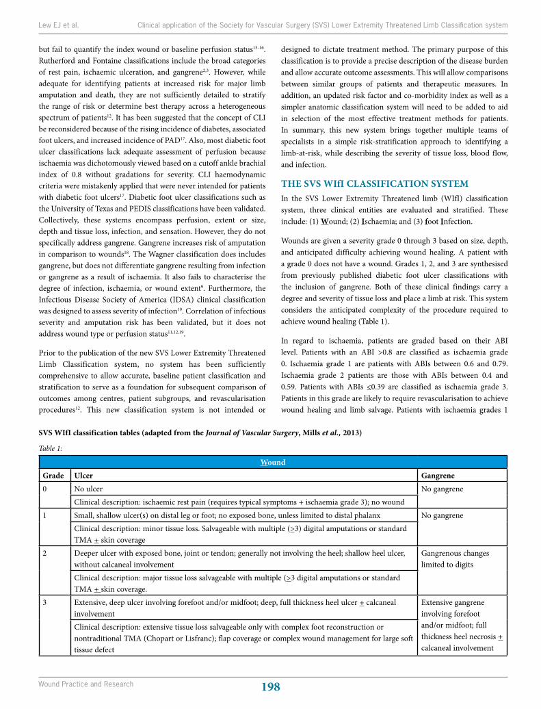
WoundGrade Ulcer Gangrene0 No ulcer No gangrene
Clinical description: ischaemic rest pain (requires typical symptoms + ischaemia grade 3); no wound1 Small, shallow ulcer(s) on distal leg or foot; no exposed bone, unless limited to distal phalanx No gangrene
Clinical description: minor tissue loss. Salvageable with multiple (>3) digital amputations or standard TMA + skin coverage
2 Deeper ulcer with exposed bone, joint or tendon; generally not involving the heel; shallow heel ulcer, without calcaneal involvement
Gangrenous changes limited to digits
Clinical description: major tissue loss salvageable with multiple (>3 digital amputations or standard TMA + skin coverage.
3 Extensive, deep ulcer involving forefoot and/or midfoot; deep, full thickness heel ulcer + calcaneal involvement
Extensive gangrene involving forefoot and/or midfoot; full thickness heel necrosis + calcaneal involvement
Clinical description: extensive tissue loss salvageable only with complex foot reconstruction or nontraditional TMA (Chopart or Lisfranc); flap coverage or complex wound management for large soft tissue defect
Table 1:
SVS WIfI classification tables (adapted from the Journal of Vascular Surgery, Mills et al., 2013)
but fail to quantify the index wound or baseline perfusion status13-16. Rutherford and Fontaine classifications include the broad categories of rest pain, ischaemic ulceration, and gangrene2,3. However, while adequate for identifying patients at increased risk for major limb amputation and death, they are not sufficiently detailed to stratify the range of risk or determine best therapy across a heterogeneous spectrum of patients12. It has been suggested that the concept of CLI be reconsidered because of the rising incidence of diabetes, associated foot ulcers, and increased incidence of PAD17. Also, most diabetic foot ulcer classifications lack adequate assessment of perfusion because ischaemia was dichotomously viewed based on a cutoff ankle brachial index of 0.8 without gradations for severity. CLI haemodynamic criteria were mistakenly applied that were never intended for patients with diabetic foot ulcers17. Diabetic foot ulcer classifications such as the University of Texas and PEDIS classifications have been validated. Collectively, these systems encompass perfusion, extent or size, depth and tissue loss, infection, and sensation. However, they do not specifically address gangrene. Gangrene increases risk of amputation in comparison to wounds18. The Wagner classification does includes gangrene, but does not differentiate gangrene resulting from infection or gangrene as a result of ischaemia. It also fails to characterise the degree of infection, ischaemia, or wound extent9. Furthermore, the Infectious Disease Society of America (IDSA) clinical classification was designed to assess severity of infection19. Correlation of infectious severity and amputation risk has been validated, but it does not address wound type or perfusion status11,12,19.
Prior to the publication of the new SVS Lower Extremity Threatened Limb Classification system, no system has been sufficiently comprehensive to allow accurate, baseline patient classification and stratification to serve as a foundation for subsequent comparison of outcomes among centres, patient subgroups, and revascularisation procedures12. This new classification system is not intended or
designed to dictate treatment method. The primary purpose of this classification is to provide a precise description of the disease burden and allow accurate outcome assessments. This will allow comparisons between similar groups of patients and therapeutic measures. In addition, an updated risk factor and co-morbidity index as well as a simpler anatomic classification system will need to be added to aid in selection of the most effective treatment methods for patients. In summary, this new system brings together multiple teams of specialists in a simple risk-stratification approach to identifying a limb-at-risk, while describing the severity of tissue loss, blood flow, and infection.
THE SVS WIfI CLASSIFICATION SYSTEMIn the SVS Lower Extremity Threatened limb (WIfI) classification system, three clinical entities are evaluated and stratified. These include: (1) Wound; (2) Ischaemia; and (3) foot Infection.
Wounds are given a severity grade 0 through 3 based on size, depth, and anticipated difficulty achieving wound healing. A patient with a grade 0 does not have a wound. Grades 1, 2, and 3 are synthesised from previously published diabetic foot ulcer classifications with the inclusion of gangrene. Both of these clinical findings carry a degree and severity of tissue loss and place a limb at risk. This system considers the anticipated complexity of the procedure required to achieve wound healing (Table 1).
In regard to ischaemia, patients are graded based on their ABI level. Patients with an ABI >0.8 are classified as ischaemia grade 0. Ischaemia grade 1 are patients with ABIs between 0.6 and 0.79. Ischaemia grade 2 patients are those with ABIs between 0.4 and 0.59. Patients with ABIs <0.39 are classified as ischaemia grade 3. Patients in this grade are likely to require revascularisation to achieve wound healing and limb salvage. Patients with ischaemia grades 1
Lew EJ et al. Clinical application of the Society for Vascular Surgery (SVS) Lower Extremity Threatened Limb Classification system
Volume 22 Number 4 – November 2014199
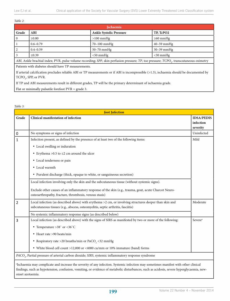
foot InfectionGrade Clinical manifestation of infection IDSA/PEDIS
infection severity
0 No symptoms or signs of infection Uninfected
1 Infection present, as defined by the presence of at least two of the following items:
• Local swelling or induration
• Erythema >0.5 to ≤2 cm around the ulcer
• Local tenderness or pain
• Local warmth
• Purulent discharge (thick, opaque to white, or sanguineous secretion)
Mild
Local infection involving only the skin and the subcutaneous tissue (without systemic signs).
Exclude other causes of an inflammatory response of the skin (e.g., trauma, gout, acute Charcot Neuro-osteoarthropathy, fracture, thrombosis, venous stasis)
2 Local infection (as described above) with erythema >2 cm, or involving structures deeper than skin and subcutaneous tissues (e.g., abscess, osteomyelitis, septic arthritis, fasciitis)
Moderate
No systemic inflammatory response signs (as described below)
3 Local infection (as described above) with the signs of SIRS as manifested by two or more of the following:
• Temperature >38˚ or <36˚C
• Heart rate >90 beats/min
• Respiratory rate >20 breaths/min or PaCO2 <32 mmHg
• White blood cell count >12,000 or <4000 cu/mm or 10% immature (band) forms
Severea
PACO2, Partial pressure of arterial carbon dioxide; SIRS, systemic inflammatory response syndrome
aIschaemia may complicate and increase the severity of any infection. Systemic infection may sometimes manifest with other clinical findings, such as hypotension, confusion, vomiting, or evidence of metabolic disturbances, such as acidosis, severe hyperglycaemia, new-onset azotaemia.
Table 3:
Table 2:
IschaemiaGrade ABI Ankle Systolic Pressure TP, TcPO20 ≥0.80 >100 mmHg ≥60 mmHg1 0.6–0.79 70–100 mmHg 40–59 mmHg2 0.4–0.59 50–70 mmHg 30–39 mmHg3 ≤0.39 <50 mmHg <30 mmHgABI, Ankle brachial index; PVR, pulse volume recording; SPP, skin perfusion pressure; TP, toe pressure; TCPO2, transcutaneous oximetryPatients with diabetes should have TP measurements.
If arterial calcification precludes reliable ABI or TP measurements or if ABI is incompressible (>1.3), ischaemia should be documented by TCPO2, SPP, or PVR.
If TP and ABI measurements result in different grades, TP will be the primary determinant of ischaemia grade.
Flat or minimally pulsatile forefoot PVR = grade 3.
Lew EJ et al. Clinical application of the Society for Vascular Surgery (SVS) Lower Extremity Threatened Limb Classification system
Wound Practice and Research 200
or 2 may benefit or even require vascular intervention to achieve wound healing. If the ABI is elevated due to incompressible vessels, toe pressures or transcutaneous oximetry measurements must be performed to stratify the degree of ischaemia. These modalities along with other non-invasive vascular studies such as pulse volume recordings, skin perfusion pressures, and quantitative indocyanine green angiography are preferred in patients with medial calcinosis leading with falsely elevated ABIs, or with previous toe or forefoot amputations (Table 2).
The risk of amputation correlates directly with increasing infection severity. The SVS WIfI classification system incorporates the IDSA clinical staging system with the PEDIS system to account for both soft tissue and bone infectious severity. The four grades are based on clinical evaluation of the affected limb with respect to systemic and metabolic observations (Table 3).
IS THERE A PROGNOSTIC VALUE OF THE SVS WIfI CLASSIFICATION?This new classification system can provide an estimated risk of amputation and possibly aid in determining whether a patient would benefit from revascularisation. A Delphi consensus process was carried out by members of the SVS. This consisted of a 12-member group of recognised experts in the field of chronic limb ischaemia. This group defined the WIfI system’s potential clinical applicability. Two questions were addressed: (1) what is the perceived
risk of amputation for each possible combination?; and (2) what is the perceived benefit from revascularisation for each possible combination? The 64 theoretically possible clinical combinations were each assigned a limb threat clinical stage as outlined below:
• Very low = VL = clinical stage 1
• Low = L = clinical stage 2
• Moderate = M = clinical stage 3
• High = H = clinical stage 4
• Clinical stage 5 would signify an unsalvageable foot
The consensus group followed these basic premises of assigning a clinical stage to each of the 64 respective clinical scenarios: (1) increase in wound class increases risk of amputation; (2) PAD and infection are synergistic — an infected wound with PAD increases likelihood revascularisation will be needed for wound healing; and (3) infection with systemic and metabolic instability (grade 3) carries a high risk of amputation regardless of other factors. Table 4 shows results of the Delphi Consensus process. These results represent the one-year risk of amputation with medical therapy alone for each of the 64 possible presentations. The purpose of this consensus exercise was to define stages of disease that may prove useful for clinical decision-making and design of prospective studies.
Lew EJ et al. Clinical application of the Society for Vascular Surgery (SVS) Lower Extremity Threatened Limb Classification system
M E D I C A LM E D I C A L
Rosidal... all you need now.
Sentry Medical Pty Ltd22 Peter Brock Drive Eastern Creek NSW 2766
Tel: 1300 995 999 | Fax: 1300 995 998 E: [email protected] W: www.sentrymedical.com.auAssuming dressing change and re-application of compression therapy occurs once per week*
Effective inelastic compression therapy in two styles
Rosidal TCS two layer cohesive compression kit
®
highly cost effective easy to apply environment-friendly 12 weeks of compression therapy*... one box, one low cost!
Rosidal sysreusable compression system
®
low profilebuilt-in skin protection layer
®
NEW
Volume 22 Number 4 – November 2014201
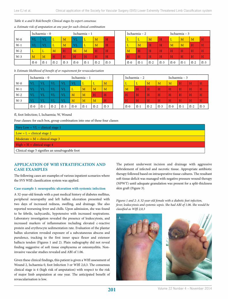
Table 4: a and b Risk/benefit: Clinical stages by expert consensus
a. Estimate risk of amputation at one year for each clinical combination
Ischaemia – 0 Ischaemia – 1
Ischaemia – 2 Ischaemia – 3
W-0 VL VL L M VL L M H L L M H L M M H
W-1 VL VL L M VL L M H L M H H M M H H
W-2 L L M H M M H H M H H H H H H H
W-3 M M H H H H H H H H H H H H H H
fI-0 fI-1 fI-2 fI-3 fI-0 fI-1 fI-2 fI-3 fI-0 fI-1 fI-2 fI-3 fI-0 fI-1 fI-2 fI-3
b. Estimate likelihood of benefit of or requirement for revascularisation
Ischaemia – 0 Ischaemia – 1
Ischaemia – 2 Ischaemia – 3
W-0 VL VL VL VL VL L L M L L M M M H H H
W-1 VL VL VL VL L M M M M H H H H H H H
W-2 VL VL VL VL M M H H H H H H H H H H
W-3 VL VL VL VL M M M H H H H H H H H H
fI-0 fI-1 fI-2 fI-3 fI-0 fI-1 fI-2 fI-3 fI-0 fI-1 fI-2 fI-3 fI-0 fI-1 fI-2 fI-3
fI, foot Infection; I, Ischaemia; W, Wound
Four classes: for each box, group combination into one of these four classes
Very Low = VL = clinical stage 1
Low = L = clinical stage 2
Moderate = M = clinical stage 3
High = H = clinical stage 4
Clinical stage 5 signifies an unsalvageable foot
Figures 1 and 2: A 32-year-old female with a diabetic foot infection, fever, leukocytosis and systemic sepsis. She had ABI of 1.06. She would be classified as WIfI 2,0,3
1. 2.
APPLICATION OF WIfI STRATIFICATION AND CASE EXAMPLESThe following cases are examples of various inpatient scenarios where the SVS WIfI classification system was applied.
Case example 1: neuropathic ulceration with systemic infection
A 32-year-old female with a past medical history of diabetes mellitus, peripheral neuropathy and left hallux ulceration presented with two days of increased redness, swelling, and drainage. She also reported worsening fever and chills. Upon admission, she was found to be febrile, tachycardic, hypotensive with increased respirations. Laboratory investigation revealed the presence of leukocytosis, and increased markers of inflammation including elevated c-reactive protein and erythrocyte sedimentation rate. Evaluation of the plantar hallux ulceration revealed exposure of a subcutaneous abscess and purulence, tracking to the first inner space flexor and extensor hallucis tendon (Figures 1 and 2). Plain radiography did not reveal finding suggestive of soft tissue emphysema or osteomyelitis. Non-invasive vascular studies revealed and ABI of 1.06.
Given these clinical findings, this patient is given a WIfI assessment of Wound 2, Ischaemia 0, foot Infection 3 or WIfI 2,0,3. The consensus clinical stage is 4 (high risk of amputation) with respect to the risk of major limb amputation at one year. The anticipated benefit of revascularisation is low.
The patient underwent incision and drainage with aggressive debridement of infected and necrotic tissue. Appropriate antibiotic therapy followed based on intraoperative tissue cultures. The resultant soft tissue deficit was managed with negative pressure wound therapy (NPWT) until adequate granulation was present for a split-thickness skin graft (Figure 3).
Lew EJ et al. Clinical application of the Society for Vascular Surgery (SVS) Lower Extremity Threatened Limb Classification system
Wound Practice and Research 202
and risk for amputation is a SVS consensus clinical stage 4 (high risk of amputation). There is also anticipated benefit of revascularisation.
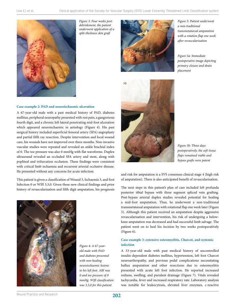
The next steps in this patient’s plan of care included left profunda posterior tibial bypass with three segment spliced vein grafting. Post-bypass arterial duplex studies revealed potential for healing a mid-foot amputation. Thus, he underwent a non-traditional transmetatarsal amputation with rotational flap one week later (Figure 5). Although this patient received an amputation despite aggressive revascularisation and intervention, his risk of undergoing a below-knee amputation was decreased and had successful limb salvage. The patient went on to heal his incision by two weeks postoperatively (Figure 6).
Case example 3: extensive osteomyelitis, Charcot, and systemic infection
A 33-year-old male with past medical history of uncontrolled insulin-dependent diabetes mellitus, hypertension, left foot Charcot neuroarthropathy, and previous pedal complications necessitating hallux amputation and other resections due to osteomyelitis presented with acute left foot infection. He reported increased redness, swelling, and purulent drainage (Figure 7). Vitals revealed tachycardia, fever and increased respiratory rate. Laboratory analysis was notable for leukocytosis, elevated liver enzymes, c-reactive
Case example 2: PAD and neuroischaemic ulceration
A 67-year-old male with a past medical history of PAD, diabetes mellitus, peripheral neuropathy presented with rest pain, a gangrenous fourth digit, and a chronic left lateral penetrating mid-foot ulceration which appeared neuroischaemic in aetiology (Figure 4). His past surgical history included superficial femoral artery (SFA) angioplasty and partial fifth ray resection. Despite intervention and local wound care, his wounds have not improved over three months. Non-invasive vascular studies were repeated and revealed an ankle brachial index of 0. The toe pressure was also 0 mmHg with flat waveforms. Duplex ultrasound revealed an occluded SFA artery and stent, along with popliteal and trifurcation occlusion. These findings were consistent with critical limb ischaemia and recurrent arterial occlusive disease. He presented without any concerns for acute infection.
This patient is given a classification of Wound 3, Ischaemia 3, and foot Infection 0 or WIfI 3,3,0. Given these new clinical findings and prior history of revascularisation and fifth digit amputation, his prognosis
Figure 4: A 67-year-old male with PAD and diabetes presented with non-healing neuroischaemic lesions to his left foot. ABI was 0 and toe pressure of 0 mmHg. WIfI classification was 3,3,0 for this patient
4.
Figure 5: Patient underwent a non-traditional transmetatarsal amputation with a rotation flap one week after revascularisation
Figure 5a: Immediate postoperative image depicting primary closure and drain placement
Figure 5b: Three days postoperatively, the soft tissue flaps remained viable and bypass grafts were patent
5a
5b
Figure 3: Four weeks post-debridement, the patient underwent application of a split-thickness skin graft
3.
Lew EJ et al. Clinical application of the Society for Vascular Surgery (SVS) Lower Extremity Threatened Limb Classification system
Volume 22 Number 4 – November 2014203
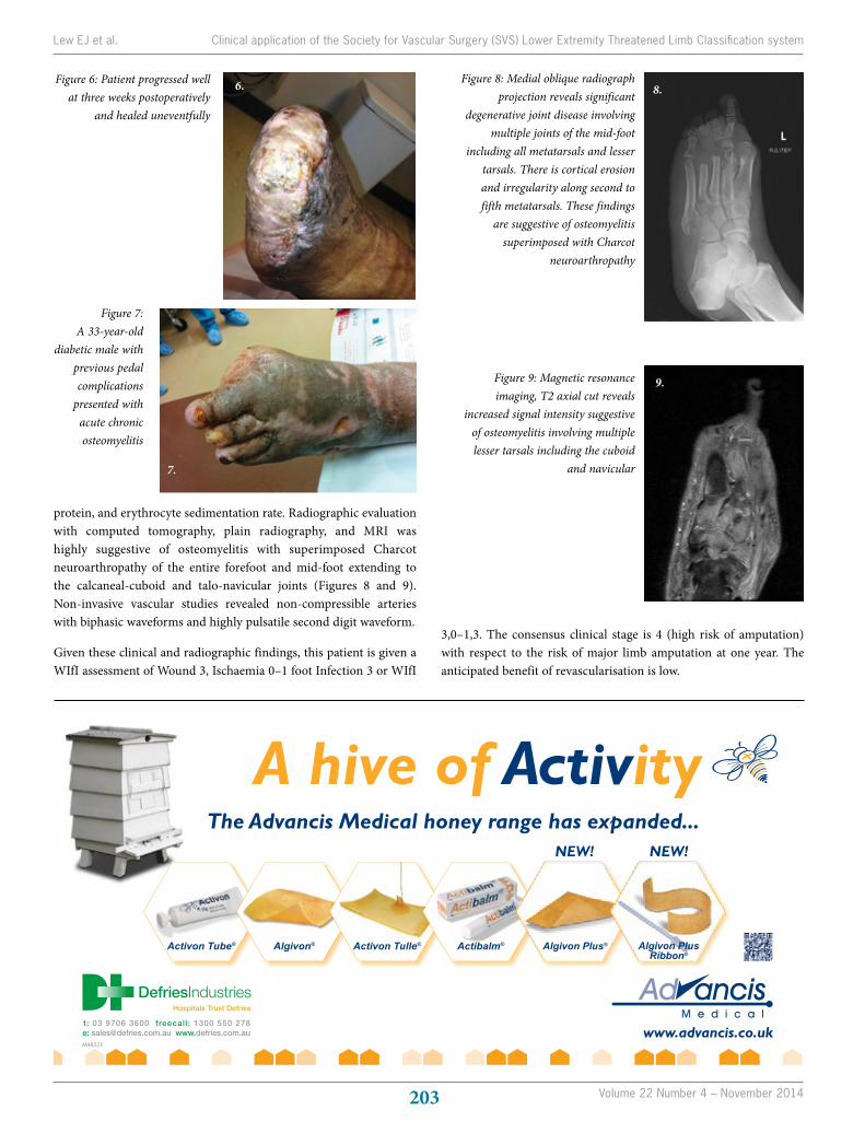
protein, and erythrocyte sedimentation rate. Radiographic evaluation with computed tomography, plain radiography, and MRI was highly suggestive of osteomyelitis with superimposed Charcot neuroarthropathy of the entire forefoot and mid-foot extending to the calcaneal-cuboid and talo-navicular joints (Figures 8 and 9). Non-invasive vascular studies revealed non-compressible arteries with biphasic waveforms and highly pulsatile second digit waveform.
Given these clinical and radiographic findings, this patient is given a WIfI assessment of Wound 3, Ischaemia 0–1 foot Infection 3 or WIfI
Figure 7: A 33-year-old
diabetic male with previous pedal complications
presented with acute chronic osteomyelitis
7.
Figure 8: Medial oblique radiograph projection reveals significant
degenerative joint disease involving multiple joints of the mid-foot
including all metatarsals and lesser tarsals. There is cortical erosion and irregularity along second to fifth metatarsals. These findings
are suggestive of osteomyelitis superimposed with Charcot
neuroarthropathy
8.
Figure 9: Magnetic resonance imaging, T2 axial cut reveals
increased signal intensity suggestive of osteomyelitis involving multiple lesser tarsals including the cuboid
and navicular
9.
Figure 6: Patient progressed well at three weeks postoperatively
and healed uneventfully
6.
3,0–1,3. The consensus clinical stage is 4 (high risk of amputation) with respect to the risk of major limb amputation at one year. The anticipated benefit of revascularisation is low.
Lew EJ et al. Clinical application of the Society for Vascular Surgery (SVS) Lower Extremity Threatened Limb Classification system
www.advancis.co.uk
A hive of Activity
MAR325
The Advancis Medical honey range has expanded...
Hospitals Trust Defries
t: 03 9706 3600 freecall: 1300 550 278e: [email protected] www.defries.com.au
Activon Tube® Algivon® Activon Tulle® Actibalm®
NEW!
Algivon Plus® Algivon Plus Ribbon®
NEW!
Honey range expanded Ad Defries.indd 1 07/11/2013 12:51
Wound Practice and Research 204
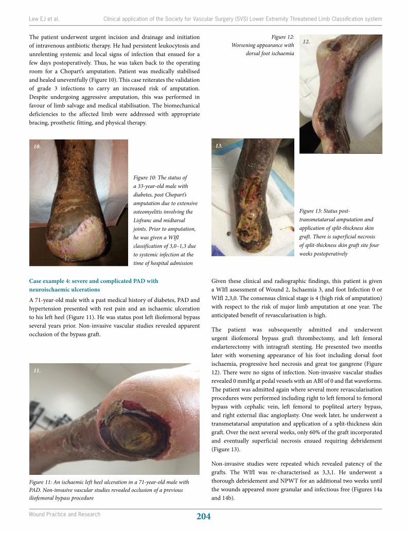
The patient underwent urgent incision and drainage and initiation of intravenous antibiotic therapy. He had persistent leukocytosis and unrelenting systemic and local signs of infection that ensued for a few days postoperatively. Thus, he was taken back to the operating room for a Chopart’s amputation. Patient was medically stabilised and healed uneventfully (Figure 10). This case reiterates the validation of grade 3 infections to carry an increased risk of amputation. Despite undergoing aggressive amputation, this was performed in favour of limb salvage and medical stabilisation. The biomechanical deficiencies to the affected limb were addressed with appropriate bracing, prosthetic fitting, and physical therapy.
Case example 4: severe and complicated PAD with neuroischaemic ulcerations
A 71-year-old male with a past medical history of diabetes, PAD and hypertension presented with rest pain and an ischaemic ulceration to his left heel (Figure 11). He was status post left iliofemoral bypass several years prior. Non-invasive vascular studies revealed apparent occlusion of the bypass graft.
Figure 10: The status of a 33-year-old male with diabetes, post Chopart’s amputation due to extensive osteomyelitis involving the Lisfranc and midtarsal joints. Prior to amputation, he was given a WIfI classification of 3,0–1,3 due to systemic infection at the time of hospital admission
10.
Figure 11: An ischaemic left heel ulceration in a 71-year-old male with PAD. Non-invasive vascular studies revealed occlusion of a previous iliofemoral bypass procedure
11.
Figure 12: Worsening appearance with
dorsal foot ischaemia
12.
Figure 13: Status post-transmetatarsal amputation and application of split-thickness skin graft. There is superficial necrosis of split-thickness skin graft site four weeks postoperatively
13.
Given these clinical and radiographic findings, this patient is given a WIfI assessment of Wound 2, Ischaemia 3, and foot Infection 0 or WIfI 2,3,0. The consensus clinical stage is 4 (high risk of amputation) with respect to the risk of major limb amputation at one year. The anticipated benefit of revascularisation is high.
The patient was subsequently admitted and underwent urgent iliofemoral bypass graft thrombectomy, and left femoral endarterectomy with intragraft stenting. He presented two months later with worsening appearance of his foot including dorsal foot ischaemia, progressive heel necrosis and great toe gangrene (Figure 12). There were no signs of infection. Non-invasive vascular studies revealed 0 mmHg at pedal vessels with an ABI of 0 and flat waveforms. The patient was admitted again where several more revascularisation procedures were performed including right to left femoral to femoral bypass with cephalic vein, left femoral to popliteal artery bypass, and right external iliac angioplasty. One week later, he underwent a transmetatarsal amputation and application of a split-thickness skin graft. Over the next several weeks, only 60% of the graft incorporated and eventually superficial necrosis ensued requiring debridement (Figure 13).
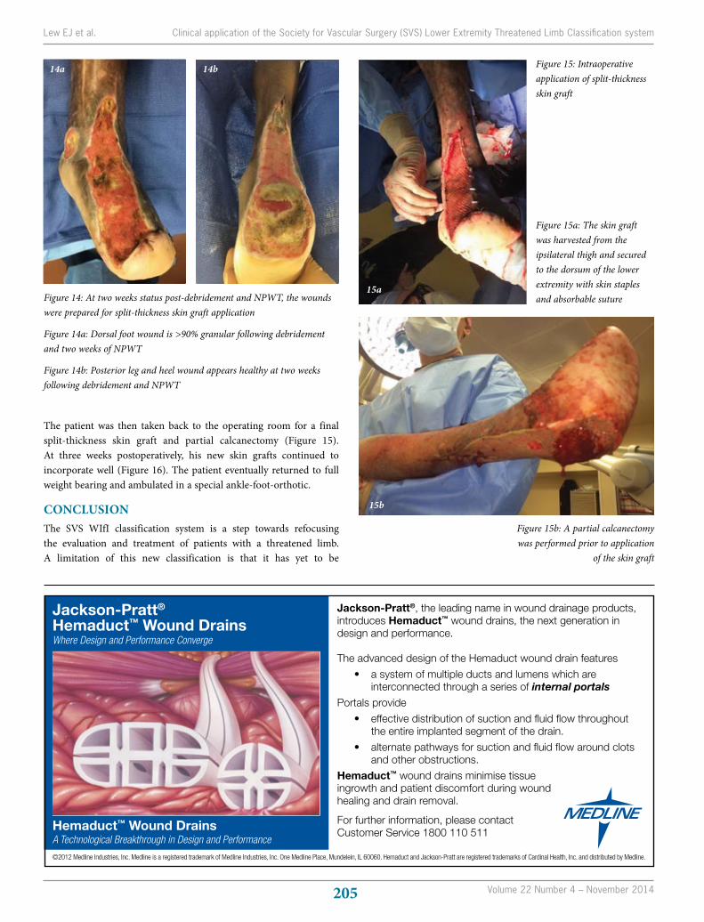
Non-invasive studies were repeated which revealed patency of the grafts. The WIfI was re-characterised as 3,3,1. He underwent a thorough debridement and NPWT for an additional two weeks until the wounds appeared more granular and infectious free (Figures 14a and 14b).
Lew EJ et al. Clinical application of the Society for Vascular Surgery (SVS) Lower Extremity Threatened Limb Classification system
Volume 22 Number 4 – November 2014205
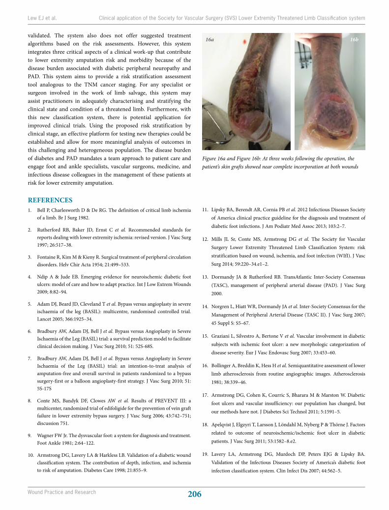
The patient was then taken back to the operating room for a final split-thickness skin graft and partial calcanectomy (Figure 15). At three weeks postoperatively, his new skin grafts continued to incorporate well (Figure 16). The patient eventually returned to full weight bearing and ambulated in a special ankle-foot-orthotic.
CONCLUSIONThe SVS WIfI classification system is a step towards refocusing the evaluation and treatment of patients with a threatened limb. A limitation of this new classification is that it has yet to be
Figure 14: At two weeks status post-debridement and NPWT, the wounds were prepared for split-thickness skin graft application
Figure 14a: Dorsal foot wound is >90% granular following debridement and two weeks of NPWT
Figure 14b: Posterior leg and heel wound appears healthy at two weeks following debridement and NPWT
14a 14b
15b
Figure 15b: A partial calcanectomy was performed prior to application
of the skin graft
Figure 15: Intraoperative application of split-thickness skin graft
Figure 15a: The skin graft was harvested from the ipsilateral thigh and secured to the dorsum of the lower extremity with skin staples and absorbable suture
15a
Lew EJ et al. Clinical application of the Society for Vascular Surgery (SVS) Lower Extremity Threatened Limb Classification system
Jackson-Pratt®
Hemaduct™ Wound DrainsWhere Design and Performance Converge
Hemaduct™ Wound DrainsA Technological Breakthrough in Design and Performance
Jackson-Pratt®, the leading name in wound drainage products,introduces Hemaduct™ wound drains, the next generation indesign and performance.
The advanced design of the Hemaduct wound drain features
• a system of multiple ducts and lumens which are interconnected through a series of internal portals
Portals provide
• effective distribution of suction and fluid flow throughout the entire implanted segment of the drain.
• alternate pathways for suction and fluid flow around clots and other obstructions.
Hemaduct™ wound drains minimise tissue ingrowth and patient discomfort during wound healing and drain removal.
For further information, please contactCustomer Service 1800 110 511
©2012 Medline Industries, Inc. Medline is a registered trademark of Medline Industries, Inc. One Medline Place, Mundelein, IL 60060. Hemaduct and Jackson-Pratt are registered trademarks of Cardinal Health, Inc. and distributed by Medline.
Medline_JP_Advt_80x180mm_Medline_JP_Advt_80x180mm.qxd 8/03/12 5:21 PM Page 1
Wound Practice and Research 206
validated. The system also does not offer suggested treatment algorithms based on the risk assessments. However, this system integrates three critical aspects of a clinical work-up that contribute to lower extremity amputation risk and morbidity because of the disease burden associated with diabetic peripheral neuropathy and PAD. This system aims to provide a risk stratification assessment tool analogous to the TNM cancer staging. For any specialist or surgeon involved in the work of limb salvage, this system may assist practitioners in adequately characterising and stratifying the clinical state and condition of a threatened limb. Furthermore, with this new classification system, there is potential application for improved clinical trials. Using the proposed risk stratification by clinical stage, an effective platform for testing new therapies could be established and allow for more meaningful analysis of outcomes in this challenging and heterogeneous population. The disease burden of diabetes and PAD mandates a team approach to patient care and engage foot and ankle specialists, vascular surgeons, medicine, and infectious disease colleagues in the management of these patients at risk for lower extremity amputation.
REFERENCES1. Bell P, Charlesworth D & De RG. The definition of critical limb ischemia
of a limb. Br J Surg 1982.
2. Rutherford RB, Baker JD, Ernst C et al. Recommended standards for reports dealing with lower extremity ischemia: revised version. J Vasc Surg 1997; 26:517–38.
3. Fontaine R, Kim M & Kieny R. Surgical treatment of peripheral circulation disorders. Helv Chir Acta 1954; 21:499–533.
4. Ndip A & Jude EB. Emerging evidence for neuroischemic diabetic foot ulcers: model of care and how to adapt practice. Int J Low Extrem Wounds 2009; 8:82–94.
5. Adam DJ, Beard JD, Cleveland T et al. Bypass versus angioplasty in severe ischaemia of the leg (BASIL): multicentre, randomised controlled trial. Lancet 2005; 366:1925–34.
6. Bradbury AW, Adam DJ, Bell J et al. Bypass versus Angioplasty in Severe Ischaemia of the Leg (BASIL) trial: a survival prediction model to facilitate clinical decision making. J Vasc Surg 2010; 51: 52S-68S.
7. Bradbury AW, Adam DJ, Bell J et al. Bypass versus Angioplasty in Severe Ischaemia of the Leg (BASIL) trial: an intention-to-treat analysis of amputation-free and overall survival in patients randomized to a bypass surgery-first or a balloon angioplasty-first strategy. J Vasc Surg 2010; 51: 5S-17S
8. Conte MS, Bandyk DF, Clowes AW et al. Results of PREVENT III: a multicenter, randomized trial of edifoligide for the prevention of vein graft failure in lower extremity bypass surgery. J Vasc Surg 2006; 43:742–751; discussion 751.
9. Wagner FW Jr. The dysvascular foot: a system for diagnosis and treatment. Foot Ankle 1981; 2:64–122.
10. Armstrong DG, Lavery LA & Harkless LB. Validation of a diabetic wound classification system. The contribution of depth, infection, and ischemia to risk of amputation. Diabetes Care 1998; 21:855–9.
11. Lipsky BA, Berendt AR, Cornia PB et al. 2012 Infectious Diseases Society of America clinical practice guideline for the diagnosis and treatment of diabetic foot infections. J Am Podiatr Med Assoc 2013; 103:2–7.
12. Mills JL Sr, Conte MS, Armstrong DG et al. The Society for Vascular Surgery Lower Extremity Threatened Limb Classification System: risk stratification based on wound, ischemia, and foot infection (WIfI). J Vasc Surg 2014; 59:220–34.e1–2.
13. Dormandy JA & Rutherford RB. TransAtlantic Inter-Society Consensus (TASC), management of peripheral arterial disease (PAD). J Vasc Surg 2000.
14. Norgren L, Hiatt WR, Dormandy JA et al. Inter-Society Consensus for the Management of Peripheral Arterial Disease (TASC II). J Vasc Surg 2007; 45 Suppl S: S5–67.
15. Graziani L, Silvestro A, Bertone V et al. Vascular involvement in diabetic subjects with ischemic foot ulcer: a new morphologic categorization of disease severity. Eur J Vasc Endovasc Surg 2007; 33:453–60.
16. Bollinger A, Breddin K, Hess H et al. Semiquantitative assessment of lower limb atherosclerosis from routine angiographic images. Atherosclerosis 1981; 38:339–46.
17. Armstrong DG, Cohen K, Courric S, Bharara M & Marston W. Diabetic foot ulcers and vascular insufficiency: our population has changed, but our methods have not. J Diabetes Sci Technol 2011; 5:1591–5.
18. Apelqvist J, Elgzyri T, Larsson J, Löndahl M, Nyberg P & Thörne J. Factors related to outcome of neuroischemic/ischemic foot ulcer in diabetic patients. J Vasc Surg 2011; 53:1582–8.e2.
19. Lavery LA, Armstrong DG, Murdoch DP, Peters EJG & Lipsky BA. Validation of the Infectious Diseases Society of America’s diabetic foot infection classification system. Clin Infect Dis 2007; 44:562–5.
Lew EJ et al. Clinical application of the Society for Vascular Surgery (SVS) Lower Extremity Threatened Limb Classification system
Figure 16a and Figure 16b: At three weeks following the operation, the patient’s skin grafts showed near complete incorporation at both wounds
16a 16b