Embed Size (px)
Citation preview
Level 2
CELL DIVISION
Remember…
*DNA more stable than RNA•* All organisms must be able to reproduce to keep life
going
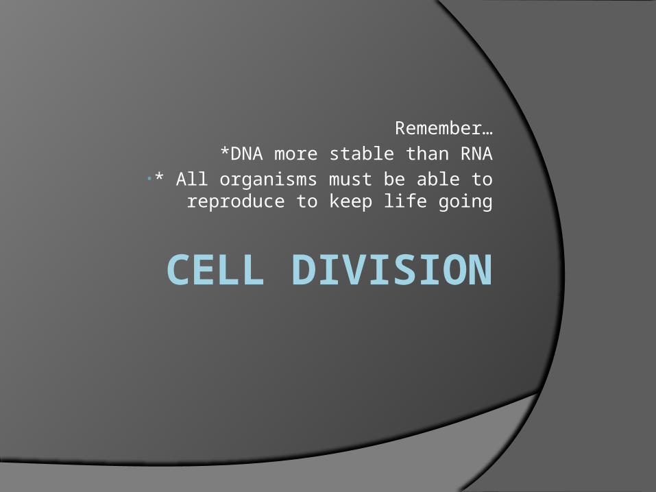
One cell becoming two
All Reproducing Cells
1. Replicate DNA in parent cell during the S phase.
2.Replicate organelles 3. Perform cytokinesis- division of
cytoplasm and cell membrane.Cyto – cell, kinesis- movement
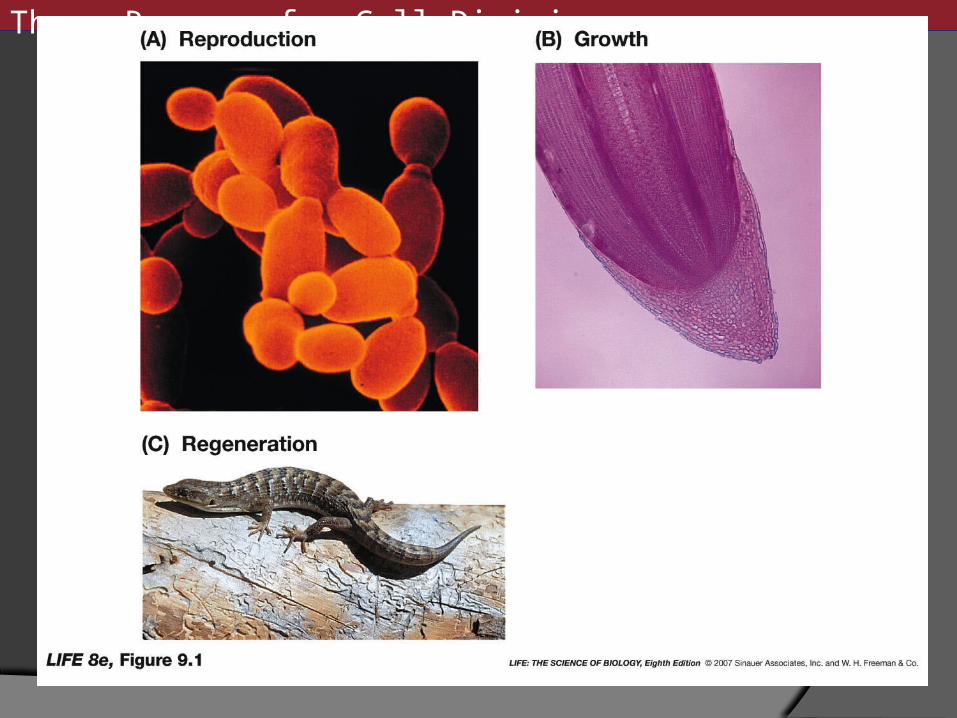
Reasons for Cell Division In unicellular organisms it is primarily for
reproduction of themselves. In multicellular organisms it is for
reproduction, growth, and repair of tissues. If cells do not divide, they get to big. Two major problems with big cells:
DNA cannot code for all of the necessary functions
Substances cannot enter and exit fast enough.
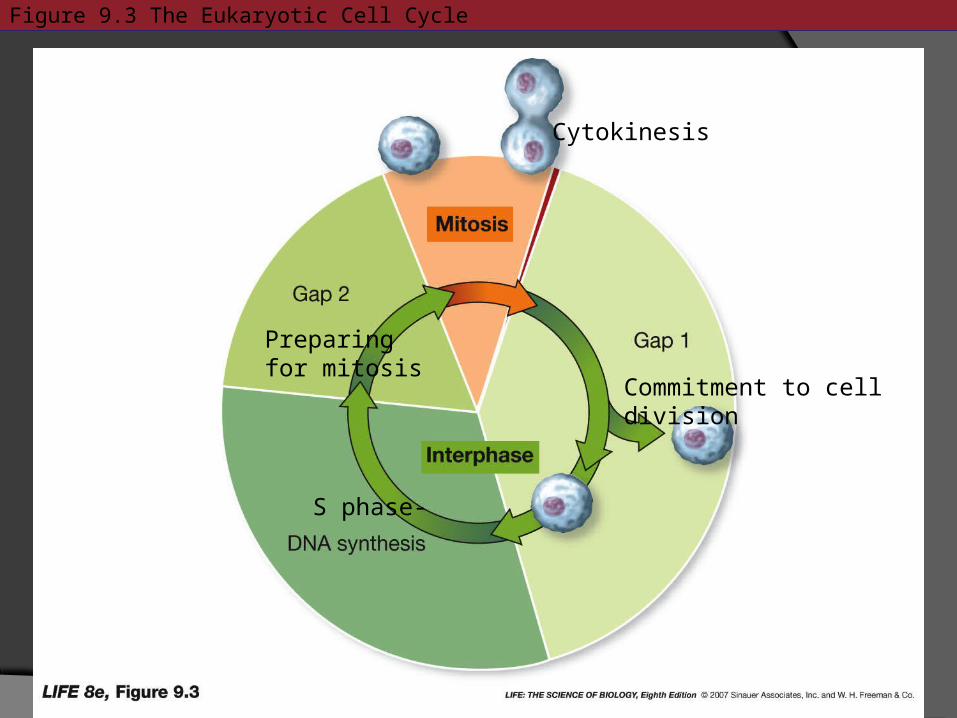
Figure 9.3 The Eukaryotic Cell Cycle
Commitment to cell division
S phase-
Preparing for mitosis
Cytokinesis
Three Reasons for Cell Division
Four Events that Must occur for Cell Division A reproductive signal
(intracellular/extracellular) to initiate division.
Replication of DNA, so the new cells match identically to old cell.
Segregation- process by which DNA is passed to each of the two resulting new cells.
Cytokinesis- process by which the cell membrane and cell wall separate into two new cells.
Binary Fission
In prokaryotes, the entire single-celled organism divides.
First it doubles in size, then duplicates its DNA, and then divides.
In prokaryotes, the initiating reproductive signal is thought to be environmental conditions and food supply
DNA Replication in Prokaryotes (S phase)
Most prokaryotes only have one chromosome, and it is circular .
Circular chromosomes are also in chloroplasts, mitochondria, and viruses.
ori- origin site of DNA replication ter- terminus of DNA replication
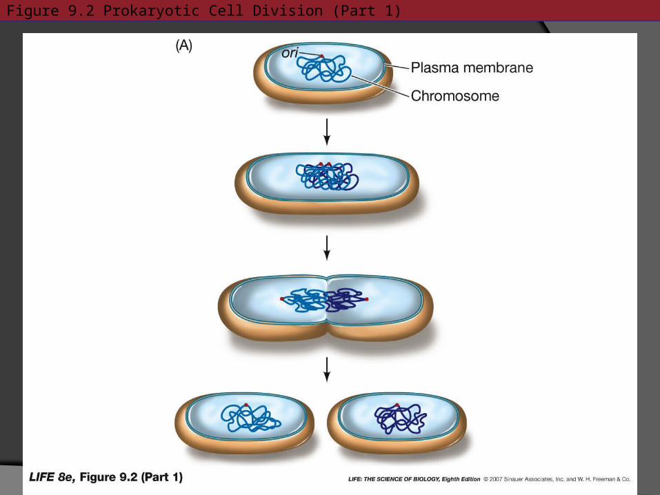
Figure 9.2 Prokaryotic Cell Division (Part 1)
Prokaryotic DNA Replication Referred to as circular or theta
replication. Cleavage furrow follows after replication The two resulting cells are clones=
identical cells. Mitosis is thought to have evolved from
binary fission…commonality: synthesis and division
Eukaryotic Cell Division
Most complex eukaryotes originate from a single cell: fertilized egg.
The formation of a multicellular organism from a fertilized egg is known as development.
Eukaryotic cell division are driven by the needs of the organism, not the environmental conditions and food supply.
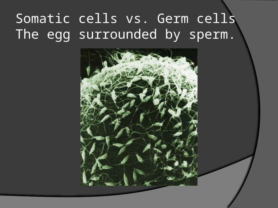
Somatic cells vs. Germ cellsThe egg surrounded by sperm.
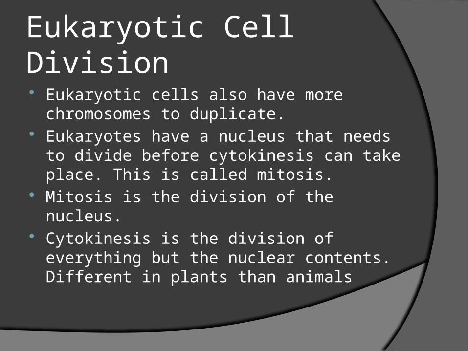
Eukaryotic Cell Division
Eukaryotic cells also have more chromosomes to duplicate.
Eukaryotes have a nucleus that needs to divide before cytokinesis can take place. This is called mitosis.
Mitosis is the division of the nucleus. Cytokinesis is the division of everything
but the nuclear contents. Different in plants than animals
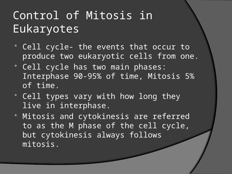
Control of Mitosis in Eukaryotes
Cell cycle- the events that occur to produce two eukaryotic cells from one.
Cell cycle has two main phases: Interphase 90-95% of time, Mitosis 5% of time.
Cell types vary with how long they live in interphase.
Mitosis and cytokinesis are referred to as the M phase of the cell cycle, but cytokinesis always follows mitosis.
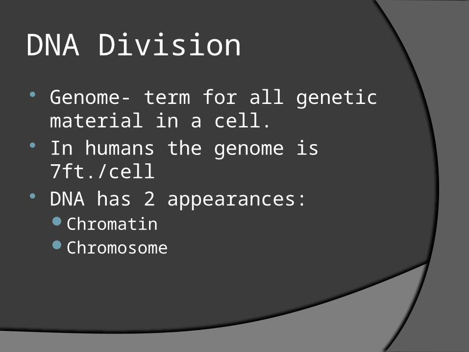
DNA Division
Genome- term for all genetic material in a cell.
In humans the genome is 7ft./cell DNA has 2 appearances:
ChromatinChromosome
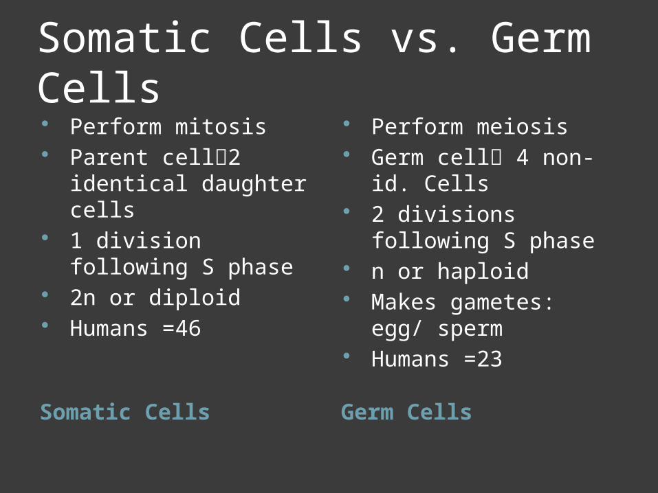
Somatic Cells vs. Germ Cells
Somatic Cells Germ Cells
Perform mitosis Parent cell2 identical
daughter cells 1 division following S
phase 2n or diploid Humans =46
Perform meiosis Germ cell 4 non-id.
Cells 2 divisions following S
phase n or haploid Makes gametes: egg/
sperm Humans =23
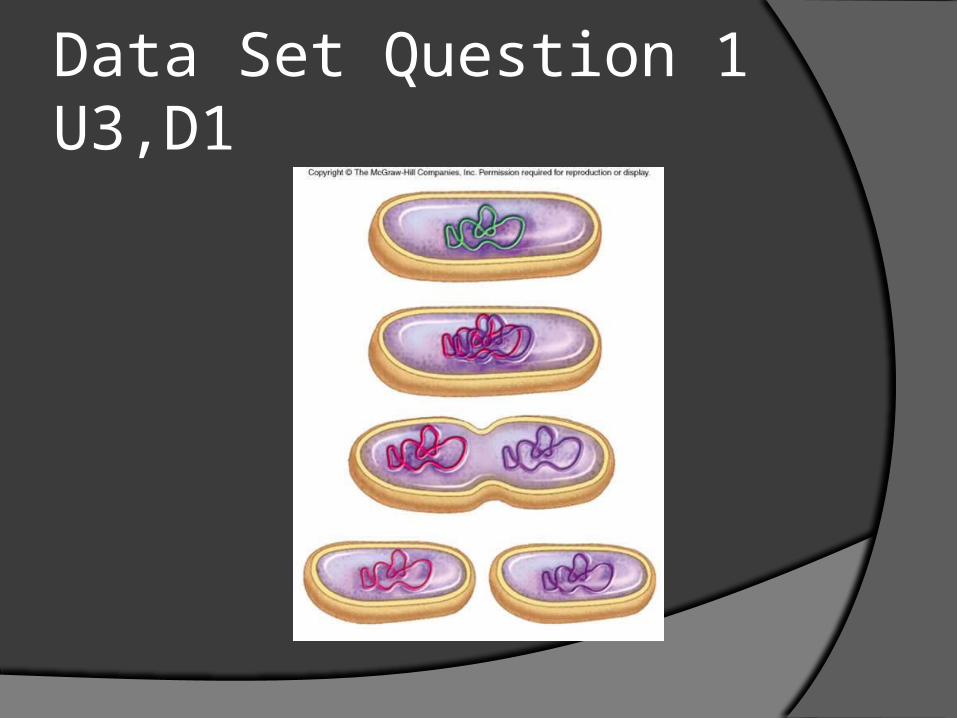
Data Set Question 1 U3,D1
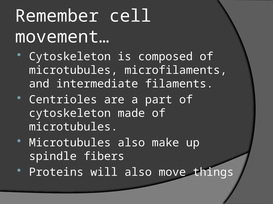
Remember cell movement… Cytoskeleton is composed of
microtubules, microfilaments, and intermediate filaments.
Centrioles are a part of cytoskeleton made of microtubules.
Microtubules also make up spindle fibers
Proteins will also move things
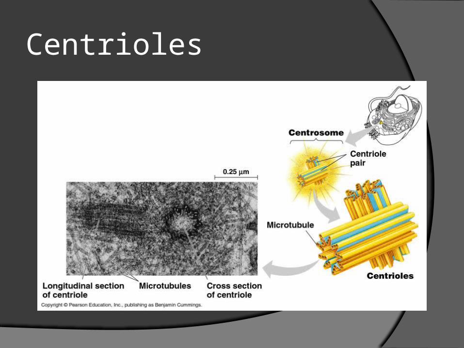
Centrioles
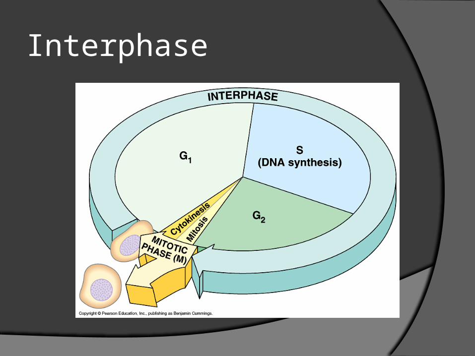
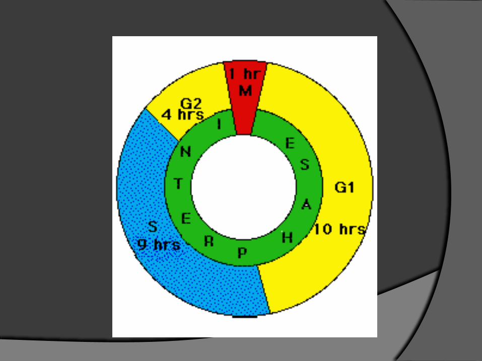
Interphase
Cell Cycle
G1- first growth, everyday activity, first checkpoint will be passed…point of no return.
S- DNA replicates, (46-92 in humans: 4n)
G2- second growth, second checkpoint where all DNA is proofed and organelles checked for cell division.
Mitosis
Mitosis is the segregation step (3) for eukaryotic cells.
Sister chromatids = replicated DNA. Chromatids held together by cohesion-
protein complex, found at centromere.
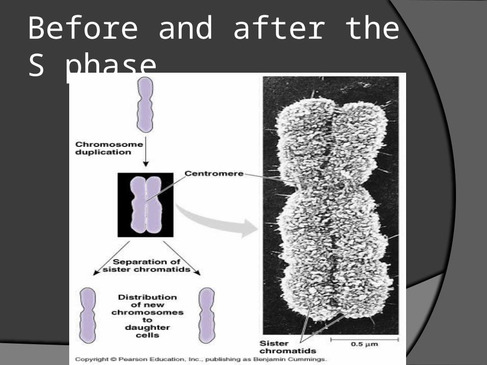
Before and after the S phase
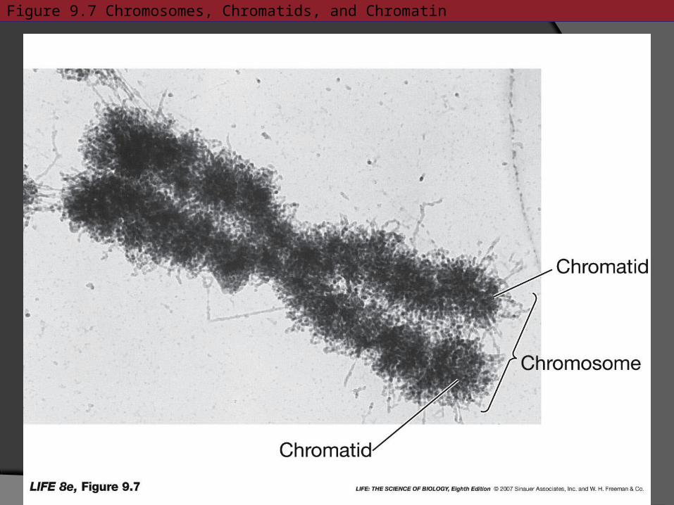
Figure 9.7 Chromosomes, Chromatids, and Chromatin
Histones Web-like proteins that have a positive
charge, and interact with the negative phosphates of DNA.
Histones form nucleosomes.Eight histone molecules.146 base pairs of DNAHistone One H1. Clamps DNA to histone core.
Chromatin will condense until chromatids move apart in anaphase.
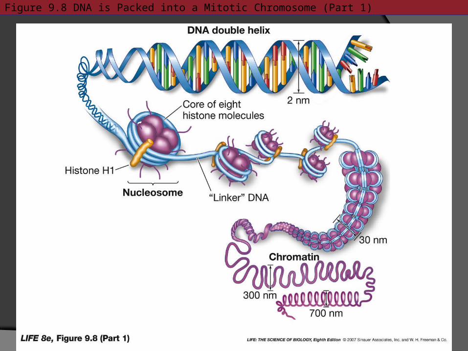
Figure 9.8 DNA is Packed into a Mitotic Chromosome (Part 1)
Centrosomes
Centrosomes consist of a pair of centrioles. Centrioles are hollow tubes consisting of nine microtubules. Each centriole pair is situated perpendicular to one another.
At G2M phase, the centrosomes migrate to opposite ends of the cell.
Plant cells do not have centrosomes, just have a microtubule organization center.
MITOSIS IS DIVISION OF THE NUCLEUS
MITOSIS ANIMATIONMITOSIS-GOOD ANIMATION
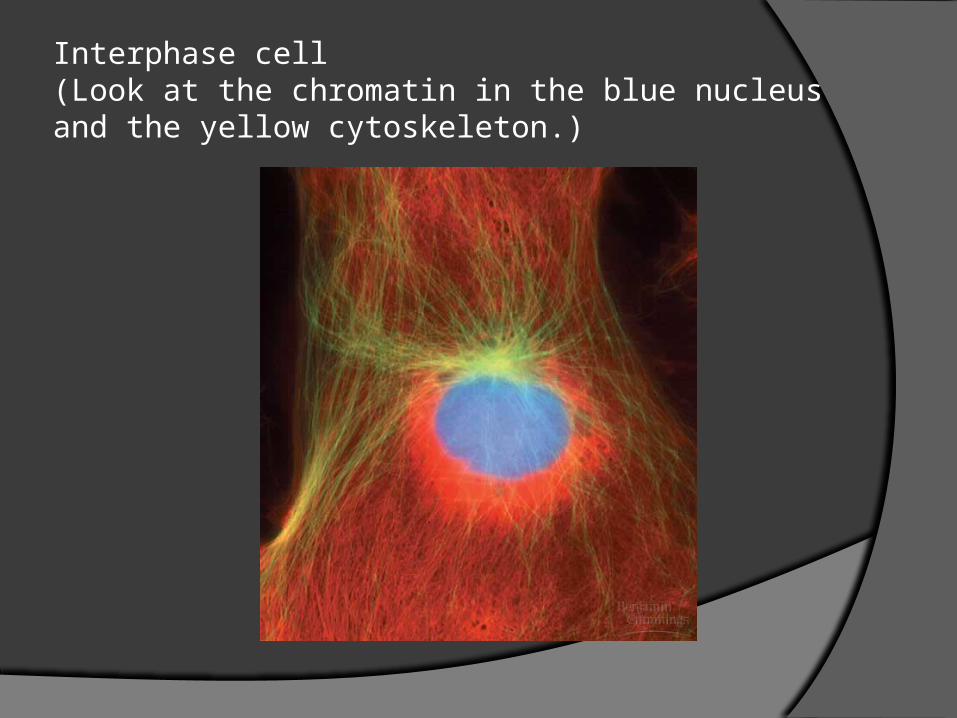
Interphase cell (Look at the chromatin in the blue nucleus and the yellow cytoskeleton.)
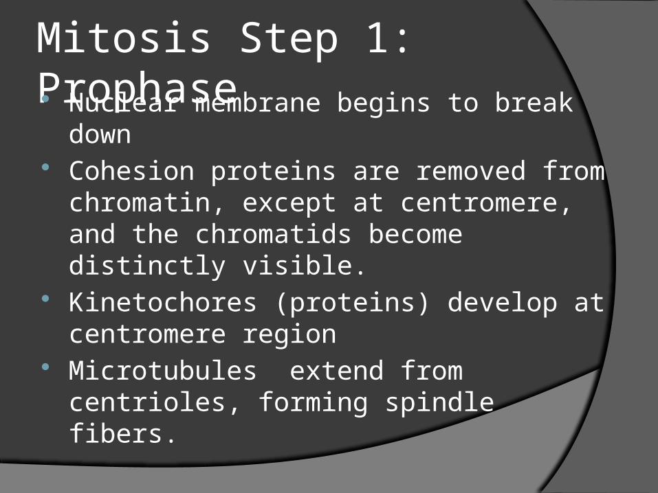
Mitosis Step 1: Prophase Nuclear membrane begins to break down Cohesion proteins are removed from
chromatin, except at centromere, and the chromatids become distinctly visible.
Kinetochores (proteins) develop at centromere region
Microtubules extend from centrioles, forming spindle fibers.
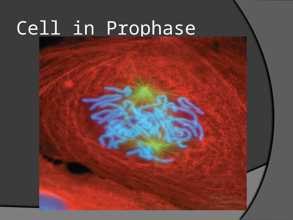
Cell in Prophase
Prometaphase
Not officially step two. Nuclear membrane completely
disappears, as well as nucleolus
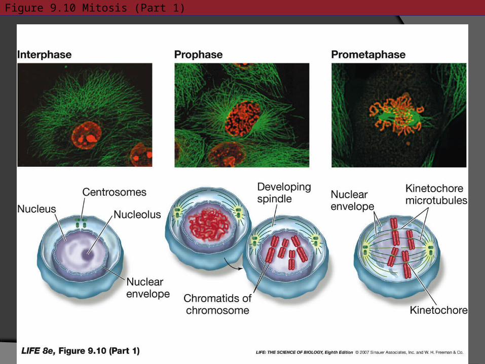
Figure 9.10 Mitosis (Part 1)
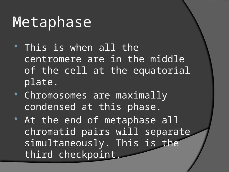
Metaphase
This is when all the centromere are in the middle of the cell at the equatorial plate.
Chromosomes are maximally condensed at this phase.
At the end of metaphase all chromatid pairs will separate simultaneously. This is the third checkpoint.
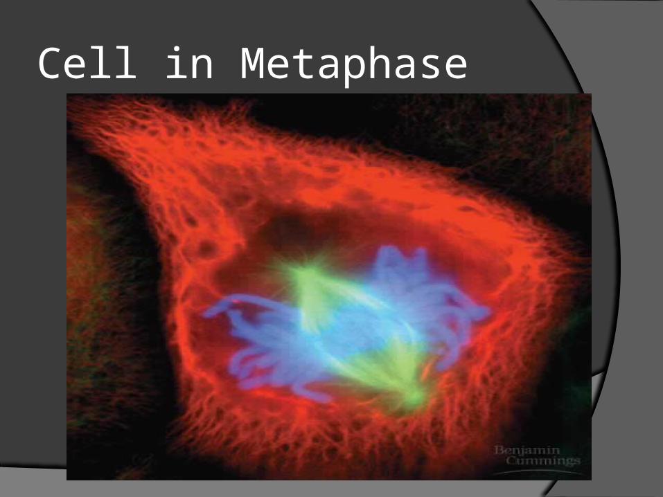
Cell in Metaphase
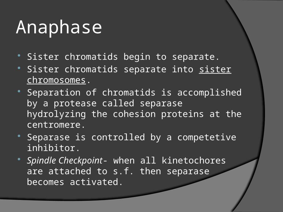
Anaphase Sister chromatids begin to separate. Sister chromatids separate into sister
chromosomes. Separation of chromatids is accomplished by a
protease called separase hydrolyzing the cohesion proteins at the centromere.
Separase is controlled by a competetive inhibitor.
Spindle Checkpoint- when all kinetochores are attached to s.f. then separase becomes activated.
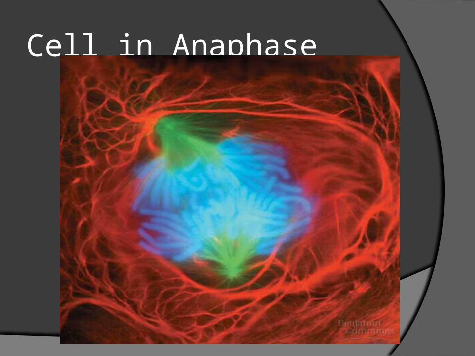
Cell in Anaphase
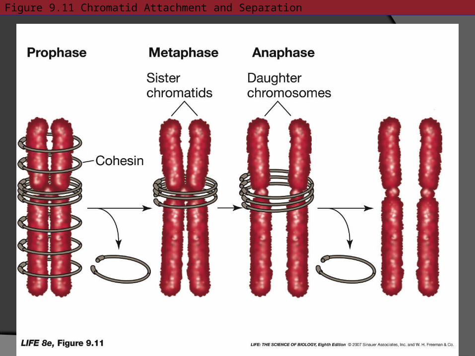
Figure 9.11 Chromatid Attachment and Separation
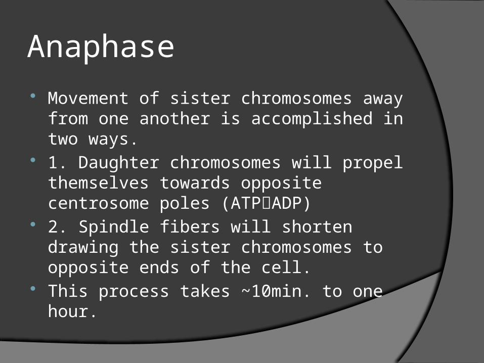
Anaphase
Movement of sister chromosomes away from one another is accomplished in two ways.
1. Daughter chromosomes will propel themselves towards opposite centrosome poles (ATPADP)
2. Spindle fibers will shorten drawing the sister chromosomes to opposite ends of the cell.
This process takes ~10min. to one hour.
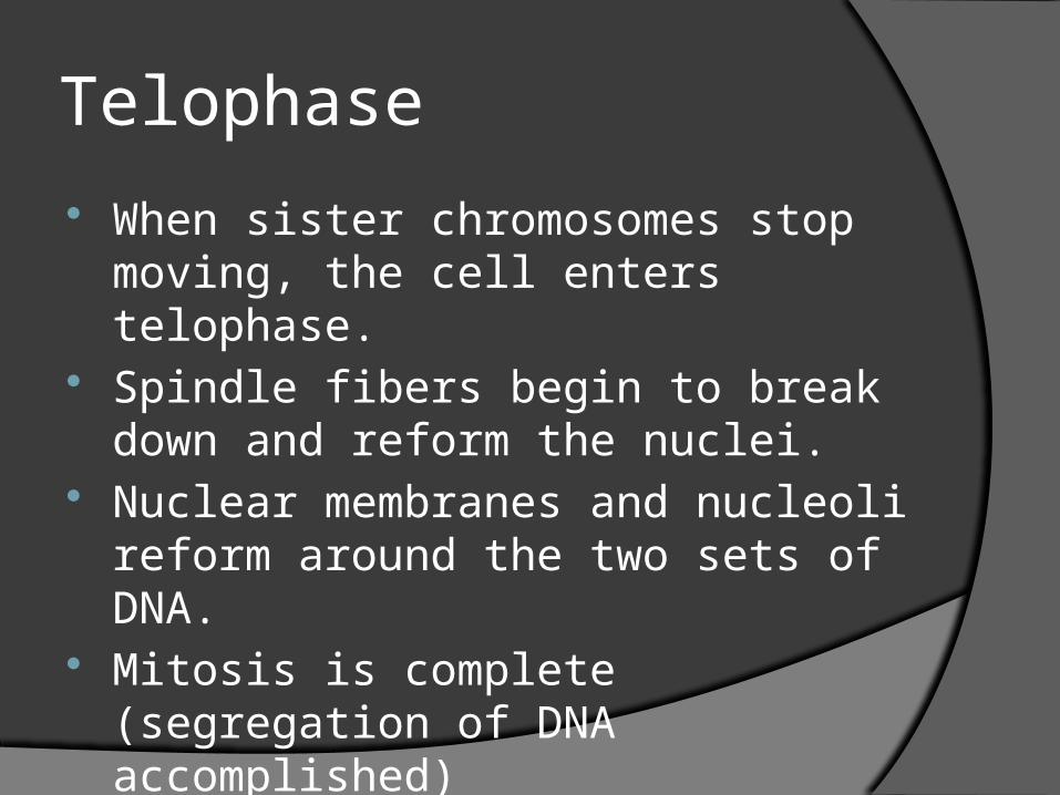
Telophase
When sister chromosomes stop moving, the cell enters telophase.
Spindle fibers begin to break down and reform the nuclei.
Nuclear membranes and nucleoli reform around the two sets of DNA.
Mitosis is complete (segregation of DNA accomplished)
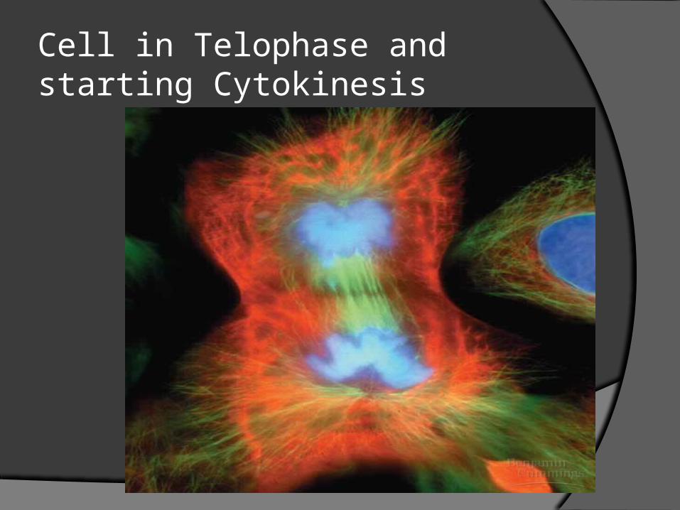
Cell in Telophase and starting Cytokinesis
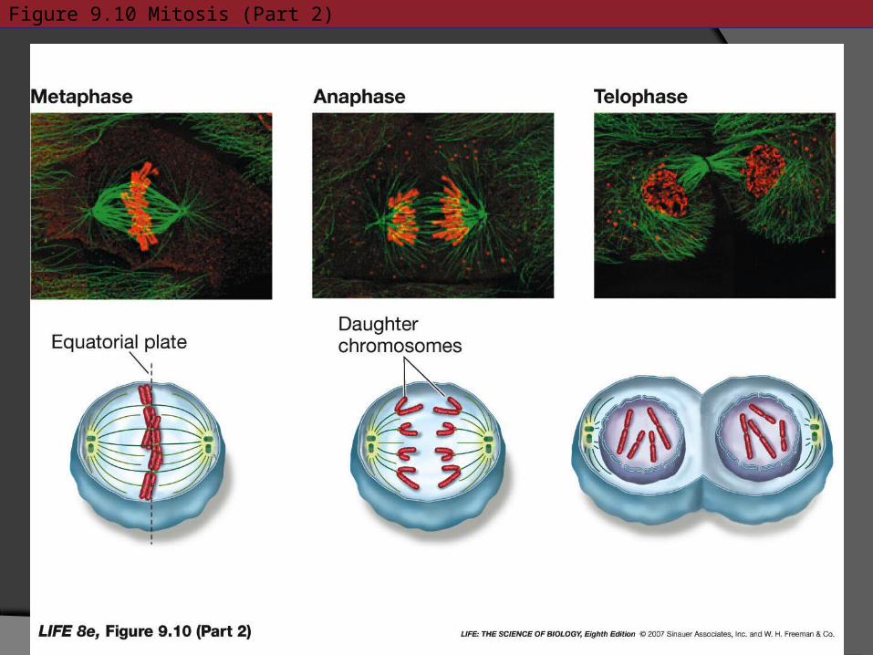
Figure 9.10 Mitosis (Part 2)
Cytokinesis The end of telophase is two nuclei in one cell;
therefore, the cell needs to divide. Cytokinesis is the process of cytoplasm
division. Animal cells divide by the cell membrane
furrowing. Contraction of actin and myosin microfilaments.
Plant cells’ vesicles from the Golgi bodies, move to equatorial plate, fuse to form new C.M. Vesicle contents also create a cell plate, which becomes new cell wall.
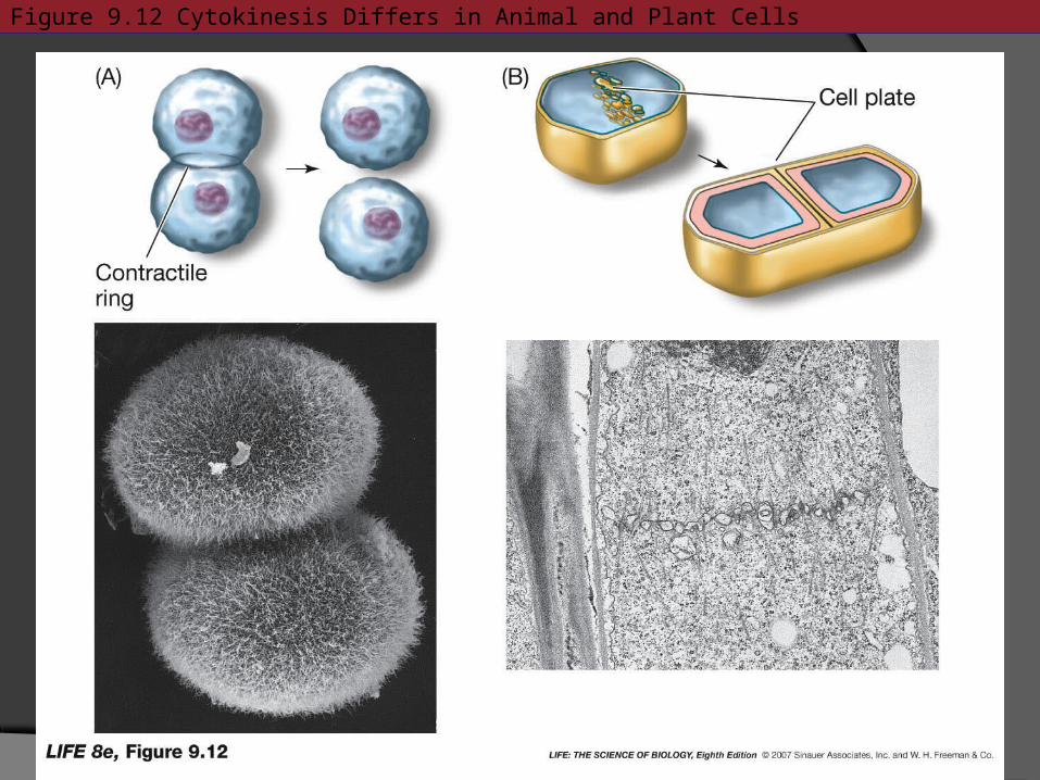
Figure 9.12 Cytokinesis Differs in Animal and Plant Cells
Cell Cycle REGULATION
Critical for normal growth and development
Controlled by proteins called cyclins. 3 Checkpoints:
Compare the difference between Theta and Eukaryotic Division
Gene # Gene Combinations Linking Genes Species Variation Inheritance from parents
Cyclins and Proteins trigger Cell Division
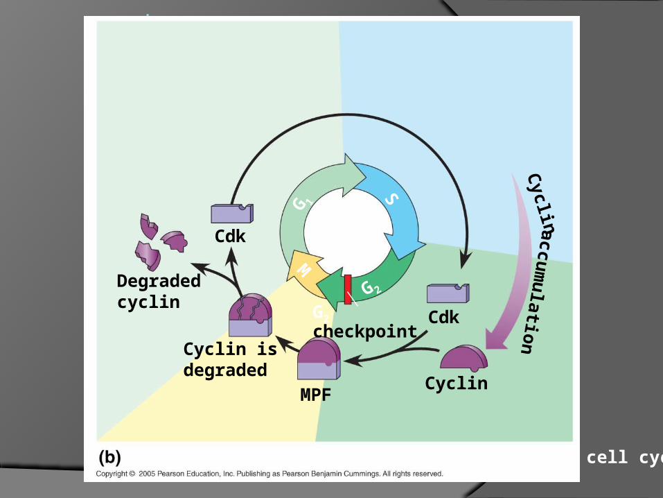
Cyclin- protein that causes G1SG2 transition. (s phase to anaphase [inc.])
Kinase is an enzyme that turn on cell processes Cyclin + Kinase = Cdk aka MPF
MPF-Maturation Promoting Factor
.
Degradedcyclin
G2
checkpoint
S
M
G 2G 1
Cdk
Cyclin isdegraded
MPFCyclin
Cdk
Molecular mechanisms that help regulate the cell cycle
accum
ulatio
nC
yclin
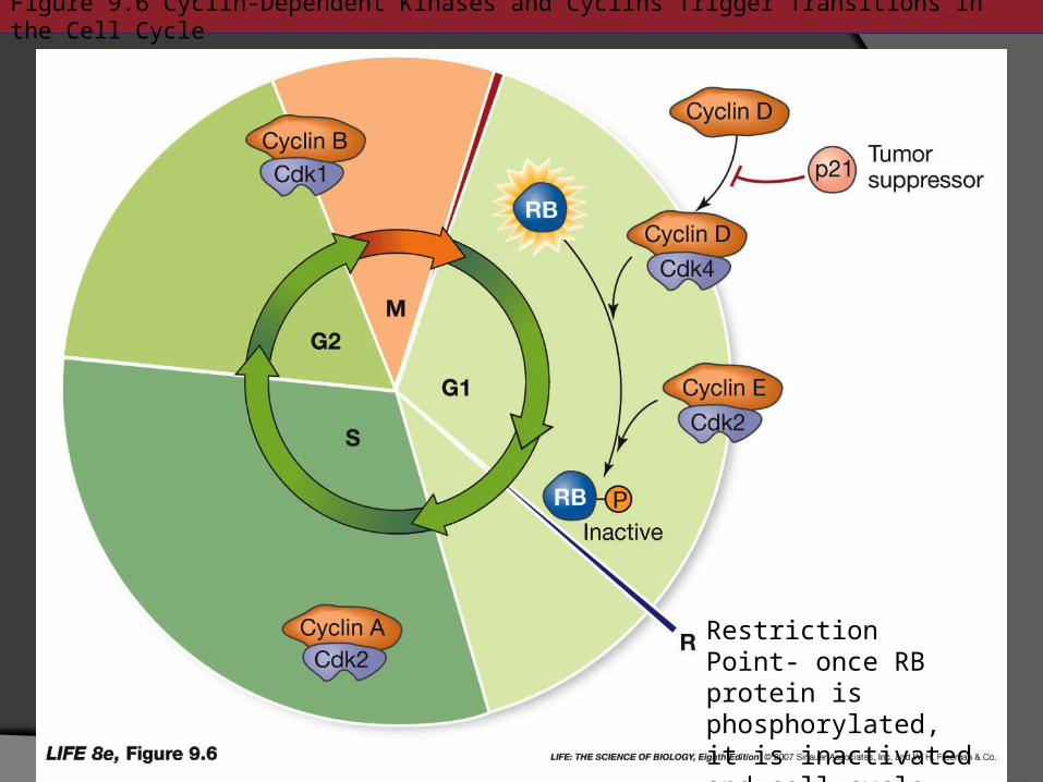
Figure 9.6 Cyclin-Dependent Kinases and Cyclins Trigger Transitions in the Cell Cycle
Restriction Point- once RB protein is phosphorylated, it is inactivated and cell cycle progresses.
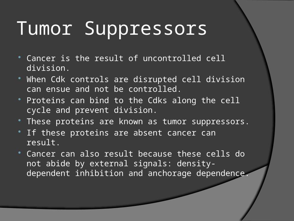
Tumor Suppressors Cancer is the result of uncontrolled cell division. When Cdk controls are disrupted cell division
can ensue and not be controlled. Proteins can bind to the Cdks along the cell
cycle and prevent division. These proteins are known as tumor
suppressors. If these proteins are absent cancer can result. Cancer can also result because these cells do
not abide by external signals: density-dependent inhibition and anchorage dependence.
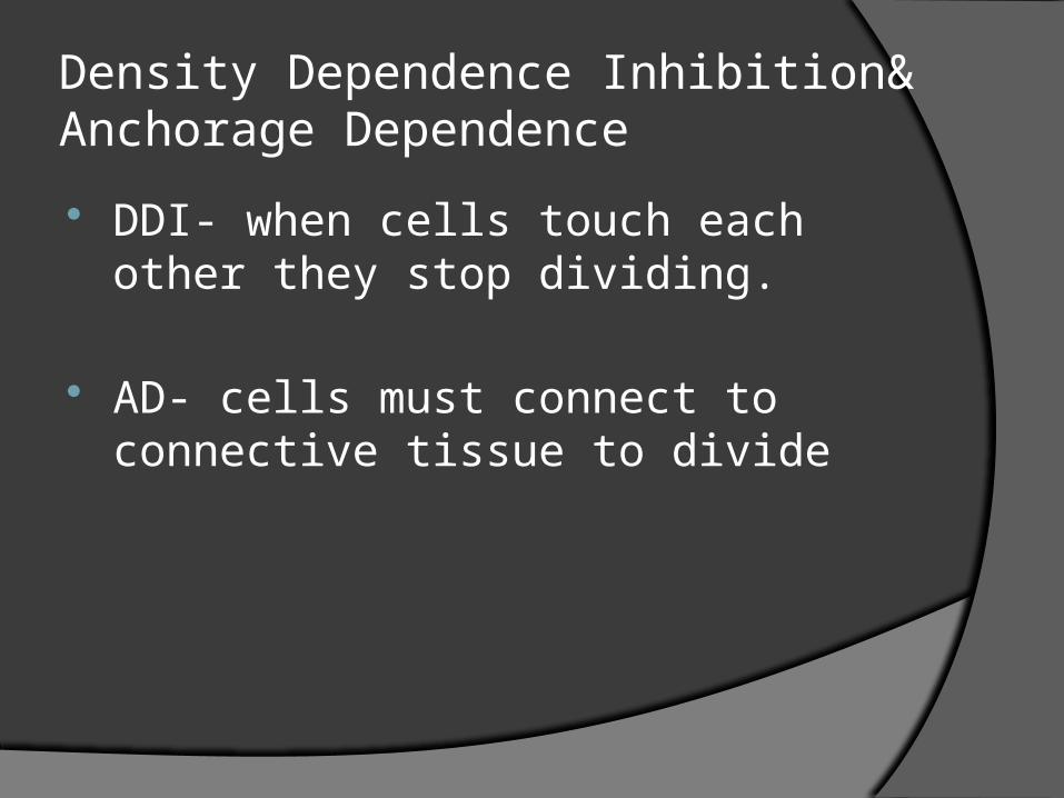
Density Dependence Inhibition& Anchorage Dependence
DDI- when cells touch each other they stop dividing.
AD- cells must connect to connective tissue to divide
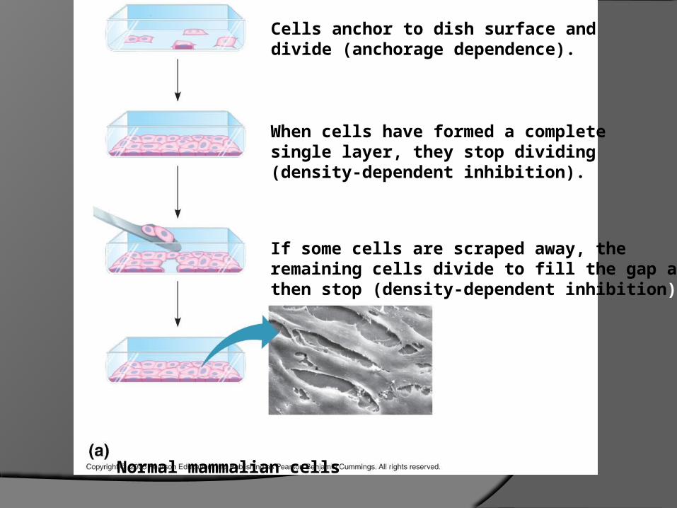
.Cells anchor to dish surface anddivide (anchorage dependence).
When cells have formed a completesingle layer, they stop dividing(density-dependent inhibition).
If some cells are scraped away, theremaining cells divide to fill the gap andthen stop (density-dependent inhibition).
25 µmNormal mammalian cells
.
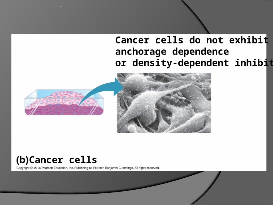
Cancer cells do not exhibitanchorage dependenceor density-dependent inhibition.
Cancer cells25 µm
Cancer Cells
Do not exhibit DDI or AD Cancer cells are immortal as long as
oxygenated blood is flowing. Performs angiogenesis
Telomerase creates cyclins, and with no checkpoints the cells divide unstoppably.
Cancer Terms
“onco” Tumor Benign Malignant Metastasis Cancer Naming Cancer causes- weak genes, environ.,
lifestyle, virus (HPV)
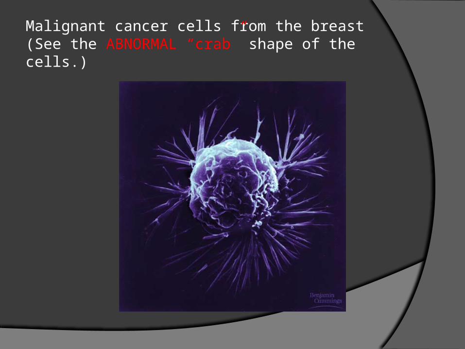
Malignant cancer cells from the breast(See the ABNORMAL “crab” shape of the cells.)
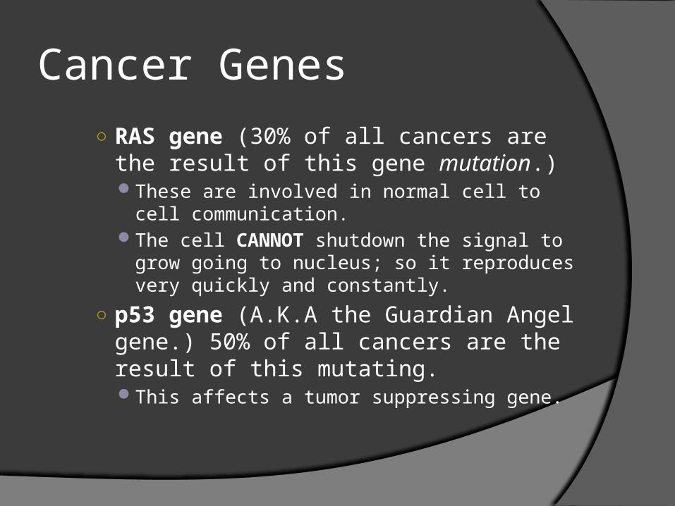
Cancer Genes○ RAS gene (30% of all cancers are the result
of this gene mutation.)These are involved in normal cell to cell
communication. The cell CANNOT shutdown the signal to grow going
to nucleus; so it reproduces very quickly and constantly.
○ p53 gene (A.K.A the Guardian Angel gene.) 50% of all cancers are the result of this mutating.This affects a tumor suppressing gene.
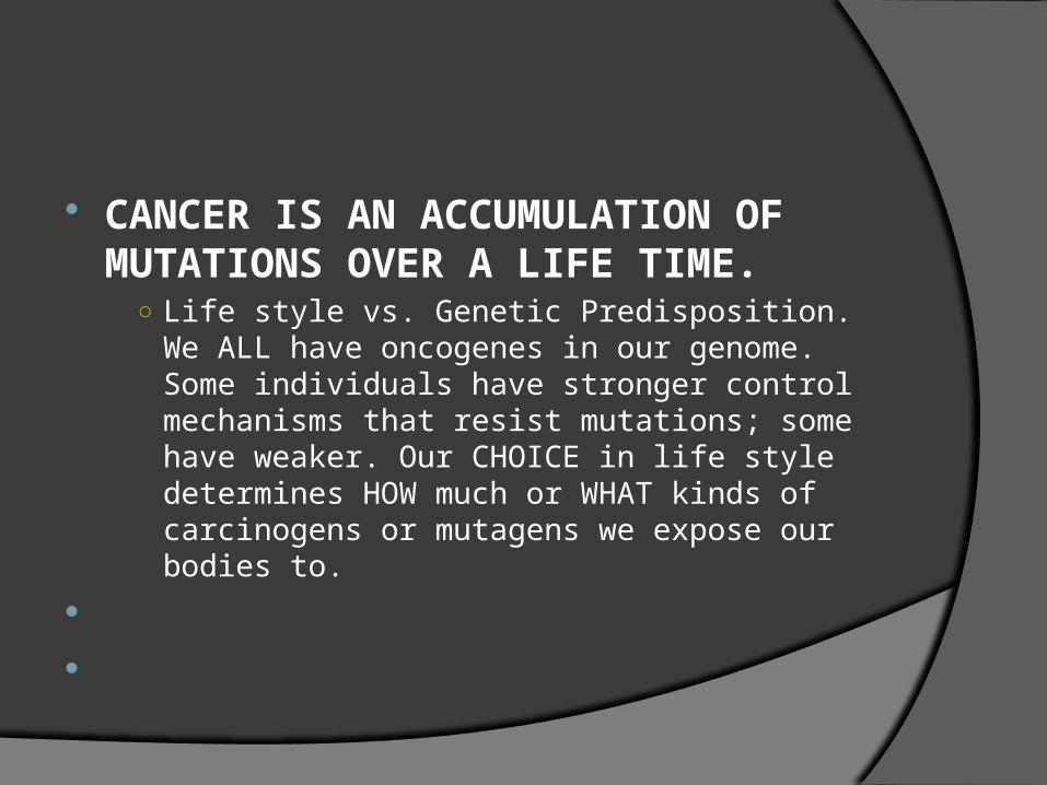
CANCER IS AN ACCUMULATION OF MUTATIONS OVER A LIFE TIME.
○ Life style vs. Genetic Predisposition. We ALL have oncogenes in our genome. Some individuals have stronger control mechanisms that resist mutations; some have weaker. Our CHOICE in life style determines HOW much or WHAT kinds of carcinogens or mutagens we expose our bodies to.
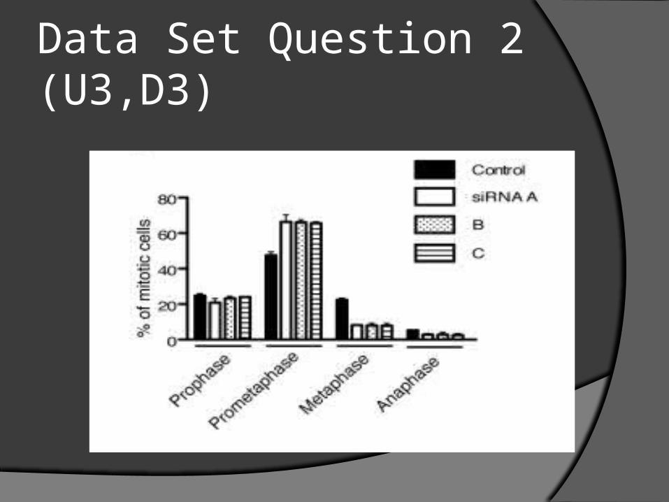
Data Set Question 2 (U3,D3)

Cell Death: Necrosis vs. Apoptosis
Necrosis Apoptosis
Cell death due to damage by toxins, O2 deprivation, or nutrient deficiency.
Cells usually swell and burst.
This usually causes inflammation due to the cell contents in the extracellular matrix
Programmed cell death Due to cell is unneeded,
or cell is aging and may be prone to genetic damage leading to cancer.
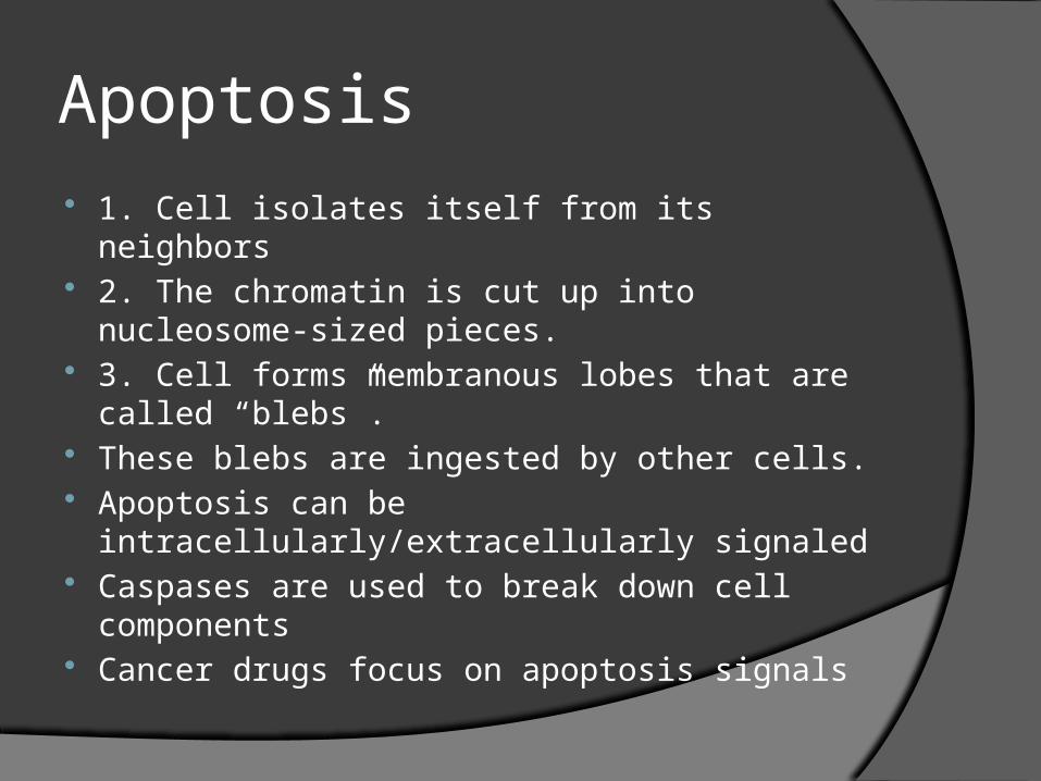
Apoptosis 1. Cell isolates itself from its neighbors 2. The chromatin is cut up into nucleosome-
sized pieces. 3. Cell forms membranous lobes that are called
“blebs”. These blebs are ingested by other cells. Apoptosis can be intracellularly/extracellularly
signaled Caspases are used to break down cell
components Cancer drugs focus on apoptosis signals
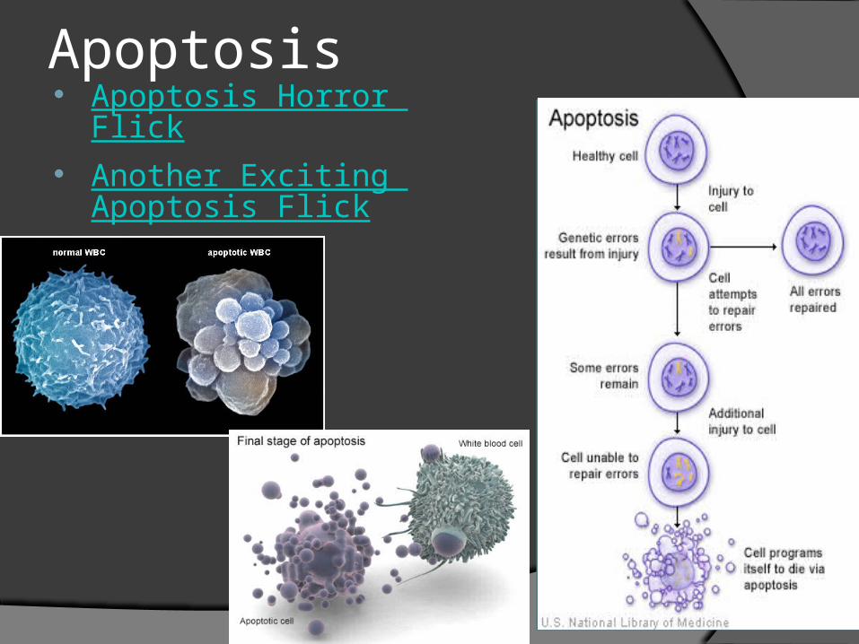
Apoptosis Apoptosis Horror Flick Another Exciting Apopt
osis Flick
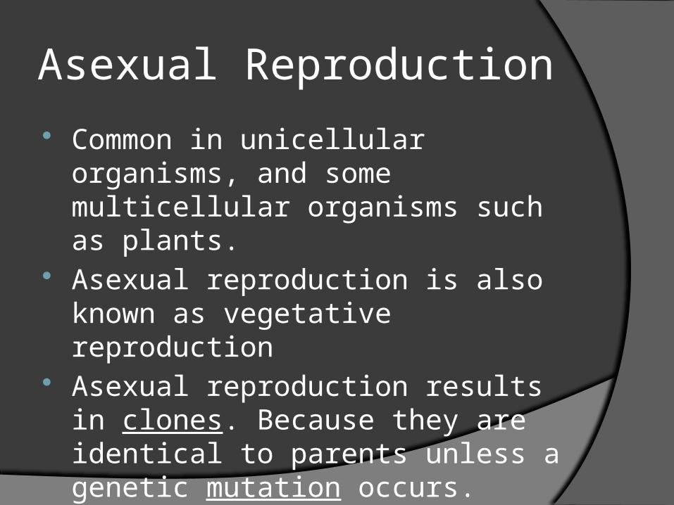
Asexual Reproduction
Common in unicellular organisms, and some multicellular organisms such as plants.
Asexual reproduction is also known as vegetative reproduction
Asexual reproduction results in clones. Because they are identical to parents unless a genetic mutation occurs.
Sexual Reproduction
Offspring dissimilar from parents. Gametes created by meiosis are
genetically different, thus creating unique offspring.
Meiosis is the raw material basis for natural selection and evolution. (Think offspring comparison of adaptation)
Meiosis is the process of gamete formation.
Fertilization
When two haploid gametes fuse to form a zygote (new organism).
Haploid gametes = sperm + egg
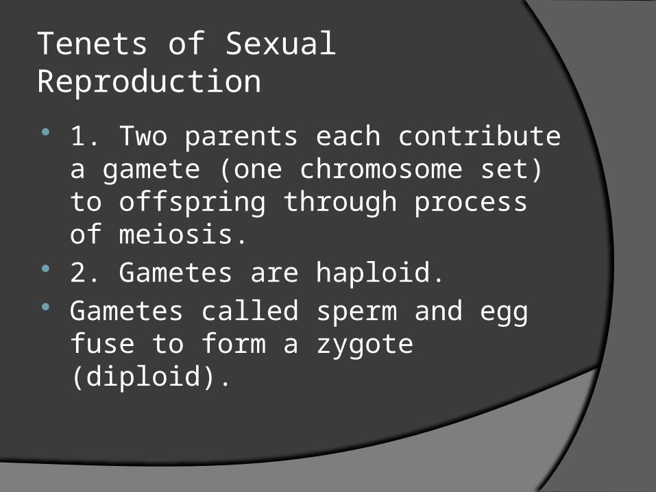
Tenets of Sexual Reproduction
1. Two parents each contribute a gamete (one chromosome set) to offspring through process of meiosis.
2. Gametes are haploid. Gametes called sperm and egg fuse to
form a zygote (diploid).
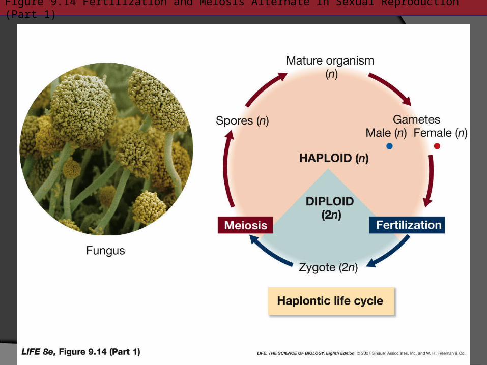
Figure 9.14 Fertilization and Meiosis Alternate in Sexual Reproduction (Part 1)

Figure 9.14 Fertilization and Meiosis Alternate in Sexual Reproduction (Part 2)
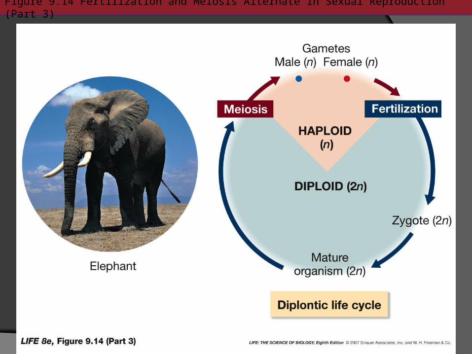
Figure 9.14 Fertilization and Meiosis Alternate in Sexual Reproduction (Part 3)
What is the purpose of mitosis?
What is the purpose of meiosis?
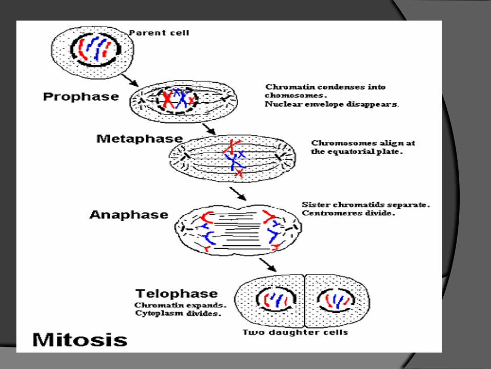
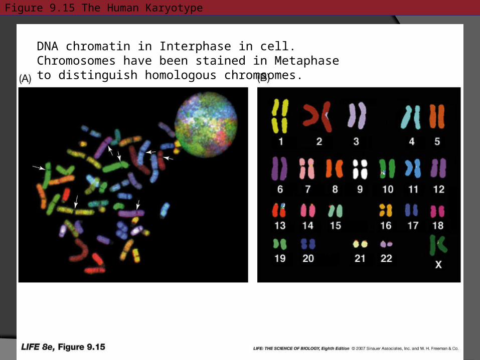
Figure 9.15 The Human Karyotype
DNA chromatin in Interphase in cell.Chromosomes have been stained in Metaphase to distinguish homologous chromsomes.

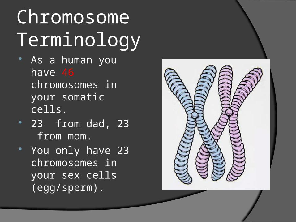
Chromosome Terminology As a human you
have 46 chromosomes in your somatic cells.
23 from dad, 23 from mom.
You only have 23 chromosomes in your sex cells (egg/sperm).
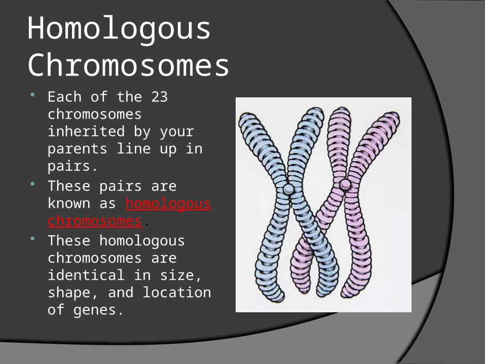
Homologous Chromosomes Each of the 23
chromosomes inherited by your parents line up in pairs.
These pairs are known as homologous chromosomes.
These homologous chromosomes are identical in size, shape, and location of genes.
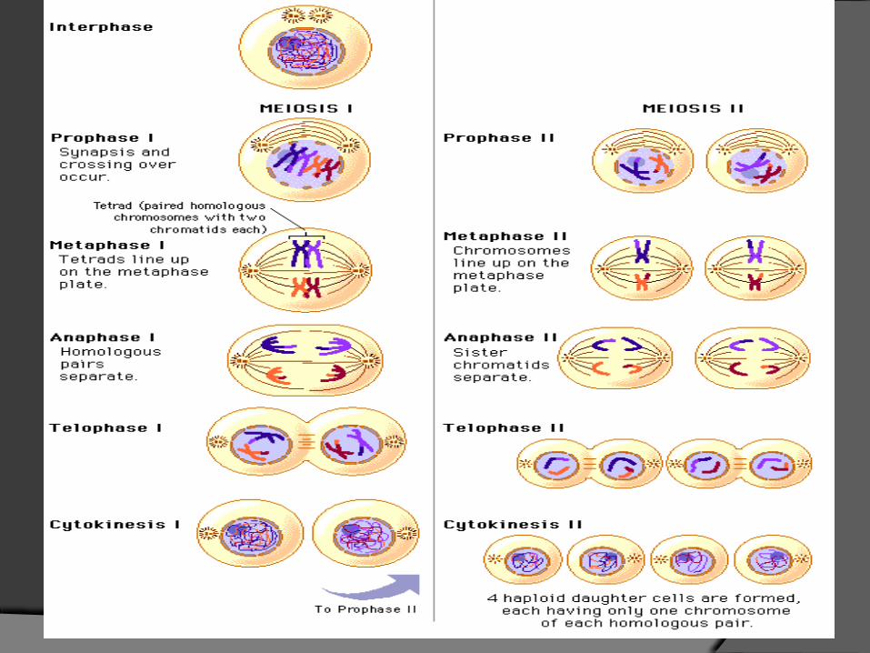
Meiosis Meiosis Anima
tion Meiosis Simul
ation
Meiosis I During Prophase I, a process called
synapsis/ chiasmata occurs. Homologous chromosomes pair by adhering at their lengths.
Proteins aid in this adhesion by forming a scaffold called a synaptonemal complex.
The four bound chromatids form a tetrad. How many chromatids are in human cells
during Meiosis I? 92
Meiosis I and Meiosis IIMeiosis I Meiosis II
Homologues will meet and form a tetrad.
Crossing over occurs: allele swapping.
Telophase I- two new cells with one homologue per cell (still replicated chromatids)
Prophase, Metaphase and Anaphase similar to Mitosis and Meiosis I.
Telophase II results in four haploid daughter cells.
One chromatid per cell
Meiosis I
The chromatids that result are known as recombinant chromatids
Not all organisms directly enter Meiosis II.
If an organism does not, it does form a nuclear membrane at the end of Telophase I
Telophase I is followed by interkinesis, which is similar to mitotic interphase
Meiosis II
Homologues are not identical like in Meiosis I because of crossing over.
The result is four haploid nuclei, with a single set of unreplicated chromosomes.
So what causes genetic diversity Synapsis, crossing over, and segregation of
homologues Aneuploidy- when there are either missing or
excessive chromosomes.MonosomyTrisomy 10-30% human zygotes show trisomy Aneuploidy, ~20% of miscarriages due to aneuploidy
(extra or missing chromosomes)Polyploidy complete extra sets of chromosomes, can
occur naturally, can be result of genetic engineeringAneuploidy Simulations- Utah Site

Cell Signaling…Remember Glycolipids and Glycoproteins Each molecules has its own distinct
shape ECM interacts with cells
If cells don’t‘ communicate with one another they will not function and eventually die .
Cells communicate via chemicals
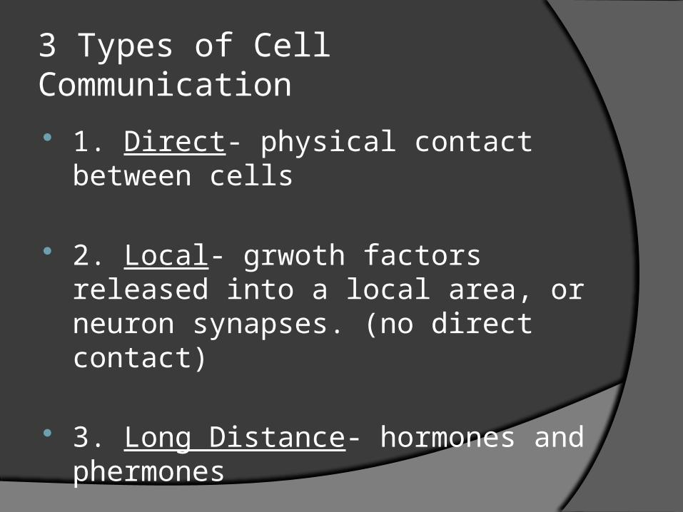
3 Types of Cell Communication
1. Direct- physical contact between cells
2. Local- grwoth factors released into a local area, or neuron synapses. (no direct contact)
3. Long Distance- hormones and phermones
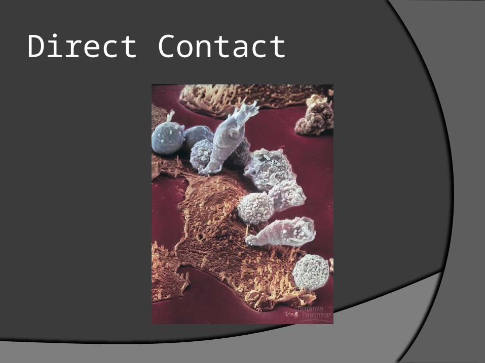
Direct Contact
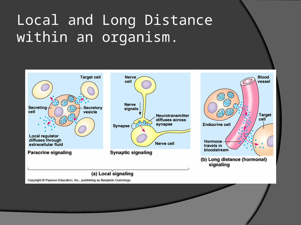
Local and Long Distance within an organism.
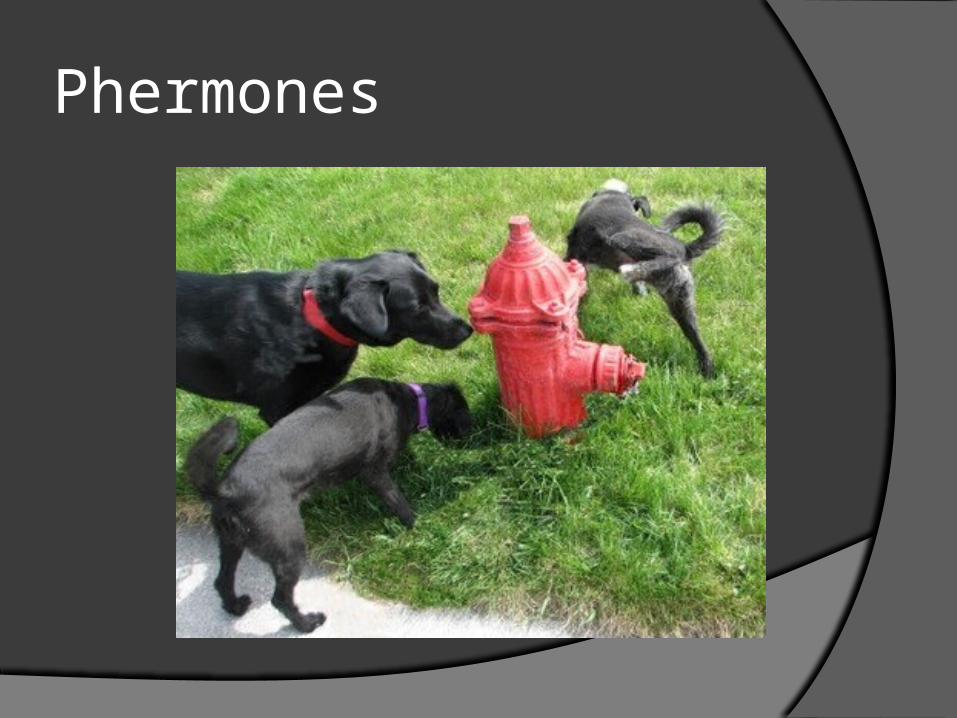
Phermones
Signal Transduction Pathway 1971, Earl Sutherland, Vanderbilt U. 1. RECEPTION 2. TRANSDUCTION 3. RESPONSE
1. RECEPTION- molecule binds to cell membrane.
2. TRANSDUCTION- occurs in cytoplasm/nucleus. Changes signal into something usable.
3. RESPONSE- usually involves DNA making protein. Turns signal into action
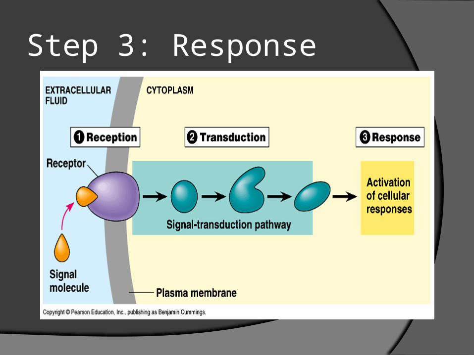
Step 3: Response
Signal Transduction
The phone call…
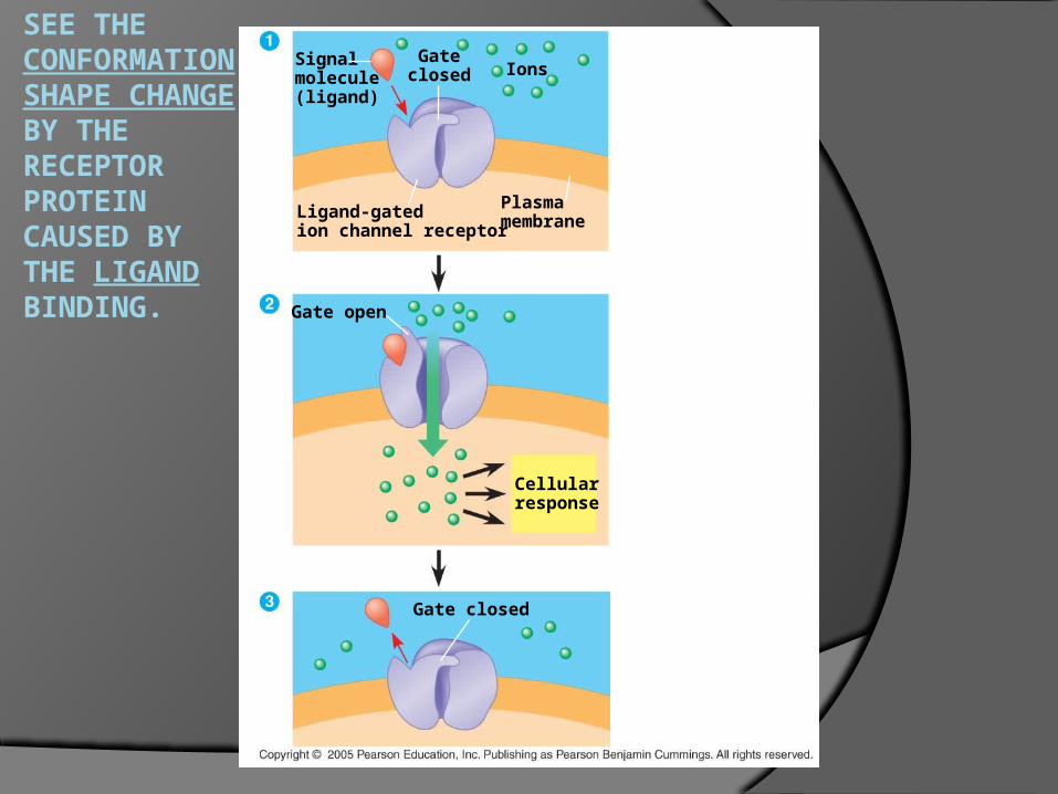
SEE THE CONFORMATION SHAPE CHANGE BY THE RECEPTOR PROTEIN CAUSED BY THE LIGAND BINDING.
Signalmolecule(ligand)
Gateclosed Ions
Ligand-gatedion channel receptor
Plasmamembrane
Gate closed
Gate open
Cellularresponse
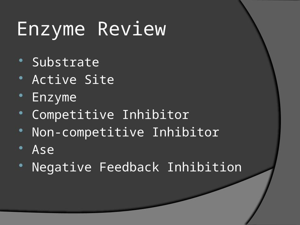
Enzyme Review
Substrate Active Site Enzyme Competitive Inhibitor Non-competitive Inhibitor Ase Negative Feedback Inhibition
Signal Transduction Pathways
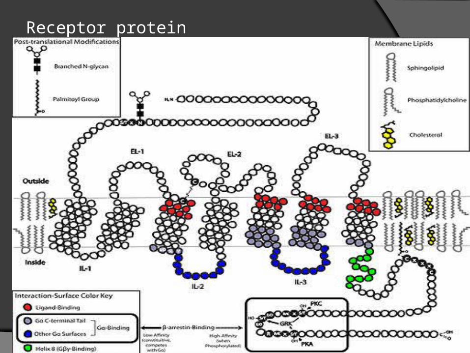
1. G Protein Linked Receptors 2. Tyrosine Kinase 3. Ion Channel Receptors 4. Intracellular Receptors

G Protein Linked Receptors On cell membrane, of ALL CELLS. Ligand binds=conformational shape
change G proteins usually phosphorylate things
to activate them.
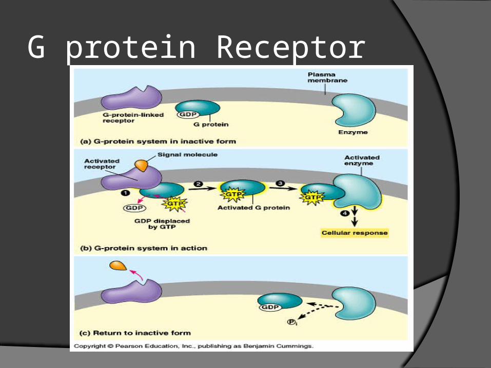
G protein Receptor
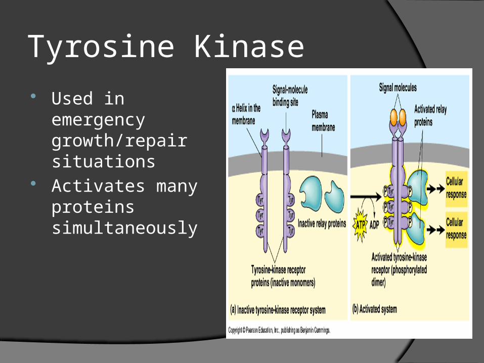
Tyrosine Kinase Used in emergency
growth/repair situations
Activates many proteins simultaneously
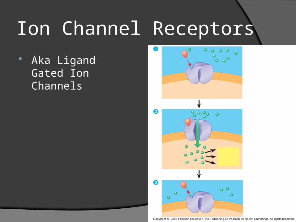
Ion Channel Receptors Aka Ligand Gated
Ion Channels
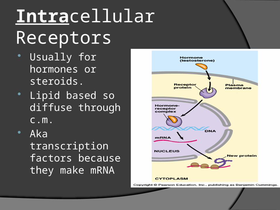
Intracellular Receptors Usually for
hormones or steroids.
Lipid based so diffuse through c.m.
Aka transcription factors because they make mRNA
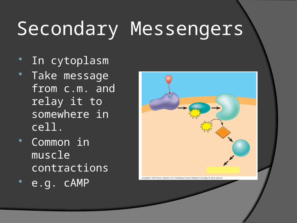
Secondary Messengers In cytoplasm Take message from
c.m. and relay it to somewhere in cell.
Common in muscle contractions
e.g. cAMP
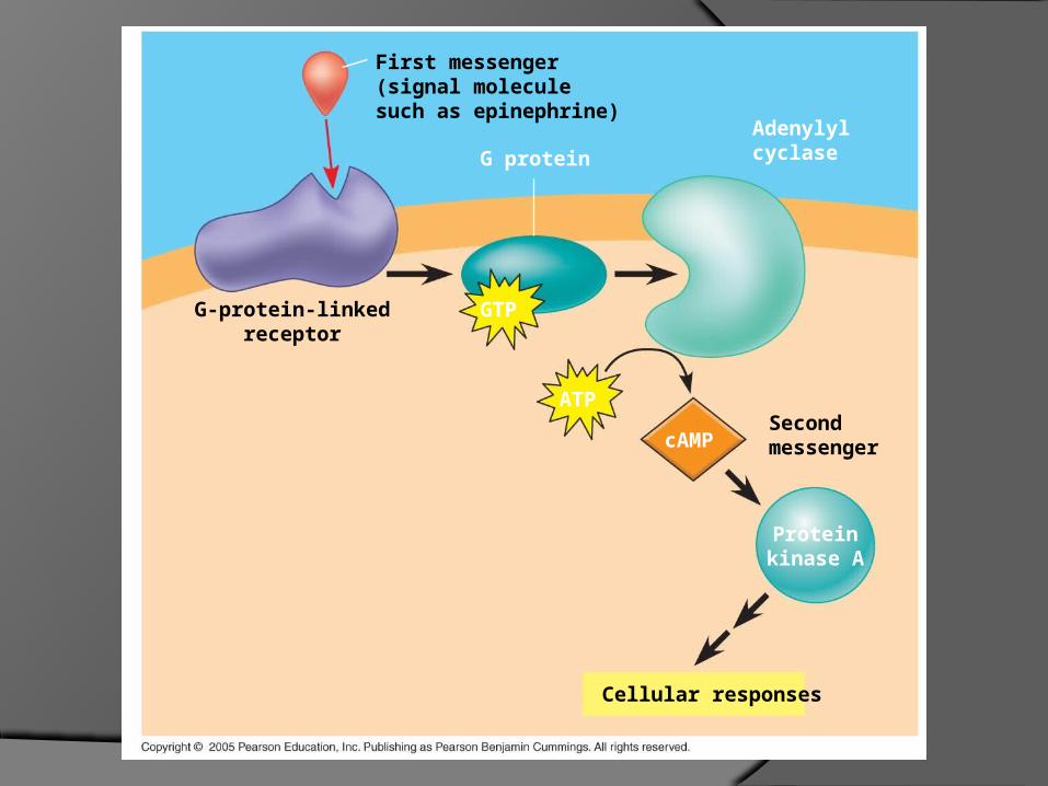
cAMP
ATPSecondmessenger
First messenger(signal moleculesuch as epinephrine)
G-protein-linkedreceptor
G protein
Adenylylcyclase
Proteinkinase A
Cellular responses
GTP
Receptor protein
Cascades
Cacade = Amplification…1 becomes 2, 2 to 4, 4 to 8…E Conservation
Protein Kinase Cascades turns ON processes by phosphorylating
Protein Phosphotase Cascades Cascades turn OFF by dephosphorylating
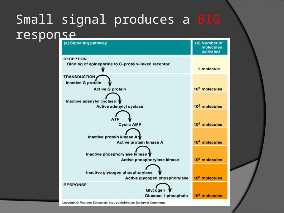
Small signal produces a BIG response
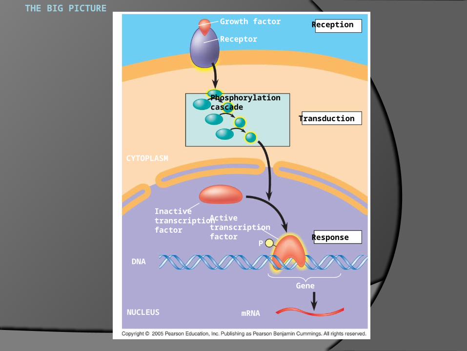
THE BIG PICTURE
ReceptionGrowth factor
Receptor
Phosphorylationcascade
Transduction
CYTOPLASM
Inactivetranscriptionfactor
Activetranscriptionfactor
PResponse
Gene
mRNA
DNA
NUCLEUS
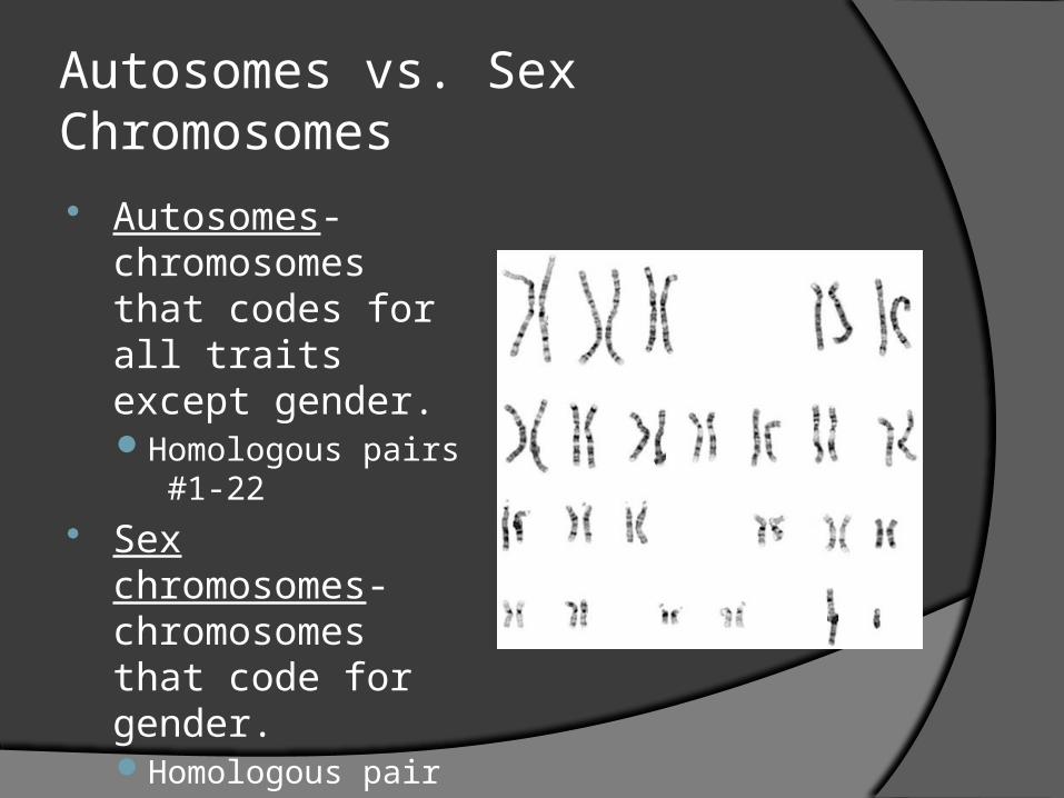
Autosomes vs. Sex Chromosomes Autosomes-
chromosomes that codes for all traits except gender.Homologous pairs
#1-22 Sex chromosomes-
chromosomes that code for gender.Homologous pair #23
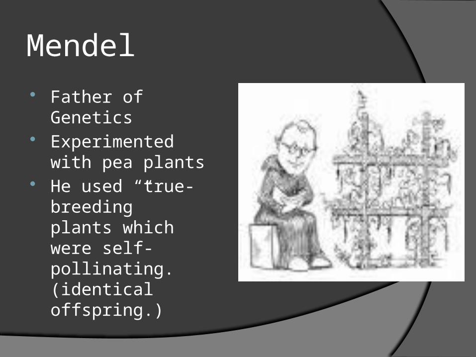
Mendel Father of Genetics Experimented with
pea plants He used “true-
breeding” plants which were self-pollinating. (identical offspring.)
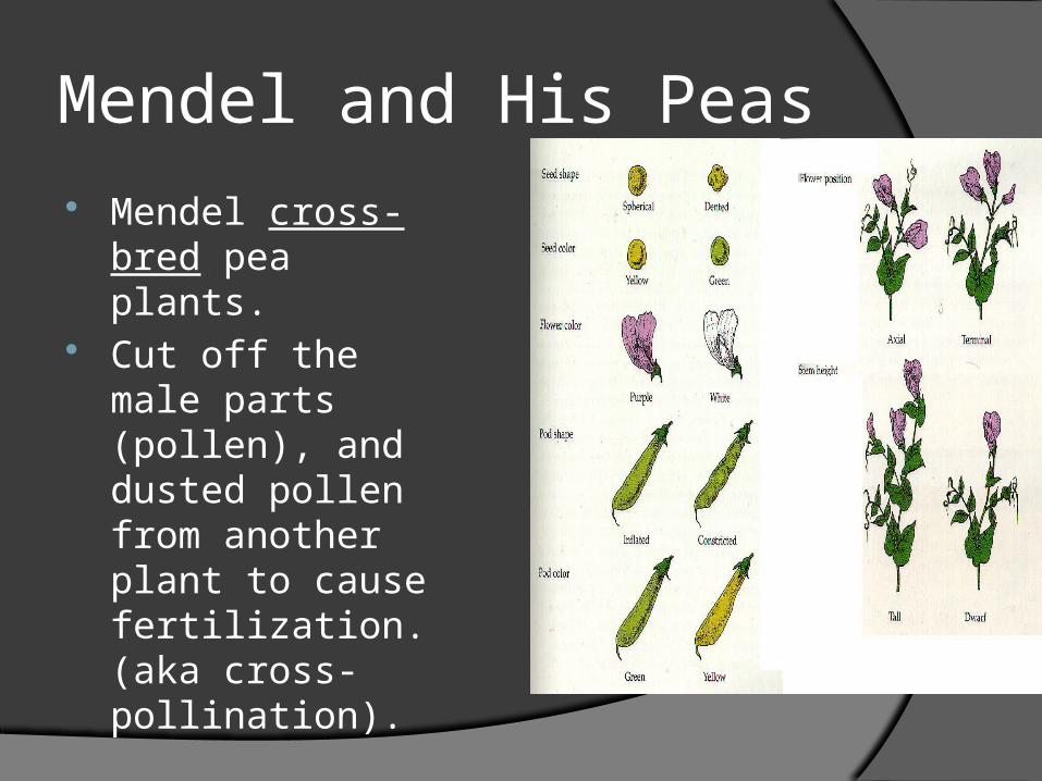
Mendel and His Peas Mendel cross-bred
pea plants. Cut off the male
parts (pollen), and dusted pollen from another plant to cause fertilization.(aka cross-pollination).
Mendel Mendel studied seven different traits. Trait- specific characteristic, like flower color. P generation- parent generation. F1 generation- offspring of the P generation. Traits are controlled by genes. Genes- segment of DNA Allele- different forms of a particular gene
Ex. Hair color, eye color, plant flower color.
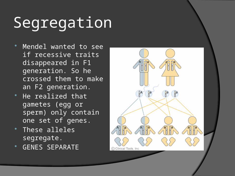
Segregation Mendel wanted to see if
recessive traits disappeared in F1 generation. So he crossed them to make an F2 generation.
He realized that gametes (egg or sperm) only contain one set of genes.
These alleles segregate. GENES SEPARATE
Law of Independent Assortment
Just because you have one dominant gene, does not mean all of your genes are dominant.
INHERITED INDEPENDENTLY
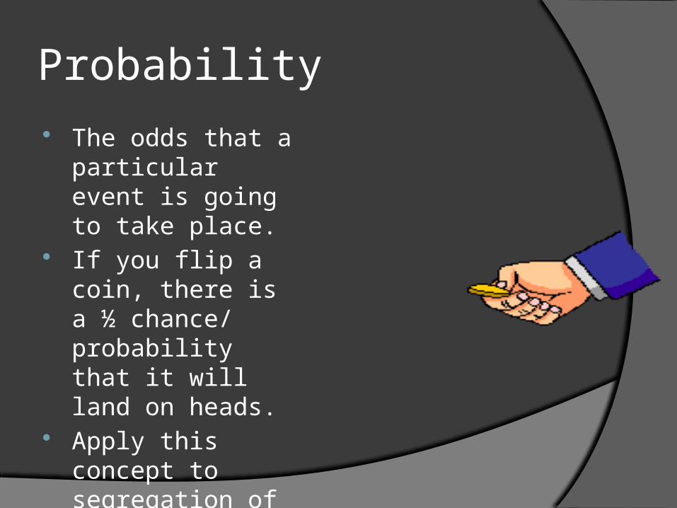
Probability The odds that a
particular event is going to take place.
If you flip a coin, there is a ½ chance/ probability that it will land on heads.
Apply this concept to segregation of alleles.
Probability and Segregation These principles can
only be viewed if there are hundreds or thousands of offspring.
You and your siblings are not enough to prove or disprove this principle.
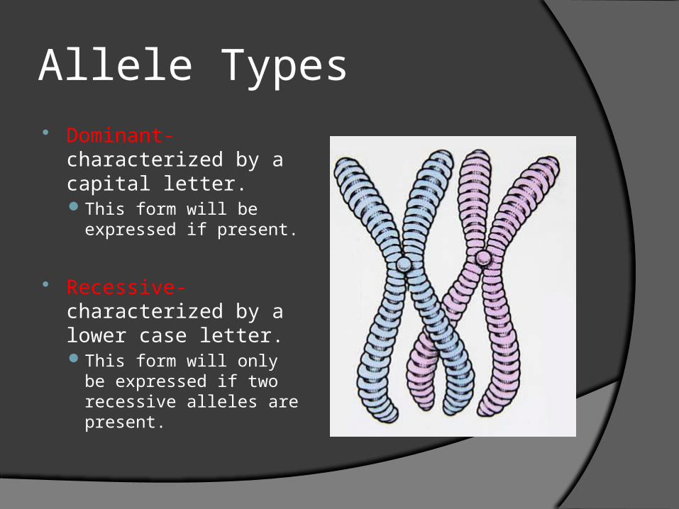
Allele Types Dominant-
characterized by a capital letter. This form will be
expressed if present.
Recessive- characterized by a lower case letter. This form will only be
expressed if two recessive alleles are present.
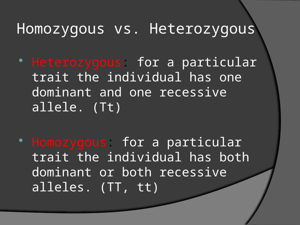
Homozygous vs. Heterozygous
Heterozygous: for a particular trait the individual has one dominant and one recessive allele. (Tt)
Homozygous: for a particular trait the individual has both dominant or both recessive alleles. (TT, tt)
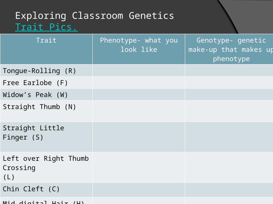
Exploring Classroom GeneticsTrait Pics.
Trait Phenotype- what you look like
Genotype- genetic make-up that makes up phenotype
Tongue-Rolling (R)
Free Earlobe (F)
Widow’s Peak (W)
Straight Thumb (N)
Straight Little Finger (S)
Left over Right Thumb Crossing (L)
Chin Cleft (C)
Mid-digital Hair (H)
Six Fingers (F)
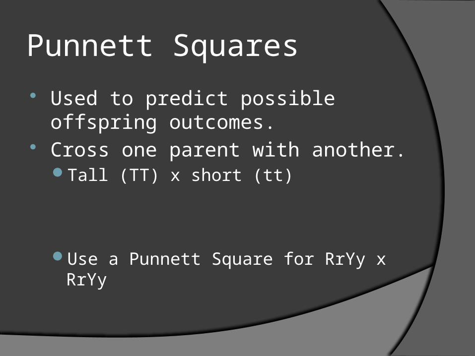
Punnett Squares
Used to predict possible offspring outcomes.
Cross one parent with another. Tall (TT) x short (tt)
Use a Punnett Square for RrYy x RrYy
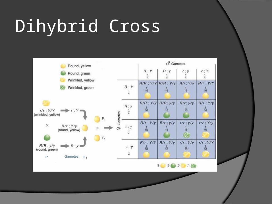
Dihybrid Cross
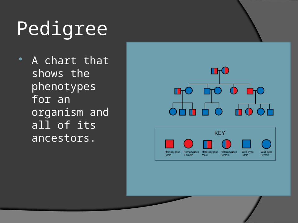
Pedigree A chart that
shows the phenotypes for an organism and all of its ancestors.
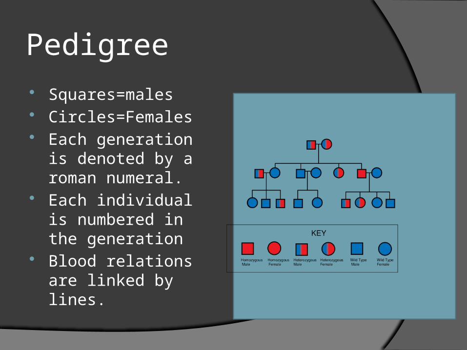
Pedigree Squares=males Circles=Females Each generation is
denoted by a roman numeral.
Each individual is numbered in the generation
Blood relations are linked by lines.
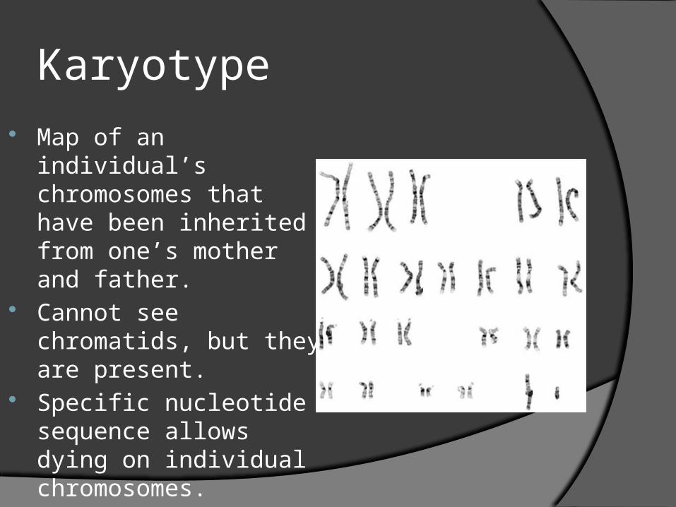
Karyotype Map of an individual’s
chromosomes that have been inherited from one’s mother and father.
Cannot see chromatids, but they are present.
Specific nucleotide sequence allows dying on individual chromosomes.

Figure 9.15 The Human Karyotype
DNA chromatin in Interphase in cell.Chromosomes have been stained in Metaphase to distinguish homologous chromsomes.
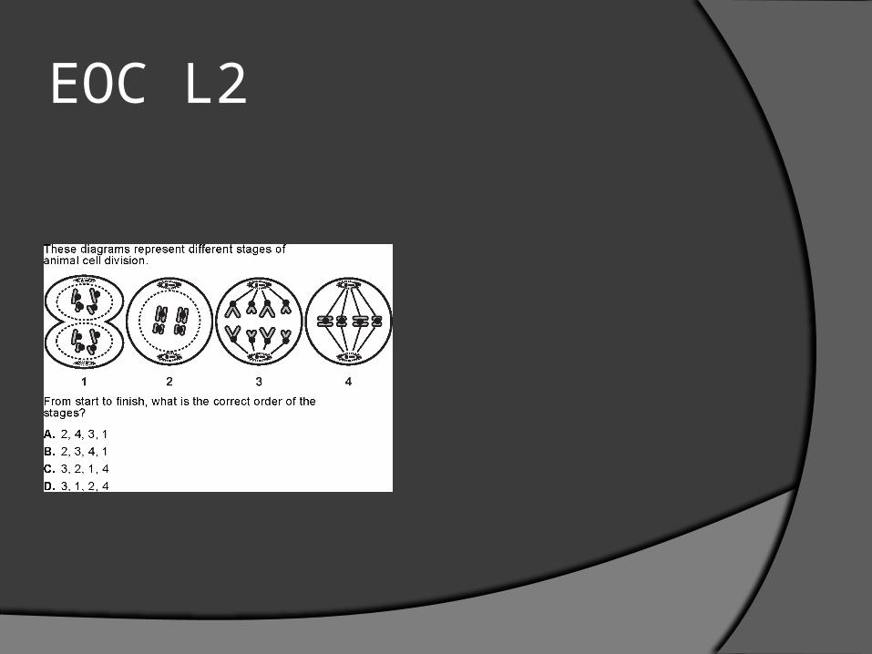
EOC L2
11-26-12
1. List the 3 Sexual Life Cycles.
2. Haploid vs. Diploid
3. Somatic vs. Sex Cell
4. Centromere…Centriole…Centrosome
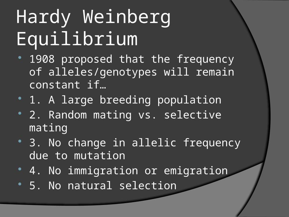
Hardy Weinberg Equilibrium 1908 proposed that the frequency of
alleles/genotypes will remain constant if…
1. A large breeding population 2. Random mating vs. selective mating 3. No change in allelic frequency due to
mutation 4. No immigration or emigration 5. No natural selection
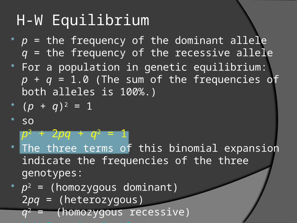
H-W Equilibrium p = the frequency of the dominant allele
q = the frequency of the recessive allele For a population in genetic equilibrium:
p + q = 1.0 (The sum of the frequencies of both alleles is 100%.)
(p + q)2 = 1 so
p2 + 2pq + q2 = 1 The three terms of this binomial expansion indicate
the frequencies of the three genotypes: p2 = (homozygous dominant)
2pq = (heterozygous)q2 = (homozygous recessive)
H-W Practice Problems