Embed Size (px)
Citation preview
Leukocyte Endothelial Interactions
Judith Berliner, Professor
Departments of Biology and Medicine, UCLA
Leukocyte/endothelial interactions are a major event in
the inflammatory process
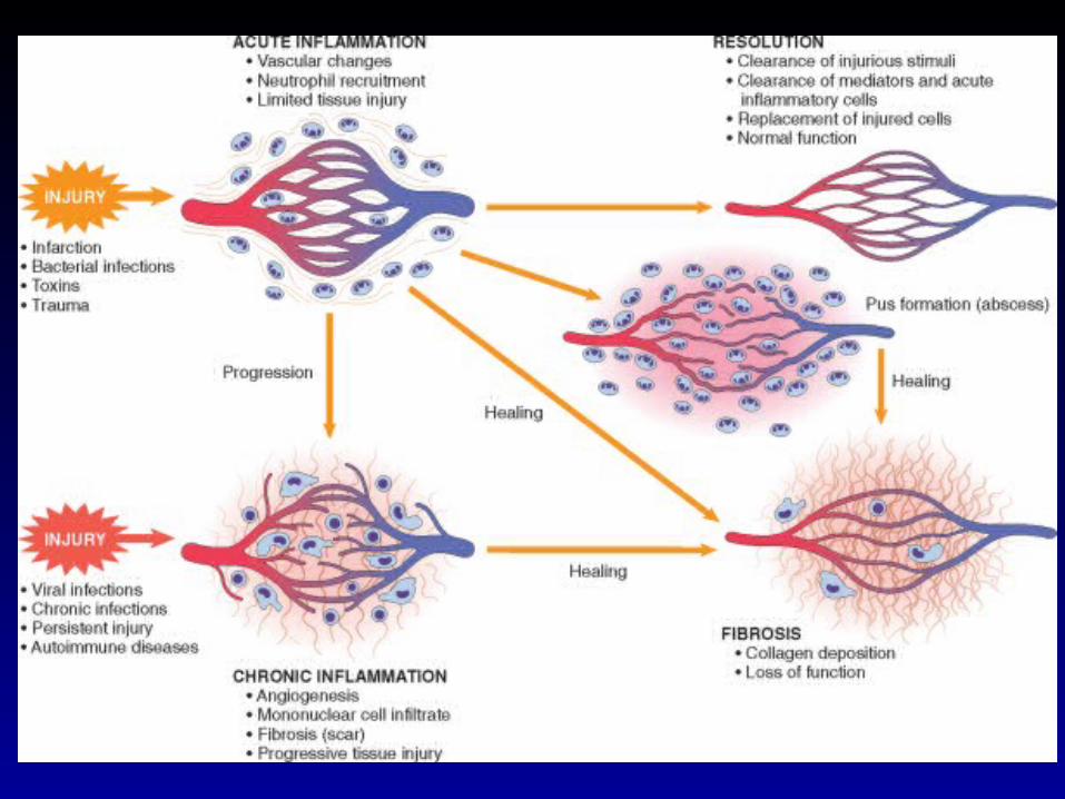
Inflammation is a reaction to injury or infection
Inflammation and repair
1. Tissue exposed to infection, toxin, trauma2. Damaged cells rapidly produce endothelial
activators3. In some cases, venules transiently increase
permeability in response to activators4. Plasma proteins enter the tissue and react
with bacteria and toxins producing more activators
Inflammation and repair
5. Activated endothelial cells upregulate leukocyte adhesion molecules and chemotactic factors
6. Leukocytes bind to and enter the vessel wall
7. Acute: Neutrophils kill and macrophages engulf bacteria and toxins. Resolution
8. Chronic: macrophages are unable to remove source of injury. Granuloma
Inflammation and repair
9. Cytokines and growth factors produced by injured cells stimulate replication of nearby cells. Fibrosis
10. Angiogenesis occurs in response to additional growth factors.
11. Tissue architecture is restored

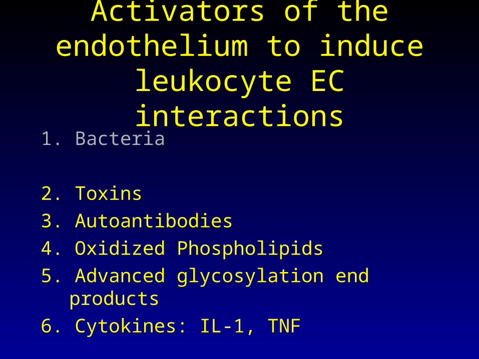
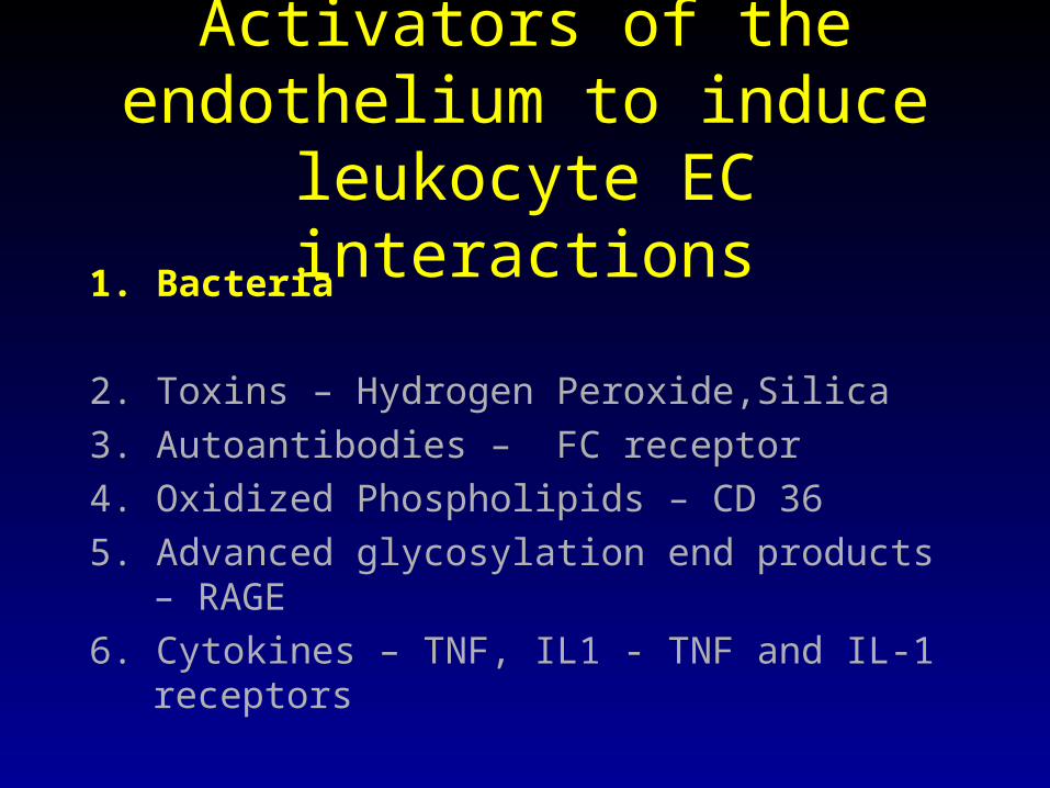
Activators of the endothelium to induce leukocyte EC interactions
1. Bacteria
2. Toxins
3. Autoantibodies
4. Oxidized Phospholipids
5. Advanced glycosylation end products
6. Cytokines: IL-1, TNF
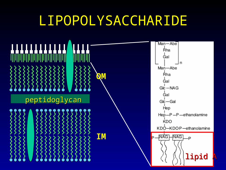
LIPOPOLYSACCHARIDE
peptidoglycanpeptidoglycan
IM
OM
lipid Alipid A
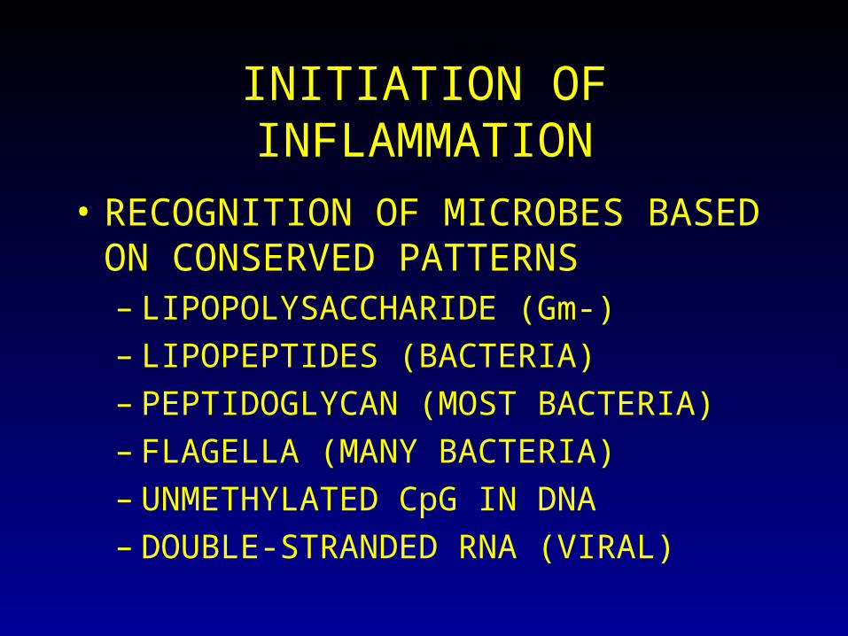
INITIATION OF INFLAMMATION
• RECOGNITION OF MICROBES BASED ON CONSERVED PATTERNS– LIPOPOLYSACCHARIDE (Gm-)– LIPOPEPTIDES (BACTERIA)– PEPTIDOGLYCAN (MOST BACTERIA)– FLAGELLA (MANY BACTERIA)– UNMETHYLATED CpG IN DNA– DOUBLE-STRANDED RNA (VIRAL)
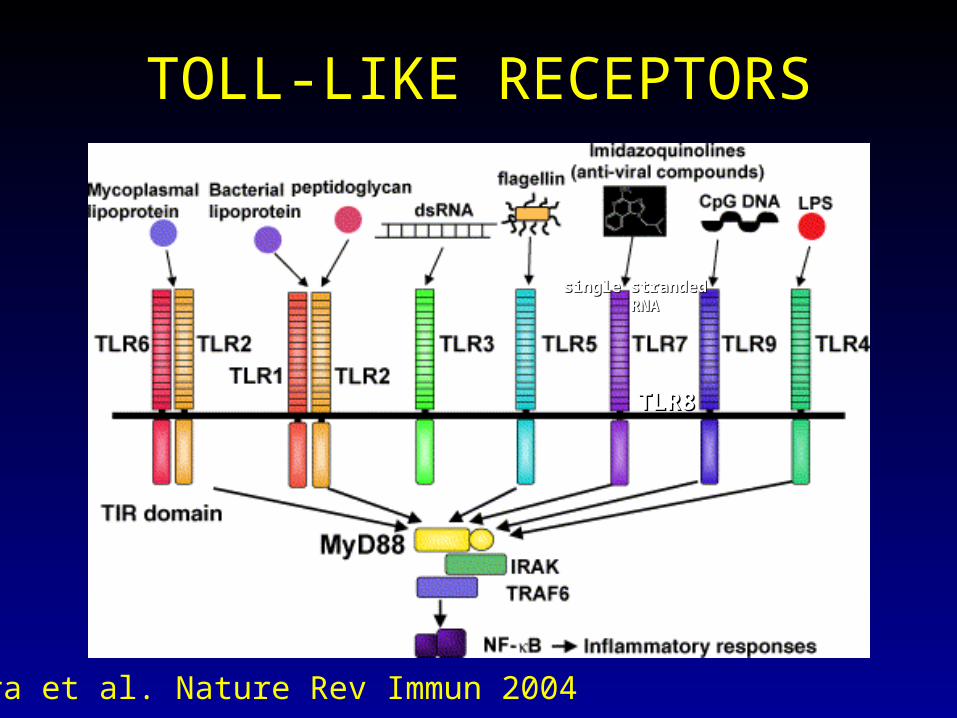
TOLL-LIKE RECEPTORS
single strandedsingle stranded RNARNA
TLR8TLR8
Akira et al. Nature Rev Immun 2004
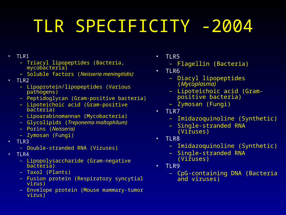
TLR SPECIFICITY -2004
• TLR1 – Triacyl lipopeptides (Bacteria, mycobacteria)– Soluble factors (Neisseria meningitidis)
• TLR2 – Lipoprotein/lipopeptides (Various pathogens)– Peptidoglycan (Gram-positive bacteria)– Lipoteichoic acid (Gram-positive bacteria)– Lipoarabinomannan (Mycobacteria)– Glycolipids (Treponema maltophilum)– Porins (Neisseria)– Zymosan (Fungi)
• TLR3 – Double-stranded RNA (Viruses)
• TLR4 – Lipopolysaccharide (Gram-negative bacteria)– Taxol (Plants)– Fusion protein (Respiratory syncytial virus)– Envelope protein (Mouse mammary-tumor
virus)
• TLR5 – Flagellin (Bacteria)
• TLR6 – Diacyl lipopeptides (Mycoplasma)– Lipoteichoic acid (Gram-positive
bacteria)– Zymosan (Fungi)
• TLR7 – Imidazoquinoline (Synthetic)– Single-stranded RNA (Viruses)
• TLR8 – Imidazoquinoline (Synthetic) – Single-stranded RNA (Viruses)
• TLR9 – CpG-containing DNA (Bacteria and
viruses)
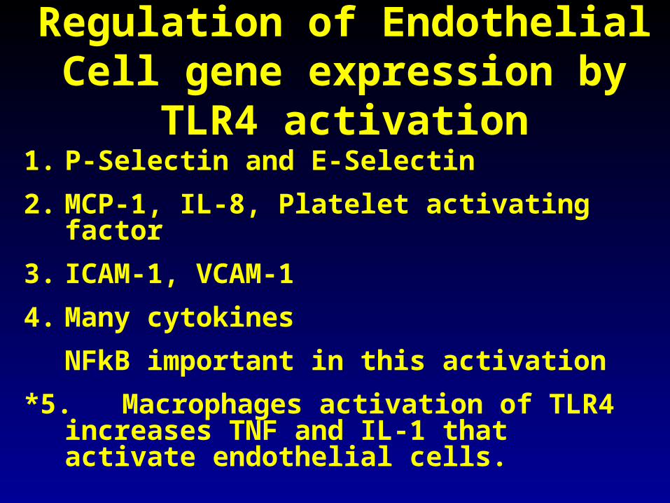
Regulation of Endothelial Cell gene expression by TLR4 activation
1. P-Selectin and E-Selectin
2. MCP-1, IL-8, Platelet activating factor
3. ICAM-1, VCAM-1
4. Many cytokines
NFkB important in this activation
*5. Macrophages activation of TLR4 increases TNF and IL-1 that activate endothelial cells.
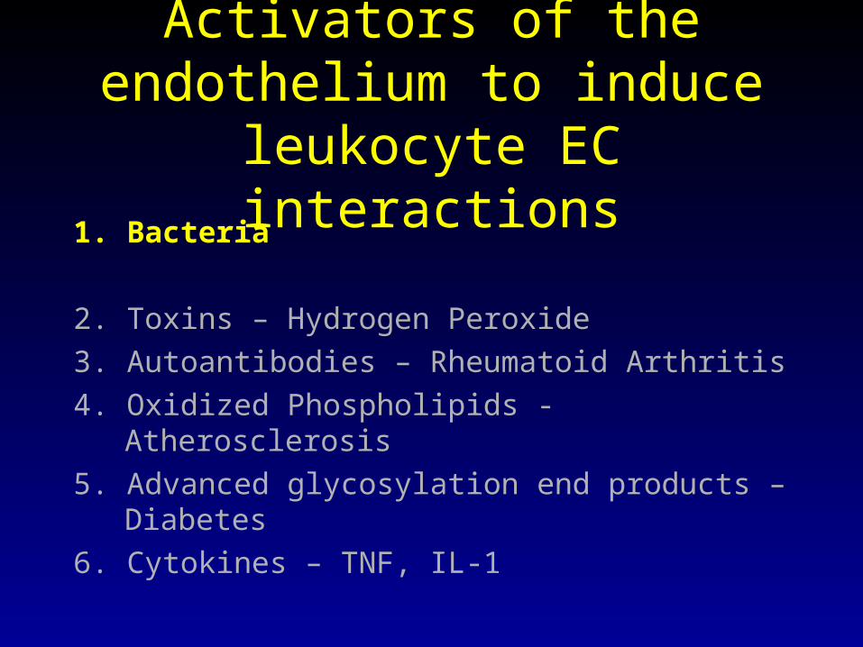
Activators of the endothelium to induce leukocyte EC interactions
1. Bacteria
2. Toxins – Hydrogen Peroxide
3. Autoantibodies – Rheumatoid Arthritis
4. Oxidized Phospholipids - Atherosclerosis
5. Advanced glycosylation end products – Diabetes
6. Cytokines – TNF, IL-1
Activators of the endothelium to induce leukocyte EC interactions
1. Bacteria
2. Toxins – Hydrogen Peroxide,Silica
3. Autoantibodies – FC receptor
4. Oxidized Phospholipids – CD 36
5. Advanced glycosylation end products – RAGE
6. Cytokines – TNF, IL1 - TNF and IL-1 receptors

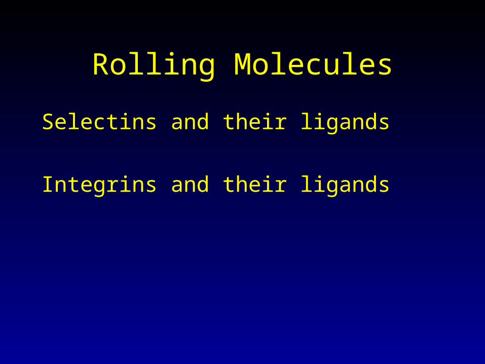
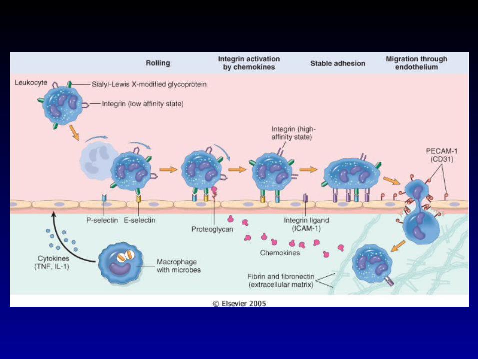
Rolling Molecules
Selectins and their ligands
Integrins and their ligands
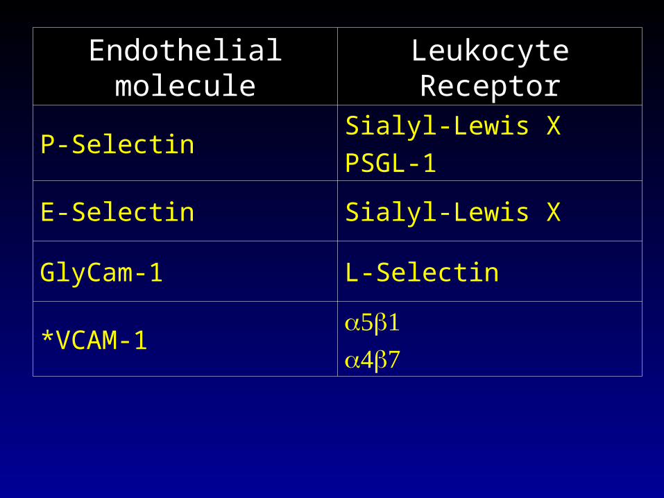
Endothelial molecule Leukocyte Receptor
P-SelectinSialyl-Lewis X
PSGL-1
E-Selectin Sialyl-Lewis X
GlyCam-1 L-Selectin
*VCAM-1
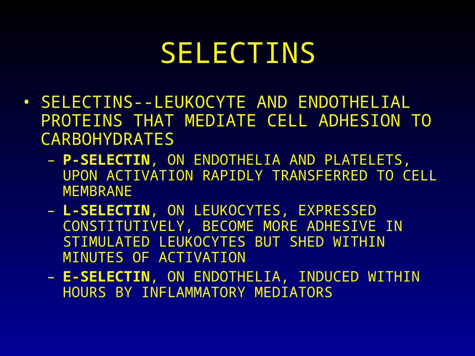
SELECTINS
• SELECTINS--LEUKOCYTE AND ENDOTHELIAL PROTEINS THAT MEDIATE CELL ADHESION TO CARBOHYDRATES– P-SELECTIN, ON ENDOTHELIA AND PLATELETS, UPON
ACTIVATION RAPIDLY TRANSFERRED TO CELL MEMBRANE
– L-SELECTIN, ON LEUKOCYTES, EXPRESSED CONSTITUTIVELY, BECOME MORE ADHESIVE IN STIMULATED LEUKOCYTES BUT SHED WITHIN MINUTES OF ACTIVATION
– E-SELECTIN, ON ENDOTHELIA, INDUCED WITHIN HOURS BY INFLAMMATORY MEDIATORS

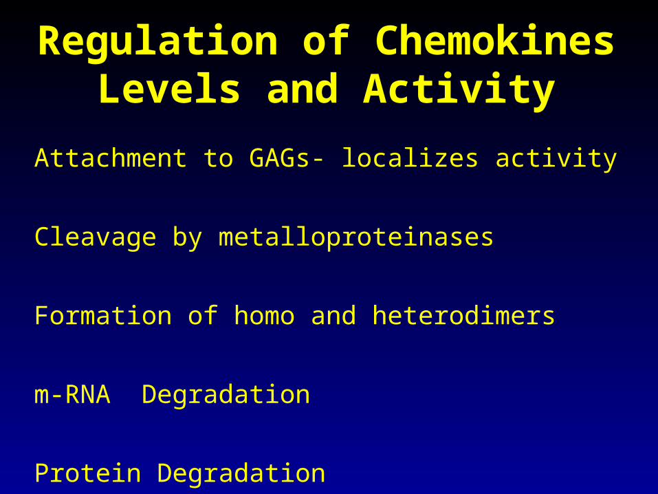
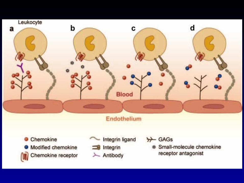
Regulation of Chemokines Levels and Activity
Attachment to GAGs- localizes activity
Cleavage by metalloproteinases
Formation of homo and heterodimers
m-RNA Degradation
Protein Degradation
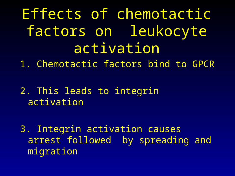
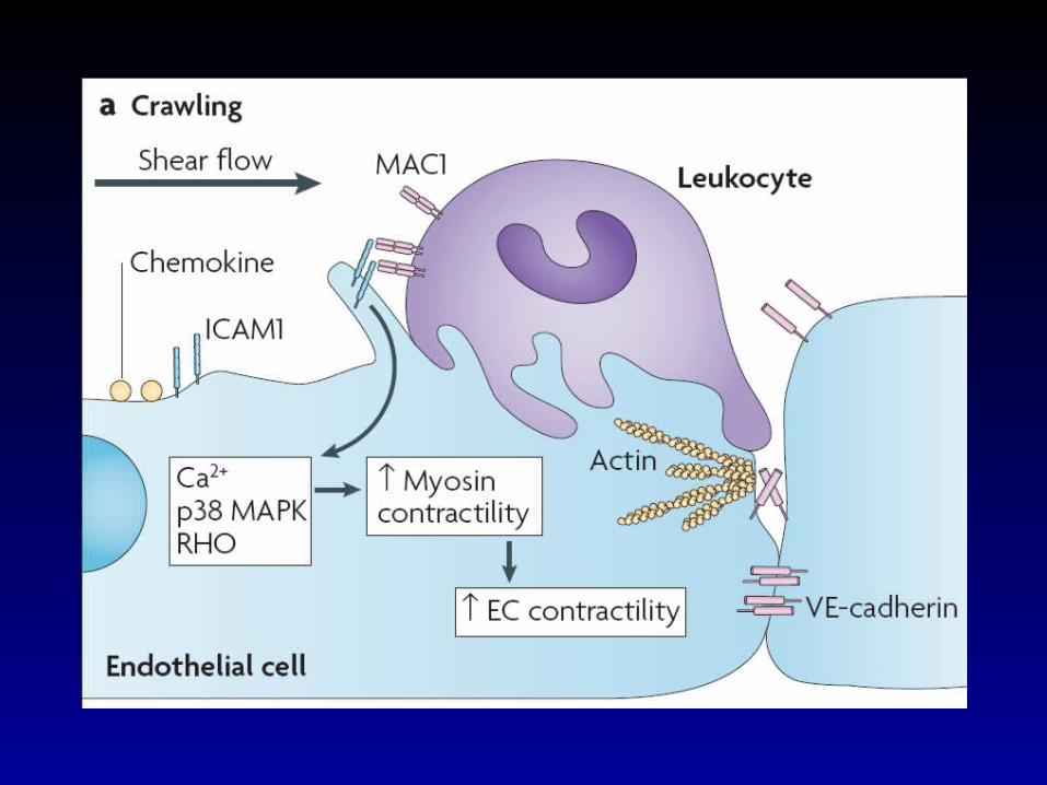
Effects of chemotactic factors on leukocyte activation
1. Chemotactic factors bind to GPCR
2. This leads to integrin activation
3. Integrin activation causes arrest followed by spreading and migration

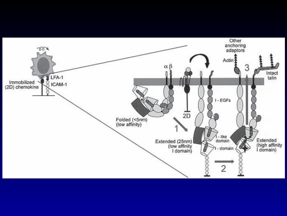
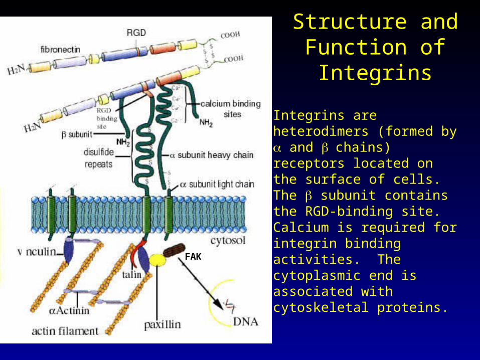
Structure and Function of Integrins
Integrins are heterodimers (formed by and chains) receptors located on the surface of cells. The subunit contains the RGD-binding site. Calcium is required for integrin binding activities. The cytoplasmic end is associated with cytoskeletal proteins.
FAK
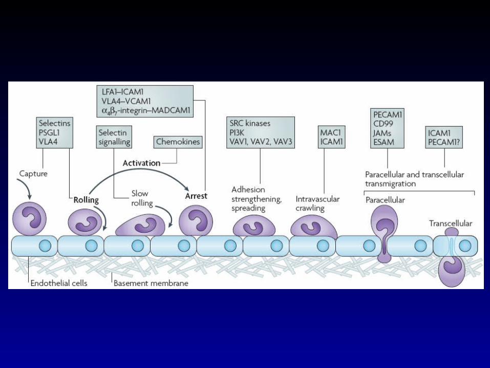
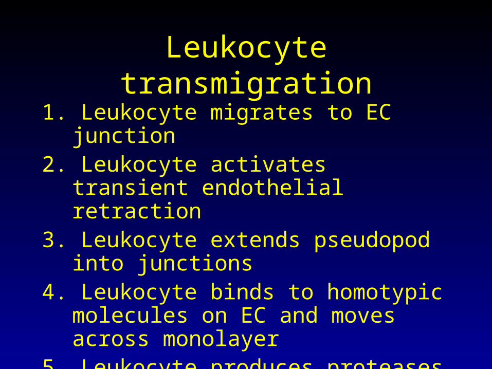
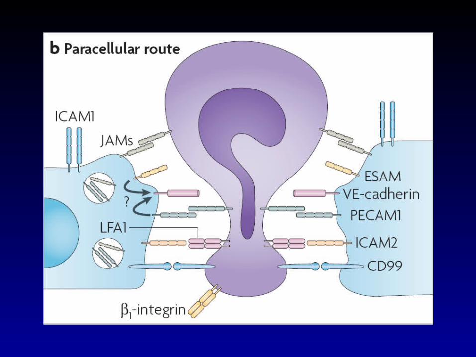
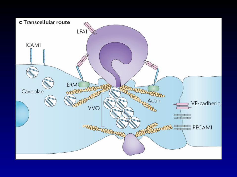
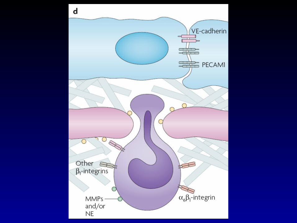
Leukocyte transmigration
1. Leukocyte migrates to EC junction2. Leukocyte activates transient endothelial
retraction3. Leukocyte extends pseudopod into
junctions4. Leukocyte binds to homotypic molecules
on EC and moves across monolayer5. Leukocyte produces proteases and migrates
across basement membrane in response to chemotactic factors
Activation of the Leukocyte for killing and phagocytosis
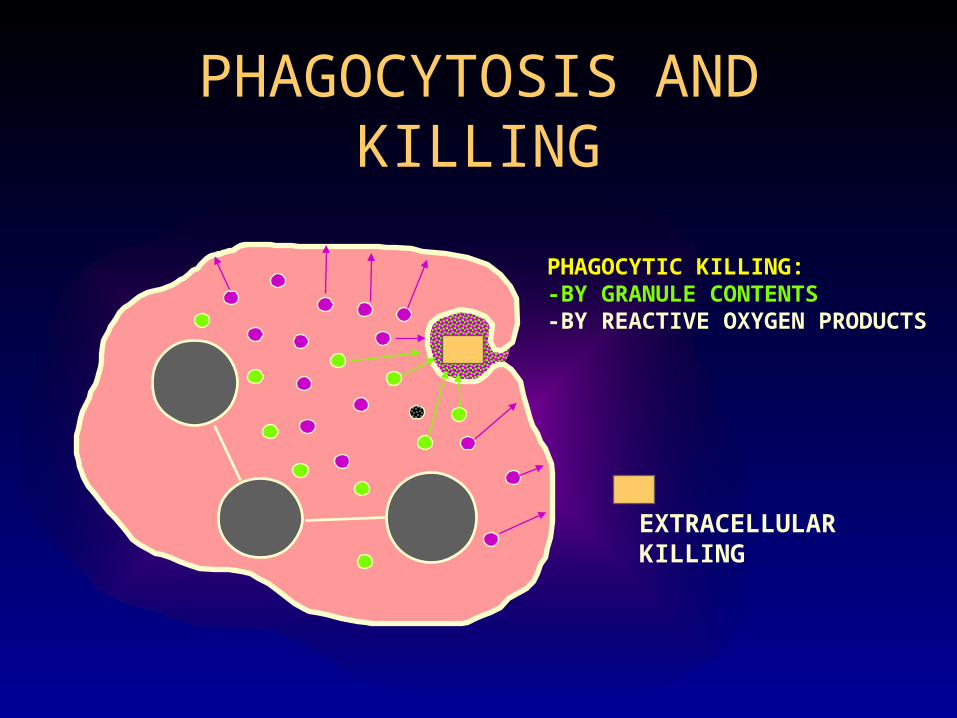
PHAGOCYTOSIS AND KILLING
EXTRACELLULARKILLING
PHAGOCYTIC KILLING:-BY GRANULE CONTENTS-BY REACTIVE OXYGEN PRODUCTS
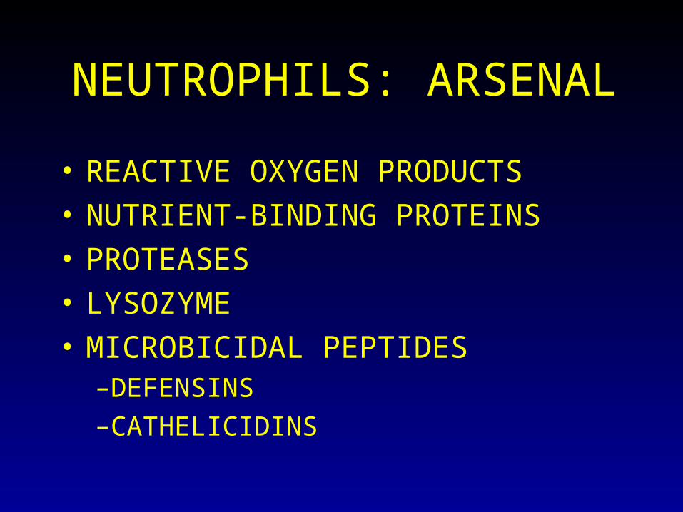
NEUTROPHILS: ARSENAL
• REACTIVE OXYGEN PRODUCTS
• NUTRIENT-BINDING PROTEINS
• PROTEASES
• LYSOZYME
• MICROBICIDAL PEPTIDES–DEFENSINS–CATHELICIDINS

Genetic Disease Defect
Leukocyte adhesion deficiency 1 -chain of CD11/CD18
Leukocyte adhesion deficiency 2Fucosyl transferase required for sialylated oligosaccharide synthesis
Chronic granulomatous disease
X-linked
Autosomal recessive
Decreased oxidative burst
NADPH oxidase (membrane)
NADPH oxidase (cytoplasm)
Myeloperoxidase deficiency Absent MPO-H2O2 system
Chediak-Higashi syndromeProtein involved in organelle membrane docking and fusion

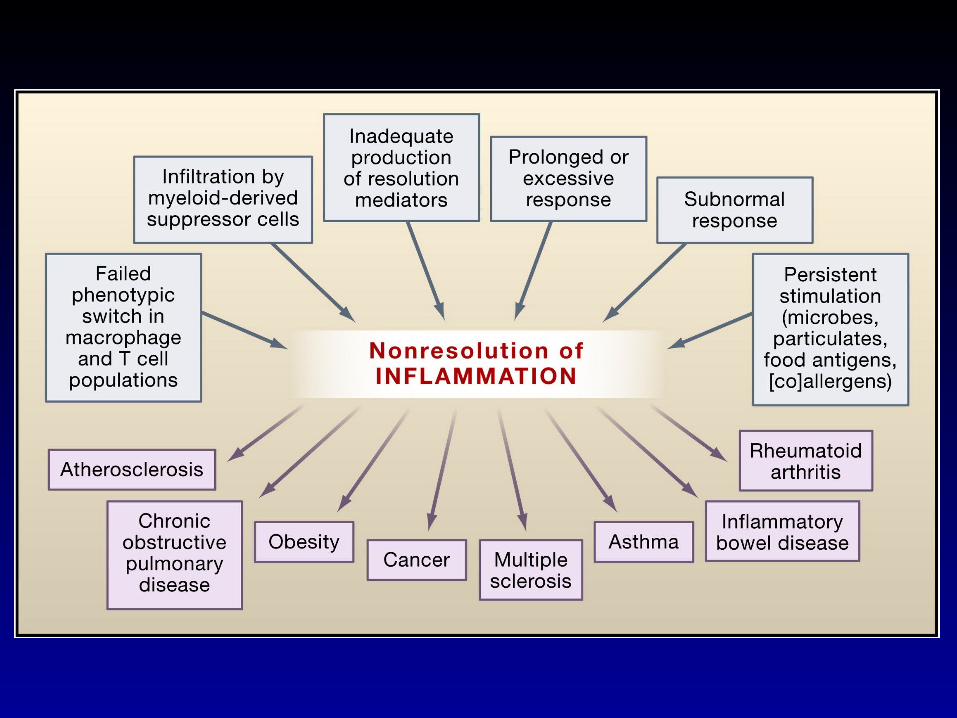
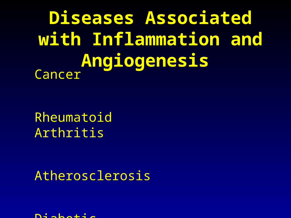
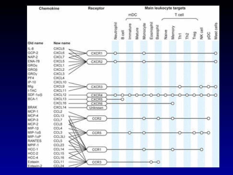
Diseases Associated with Inflammation and Angiogenesis
Cancer
Rheumatoid Arthritis
Atherosclerosis
Diabetic Retinopathy
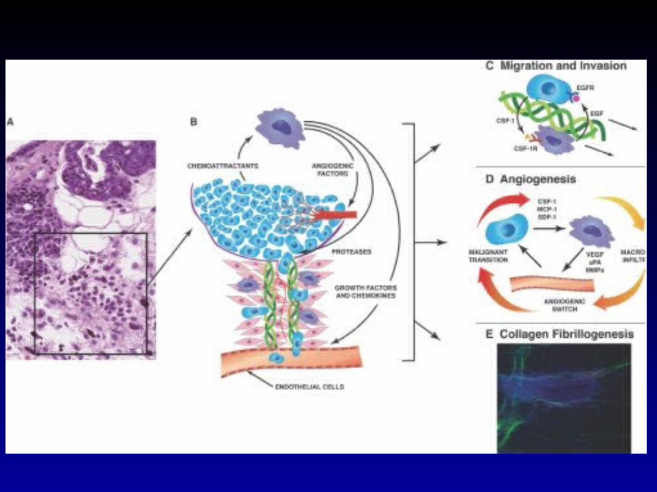
Sources of Inflammatory Molecules in Tumors
Extrinsic Pathway – Inflammation due to infection of cell injury
Intrinsic Pathway – Oncogene Activation
Both lead to increased macrophages in tumors

Macrophage Phenotypes and Cancer
M1 M2
Markers: IL12, TNF, IL6,ROS IL-4, IL-10, TGFB
Functions: Attract lymphocytes Decrease lymphocyte entry
Activate lymphocytes Decrease in lymphocyte activation
Kill tumor cells Increase in angiogenesis
Increase tumor cell growth