Embed Size (px)
Citation preview

Published Ahead of Print 31 March 2014. 10.1128/MCB.00914-13.
2014, 34(12):2147. DOI:Mol. Cell. Biol. Marius Ueffing and Christian Johannes GloecknerWeinkauf, Michael Sattler, Carlo Sala, Michela Matteoli, Flavia Antonucci, Christina Kiel, Mingjie Zhang, Sevilvon Zweydorf, Andreas Vogt, Florian Giesert, Lifeng Pan, Kastenmüller, Francesca Pischedda, Antonella Marte, FelixChristoph J. O. Kaiser, Pravinkumar Jagtap, Andreas Giovanni Piccoli, Franco Onofri, Maria Daniela Cirnaru, WD40 DomainInteractions Mediated by Its C-TerminalNeuronal Vesicles through Protein Leucine-Rich Repeat Kinase 2 Binds to
http://mcb.asm.org/content/34/12/2147Updated information and services can be found at:
These include:
SUPPLEMENTAL MATERIAL Supplemental material
REFERENCEShttp://mcb.asm.org/content/34/12/2147#ref-list-1at:
This article cites 64 articles, 21 of which can be accessed free
CONTENT ALERTS more»articles cite this article),
Receive: RSS Feeds, eTOCs, free email alerts (when new
http://journals.asm.org/site/misc/reprints.xhtmlInformation about commercial reprint orders: http://journals.asm.org/site/subscriptions/To subscribe to to another ASM Journal go to:
on Novem
ber 5, 2014 by FU
DA
N U
NIV
ER
SIT
Yhttp://m
cb.asm.org/
Dow
nloaded from
on Novem
ber 5, 2014 by FU
DA
N U
NIV
ER
SIT
Yhttp://m
cb.asm.org/
Dow
nloaded from

Leucine-Rich Repeat Kinase 2 Binds to Neuronal Vesicles throughProtein Interactions Mediated by Its C-Terminal WD40 Domain
Giovanni Piccoli,a,b Franco Onofri,c Maria Daniela Cirnaru,b Christoph J. O. Kaiser,d Pravinkumar Jagtap,d,e Andreas Kastenmüller,d
Francesca Pischedda,b Antonella Marte,c Felix von Zweydorf,f Andreas Vogt,a,f Florian Giesert,g Lifeng Pan,h Flavia Antonucci,i
Christina Kiel,j Mingjie Zhang,h Sevil Weinkauf,d Michael Sattler,d,e Carlo Sala,b Michela Matteoli,i Marius Ueffing,a,f
Christian Johannes Gloecknera,f
Helmholtz Zentrum München, German Research Center for Environmental Health, Research Unit Protein Science, Neuherberg, Germanya; Institute of Neuroscience,National Research Council, Milan, Italyb; Department of Experimental Medicine, University of Genoa, Genoa, Italyc; Center for Integrated Protein Science Munich andTechnische Universität München, Department of Chemistry, Garching, Germanyd; Helmholtz Zentrum München, German Research Center for Environmental Health,Institute of Structural Biology, Neuherberg, Germanye; Eberhard Karls University Tübingen, Institute for Ophthalmic Research, Medical Proteome Center, Tübingen,Germanyf; Helmholtz Zentrum München, German Research Center for Environmental Health, Institute of Developmental Genetics, Neuherberg, Germanyg; Division of LifeScience, Centre of Systems Biology and Human Health, Institute for Advanced Study and School of Science, Hong Kong University of Science and Technology, ClearWater Bay, Kowloon, Hong Kongh; Department of Biotechnology and Translational Medicine, University of Milan and Humanitas Clinical and Research Center, Rozzano,Italyi; CRG-EMBL System Biology Program, Centre de Regulació Genòmica UPF, Barcelona, Spainj
Mutations in the leucine-rich repeat kinase 2 gene (LRRK2) are associated with familial and sporadic Parkinson’s disease (PD).LRRK2 is a complex protein that consists of multiple domains, including predicted C-terminal WD40 repeats. In this study, weanalyzed functional and molecular features conferred by the WD40 domain. Electron microscopic analysis of the purifiedLRRK2 C-terminal domain revealed doughnut-shaped particles, providing experimental evidence for its WD40 fold. We demon-strate that LRRK2 WD40 binds and sequesters synaptic vesicles via interaction with vesicle-associated proteins. In fact, a do-main-based pulldown approach combined with mass spectrometric analysis identified LRRK2 as being part of a highly specificprotein network involved in synaptic vesicle trafficking. In addition, we found that a C-terminal sequence variant associatedwith an increased risk of developing PD, G2385R, correlates with a reduced binding affinity of LRRK2 WD40 to synaptic vesicles.Our data demonstrate a critical role of the WD40 domain within LRRK2 function.
Parkinson’s disease (PD) is the second most common age-re-lated neurodegenerative disease and is clinically characterized
by movement impairments, bradykinesia, rigidity, and restingtremor and pathologically by the progressive loss of dopaminergicneurons in the substantia nigra and the formation of Lewy bodies(1, 2). Although the majority of cases are sporadic, mutations inthe leucine-rich repeat kinase 2 (LRRK2) gene (PARK8; OnlineMendelian Inheritance in Man [OMIM] accession number609007) had been unequivocally linked to late-onset autosomaldominant PD. LRRK2 mutations account for up to 13% of famil-ial PD cases compatible with dominant inheritance and are alsofound in 1 to 2% of sporadic PD patients (62–64). LRRK2 is acomplex 286-kDa protein that consists of multiple domains, in-cluding (in order, from the amino to carboxyl terminus) arma-dillo, ankyrin, and the namesake leucine-rich repeats (LRRs), fol-lowed by an ROC (Ras of complex proteins) GTPase domain, aCOR (C-terminal of ROC) dimerization domain, a kinase do-main, and a predicted C-terminal WD40 repeat domain (4–6).Several single-nucleotide alterations have been identified inLRRK2, but only five missense mutations within the ROC, COR,and kinase domains clearly segregate with PD in large family stud-ies (7, 8). It has recently been shown that the WD40 domain isrequired to stabilize the LRRK2 dimer and to execute LRRK2-associated kinase activity as well as neurotoxicity (9, 10), but therole of this domain within LRRK2 physiological and pathologicalfunction has not yet been completely defined. The beta-propeller-forming WD40 domains are among the 10 most abundant do-main types across eukaryotic proteomes (11) and constitute plat-forms where multiprotein complexes assemble reversibly (12).Here, we systematically analyzed the protein-protein interactions
conferred by the LRRK2 WD40 domain. The nature of the LRRK2WD40 interactors and the finding that the LRRK2 WD40 domainis able to bind to synaptic vesicles (SV) contribute to accumulatingevidence suggesting that LRRK2 serves as a scaffold protein con-necting vesicle trafficking and cytoskeleton (13). Strong geneticassociation indicates that the substitution of arginine for glycine2385 (G2385R) within the LRRK2 WD40 domain is a pathologi-cally relevant variant (14). This variant is considered a commonrisk factor for sporadic PD in Chinese Han and Korean ethnicity,but its functional impact is largely unknown (15, 16). We demon-strate that the G2385R variant alters LRRK2 WD40 binding tosynaptic vesicles. Altogether, these data suggest that the LRRK2WD40 domain is a determinant for LRRK2 physiological andpathological activities.
MATERIALS AND METHODSCell cultures. Cortical neuron cultures were prepared from embryonicday 15.5 to 16.5 mouse cortexes (C57BL/6). Medium-density (150 to 200
Received 23 July 2013 Returned for modification 14 August 2013Accepted 18 March 2014
Published ahead of print 31 March 2014
Address correspondence to Christian Johannes Gloeckner,[email protected].
M.U. and C.J.G. contributed equally to this work
Supplemental material for this article may be found at http://dx.doi.org/10.1128/MCB.00914-13.
Copyright © 2014, American Society for Microbiology. All Rights Reserved.
doi:10.1128/MCB.00914-13
June 2014 Volume 34 Number 12 Molecular and Cellular Biology p. 2147–2161 mcb.asm.org 2147
on Novem
ber 5, 2014 by FU
DA
N U
NIV
ER
SIT
Yhttp://m
cb.asm.org/
Dow
nloaded from

cells/mm2) neuron cultures were plated and grown in Neurobasal culturemedium supplemented with 2% B27 and 0.1% gentamicin on 24-wellplastic tissue culture plates (Iwaki; Bibby Sterilin, Staffordshire, UnitedKingdom). In such cultures glial growth is reduced to less than 0.5% of thenearly pure neuronal population (17). HEK293T (ATCC CRL-11268)cells were cultured in Dulbecco’s modified Eagle’s medium (DMEM) sup-plemented with 10% fetal bovine serum (FBS) and 1% penicillin-strepto-mycin mix according to standard protocols. All media were purchasedfrom Life Technologies.
Plasmids and transfection. Human LRRK2 LRR (amino acids [aa]921 to 1356), LRRK2 WD40 (aa 2124 to 2527), full-length RACK1, andfull-length human LRRK2 were subcloned into pDEST-15 (N-terminalglutathione S-transferase [GST] tag) and/or pDEST-733 (N-terminal redfluorescent protein [RFP] tag) using the Gateway system (Life Technolo-gies) as described previously (18). The LRRK2 WD40 G2385R variant aswell as FLAG- and RFP-tagged LRRK2 consisting of residues 1 to 2141(hereinafter termed LRRK2 1–2141) were generated by site-directed mu-tagenesis using a QuikChange mutagenesis kit (Stratagene). Cloning ofpGEX-RACK1 and full-length FLAG-LRRK2 was described previously(18–20). LRRK2 WD40 consisting of aa 2148 to 2527 with six copies of aHis tag (6�His) was cloned into pETM11. pGFP (where GFP is greenfluorescent protein) and pDsRed2-1 were purchased from Clontech lab-oratories. Neurons were transfected at 10 days of in vitro culture (DIV10)with GFP and RFP-tagged constructs or DsRed in a 1:3 ratio by calciumphosphate precipitation as previously described (21) and processed whenindicated in the text.
Purification of GST- and His-tagged proteins. GST fusion proteinscontaining single LRRK2 domains and GST-RACK1 were expressed in theEscherichia coli BL21 strain (Life Technologies), purified as described ear-lier (22). The purification procedure for the N-terminal 6�His-taggedLRRK2 WD40 construct (LRRK2 2148-end), used for electron micros-copy (EM) analysis, has been adapted to that used for the GST fusionproteins. Briefly, for expression, E. coli cells were grown in terrific broth(TB) medium with 0.1 mM isopropyl-�-D-thiogalactopyranoside (IPTG)induction overnight at 18°C. Cells were lysed in 20 mM Tris, pH 7.5, 150mM NaCl, 2 mM beta-mercaptoethanol (BME), 0.7% Sarkosyl, and 2%Triton X-100 by sonication. The lysate was loaded onto a nickel column,and the column was then washed with 20 column volumes each of lysisbuffer and high-salt buffer (20 mM Tris, pH 7.5, 150 mM NaCl, 350 mMKCl, 5 mM MgCl2, 1 mM ATP). The His-tagged protein was finally elutedwith lysis buffer containing 150 mM imidazole. The protein was thendialyzed overnight against 20 mM Tris, pH 7.5, 150 mM NaCl, and 5 mMdithiothreitol (DTT).
Subcellular fractionation and synaptic vesicle binding assay. Sub-cellular fractionation of rat forebrain tissue was carried out as previouslydescribed in the presence of protease inhibitors (23). Briefly, the freshlydissected cerebral cortex was homogenized with a glass-Teflon homoge-nizer in ice-cold buffered sucrose (0.32 M sucrose, 5 mM HEPES, pH 7.4)(homogenate) and centrifuged at 800 � g for 10 min. The nuclear pelletwas discarded, and the postnuclear supernatant (containing cell mem-brane, cytosol, and organelles; S1 fraction) was centrifuged at 9,200 � gfor 15 min to give a supernatant fraction (containing cytosol and micro-somes; S2 fraction) and a crude mitochondrial pellet (containing mito-chondria and synaptosomes; P2 fraction). The P2 fraction was subjectedto osmotic lysis by homogenization in 10 volumes of ice-cold water andcentrifuged at 25,000 � g for 20 min to yield a lysate pellet (LP1) enrichedin presynaptic plasma membranes and a lysate supernatant (LS1). TheLS1 fraction was further centrifuged at 16,500 � g for 2 h to yield asynaptosolic fraction (LS2) and a crude SV pellet (LP2) containing syn-aptic vesicles and small presynaptic plasma membranes. The LP2 fractionwas further fractionated by centrifugation through a continuous sucrosegradient and chromatography through a controlled-pore glass column toyield highly purified SV (untreated SV [US]) and a column flowthrough(FT). When required, purified SV were partially depleted of endogenousproteins by dilution in 0.2 M NaCl (salt-treated SV, SSV). SV were cen-
trifuged at 200,000 � g for 2 h after 2 h of incubation at 0°C. After cen-trifugation, SV were resuspended in 0.3 M glycine, 5 mM HEPES-NaOH,pH 7.4, at a protein concentration of 1.5 to 2 mg/ml. The binding of GSTfusion proteins to SV was carried out using a high-speed sedimentationassay (24). Briefly, SV (5 to 10 �g of total protein) were incubated for 1 hat 0°C with increasing amounts of a GST fusion protein in a buffer con-taining 220 mM glycine, 30 mM NaCl, 5 mM Tris-HCl, 4 mM HEPES (pH7.4), 0.22 mM NaN3, and 100 �g/ml of bovine serum albumin (BSA).After the incubation, GST fusion protein which bound to SV was sepa-rated by high-speed centrifugation (400,000 � g for 30 min). Aliquots ofthe resuspended pellets were subjected to SDS-PAGE and subsequentWestern blotting with GST-specific antibodies. The amount of GST pro-tein was determined as a function of optical density in comparison toknown amounts of fusion proteins. The recovery of SV, used to correct theamounts of fusion protein bound to SV, was determined by Westernblotting with antisynaptophysin antibodies. FLAG-LRRK2 was purifiedvia affinity chromatography using FLAG-M2 agarose beads (Sigma-Al-drich) from HEK293T cells transfected by lipofection using Lipo-fectamine 2000 (Life Technologies) according to the manufacturer’s in-structions. The binding of FLAG-LRRK2 to SV was performed asdescribed above with minor modifications: only one concentration offusion protein (50 nM) was assayed, and FLAG-LRRK2 yield was evalu-ated via Western blotting with FLAG-specific antibodies.
Pulldown, immunoprecipitation, and antibodies. For pulldowns, 5�g of each GST fusion protein was loaded onto glutathione-Sepharoseresin (GE Healthcare) and coincubated with adult mouse brain lysate orthe LS1 fraction (1 mg of total protein). In immunoprecipitation assays,10 �g of 1E11 anti-LRRK2 antibody was incubated with 1 mg of proteinlysate and loaded onto protein G-Sepharose resin (GE Healthcare). Inboth procedures, resins were extensively washed in Tris-EDTA buffer (10mM Tris-HCl, pH 8.0, 1 mM EDTA, 150 mM NaCl, 0.2% Triton X-100),followed by final elution of the samples with Laemmli buffer. For proteinidentification by Western blotting, samples were loaded onto 4 to 12%NuPAGE gels (Invitrogen); the proteins were transferred onto a nitrocel-lulose membrane (Sigma-Aldrich) at 80 V for 120 min at 4°C. The pri-mary antibodies were applied overnight in blocking buffer (20 mM Tris,pH 7.4, 150 mM NaCl, 0.1% Tween 20, and 5% nonfat dry milk); primaryantibodies (source in parentheses) included rat monoclonal anti-LRRK21E11 at 1:1,000 (that recognizes an epitope within the LRRK2 kinase do-main [25]), mouse anti-synapsin I at 1:1,000 (Synaptic System), mouseanti-SNAP-25 at 1:1,000 (Chemicon), rabbit anti-MAP2 at 1:1,000,mouse anti-MAP6 at 1:1,000, rabbit anti-N-ethylmaleimide-sensitive fac-tor (anti-NSF; 1:1,000 [Cell Signaling]), mouse anti-FLAG at 1:2,000,mouse anti-Rac1 at 1:1,000, mouse anti-�-actin at 1:1,000 mouse anti-syntaxin 1A at 1:1,000, mouse anti-Rab3A at 1:1,000, and mouse anti-�-tubulin at 1:1,000 (Sigma-Aldrich). Secondary antibodies (horseradishperoxidase [HRP]-conjugated anti-mouse, anti-rabbit, or anti-rat anti-bodies; Jackson ImmunoResearch) were used in a ratio of 1:8,000. Thesignal was detected using an enhanced chemiluminescence (ECL) detec-tion system (GE Healthcare). Films (Hyperfilm ECL; GE Healthcare) weredigitalized using a GS-800 densitometer (Bio-Rad) calibrated accordingto the manufacturer’s instructions, and protein abundance was estimatedas a function of the optical density of a specific band quantified by ImageJsoftware (NIH).
MS/MS identification. Liquid chromatography-tandem mass spec-trometry (LC-MS/MS) analysis was performed on an Ultimate 3000 Nanohigh-performance liquid chromatography (HPLC) system (Dionex) cou-pled to a linear trap quadrupole (LTQ) OrbitrapXL mass spectrometer(Thermo Fisher Scientific) by a nanospray ion source. Tryptic peptidemixtures were automatically injected and loaded at a flow rate of 30 �l/min in 95% buffer C (0.5% trifluoroacetic acid in HPLC-grade water) and5% buffer B (98% acetonitrile and 0.1% formic acid in HPLC-grade wa-ter) onto a nanotrap column (100-�m interior diameter [i.d.] by 2 cm;Acclaim PepMap100 C18 column, 5-�m particle diameter, 100-Å poresize [Dionex]). After 5 min, peptides were eluted and separated on the
Piccoli et al.
2148 mcb.asm.org Molecular and Cellular Biology
on Novem
ber 5, 2014 by FU
DA
N U
NIV
ER
SIT
Yhttp://m
cb.asm.org/
Dow
nloaded from
analytical column (75-�m i.d. by 15 cm; Acclaim PepMap100 C18 col-umn, 3-�m particle diameter, 100-Å pore size [Dionex]) by a linear gra-dient from 5% to 40% of buffer B in buffer A (2% acetonitrile and 0.1%formic acid in HPLC grade water) at a flow rate of 300 nl/min over 90 min.Remaining peptides were eluted by a short gradient from 40% to 100%buffer B in 5 min. The eluted peptides were analyzed by the LTQ Or-bitrapXL mass spectrometer. From the high-resolution mass spectrome-try prescan with a mass range of 300 to 1,500, the 10 most intense peptideions were selected for fragment analysis in the linear ion trap if they ex-ceeded an intensity of at least 200 counts and if they were at least doublycharged. The normalized collision energy for collision-induced dissocia-tion was set to a value of 35, and the resulting fragments were detectedwith normal resolution in the linear ion trap. The lock mass option wasactivated; the background signal with a mass of 445.12002 was used as lockmass (26). Every ion selected for fragmentation was excluded for 30 s bydynamic exclusion. The acquired spectra were processed and analyzed byusing Mascot Daemon (version 2.4.0) in combination with ExtractMSN(Thermo-Fisher) with the following settings: cysteine carbamidomethy-lation as a fixed modification and methionine oxidation and asparagine/glutamine deamidation as variables with a maximum of three modifica-tions per peptide allowed. Mass tolerances for parent and fragmentpeptides were set to 10 ppm and 1.00 Da, respectively. The database usedconsisted of a combined set of mouse and E. coli subsets of the Swiss-Protdatabase (UniProt release 2012_7, published on 11 July 2012; 20,847 en-tries plus decoy) with spiked-in bait proteins (human LRRK2 and GST ofSchistosoma japonicum). Reversed sequences generated by Scaffold wereused as a decoy. Mascot result files were analyzed by the Scaffold software(version 4.1.1) (Proteome Software, Inc., Portland, OR) to validate MS/MS-based peptide and protein identifications. Peptide identificationswere accepted if they could be established at greater than 90% probabilityas specified by the Peptide Prophet algorithm (27). Protein identificationswere accepted if they could be established at greater than 95% probabilityand contained at least two identified unique peptides. Protein probabili-ties were assigned by the Protein Prophet algorithm (28). Proteins thatcontained similar peptides and could not be differentiated based onMS/MS analysis alone were grouped to satisfy the principles of parsimony.The final data sets describing the LRRK2 interactome were generatedbased on at least four independent experiments for each bait (LRRK2WD40, LRRK2 LRR, and GST control). Proteins were considered specificLRRK2 WD40 interactors if they were found in at least two independentpulldown experiments and if they were, based on spectral counting, atleast 2-fold enriched in the LRRK2 WD40 pulldown set compared to theGST control set. Typical contaminants, including E. coli proteins, ribo-somal proteins, and keratins, were excluded.
Exoendocytotic assay, immunocytochemistry, and quantification.The endocytosis assay to monitor SV recycling was performed as de-scribed previously with minor modifications (13, 29) in neurons trans-fected at DIV12 with RFP-WD40 or RFP-RACK1 or DsRed plus GFP tovisualize cellular processes. Briefly, rabbit polyclonal antibodies directedagainst the intravesicular domain of synaptotagmin 1 (Synaptic System)were diluted 1:400 in Tyrode solution (124 mM NaCl, 5 mM KCl, 5 mMCaCl2, 1 mM MgCl2, 30 mM glucose, 25 mM HEPES, pH 7.4) and appliedfor 5 min at room temperature (RT) on living cultures. After extensivewashing with Tyrode solution, neurons were fixed in 4% paraformalde-hyde and 4% sucrose at room temperature. Where indicated in the figurelegends, rat anti-FLAG (1:100; kindly provided by E. Kremmer, Instituteof Molecular Immunology, Helmholtz Zentrum München), rabbit anti-SV2A (1:400; Synaptic System), or mouse anti-SNAP-25 (1:1,000; Sigma-Aldrich) antibody was applied in GDB buffer (30 mM phosphate buffer,pH 7.4, containing 0.2% gelatin, 0.5% Triton X-100, and 0.8 M NaCl)overnight at 4°C. Cy5-coupled secondary antibodies and 4=,6=-di-amidino-2-phenylindole (DAPI; Life Technologies) were diluted 1:1,000in GDB buffer and applied for 1 h. Transfected neurons were randomlychosen in at least four independent experiments for each condition. Thefluorescence images were acquired using an LSM Zeiss 510 confocal mi-
croscope with a Zeiss 63� objective (Karl Zeiss, Jena, Germany) at aresolution of 1 pixel equal to 0.098 �m. Image analysis was performedusing ImageJ (NIH). To quantify synaptotagmin- or SV2A-positive clus-ters, GFP-positive processes were manually tracked, and the number ofpositive clusters in the region of interest was automatically counted. Toevaluate neuron morphology, neurites were randomly selected and man-ually traced, and length and number of processes were automatically de-termined and logged into Microsoft Excel as described previously (30).
Electron microscopy. Purified GST- or 6�His-tagged fusion proteinswere adsorbed onto carbon-coated grids that were glow discharged in airbefore the application of 5 �l of protein solution. Excess protein solutionwas blotted off after 2 min. The adsorbed molecules were negativelystained for 30 s using 5 �l of stain solution as indicated in the figurelegends. Micrographs were recorded on Kodak SO-163 image film (Sigma-Aldrich) using JEOL JEM 2010 and JEOL JEM 100CX electron micro-scopes operated at 120 and 100 kV, respectively, at defocus values of 600 to900 nm. Suitable micrographs were selected by optical diffraction anddigitized using a Flextight Precision II (Hasselblad) scanner, resulting inpixel sizes of 1.59 or 1.28 Å at the specimen level. Particles were selectedand extracted using EMAN1 (31) and classified using IMAGIC, version5.0 (Image Science Software).
Homology modeling. Modeling templates were identified in the Pro-tein Data Bank (PDB) (32, 33) using the profile-profile alignment pro-gram Phyre2 (3). The initial homology model was built based on theWD40 domain structure of RACK1 from Arabidopsis thaliana (PDB code3DM0) (34). The model structure was further refined using the YASARAenergy minimization server to increase the model accuracy (35). Thequality of the final structure model was validated using PROCHECK (36).PyMOL was used for visualization of the final model.
Statistical analysis. All data are expressed as means � standard errorof the means (SE). Data were analyzed with an unpaired Student’s t test(two classes) or by analysis of variance (ANOVA) followed by Tukey’s posthoc test (more than two classes). The indication of the number of exper-iments (n) and level of significance (P) are given throughout the text.
Database accession number. The interactome data set has been sub-mitted to the IntAct database under accession number IM-20537 (http://www.ebi.ac.uk/intact/search/do/search?searchString�imex:IM-20537).
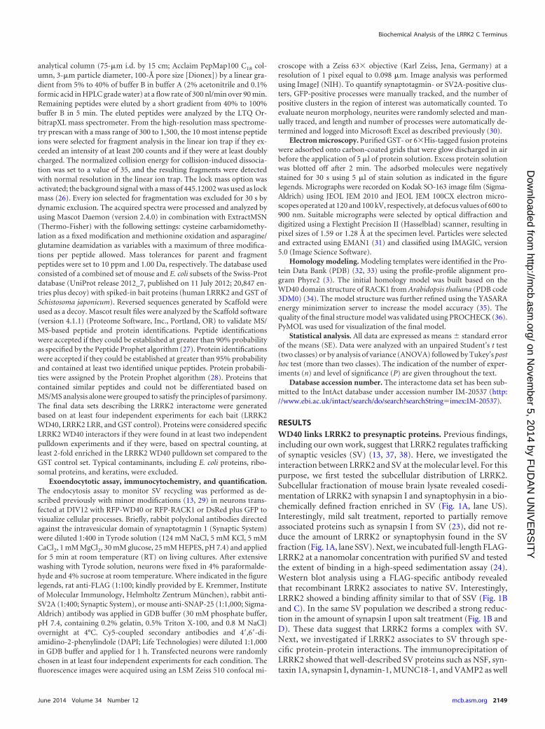
RESULTSWD40 links LRRK2 to presynaptic proteins. Previous findings,including our own work, suggest that LRRK2 regulates traffickingof synaptic vesicles (SV) (13, 37, 38). Here, we investigated theinteraction between LRRK2 and SV at the molecular level. For thispurpose, we first tested the subcellular distribution of LRRK2.Subcellular fractionation of mouse brain lysate revealed cosedi-mentation of LRRK2 with synapsin I and synaptophysin in a bio-chemically defined fraction enriched in SV (Fig. 1A, lane US).Interestingly, mild salt treatment, reported to partially removeassociated proteins such as synapsin I from SV (23), did not re-duce the amount of LRRK2 or synaptophysin found in the SVfraction (Fig. 1A, lane SSV). Next, we incubated full-length FLAG-LRRK2 at a nanomolar concentration with purified SV and testedthe extent of binding in a high-speed sedimentation assay (24).Western blot analysis using a FLAG-specific antibody revealedthat recombinant LRRK2 associates to native SV. Interestingly,LRRK2 showed a binding affinity similar to that of SSV (Fig. 1Band C). In the same SV population we described a strong reduc-tion in the amount of synapsin I upon salt treatment (Fig. 1B andD). These data suggest that LRRK2 forms a complex with SV.Next, we investigated if LRRK2 associates to SV through spe-cific protein-protein interactions. The immunoprecipitation ofLRRK2 showed that well-described SV proteins such as NSF, syn-taxin 1A, synapsin I, dynamin-1, MUNC18-1, and VAMP2 as well
Biochemical Analysis of the LRRK2 C Terminus
June 2014 Volume 34 Number 12 mcb.asm.org 2149
on Novem
ber 5, 2014 by FU
DA
N U
NIV
ER
SIT
Yhttp://m
cb.asm.org/
Dow
nloaded from

as actin and tubulin are LRRK2-interacting proteins (Fig. 1E) (13,39). LRRK2 contains two domains that typically mediate protein-protein interactions, an N-terminal LRR and a C-terminal WD40domain (5, 10, 37). Thus, we proceeded with a systematic analysisof protein-protein interactions conferred by LRRK2 LRR andWD40 domains. To this aim, we expressed the LRRK2 LRR andWD40 domains as GST fusion proteins (herein termed LRRK2LRR and LRRK2 WD40) (see Fig. S1A in the supplemental mate-rial). GST-only served as a control in order to detect false positivescaused by unspecific binding to the affinity tag or matrix. Equalamounts of each protein (baits and control) were loaded ontoglutathione-Sepharose resin and incubated with adult mousebrain lysates. Interacting proteins were identified by silver staining(see Fig. S1B), Western blot assays (see Fig. S1C), and MS/MSspectrometry (see Table S1 in the supplemental material). This
combined approach revealed a panel of 17 and 42 putative inter-actors for LRRK2 LRR and LRRK2 WD40, respectively, and sug-gested the C-terminal WD40 domain as an important hub forprotein-protein interaction within LRRK2. Thus, we performed amore detailed investigation of the interactions conferred byLRRK2 WD40 combining Western blot assays and MS/MS spec-trometry. As a protein source, we used mouse brain lysate or theLS1 fraction, partially enriched in SV. Our analysis identified 84putative LRRK2 WD40 interactors (Table 1); in particular, wedemonstrated that LRRK2 WD40 has affinity for NSF, syntaxin1A, synapsin I, VAMP2, Rab3A, MAP2, Rac1, actin, tubulin, andHsp90 but not for synaptophysin (Fig. 2). Surprisingly, we foundthat LRRK2 WD40 binds endogenous LRRK2, implying a role forthis domain in LRRK2 dimerization (10). WD40 repeats com-monly constitute a molecular platform for protein-protein inter-
FIG 1 LRRK2 binds presynaptic proteins. (A) Distribution of LRRK2, synapsin I, and synaptophysin immunoreactivities in subcellular fractions of rat forebrain.LRRK2 is present in highly purified vesicle fractions (US), and its association with the vesicle membrane is not affected by salt treatment (SSV). H, homogenate.(B) LRRK2 binds to SV. Full-length FLAG-LRRK2, purified from transfected HEK293T cells, was incubated with unstripped SV (US) or salt-stripped SV (SSV)before high-speed sedimentation. Representative Western blots stained with anti-FLAG antibody show the initial amount of FLAG-LRRK2 protein (total), theyield of protein precipitated by US or SSV (bound), and the amount of endogenous synapsin I associated to SV. Fusion proteins were incubated with equalamounts of SV (monitored by antisynaptophysin staining). (C) The graph reports the yield of FLAG-LRRK2 precipitated by US and SSV, expressed as a fractionof FLAG-LRRK2 total protein and normalized against the SV total protein amount. (D) The graph reports the amount of endogenous synapsin I associated to USor SSV. Data are expressed as optical density (OD) in arbitrary units. *, P � 0.05, n � 4 (Student’s t test). (E) Immunoprecipitation of endogenous LRRK2 fromadult brain lysate confirms the interaction between LRRK2 and selected presynaptic and cytoskeletal proteins.
Piccoli et al.
2150 mcb.asm.org Molecular and Cellular Biology
on Novem
ber 5, 2014 by FU
DA
N U
NIV
ER
SIT
Yhttp://m
cb.asm.org/
Dow
nloaded from

TABLE 1 Identification of LRRK2 WD40 interactors
Accession no.a Protein name Gene Experiment(s)b
Mass(kDa)
Coverage(%)c
No. of WD40repeatsd Sourcee
P60711 (P60710) Actin, beta Actb MS, WB, IP 42 46.0 17 LS1, total brainP85515 Alpha-centractin Actr1a MS, WB 43 16.0 4 LS1 fractionP39069 Adenylate kinase 1 Ak1 MS 22 14.0 2 LS1 fractionQ9DBG3 Adaptor-related protein complex 2, beta 1 subunit Ap2b1 MS 105 4.0 4 Total brainQ08163 Adenylyl cyclase-associated protein 1 Cap1 MS 52 6.0 2 LS1 fractionP84079 ADP-ribosylation factor 1 Arf1 MS 21 15.0 2 LS1 fractionQ5XI73 Rho GDP dissociation inhibitor (GDI) alpha Arhgdia MS 23 33.0 4 LS1 fractionQ8VDN2 ATPase, Na/K transporting, alpha 1 polypeptide Atp1a1 MS 113 17.0 4 Total brainQ6PIE5 ATPase, Na/K transporting, alpha 2 polypeptide Atp1a2 MS 112 17.0 5 Total brainP06687 ATPase, Na/K transporting, alpha 3 polypeptide Atp1a3 MS 112 12.0 10 LS1 fractionQ9R0K7 ATPase, Ca2 transporting, plasma membrane 2 Atp2b2 MS 133 4.0 3 Total brainQ03265 ATP synthase alpha subunit, isoform 1 Atp5a1 MS 60 21.0 12 LS1 fractionP56480 (P10719) ATP synthase beta subunit Atp5b MS 56 13.0 5 LS1 fraction, total
brainP62815 V-type proton ATPase subunit B, brain isoform Atp6v1b2 MS 57 36.0 14 LS1 fractionQ6PCU2 ATPase, H transporting, lysosomal V1 subunit E1 Atp6v1e1 MS 26 15.0 4 LS1 fractionP47728 Calbindin 2 Calb2 MS 31 14.0 3 LS1 fractionP28480 T-complex protein 1 subunit alpha Tcp1 MS 60 11.0 5 LS1 fractionQ5XIM9 T-complex protein 1 subunit beta Cct2 MS 57 18.0 6 LS1 fractionQ68FD5 Clathrin, heavy polypeptide (Hc) Cltc MS 192 24.0 28 Total brainP63041 Complexin 1 Cplx1 MS 15 17.0 2 LS1 fractionP39053 Dynamin 1 Dnm1 MS, IP 98 8.0 7 Total brainQ62952 Dihydropyrimidinase-like 3 Dpysl3 MS 62 31.0 7 LS1 fractionP38650 Dynein cytoplasmic 1 heavy chain 1 Dync1h1 MS 532 18.0 4 LS1 fractionP85845 Fascin homolog Fscn1 MS 54 9.0 3 LS1 fractionP50398 Rab GDP dissociation inhibitor alpha Gdi1 MS 51 49.0 16 LS1 fractionP50399 Rab GDP dissociation inhibitor beta Gdi2 MS 51 25.0 5 LS1 fractionP59215 (P18872) Guanine nucleotide-binding protein G(o) subunit
alphaGnao1 MS 40 12.0 3 LS1 fraction, total
brainP07901 Heat shock protein 90, alpha Hsp90aa1 MS, WB 85 5.0 2 Total brainP11499 Heat shock protein 90, beta Hsp90ab1 MS, WB 83 7.0 2 Total brainO35814 Stress-induced phosphoprotein 1 Stip1 MS 63 10.0 4 LS1 fractionQ2PQA9 Kinesin-1 heavy chain Kif5b MS 110 4.0 3 LS1 fractionQ9WV63 Kinesin-like protein KIF2A Kif2a MS 80 5.0 3 LS1 fractionP63086 Mitogen-activated protein kinase 1 Mapk1 MS 41 20.0 6 LS1 fractionQ3B8Q0 Microtubule-associated protein RP/EB family
member 2Mapre2 MS 37 11.0 2 LS1 fraction
P19332 Microtubule-associated protein tau Mapt MS 76 7.0 5 LS1 fractionP34926 Microtubule-associated protein 1A Map1a MS 300 2.0 3 LS1 fractionP15205 Microtubule-associated protein 1B Map1b MS 270 2.0 5 LS1 fractionP15146 (P20357) Microtubule-associated protein 2 Map2 MS 199 6.0 6 LS1 fraction, total
brainQ9QYF3 Myosin VA Myo5a MS 212 5.0 8 LS1 fractionQ63610 Tropomyosin alpha-3 Tpm3 MS 29 19.0 4 LS1 fractionP85969 Beta-soluble NSF attachment protein Napb MS 34 11.0 3 LS1 fractionQ9Z0E0 Neurochondrin Ncdn MS 79 4.0 2 LS1 fractionP46460 N-Ethylmaleimide sensitive fusion protein Nsf MS, WB, IP 83 10.0 6 Total brainQ9QXU9 ProSAAS Pcsk1n MS 27 18.0 5 LS1 fractionQ9EPC6 Profilin 2 Pfn2 MS 15 18.0 3 LS1 fractionP60203 Proteolipid protein (myelin) 1 Plp1 MS 30 9.0 2 LS1 fractionP63329 PP2B catalytic subunit alpha isoform Ppp3ca MS 59 28.0 11 LS1 fractionQ63716 Peroxiredoxin 1 Prdx1 MS 22 20.0 3 LS1 fractionP35704 Peroxiredoxin 2 Prdx2 MS 22 31.0 8 LS1 fractionO35244 Similar to peroxiredoxin 6 Prdx6 MS 25 32.0 5 LS1 fractionP63011 Ras-related protein Rab-3A Rab3a WB 25 31.0 7 Total brainQ9WU34 Neuron-specific septin-3 Sept3 MS 41 11.0 4 LS1 fractionQ9JJM9 Septin 5 Sept5 MS 43 10.0 3 LS1 fractionQ9WVC0 Septin 7 Sept7 MS 51 15.0 5 LS1 fractionO35179 Endophilin-A1 Sh3gl2 MS 40 29.0 9 LS1 fractionQ5PPJ9 Endophilin-B2 Sh3glb2 MS 45 8.0 3 LS1 fraction
(Continued on following page)
Biochemical Analysis of the LRRK2 C Terminus
June 2014 Volume 34 Number 12 mcb.asm.org 2151
on Novem
ber 5, 2014 by FU
DA
N U
NIV
ER
SIT
Yhttp://m
cb.asm.org/
Dow
nloaded from

actions (12). In order to determine the specificity/exclusiveness ofthe protein interactions determined by LRRK2 WD40, we inves-tigated the interactome of RACK1, an unrelated protein encom-passing a well-defined WD40 repeat domain (40). We performedpulldown experiments using full-length RACK1 fused to GST as abait protein and analyzed the coprecipitated proteins by Westernblotting (see Fig. S2A and B in the supplemental material) andmass spectrometry (see Table S2). As a result of this analysis, wefound that only seven putative LRRK2 WD40 binding partnersdemonstrated affinity also for RACK1. Notably, among them, wedid not identify LRRK2 itself, MAP2, actin, syntaxin 1A, and NSF,indicating that these proteins are bona fide specific binders ofLRRK2 WD40.
For further analysis, we elaborated the interactome conferredto LRRK2 by its WD40 domain, in silico, using the STRING pro-tein database tools (accessible online at http://string-db.org) (41).The resulting scale-free network was visualized by Cytoscape soft-ware (42). The network included 85 nodes (84 interactors plusLRRK2) connected by 160 edges (see Fig. S3 and Table S3 in thesupplemental material). In order to identify major hubs within thenetwork, the complete data set was filtered for those nodes quali-fied by a degree of connectivity higher than four. The filtered data
set formed a subnetwork of 21 nodes associated through 62 edges(Fig. 3A). Interestingly, proteins represented in this subnetworkare key determinants of SV trafficking (Fig. 3B). Taken together,our data suggest that the C-terminal WD40 domain of LRRK2serves as a major hub for its interaction with other proteins andthat LRRK2 is part of a highly interconnected protein networkinvolved in synaptic vesicle trafficking.
The LRRK2 WD40 domain induces neurotoxicity. It has beenreported that the C-terminal LRRK2 WD40 domain is requiredfor LRRK2-associated neurotoxicity (10). To gain a better under-standing of this phenomenon, we cotransfected cortical neuronsat DIV3 with GFP and vectors expressing either DsRed, the iso-lated LRRK2 WD40 domain (RFP-LRRK2 WD40), truncatedLRRK2 lacking the C-terminal domain (RFP LRRK2 1–2141),full-length LRRK2 (RFP-LRRK2), or full-length RACK1 (RFP-RACK1) and analyzed neuron morphology at DIV16 (Fig. 4). Theoverexpression of the full-length RFP LRRK2 severely reduced thenumber of processes compared to control neurons expressingDsRed. This outcome might be related to the high expression ofrecombinant full-length LRRK2 achieved in our model (43). In-terestingly, while ectopic LRRK2 1–2141 expression did not sig-nificantly alter neuron morphology, overexpression of the single
TABLE 1 (Continued)
Accession no.a Protein name Gene Experiment(s)b
Mass(kDa)
Coverage(%)c
No. of WD40repeatsd Sourcee
P60881 Synaptosome-associated protein 25 Snap25 MS, WB 23 14.0 2 LS1 fractionQ61548 Synaptosome-associated protein 91 Snap91 MS 92 4.0 3 Total brainP37377 Synuclein, alpha Snca MS 14 39.0 3 LS1 fractionQ91ZZ3 Synuclein, beta Sncb MS 14 50.0 5 LS1 fractionP16546 Spectrin alpha 2 Spna2 MS 285 4.0 9 Total brainQ62261 Spectrin beta 2 Spnb2 MS 274 1.0 2 Total brainP63039 60-kDa heat shock protein Hspd1 MS 61 46.0 25 LS1 fractionO35814 Stress-induced phosphoprotein 1 Stip1 MS 63 10.0 4 LS1 fractionP13668 Stathmin Stmn1 MS 17 36.0 5 LS1 fractionP61264 Syntaxin 1B Stx1b MS 33 13.0 3 Total brainP61765
(O08599)Syntaxin-binding protein 1 Stxbp1 MS, IP 68 19.0 10 LS1 fraction, total
brainQ9JIS5 Synaptic vesicle glycoprotein 2A Sv2a MS 83 5.0 2 Total brainO88935 Synapsin-1 Syn1 WB, IP 75 16.0 5 Total brainQ63537
(Q64332)Synapsin-2 Syn2 WB, IP 63 17.0 7 LS1 fraction, total
brainQ62910 Synaptojanin 1 Synj1 MS 173 4.0 5 LS1 fractionQ6P9V9
(Q99KA2)Tubulin alpha-1B chain Tuba1b MS, WB, IP 50 45.0 21 LS1 fraction, total
brainQ5XIF6 Tubulin alpha-4A chain Tuba4a MS 50 36.0 5 LS1 fractionP85108 Tubulin beta-2A chain Tubb2a MS 50 64.0 2 LS1 fractionQ3KRE8 Tubulin beta-2B chain Tubb2b MS 50 64.0 10 LS1 fractionQ4QRB4 Tubulin beta-3 chain Tubb3b MS 50 66.0 12 LS1 fractionQ6P9T8 Tubulin beta-4B chain Tubb4b MS 50 63.0 28 LS1 fractionP69897 Tubulin beta-5 chain Tubb5b MS 50 63.0 6 LS1 fractionQ00981 Ubiquitin carboxyl-terminal hydrolase isozyme L1 Uchl1 MS 25 37.0 6 LS1 fractionP63045 Vesicle-associated membrane protein 2 Vamp2 MS, WB, IP 13 46.0 6 LS1 fractionP35213 14-3-3 protein beta polypeptide Ywhab MS 28 30.0 2 LS1 fractionP62259 14-3-3 protein epsilon polypeptide Ywhae MS 29 16.0 2 Total brainP68511 14-3-3 protein eta polypeptide Ywhah MS 28 52.0 7 LS1 fractionP68255 14-3-3 protein theta Ywhaq MS 28 30.0 4 LS1 fractiona UniProtKB/Swiss-Prot accession numbers.b Interactors were found by immunoprecipitation of endogenous LRRK2 (IP) or LRRK2 WD40 pulldown assays analyzed by Western blotting (WB) and mass spectrometry (MS).c Sequence coverage for proteins identified by LC-MS/MS.d Number of identified unique peptides.e Lysate from adult mouse brain (total brain) or the LS1 fraction.
Piccoli et al.
2152 mcb.asm.org Molecular and Cellular Biology
on Novem
ber 5, 2014 by FU
DA
N U
NIV
ER
SIT
Yhttp://m
cb.asm.org/
Dow
nloaded from

LRRK2 WD40 domain significantly impaired process number.Given the severe neurotoxicity observed upon expression of RFPWD40 and RFP LRRK2 from DIV3 to DIV16, we modified ourexperimental setup and investigated the effect of ectopic LRRK2construct expression in neurons transfected at DIV10 and imagedat DIV16 (Fig. 5A). Given that we experienced an extremely lowefficiency when transfecting full-length LRRK2 in primary cul-tures at DIV10 (data not shown), we excluded this condition insubsequent experiments. While ectopic DsRed, RACK1, andLRRK2 1–2141 were diffusely distributed in neuronal soma andalong neuronal processes, partially colocalizing with the synapticmarker SNAP-25, we found that the ectopically expressed LRRK2WD40 domain was mainly localized in a perinuclear somatic re-gion, with low colocalization with SNAP-25 (see Fig. S4 in thesupplemental material). Next, we considered neuron morphol-ogy. The overexpression of RFP-LRRK2 WD40 significantly re-duced the number of processes and increased the amount of swol-len or fragmented neurites compared to control neurons. Incontrast, ectopic LRRK2 1–2141 expression did not influence totalneurite number and was associated to a milder increase of swollenprocesses than LRRK2 WD40. Finally, we found an increasednumber of processes in RACK1-overexpressing neurons (Fig. 5Band C). Given that neuronal fragmentation represents the firstsign of neuronal sufferance (44), our data indicate that the pro-longed expression of the isolated LRRK2 WD40 domain inducestoxicity in neurons.
LRRK2 WD40 domain and RACK1 alter SV trafficking.Given the interactions described between the LRRK2 WD40 do-main and presynaptic proteins, we wondered whether the LRRK2
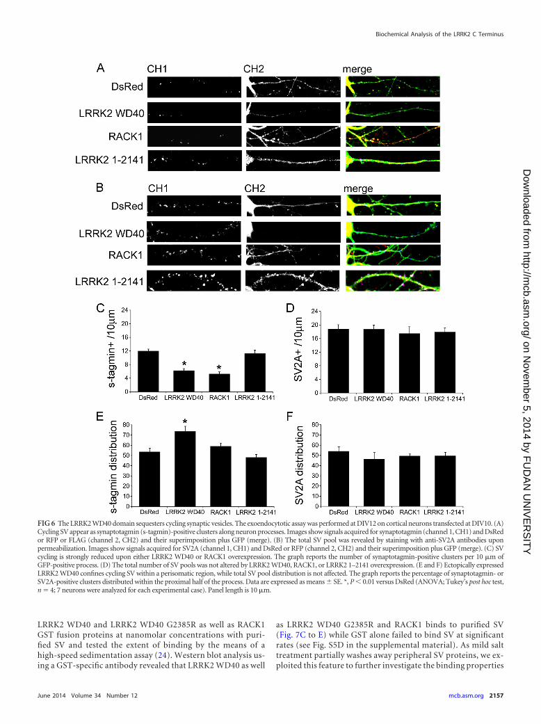
WD40 domain affects proper SV trafficking and distribution. Totest this hypothesis, we cotransfected cortical neurons at DIV10with GFP and either DsRed, RFP-LRRK2 WD40, RFP-RACK1, orLRRK2 1–2141 expression vectors. At DIV12 we determined theSV exoendocytic rate by exposing living cortical neurons to anantisynaptotagmin antibody as previously described (13). Trans-fected neurons were then tracked via laser confocal microscopy.Recycling vesicles appeared as synaptotagmin-positive clustersalong GFP-positive neuronal processes (Fig. 6A). The total vesiclepool was estimated by staining with antibodies directed against anintegral SV protein (SV2A) after fixation and permeabilization ofthe cells (Fig. 6B). While ectopic LRRK2 1–2141 did not influencesynaptotagmin uptake, the overexpression of RFP-LRRK2 WD40as well as RFP-RACK1 induced a significant decrease in the num-ber of synaptotagmin-positive clusters (Fig. 6C). At the same time,neither LRRK2 WD40 nor RACK1 nor LRRK2 1–2141 overex-pression affected the amount of SV2A-positive clusters comparedto DsRed transfected neurons (Fig. 6D). Next, we examined if theexpression of LRRK2 WD40, RACK1, or LRRK2 1–2141 had animpact on the distribution of cycling or total SV pools. To this end,we tracked the distribution of synaptotagmin (cycling SV) andSV2A-positive (total SV) clusters along GFP-positive processes.Interestingly, in LRRK2 WD40 transfected neurons, synaptotag-min-positive clusters were mainly distributed proximally to thecellular soma while SV2A-positive clusters were homogeneouslydiffused along the entire length of the neurites (Fig. 6E and F).Taken together, these data strongly indicate that ectopic expres-sion of the LRRK2 WD40 domain influences trafficking, distribu-tion, and topology of the SV cycling pool.
FIG 2 The C-terminal WD40 domain of LRRK2 represents a hub for protein-protein interactions. Western blots are shown, confirming the specific interactionof proteins following a domain-based pulldown assay for the LRRK2 WD40 domain. s-physin, synaptophysin.
Biochemical Analysis of the LRRK2 C Terminus
June 2014 Volume 34 Number 12 mcb.asm.org 2153
on Novem
ber 5, 2014 by FU
DA
N U
NIV
ER
SIT
Yhttp://m
cb.asm.org/
Dow
nloaded from

The G2385R PD risk variant alters LRRK2 WD40 bindingproperties to synaptic vesicles. The critical role conferred by theLRRK2 WD40 domain is suggested by the existence of the G2385Rpolymorphism, described as a risk factor for the development ofPD (14–16).
Sequence alignments and secondary structure predictions sug-gest that the LRRK2 C terminus contains a WD40 propeller do-main composed of seven �-blades. We assessed the structure ofthe region comprising LRRK2 residues 2124 to 2499 using homol-
ogy modeling in order to gain information about the structuralfeatures of the LRRK2 C terminus (Fig. 7A; see also Fig. S5A in thesupplemental material). Our model, based on the structure of theWD40 protein RACK1 from Arabidopsis thaliana (34), shows thatthe LRRK2 C-terminal domain is compatible with the character-istic structure of WD40 domains, i.e., seven �-propeller repeatscombined to a cleft of basic residues (5, 15). A more detailedanalysis of the region surrounding the residue G2385 revealed thatG2385 lies in close vicinity of two hydrophobic residues, V2375
FIG 3 LRRK2 interacts with presynaptic proteins. (A) The network of LRRK2 interactors was modeled from STRING annotation on the Cytoscape represen-tation. annotated, interactions annotated on STRING; experimental, interactions described in the manuscript. (B) Gene symbols, names, and gene ontology(GO) terms for the proteins included in the network.
Piccoli et al.
2154 mcb.asm.org Molecular and Cellular Biology
on Novem
ber 5, 2014 by FU
DA
N U
NIV
ER
SIT
Yhttp://m
cb.asm.org/
Dow
nloaded from

and L2439, on one side and with R2442 and R2443 on the otherside. Even with its limitations, the structural model suggests thatthe substitution of G2385 with a bulky and positively chargedarginine residue would lead to unfavorable charge repulsions andpotentially conformational changes of the protein near the inter-face between �-blades 5 and 6. This might not only alter the localconformation but also affect the binding surface of WD40 forinteraction with interacting proteins and thus impair LRRK2 ac-tivity. In order to experimentally assess the structural properties ofthe LRRK2 C terminus and the alterations caused by the G2385Rvariant, purified GST fusion proteins LRRK2 WD40 and LRRK2WD40 G2385R were analyzed by transmission electron micros-copy (EM). To meet the demand of EM for highly pure material,purity and concentration of the GST fusion proteins were con-
firmed by SDS-PAGE (see Fig. S2A in the supplemental material).While particles with distinct shapes were discernible on electronmicrographs of negatively stained LRRK2 WD40 fusion proteins,similar structures were not visible for the GST tag alone (see Fig.S5B). Single-particle classification and averaging indicated the ex-istence of particle populations with an annular, doughnut-likeappearance and diameters ranging from 5 to 8 nm for LRRK2WD40 (Fig. 7B). Structures similar in size and shape were alsodetected for the GST-RACK1 fusion protein (Fig. 7B), indicatingthat the fold of the LRRK2 WD40 domain is comparable despitelimited sequence homology. To ensure that the fusion of theWD40 domains to the GST tag does not induce ring-like struc-tures, N-terminal 6�His-tagged LRRK2 WD40 was subjected toEM (see Fig. S5C in the supplemental material). Although the
FIG 4 Expression of an LRRK2 WD40 domain construct is sufficient to induce neurotoxicity in primary neurons. Neurons were transfected at DIV3 andimaged at DIV16. Long-lasting overexpression of RFP-LRRK2 WD40 and full-length RFP-LRRK2 significantly reduced the number of processes andincreased the amount of swollen or fragmented neurites compared to levels in DsRed-, RFP LRRK2 1–2141-, or RFP-RACK1-transfected neurons. Imagesshow signals acquired for GFP (channel 1, CH1), DsRed or RFP (channel 2, CH2), superimposed channel signals (merge), and tracings. Arrow headsindicate an RFP-LRRK2 WD40-positive cell. Numbers in the tracing panel indicate total process numbers and are expressed as means � SE. **, P � 0.01versus DsRed; #, P � 0.01 versus full-length RFP-LRRK2 (ANOVA; Tukey’s post hoc test, n � 3; 8 neurons were analyzed for each experimental case). Panelsize shown is 200 by 200 �m.
Biochemical Analysis of the LRRK2 C Terminus
June 2014 Volume 34 Number 12 mcb.asm.org 2155
on Novem
ber 5, 2014 by FU
DA
N U
NIV
ER
SIT
Yhttp://m
cb.asm.org/
Dow
nloaded from

protein lacking the GST tag turned out to be more aggregationprone, doughnut-like structures similar in size and appearance tothose observed in GST fusion proteins were still present. The anal-ysis of the LRRK2 WD40 G2385R variant by EM revealed strongsimilarity to the other WD40 domain constructs, however, with awider size distribution ranging from 5 to 13 nm. This larger sizeheterogeneity may be due to structural alterations in WD40 in-duced by electrostatic repulsion in the �-propeller fold, as sug-gested by the structural model.
In order to further characterize the G2385R variant, we inves-tigated the impact of LRRK2 WD40 G2385R overexpression onneurons. Ectopic LRRK2 WD40 G2385R induced neuronal toxic-ity and sequestered cycling SV to a similar extent as LRRK2 WD40(see Fig. S6 in the supplemental material).
The data presented above suggest that the WD40 domainplays a major role in tethering LRRK2 to SV via protein inter-actions; thus, we asked if the G2385R variant can influenceLRRK2 binding to SV. To explore this hypothesis, we incubated
FIG 5 Expression of an LRRK2 WD40 domain construct induces neurotoxicity in primary neurons. (A) Neurons were transfected at DIV10 and imaged atDIV16. Long-lasting overexpression of RFP-LRRK2 WD40 significantly reduced the number of processes and increased the amount of swollen or fragmentedneurites compared to levels in DsRed-, LRRK2 1–2141-, or RFP-RACK1-transfected neurons. Images show signals acquired for GFP (channel 1, CH1), DsRedor RFP (channel 2, CH2), superimposed channel signals (merge), and tracings. Graphs report the number of total processes (B) and number of fragmentedprocesses (C) (means � SE). *, P � 0.05 versus DsRed; **, P � 0.01 versus DsRed; #, P � 0.05 versus LRRK2 WD40 (ANOVA; Tukey’s post hoc test, n � 4; 7neurons were analyzed for each experimental case). Panel size shown is 200 by 200 �m.
Piccoli et al.
2156 mcb.asm.org Molecular and Cellular Biology
on Novem
ber 5, 2014 by FU
DA
N U
NIV
ER
SIT
Yhttp://m
cb.asm.org/
Dow
nloaded from
LRRK2 WD40 and LRRK2 WD40 G2385R as well as RACK1GST fusion proteins at nanomolar concentrations with puri-fied SV and tested the extent of binding by the means of ahigh-speed sedimentation assay (24). Western blot analysis us-ing a GST-specific antibody revealed that LRRK2 WD40 as well
as LRRK2 WD40 G2385R and RACK1 binds to purified SV(Fig. 7C to E) while GST alone failed to bind SV at significantrates (see Fig. S5D in the supplemental material). As mild salttreatment partially washes away peripheral SV proteins, we ex-ploited this feature to further investigate the binding properties
FIG 6 The LRRK2 WD40 domain sequesters cycling synaptic vesicles. The exoendocytotic assay was performed at DIV12 on cortical neurons transfected at DIV10. (A)Cycling SV appear as synaptotagmin (s-tagmin)-positive clusters along neuron processes. Images show signals acquired for synaptotagmin (channel 1, CH1) and DsRedor RFP or FLAG (channel 2, CH2) and their superimposition plus GFP (merge). (B) The total SV pool was revealed by staining with anti-SV2A antibodies uponpermeabilization. Images show signals acquired for SV2A (channel 1, CH1) and DsRed or RFP (channel 2, CH2) and their superimposition plus GFP (merge). (C) SVcycling is strongly reduced upon either LRRK2 WD40 or RACK1 overexpression. The graph reports the number of synaptotagmin-positive clusters per 10 �m ofGFP-positive process. (D) The total number of SV pools was not altered by LRRK2 WD40, RACK1, or LRRK2 1–2141 overexpression. (E and F) Ectopically expressedLRRK2 WD40 confines cycling SV within a perisomatic region, while total SV pool distribution is not affected. The graph reports the percentage of synaptotagmin- orSV2A-positive clusters distributed within the proximal half of the process. Data are expressed as means � SE. *, P � 0.01 versus DsRed (ANOVA; Tukey’s post hoc test,n � 4; 7 neurons were analyzed for each experimental case). Panel length is 10 �m.
Biochemical Analysis of the LRRK2 C Terminus
June 2014 Volume 34 Number 12 mcb.asm.org 2157
on Novem
ber 5, 2014 by FU
DA
N U
NIV
ER
SIT
Yhttp://m
cb.asm.org/
Dow
nloaded from

of the three fusion proteins. Interestingly, the salt treatmentdid not alter the SV association of LRRK2 WD40 and RACK1,whereas the affinity/binding strength of the LRRK2 WD40G2385R domain to salt-treated SV was significantly reduced(Fig. 7C to E).
Taken together, these results suggest that the structural altera-tion induced by the G2385R substitution functionally disturbsLRRK2 WD40 binding properties to SV.
DISCUSSION
Since the first description of LRRK2 as a PD-causative gene, majorattention has been devoted to its GTPase and kinase activity,linking disease-associated mutations to altered functional and
(patho)physiological enzymatic properties of the protein (45–50).However, LRRK2 C-terminal deletion mutants fail to induceapoptosis and toxicity and demonstrate a reduced kinase activity(Fig. 4 and 5) (10, 51). These reports, however, do not address thequestion of whether the kinase domain or WD40 domain or bothare causative for PD pathology. We, on the other hand, observedthat the severe toxicity induced by overexpression of full-lengthLRRK2 in primary cultures is mimicked by ectopic expression of aconstruct containing only the C-terminal WD40 domain ofLRRK2. Noticeably, in contrast to LRRK2 WD40, RACK1, an-other WD40 protein, has a positive effect on neurite complexity.These data suggest that the C-terminal WD40 domain has a majorrole in LRRK2-associated toxicity (43).
FIG 7 The G2385R substitution impacts the local structure of LRRK2 WD40. (A) Combined stick-and-ribbon representation showing a structuralhomology model (based on RACK1 from Arabidopsis thaliana, PDB 3DM0) for the LRRK2 WD40 domain. The N terminus, C terminus, and the position ofG2385 (red sphere) are indicated. (B) Transmission EM images of negatively stained (1% uranyl acetate) LRRK2 WD40, LRRK2 WD40 G2385R, and RACK1proteins show doughnut-shaped particles consistent with the characteristic structure of WD40 folds. Four representative averaged single-particle two-dimen-sional projections are shown for each protein (scale bar, 5 nm). (C) LRRK2 WD40 binds SV. Increasing nanomolar amounts of LRRK2 WD40, LRRK2 WD40G2385R domains, and RACK1 protein were incubated with unstripped SV (US) or salt-stripped SV (SSV) before high-speed sedimentation. RepresentativeWestern blots show initial amount of fusion protein (total) and the yield of GST fusion proteins precipitated by US or SSV (bound). Fusion proteins wereincubated with equal amounts of SV (monitored by antisynaptophysin staining). (D) The table reports the dissociation constant (KD) describing the bindingbetween the indicated fusion protein and US or SSV. Data are expressed as means � SE. *, P � 0.05 versus LRRK2 WD40 G2385R binding to US; °, P � 0.01 versusLRRK2 WD40 binding to SSV (ANOVA; Tukey’s post hoc test, n � 4). (E) The graph reports the yield of precipitated GST fusion protein normalized versus theSV total protein amount (average data plus fitting) on the y axis and the initial amount of GST fusion protein on the x axis.
Piccoli et al.
2158 mcb.asm.org Molecular and Cellular Biology
on Novem
ber 5, 2014 by FU
DA
N U
NIV
ER
SIT
Yhttp://m
cb.asm.org/
Dow
nloaded from

From structural prediction it appears likely that the LRRK2WD40 domain folds as a seven-bladed propeller (15, 52). How-ever, sequence homology is considerably low, and no experimen-tal evidence for this assumption has existed so far. According toour EM analysis, the purified LRRK2 WD40 fusion protein formsdoughnut-like structures with an average diameter of 5 to 8 nm,resembling EM structures reported for known WD40-containingproteins such as the splicing factor Prp19p (53) or RACK1 (thispaper). These findings suggest that the LRRK2 C terminus mayindeed form a propeller-like structure, in agreement with a WD40fold. WD repeat-containing proteins execute a broad spectrum ofcritical functions. They participate in organizing cytoskeleton as-sembly, mitotic spindle formation, and vesicular trafficking (11,12, 54, 55). Furthermore, increasing evidence describes WD40repeat domains as molecular hubs orchestrating complex protein-protein interactions. Structurally, this feature is based on the evo-lutionary principle to generate binding epitopes with differentspecificities by concatenation of stable folded repeats and loopswith variable sequences (11). Following the “guilt by association”principle (56), we systematically analyzed the domain-specific in-teractome of the LRRK2 C terminus in order to assign specificphysiological functions to this domain. Through its WD40 do-main, LRRK2 interacts with critical players of the SV cycle such asNSF, syntaxin 1A, synapsin I, dynamin-1, MUNC18-1, VAMP2,synaptojanin, and synuclein. Thus, the ability of LRRK2 to influ-ence vesicle trafficking (13, 37, 38) is likely to involve its WD40domain. Accordingly, we observed a severe reduction in the trans-port of synaptotagmin-labeled (cycling) SV to distal parts of neu-ronal processes upon overexpression of LRRK2 WD40 domainconstructs; such output might be read as a dominant negativeeffect of ectopic LRRK2 WD40 executed on endogenous LRRK2function. RACK1 is a seven-bladed WD40 propeller protein (19,40), reported to bind and anchor recycling endosomal vesicles tocentrosomes (57). Accordingly, we reported that RACK1 signifi-cantly reduces SV recycling (but not spatial segregation) once it isoverexpressed in neurons and that it cosediments with pure SV.Thus, the ability to bind and sequester SV might be a biochemicalproperty common to several WD40-containing proteins or, alter-natively, might arise as an in vitro effect due to high local concen-trations of WD40 domains. Even if we cannot completely rule outthe latter explanation, our data suggest a peculiar physiologicalrole for LRRK2 at the synaptic site. In fact, we demonstrated herethat endogenous LRRK2 can be detected in a pure SV fraction,that full-length LRRK2 binds SV, and that LRRK2 WD40, but notLRRK2 LRR or full-length RACK1, interacts with several proteinsinvolved in SV trafficking. Thus, we propose that LRRK2 associa-tion with SV is mediated by the interaction of its WD40 domainwith SV-integral and -associated proteins.
The description of the G2385R point mutation within theWD40 domain as the main PD risk variant in the Chinese Han andKorean population further underlines the functional and patho-logical role of this domain (14). G2385R carrier patients demon-strate clinical features similar to noncarrier patients; however, theG2385R variant does correlate to a small but significant effect inlowering the age of PD onset (16). It has recently been reportedthat G2385R has a mild impact on LRRK2 biochemical properties,such as reduced LRRK2 kinase activity and interactions with otherproteins (9), while it neither influences LRRK2 toxicity in neuroncultures nor affects overall autophosphorylation (46). Accord-ingly, we reported here that LRRK2 WD40 G2385R behaved sim-
ilarly to LRRK2 WD40 once it was overexpressed in primary neu-rons. Comparative protein models predict that the glycine 2385residue stays at the surface of the WD40 domain (Fig. 7A) (15).The G2385R variant replaces the glycine with a long, positivelycharged arginine residue, thus increasing the net positive charge ofthe domain and likely inducing an electrostatic repulsion betweenWD40 repeats 5 and 6. This may explain the increased mean di-ameter observed for LRRK2 WD40 G2385R in our EM analysis.The changes in surface charge and local structural features in theWD40 fold are expected to result in altered biochemical propertieswhich affect protein-protein interaction strength and quality. In-deed, we reported that the association with SV is partially im-paired by the glycine-to-arginine change. In particular, whileLRRK2 WD40 showed a high affinity toward both native and salt-treated SV, LRRK2 WD40 G2385R binding to SV was significantlysensitive to salt treatment. In line with our proposal of LRRK2 as asynaptic scaffold protein involved in vesicular trafficking and ves-icle storage, our data strongly support the idea that in addition toincreased kinase activity, other molecular mechanisms, such asaltered protein binding, may underlie LRRK2-associated forms ofPD. In particular, given the recent independent evidence linkingLRRK2 dysfunction to neurotransmission defects in PD models(58–60) and in patients carrying LRRK2 mutations (61), an al-tered presynaptic vesicle transport, storage, and release kineticsmay arise as a common pathway disturbed by the different LRRK2pathological mutations described so far and become a future tar-get for pharmacological treatment.
ACKNOWLEDGMENTS
We are grateful to Nathalie Théret (INSERM, Rennes, France) for re-agents, to Sandra Helm for technical assistance, and to Peijian Zou andArie Geerlof for initial bacterial expression clones of the WD40 domain.In addition we thank Pablo Porras and Henning Hermjakob (EMBL-EBI)for data integration into the IntAct database.
This work was supported by the National Genome Research Frame-work program NGFN-Plus (grant 01GS08140, subproject 12), the Helm-holtz Alliance for Mental Health in an Aging Society (grant HA-215, topic3 WP11), the European Community’s Seventh Framework Program FP7/2009 under grant agreement number 241955, number 278568, PRIMES,and number 241481, AFFINOMICS (to M.U.), and the LRRK2 BiologyLEAPS 2012 award of the Michael J. Fox Foundation to G.P., C.J.G., M.S.,and M.U. G.P. and F.O. are supported by Fondazione Cariplo (grant2011-0540), MJFF, and Fondazione Telethon (grant GGP12237). G.P. issupported also by the FIRB program (grant RBFR08F82X_002) andFondazione Grigioni per il morbo di Parkinson. F.O. is grateful to PRIN2010-11. M.M. is supported by Fondazione Cariplo (grant 2008-3184).
Author contributions are as follows. G.P., C.J.G., F.O. and M.U. de-signed the study. G.P., C.J.G., and M.U. wrote the paper. G.P., M.D.C.,A.M., F.P., F.A., F.G., P.J., C.J.K., F.V.Z., and A.K. performed experi-ments. G.P., C.J.G., C.S., C.J.O.K., M.M., A.V., C.J.O.K., L.P., M.Z, S.W.,M.S., and M.U. analyzed the data and/or provided data analysis expertise.
REFERENCES1. Clarke C, Moore AP. 2005. Parkinson’s disease. Clin. Evid. 13:1658 –
1677.2. Fahn S. 2003. Description of Parkinson’s disease as a clinical syndrome.
Ann. N. Y. Acad. Sci. 991:1–14. http://dx.doi.org/10.1111/j.1749-6632.2003.tb07458.x.
3. Kelley LA, Sternberg MJE. 2009. Protein structure prediction on theWeb: a case study using the Phyre server. Nat. Protoc. 4:363–371. http://dx.doi.org/10.1038/nprot.2009.2.
4. Bosgraaf L, Van Haastert PJ. 2003. Roc, a Ras/GTPase domain in com-plex proteins. Biochim. Biophys. Acta 1643:5–10. http://dx.doi.org/10.1016/j.bbamcr.2003.08.008.
Biochemical Analysis of the LRRK2 C Terminus
June 2014 Volume 34 Number 12 mcb.asm.org 2159
on Novem
ber 5, 2014 by FU
DA
N U
NIV
ER
SIT
Yhttp://m
cb.asm.org/
Dow
nloaded from

5. Mills RD, Mulhern TD, Cheng H-C, Culvenor JG. 2012. Analysis ofLRRK2 accessory repeat domains: prediction of repeat length, numberand sites of Parkinson’s disease mutations. Biochem. Soc. Trans. 40:1086 –1089. http://dx.doi.org/10.1042/BST20120088.
6. Guo L, Wang W, Chen SG. 2006. Leucine-rich repeat kinase 2: relevanceto Parkinson’s disease. Int. J. Biochem. Cell Biol. 38:1469 –1475. http://dx.doi.org/10.1016/j.biocel.2006.02.009.
7. Goldwurm S, Zini M, Di Fonzo A, De Gaspari D, Siri C, Simons EJ, vanDoeselaar M, Tesei S, Antonini A, Canesi M, Zecchinelli A, Mariani C,Meucci N, Sacilotto G, Cilia R, Isaias IU, Bonetti A, Sironi F, Ricca S,Oostra BA, Bonifati V, Pezzoli G. 2006. LRRK2 G2019S mutation andParkinson’s disease: a clinical, neuropsychological and neuropsychiatricstudy in a large Italian sample. Parkinsonism Relat. Disord. 12:410 – 419.http://dx.doi.org/10.1016/j.parkreldis.2006.04.001.
8. Bonifati V. 2006. Parkinson’s disease: the LRRK2–G2019S mutation:opening a novel era in Parkinson’s disease genetics. Eur. J. Hum. Genet.14:1061–1062. http://dx.doi.org/10.1038/sj.ejhg.5201695.
9. Rudenko IN, Kaganovich A, Hauser DN, Beylina A, Chia R, Ding J,Maric D, Jaffe H, Cookson MR. 2012. The G2385R variant of leucine-rich repeat kinase 2 associated with Parkinson’s disease is a partial loss-of-function mutation. Biochem. J. 446:99 –111. http://dx.doi.org/10.1042/BJ20120637.
10. Jorgensen ND, Peng Y, Ho CC-Y, Rideout HJ, Petrey D, Liu P, DauerWT. 2009. The WD40 domain is required for LRRK2 neurotoxicity. PLoSOne 4:e8463. http://dx.doi.org/10.1371/journal.pone.0008463.
11. Stirnimann CU, Petsalaki E, Russell RB, Müller CW. 2010. WD40proteins propel cellular networks. Trends Biochem. Sci. 35:565–574. http://dx.doi.org/10.1016/j.tibs.2010.04.003.
12. Smith TF. 2008. Diversity of WD-repeat proteins. Subcell. Biochem. 48:20 –30. http://dx.doi.org/10.1007/978-0-387-09595-0_3.
13. Piccoli G, Condliffe SB, Bauer M, Giesert F, Boldt K, De Astis S,Meixner A, Sarioglu H, Vogt-Weisenhorn DM, Wurst W, Gloeckner CJ,Matteoli M, Sala C, Ueffing M. 2011. LRRK2 controls synaptic vesiclestorage and mobilization within the recycling pool. J. Neurosci. 31:2225–2237. http://dx.doi.org/10.1523/JNEUROSCI.3730-10.2011.
14. Tan E-K. 2006. Identification of a common genetic risk variant (LRRK2Gly2385Arg) in Parkinson’s disease. Ann. Acad. Med. Singapore 35:840 –842.
15. Tan EK, Zhao Y, Skipper L, Tan MG, Di Fonzo A, Sun L, Fook-Chong S,Tang S, Chua E, Yuen Y, Tan L, Pavanni R, Wong MC, Kolatkar P, Lu CS,Bonifati V, Liu JJ. 2007. The LRRK2 Gly2385Arg variant is associated withParkinson’s disease: genetic and functional evidence. Hum. Genet. 120:857–863. http://dx.doi.org/10.1007/s00439-006-0268-0.
16. Tan EK, Peng R, Wu YR, Wu RM, Wu-Chou YH, Tan LC, An XK, ChenCM, Fook-Chong S, Lu CS. 2009. LRRK2 G2385R modulates age at onsetin Parkinson’s disease: A multi-center pooled analysis. Am. J. Med. Genet.B Neuropsychiatr. Genet. 150B:1022–1023. http://dx.doi.org/10.1002/ajmg.b.30923.
17. Brewer GJ, Torricelli JR, Evege EK, Price PJ. 1993. Optimized survival ofhippocampal neurons in B27-supplemented Neurobasal, a new serum-free medium combination. J. Neurosci. Res. 35:567–576. http://dx.doi.org/10.1002/jnr.490350513.
18. Gloeckner CJ, Boldt K, Schumacher A, Ueffing M. 2009. Tandemaffinity purification of protein complexes from mammalian cells by theStrep/FLAG (SF)-TAP tag. Proteomics 564:359 –372. http://dx.doi.org/10.1007/978-1-60761-157-8_21.
19. Bourd-Boittin K, Le Pabic H, Bonnier D, L’Helgoualc’h A, Théret N.2008. RACK1, a new ADAM12 interacting protein. Contribution to liverfibrogenesis. J. Biol. Chem. 283:26000 –26009. http://dx.doi.org/10.1074/jbc.M709829200.
20. Gloeckner CJ, Kinkl N, Schumacher A, Braun RJ, O’Neill E, MeitingerT, Kolch W, Prokisch H, Ueffing M. 2006. The Parkinson disease causingLRRK2 mutation I2020T is associated with increased kinase activity.Hum. Mol. Genet. 15:223–232. http://dx.doi.org/10.1093/hmg/ddi439.
21. Xia Z, Dudek H, Miranti CK, Greenberg ME. 1996. Calcium influx viathe NMDA receptor induces immediate early gene transcription by a MAPkinase/ERK-dependent mechanism. J. Neurosci. 16:5425–5436.
22. Frangioni JV, Neel BG. 1993. Solubilization and purification of enzymat-ically active glutathione S-transferase (pGEX) fusion proteins. Anal.Biochem. 210:179 –187. http://dx.doi.org/10.1006/abio.1993.1170.
23. Huttner WB, Schiebler W, Greengard P, De Camilli P. 1983. SynapsinI (protein I), a nerve terminal-specific phosphoprotein. III. Its association
with synaptic vesicles studied in a highly purified synaptic vesicle prepa-ration. J. Cell Biol. 96:1374 –1388.
24. Messa M, Congia S, Defranchi E, Valtorta F, Fassio A, Onofri F,Benfenati F. 2010. Tyrosine phosphorylation of synapsin I by Src regu-lates synaptic-vesicle trafficking. J. Cell Sci. 123:2256 –2265. http://dx.doi.org/10.1242/jcs.068445.
25. Gillardon F, Kremmer E, Froehlich T, Ueffing M, Hengerer B, Gloeck-ner CJ. 2013. ATP-competitive LRRK2 inhibitors interfere with mono-clonal antibody binding to the kinase domain of LRRK2 under nativeconditions. A method to directly monitor the active conformation ofLRRK2? J. Neurosci. Methods 214:62– 68. http://dx.doi.org/10.1016/j.jneumeth.2012.12.015.
26. Olsen JV. 2005. Parts per million mass accuracy on an Orbitrap massspectrometer via lock mass injection into a C-trap. Mol. Cell. Proteomics4:2010 –2021. http://dx.doi.org/10.1074/mcp.T500030-MCP200.
27. Keller A, Nesvizhskii AI, Kolker E, Aebersold R. 2002. Empirical statis-tical model to estimate the accuracy of peptide identifications made byMS/MS and database search. Anal. Chem. 74:5383–5392. http://dx.doi.org/10.1021/ac025747h.
28. Nesvizhskii AI, Keller A, Kolker E, Aebersold R. 2003. A statistical modelfor identifying proteins by tandem mass spectrometry. Anal. Chem. 75:4646 – 4658. http://dx.doi.org/10.1021/ac0341261.
29. Matteoli M, Takei K, Perin MS, Sudhof TC, De Camilli P. 1992.Exo-endocytotic recycling of synaptic vesicles in developing processes ofcultured hippocampal neurons. J. Cell Biol. 117:849 – 861. http://dx.doi.org/10.1083/jcb.117.4.849.
30. Piccoli G, Verpelli C, Tonna N, Romorini S, Alessio M, Nairn AC, BachiA, Sala C. 2007. Proteomic analysis of activity-dependent synaptic plas-ticity in hippocampal neurons. J. Prot. Res. 6:3203–3215. http://dx.doi.org/10.1021/pr0701308.
31. Ludtke SJ, Baldwin PR, Chiu W. 1999. EMAN: semiautomated softwarefor high-resolution single-particle reconstructions. J. Struct. Biol. 128:82–97. http://dx.doi.org/10.1006/jsbi.1999.4174.
32. Berman HM, Bhat TN, Bourne PE, Feng Z, Gilliland G, Weissig H,Westbrook J. 2000. The Protein Data Bank and the challenge of structuralgenomics. Nat. Struct. Biol. 7(Suppl):957–959. http://dx.doi.org/10.1038/80734.
33. Berman HM, Battistuz T, Bhat TN, Bluhm WF, Bourne PE, BurkhardtK, Feng Z, Gilliland GL, Iype L, Jain S, Fagan P, Marvin J, Padilla D,Ravichandran V, Schneider B, Thanki N, Weissig H, Westbrook JD,Zardecki C. 2002. The Protein Data Bank. Acta Crystallogr. D. Biol. Crys-tallogr. 58:899 –907. http://dx.doi.org/10.1107/S0907444902003451.
34. Ullah H, Scappini EL, Moon AF, Williams LV, Armstrong DL, PedersenLC. 2008. Structure of a signal transduction regulator, RACK1, from Ara-bidopsis thaliana. Protein Sci. 17:1771–1780. http://dx.doi.org/10.1110/ps.035121.108.
35. Krieger E, Koraimann G, Vriend G. 2002. Increasing the precision ofcomparative models with YASARA NOVA-a self-parameterizing forcefield. Proteins 47:393– 402. http://dx.doi.org/10.1002/prot.10104.
36. Laskowski R, Rullmann JA, MacArthur M, Kaptein R, Thornton J.1996. AQUA and PROCHECK-NMR: programs for checking the qualityof protein structures solved by NMR. J. Biomol. NMR 8:477– 486.
37. Shin N, Jeong H, Kwon J, Heo HY, Kwon JJ, Yun HJ, Kim C-H, HanBS, Tong Y, Shen J, Hatano T, Hattori N, Kim KS, Chang S, Seol W.2008. LRRK2 regulates synaptic vesicle endocytosis. Exp. Cell Res. 314:2055–2065. http://dx.doi.org/10.1016/j.yexcr.2008.02.015.
38. Xiong Y, Coombes CE, Kilaru A, Li X, Gitler AD, Bowers WJ, DawsonVL, Dawson TM, Moore DJ. 2010. GTPase activity plays a key role in thepathobiology of LRRK2. PLoS Genet. 6:e1000902. http://dx.doi.org/10.1371/journal.pgen.1000902.
39. Meixner A, Boldt K, Van Troys M, Askenazi M, Gloeckner CJ, Bauer M,Marto JA, Ampe C, Kinkl N, Ueffing M. 2011. A QUICK screen for Lrrk2interaction partners-leucine-rich repeat kinase 2 is involved in actin cyto-skeleton dynamics. Mol. Cell Proteomics 10:M110.001172. http://dx.doi.org/10.1074/mcp.M110.001172.
40. Sengupta J, Nilsson J, Gursky R, Spahn CMT, Nissen P, Frank J. 2004.Identification of the versatile scaffold protein RACK1 on the eukaryoticribosome by cryo-EM. Nat. Struct. Mol. Biol. 10:957–962. http://dx.doi.org/10.1038/nsmb822.
41. Szklarczyk D, Franceschini A, Kuhn M, Simonovic M, Roth A, MinguezP, Doerks T, Stark M, Muller J, Bork P, Jensen LJ, von Mering C. 2011.The STRING database in 2011: functional interaction networks of pro-
Piccoli et al.
2160 mcb.asm.org Molecular and Cellular Biology
on Novem
ber 5, 2014 by FU
DA
N U
NIV
ER
SIT
Yhttp://m
cb.asm.org/
Dow
nloaded from

teins, globally integrated and scored. Nucleic Acids Res. 39:D561–D568.http://dx.doi.org/10.1093/nar/gkq973.
42. Shannon P, Markiel A, Ozier O, Baliga NS, Wang JT, Ramage D, AminN, Schwikowski B, Ideker T. 2003. Cytoscape: a software environment forintegrated models of biomolecular interaction networks. Genome Res.13:2498 –2504. http://dx.doi.org/10.1101/gr.1239303.
43. Skibinski G, Nakamura K, Cookson MR, Finkbeiner S. 2014. MutantLRRK2 toxicity in neurons depends on LRRK2 levels and synuclein butnot kinase activity or inclusion bodies. J. Neurosci. 34:418 – 433. http://dx.doi.org/10.1523/JNEUROSCI.2712-13.2014.
44. Chernova T, Steinert JR, Guerin CJ, Nicotera P, Forsythe ID, SmithAG. 2007. Neurite degeneration induced by heme deficiency mediated viainhibition of NMDA receptor-dependent extracellular signal-regulatedkinase 1/2 activation. J. Neurosci. 27:8475– 8485. http://dx.doi.org/10.1523/JNEUROSCI.0792-07.2007.
45. Gloeckner CJ, Schumacher A, Boldt K, Ueffing M. 2009. The Parkinsondisease-associated protein kinase LRRK2 exhibits MAPKKK activity andphosphorylates MKK3/6 and MKK4/7, in vitro. J. Neurochem. 109:959 –968. http://dx.doi.org/10.1111/j.1471-4159.2009.06024.x.
46. West AB, Moore DJ, Choi C, Andrabi SA, Li X, Dikeman D, Biskup S,Zhang Z, Lim KL, Dawson VL, Dawson TM. 2007. Parkinson’s disease-associated mutations in LRRK2 link enhanced GTP-binding and kinaseactivities to neuronal toxicity. Hum. Mol. Genet. 16:223–232. http://dx.doi.org/10.1093/hmg/ddl471.
47. Lee BD, Shin J-H, VanKampen J, Petrucelli L, West AB, Ko HS, Lee Y-I,Maguire-Zeiss KA, Bowers WJ, Federoff HJ, Dawson VL, Dawson TM.2010. Inhibitors of leucine-rich repeat kinase-2 protect against models ofParkinson’s disease. Nat. Med. 16:998 –1000. http://dx.doi.org/10.1038/nm.2199.
48. Kett LR, Boassa D, Ho CC-Y, Rideout HJ, Hu J, Terada M, Ellisman M,Dauer WT. 2012. LRRK2 Parkinson disease mutations enhance its micro-tubule association. Hum. Mol. Genet. 21:890 – 899. http://dx.doi.org/10.1093/hmg/ddr526.
49. Greggio E, Jain S, Kingsbury A, Bandopadhyay R, Lewis P, KaganovichA, van der Brug MP, Beilina A, Blackinton J, Thomas KJ, Ahmad R,Miller DW, Kesavapany S, Singleton A, Lees A, Harvey RJ, Harvey K,Cookson MR. 2006. Kinase activity is required for the toxic effects ofmutant LRRK2/dardarin. Neurobiol. Dis. 23:329 –341. http://dx.doi.org/10.1016/j.nbd.2006.04.001.
50. Imai Y, Gehrke S, Wang HQ, Takahashi R, Hasegawa K, Oota E, Lu B.2008. Phosphorylation of 4E-BP by LRRK2 affects the maintenance ofdopaminergic neurons in Drosophila. EMBO J. 27:2432–2443. http://dx.doi.org/10.1038/emboj.2008.163.
51. Iaccarino C, Crosio C, Vitale C, Sanna G, Carri MT, Barone P. 2007.Apoptotic mechanisms in mutant LRRK2-mediated cell death. Hum.Mol. Genet. 16:1319 –1326. http://dx.doi.org/10.1093/hmg/ddm080.
52. Wang Y, Jiang F, Zhuo Z, Wu X-H, Wu Y-D. 2013. A method for WD40
repeat detection and secondary structure prediction. PLoS One 8:e65705.http://dx.doi.org/10.1371/journal.pone.0065705.
53. Ohi MD, Kooi CWV, Rosenberg JA, Ren L, Hirsch JP, Chazin WJ, WalzT, Gould KL. 2005. Structural and functional analysis of essential pre-mRNA splicing factor Prp19p. Mol. Cell. Biol. 25:451– 460. http://dx.doi.org/10.1128/MCB.25.1.451-460.2005.
54. Pryer NK, Salama NR, Schekman R, Kaiser CA. 1993. Cytosolic Sec13pcomplex is required for vesicle formation from the endoplasmic reticulum invitro. J. Cell Biol. 120:865–875. http://dx.doi.org/10.1083/jcb.120.4.865.
55. Vaisman N, Tsouladze A, Robzyk K, Ben-Yehuda S, Kupiec M, KassirY. 1995. The role of Saccharomyces cerevisiae Cdc40p in DNA replicationand mitotic spindle formation and/or maintenance. Mol. Gen. Genet.247:123–136. http://dx.doi.org/10.1007/BF00705642.
56. Wang PI, Marcotte EM. 2010. It’s the machine that matters: predictinggene function and phenotype from protein networks. J. Proteomics 73:2277–2289. http://dx.doi.org/10.1016/j.jprot.2010.07.005.
57. Ai E, Skop AR. 2009. Endosomal recycling regulation during cytoki-nesis. Commun. Integr. Biol. 2:444 – 447. http://dx.doi.org/10.4161/cib.2.5.8931.
58. Li X, Patel JC, Wang J, Avshalumov MV, Nicholson C, Buxbaum JD,Elder GA, Rice ME, Yue Z. 2010. Enhanced striatal dopamine transmis-sion and motor performance with LRRK2 overexpression in mice is elim-inated by familial Parkinson’s disease mutation G2019S. J. Neurosci. 30:1788 –1797. http://dx.doi.org/10.1523/JNEUROSCI.5604-09.2010.
59. Tong Y, Pisani A, Martella G, Karouani M, Yamaguchi H, Pothos EN,Shen J. 2009. R1441C mutation in LRRK2 impairs dopaminergic neu-rotransmission in mice. Proc. Natl. Acad. Sci. 106:14622–14627. http://dx.doi.org/10.1073/pnas.0906334106.
60. Li Y, Liu W, Oo TF, Wang L, Tang Y, Jackson-Lewis V, Zhou C,Geghman K, Bogdanov M, Przedborski S, Beal MF, Burke RE, Li C.2009. Mutant LRRK2R1441G BAC transgenic mice recapitulate cardinalfeatures of Parkinson’s disease. Nat. Neurosci. 12:826 – 828. http://dx.doi.org/10.1038/nn.2349.
61. Sossi V, de la Fuente-Fernández R, Nandhagopal R, Schulzer M,McKenzie J, Ruth TJ, Aasly JO, Farrer MJ, Wszolek ZK, Stoessl JA. 2010.Dopamine turnover increases in asymptomatic LRRK2 mutations carriers.Mov. Disord. 25:2717–2723. http://dx.doi.org/10.1002/mds.23356.
62. Berg D, Schweitzer KJ, Leitner P, Zimprich A, Lichtner P, Belcredi P,Brüssel T, Schulte C, Maass S, Nägele T, Wszolek ZK, Gasser T. 2005.Type and frequency of mutations in the LRRK2 gene in familial and spo-radic Parkinson’s disease*. Brain 128:3000 –3011. http://dx.doi.org/10.1093/brain/awh666.
63. Brice A. 2005. Genetics of Parkinson’s disease: LRRK2 on the rise. Brain128:2760 –2762. http://dx.doi.org/10.1093/brain/awh676.
64. Hardy J, Lewis P, Revesz T, Lees A, Paisan-Ruiz C. 2009. The genetics ofParkinson’s syndromes: a critical review. Curr. Opin. Genet. Dev. 19:254 –265. http://dx.doi.org/10.1016/j.gde.2009.03.008.
Biochemical Analysis of the LRRK2 C Terminus
June 2014 Volume 34 Number 12 mcb.asm.org 2161
on Novem
ber 5, 2014 by FU
DA
N U
NIV
ER
SIT
Yhttp://m
cb.asm.org/
Dow
nloaded from