Embed Size (px)
Citation preview
© Our Dermatol Online 4.2015 481
How to cite this article: Shricharith S, Anuradha J, Raghavendra R, Pai S. Entodermoscope: A tool to diagnose and monitor pediculosis captitis. Our Dermatol Online. 2015;6(4):481-482.Submission: 01.03.2015; Acceptance: 05.05.2015DOI:10.7241/ourd.20153.133
Entodermoscope: A tool to diagnose and monitor pediculosis captitisShetty Shricharith, Jindal Anuradha, Rao Raghavendra, Sathish Pai
Department of Dermatology, Kasturba Medical College, Manipal University, Manipal, Karnataka, India
Corresponding author: Dr. Shetty Shricharith, E-mail: [email protected]
Sir,
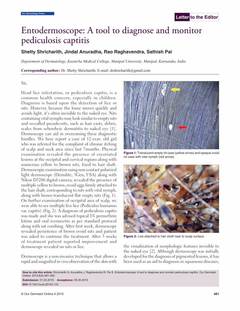
Head lice infestation, or pediculosis capitis, is a common health concern, especially in children. Diagnosis is based upon the detection of lice or nits. However, because the louse moves quickly and avoids light, it’s often invisible to the naked eye. Nits containing vital nymphs may look similar to empty nits and so-called pseudo-nits, such as hair casts, debris, scales from seborrheic dermatitis to naked eye [1]. Dermoscopy can aid in overcoming these diagnostic hurdles. We here report a case of 12-year- old girl who was referred for the complaint of chronic itching of scalp and neck area since last 7months. Physical examination revealed the presence of excoriated lesions at the occipital and cervical regions along with numerous yellow to brown nits, fixed to hair shaft. Dermoscopic examination using non-contact polarized light dermoscope (Dermlite; 3Gen, USA) along with Nikon D3200 digital camera, revealed the presence of multiple yellow to brown, ovoid eggs firmly attached to the hair shaft, corresponding to nits with vital nymph, along with brown translucent flat empty nits (Fig. 1). On further examination of occipital area of scalp, we were able to see multiple live lice (Pediculus humanus var. capitis) (Fig. 2). A diagnosis of pediculosis capitis was made and she was advised topical 1% permethrin lotion and oral ivermectin as per standard protocol along with nit combing. After first week, dermoscopy revealed persistence of brown ovoid nits and patient was asked to continue the treatment. After 3 weeks of treatment patient reported improvement and dermoscopy revealed no nits or lice.
Dermoscopy is a non-invasive technique that allows a rapid and magnified in vivo observation of the skin with
the visualization of morphologic features invisible to the naked eye [2]. Although dermoscopy was initially developed for the diagnosis of pigmented lesions, it has been used as an aid to diagnosis in squamous diseases,
Letter to the Editor
Figure 1: Translucent empty nit case (yellow arrow) and opaque ovoid nit case with vital nymph (red arrow).
Figure 2: Lice attached to hair shaft near to scalp surface.
www.odermatol.com
© Our Dermatol Online 4.2015 482
depigmenting diseases, infections and infestations [3]. The term “entodermoscopy” was coined to refer to the use of dermoscopy as an aid in the diagnosis and follow-up of treatment of infestations such as scabies, pediculosis, tungiasis, cutaneous larva migrans and tick infestations [3,4]. New generation non-contact dermoscope using polarized light prevents the possible risk of transfection in the latter cases. Dermoscope hence not only provides an easy way to establish confirmed diagnosis but also help in monitoring the disease after starting the treatment, especially in the era where resistance to pediculicides becoming an emerging problem in many parts of the world.
CONSENT
The examination of the patient was conducted according to the Declaration of Helsinki principles.
REFERENCES
1. Di Stefani A, Hofmann-Wellenhof R, Zalaudek I. Dermoscopy for diagnosis and treatment monitoring of pediculosis capitis. J Am Acad Dermatol. 2006;54:909-11.
2. Micali G, Tedeschi A, West DP, Dinotta F, Lacarrubba F. The use of videodermatoscopy to monitor treatment of scabies and pediculosis. J Dermatolog Treat. 2011;22:133-37.
3. Zalaudek I, Giacomel J, Cabo H, Di Stefani A, Ferrara G, Hofmann-Wellenhof R, et al. Entodermoscopy: a new tool for diagnosing skin infections and infestations. Dermatology. 2008;216:14–23.
4. Tschandl P, Argenz iano G, Bakos R, Gourhant JY, Hofmann-Wellenhof R, Kittler H, et al. Dermoscopy and entomology (entomodermoscopy). J Dtsch Dermatol Ges. 2009;7:589–96.
Copyright by Shetty Shricharith, et al. This is an open access article distributed under the terms of the Creative Commons Attribution License, which permits unrestricted use, distribution, and reproduction in any medium, provided the original author and source are credited.Source of Support: Nil, Conflict of Interest: None declared.