Embed Size (px)
Citation preview
Cell Cycle - Regulation
Institute of Lifelong Learning, University of Delhi 0
Lesson: Cell Cycle: Regulation
Lesson Developer: Rama Sisodia
College/ Department: Department of Botany, Maitreyi College, University
of Delhi
Cell Cycle - Regulation
Institute of Lifelong Learning, University of Delhi 1
Table of Contents
Chapter: Cell Cycle - Regulation
Introduction
Cell cycle regulation by internal check points
G1 check point
G2 check point
M check point
Regulator molecules of the cell cycle
Cyclin dependent kinases
Identification
Regulation
Classes of Cdks and Cyclins
Cdk regulation of M phase
Extracellular signals
Checkpoint control
DNA damage induced check points
Spindle assembly check point
Regulation of meiosis
Fertilization
Summary
Exercise/ Practice
Glossary
References/ Bibliography/ Further Readings
Cell Cycle - Regulation
Institute of Lifelong Learning, University of Delhi 2
Introduction
An understanding of the mechanisms that control cell cycle is important not only for basic
biology but also since it underlies the basis of studying cancer- the uncontrolled proliferation
of cells. The transition from one phase to other of the cell cycle is very well coordinated and
is under the control of extracellular signals as well as certain internal signals.
Cell cycle regulation by internal checkpoints
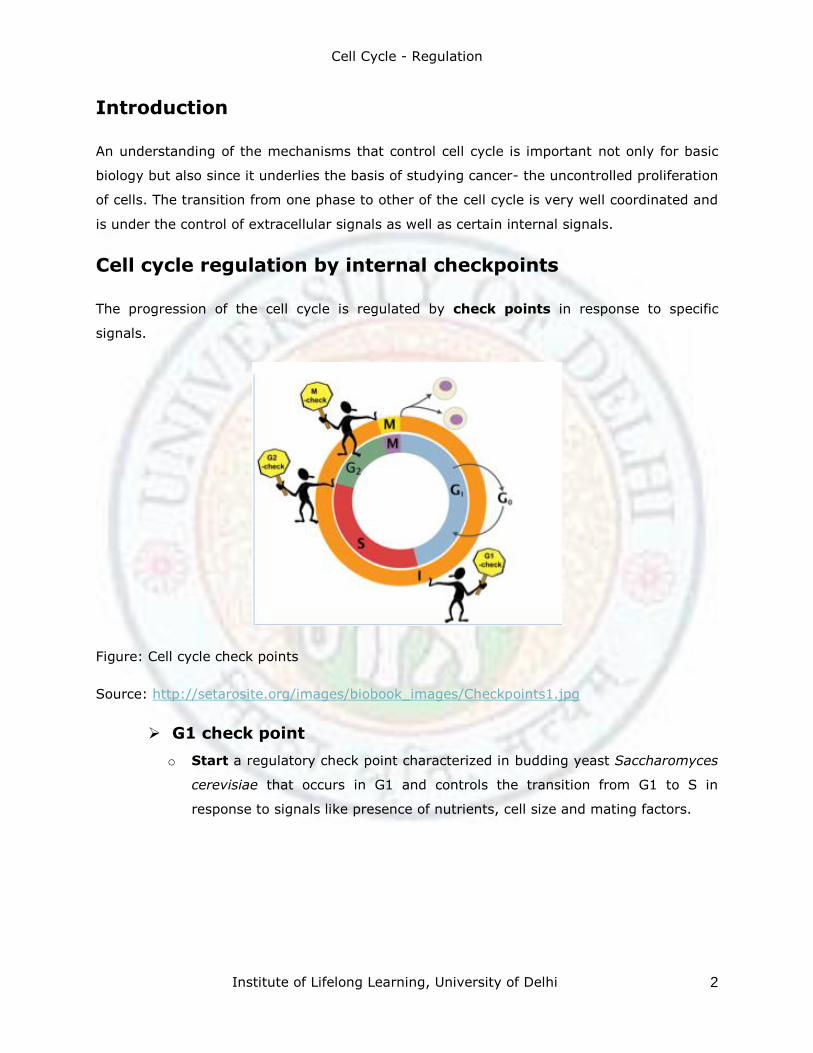
The progression of the cell cycle is regulated by check points in response to specific
signals.
Figure: Cell cycle check points
Source: http://setarosite.org/images/biobook_images/Checkpoints1.jpg
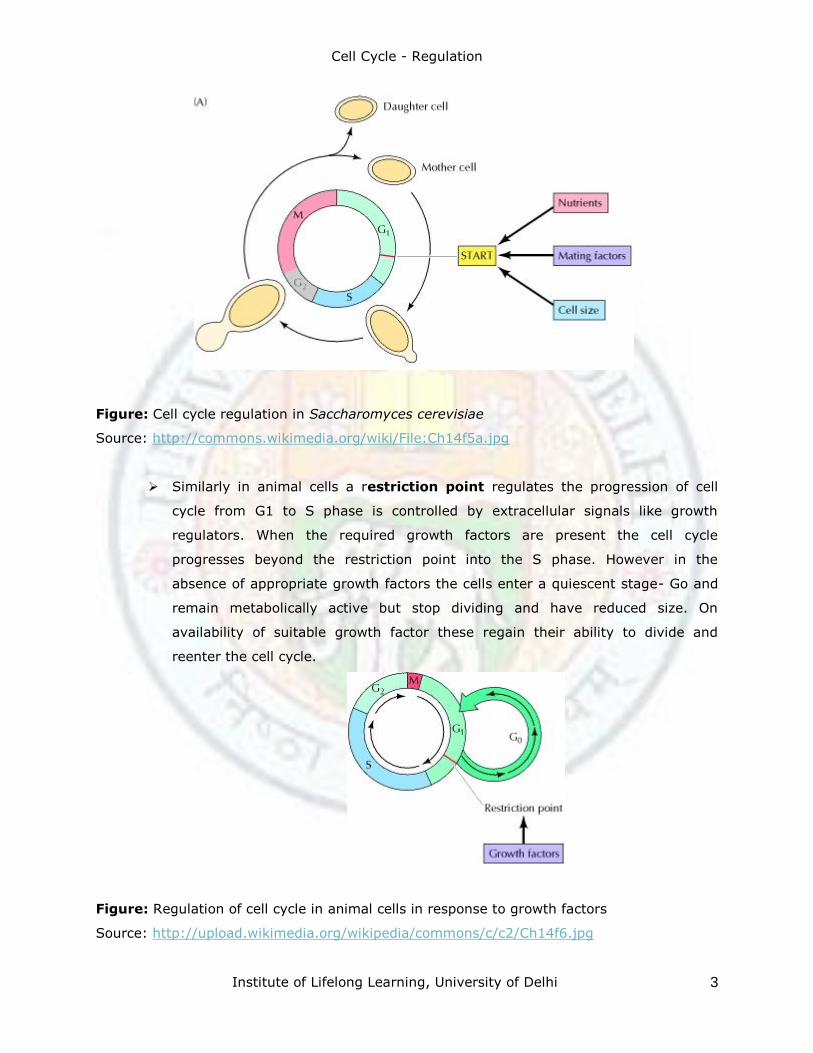
G1 check point
o Start a regulatory check point characterized in budding yeast Saccharomyces
cerevisiae that occurs in G1 and controls the transition from G1 to S in
response to signals like presence of nutrients, cell size and mating factors.
Cell Cycle - Regulation
Institute of Lifelong Learning, University of Delhi 3
Figure: Cell cycle regulation in Saccharomyces cerevisiae
Source: http://commons.wikimedia.org/wiki/File:Ch14f5a.jpg
Similarly in animal cells a restriction point regulates the progression of cell
cycle from G1 to S phase is controlled by extracellular signals like growth
regulators. When the required growth factors are present the cell cycle
progresses beyond the restriction point into the S phase. However in the
absence of appropriate growth factors the cells enter a quiescent stage- Go and
remain metabolically active but stop dividing and have reduced size. On
availability of suitable growth factor these regain their ability to divide and
reenter the cell cycle.
Figure: Regulation of cell cycle in animal cells in response to growth factors
Source: http://upload.wikimedia.org/wikipedia/commons/c/c2/Ch14f6.jpg
Cell Cycle - Regulation
Institute of Lifelong Learning, University of Delhi 4
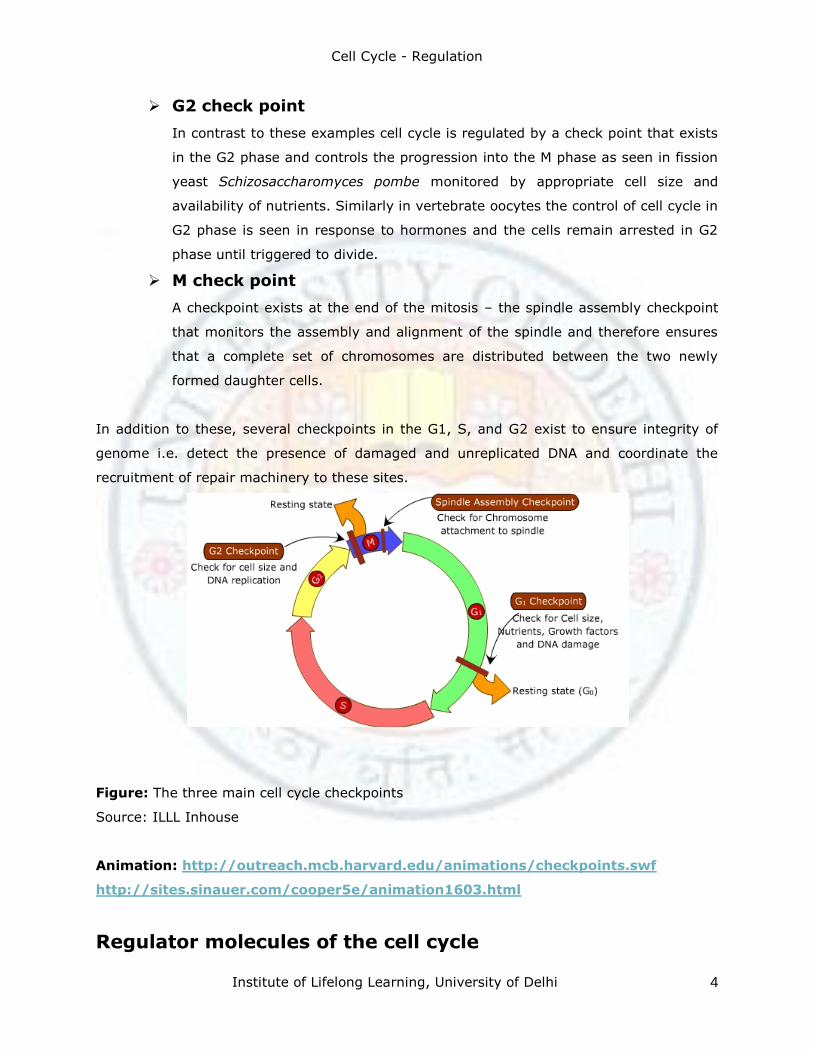
G2 check point
In contrast to these examples cell cycle is regulated by a check point that exists
in the G2 phase and controls the progression into the M phase as seen in fission
yeast Schizosaccharomyces pombe monitored by appropriate cell size and
availability of nutrients. Similarly in vertebrate oocytes the control of cell cycle in
G2 phase is seen in response to hormones and the cells remain arrested in G2
phase until triggered to divide.
M check point
A checkpoint exists at the end of the mitosis – the spindle assembly checkpoint
that monitors the assembly and alignment of the spindle and therefore ensures
that a complete set of chromosomes are distributed between the two newly
formed daughter cells.
In addition to these, several checkpoints in the G1, S, and G2 exist to ensure integrity of
genome i.e. detect the presence of damaged and unreplicated DNA and coordinate the
recruitment of repair machinery to these sites.
Figure: The three main cell cycle checkpoints
Source: ILLL Inhouse
Animation: http://outreach.mcb.harvard.edu/animations/checkpoints.swf
http://sites.sinauer.com/cooper5e/animation1603.html
Regulator molecules of the cell cycle
Cell Cycle - Regulation
Institute of Lifelong Learning, University of Delhi 5
Cyclin dependent kinases
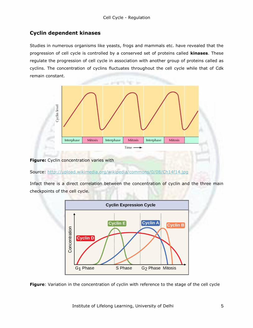
Studies in numerous organisms like yeasts, frogs and mammals etc. have revealed that the
progression of cell cycle is controlled by a conserved set of proteins called kinases. These
regulate the progression of cell cycle in association with another group of proteins called as
cyclins. The concentration of cyclins fluctuates throughout the cell cycle while that of Cdk
remain constant.
Figure: Cyclin concentration varies with
Source: http://upload.wikimedia.org/wikipedia/commons/0/08/Ch14f14.jpg
Infact there is a direct correlation between the concentration of cyclin and the three main
checkpoints of the cell cycle.
Figure: Variation in the concentration of cyclin with reference to the stage of the cell cycle
Cell Cycle - Regulation
Institute of Lifelong Learning, University of Delhi 6
Source: OpenStax College, http://cnx.org/content/m44466/1.3/
Source: http://sgugenetics.pbworks.com/f/1269908653/cyclinCDK.jpg
Identification
Molecules that control the progression of cell cycle were initially identified from cell fusion
experiments. When cultured mammalian cells arrested in different stages of cell cycle were
fused to form heterokaryons (a single cell with two nuclei) the progression to the next stage
was possible indicating the presence of molecules controlling the progression to the next
stage( Rao and Johnson, 1970). For example a fusion of a cell in S phase with a cell in G1
phase resulted in a heterokaryon which showed transition to S phase and the inititation of
DNA replication indicating the S phase cells contained the molecules that triggers the
progression from G1 to S phase.
In 1971, investigations involving frog oocytes ( Masui,Y. and Markert, C.; Smith,D and
Cell Cycle - Regulation
Institute of Lifelong Learning, University of Delhi 7
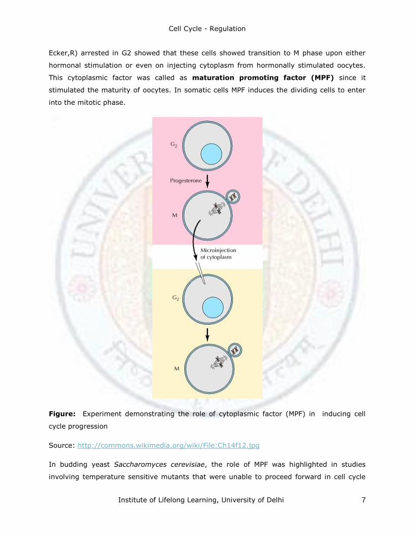
Ecker,R) arrested in G2 showed that these cells showed transition to M phase upon either
hormonal stimulation or even on injecting cytoplasm from hormonally stimulated oocytes.
This cytoplasmic factor was called as maturation promoting factor (MPF) since it
stimulated the maturity of oocytes. In somatic cells MPF induces the dividing cells to enter
into the mitotic phase.
Figure: Experiment demonstrating the role of cytoplasmic factor (MPF) in inducing cell
cycle progression
Source: http://commons.wikimedia.org/wiki/File:Ch14f12.jpg
In budding yeast Saccharomyces cerevisiae, the role of MPF was highlighted in studies
involving temperature sensitive mutants that were unable to proceed forward in cell cycle
Cell Cycle - Regulation
Institute of Lifelong Learning, University of Delhi 8
and were arrested at particular
r points of cell cycle. For example the cell divison cycle mutants called cdc28 were arrested
at the check point START and only in the presence of the protein Cdc28 the progression to
the next phase was achieved. Similarly in fission yeast Schizosaccaromyces pombe the
product of cdc2 gene was required for the transition of cell cycle through the G1 START and
the G2 to M transition. It was later revealed that the genes cdc 28 and cdc2 encode a
protein kinase Cdk1. Related genes were identified in higher eukaryotes including human
beings.
Cell divisions in early embryo development in sea urchin were found to be
accompanied with new protein synthesis
Periodic variation in accumulation of proteins-cyclins.
Microinjection of cyclin in frog oocytes was found to trigger G2 to M transition.
MPF purified from frog eggs in 1988, James Maller
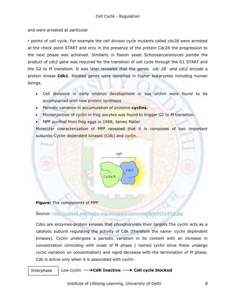
Molecular characterization of MPF revealed that it is composes of two important
subunits-Cyclin dependent kinases (Cdk) and cyclin.
Figure: The components of MPF
Source: http://upload.wikimedia.org/wikipedia/commons/6/60/Ch14f15.jpg
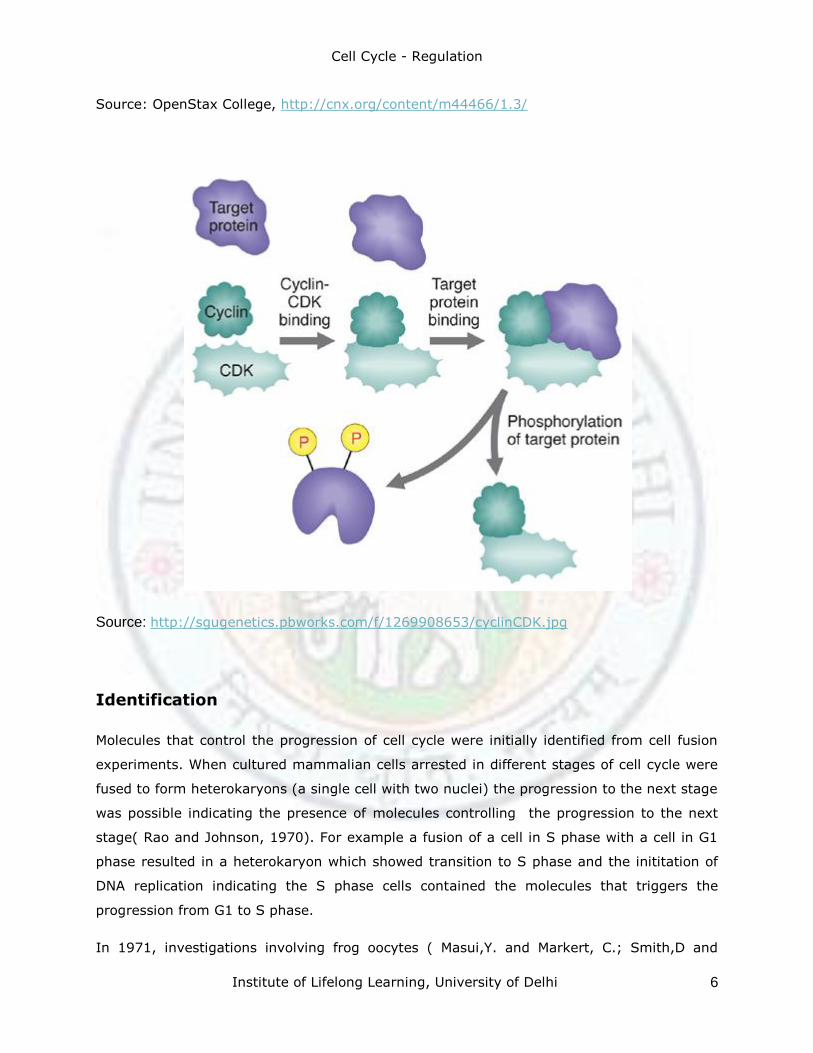
Cdks are enzymes-protein kinases that phosphorylate their targets.The cyclin acts as a
catalytic subunit regulating the activity of Cdk (therefore the name- cyclin dependent
kinases). Cyclin undergoes a periodic variation in its content with an increase in
concentration coinciding with onset of M phase ( named cyclin since these undergo
cyclic variation on concentration) and rapid decrease with the termination of M phase.
Cdk is active only when it is associated with cyclin.
– Low cyclin CdK Inactive Cell cycle blocked Interphase
Cell Cycle - Regulation
Institute of Lifelong Learning, University of Delhi 9
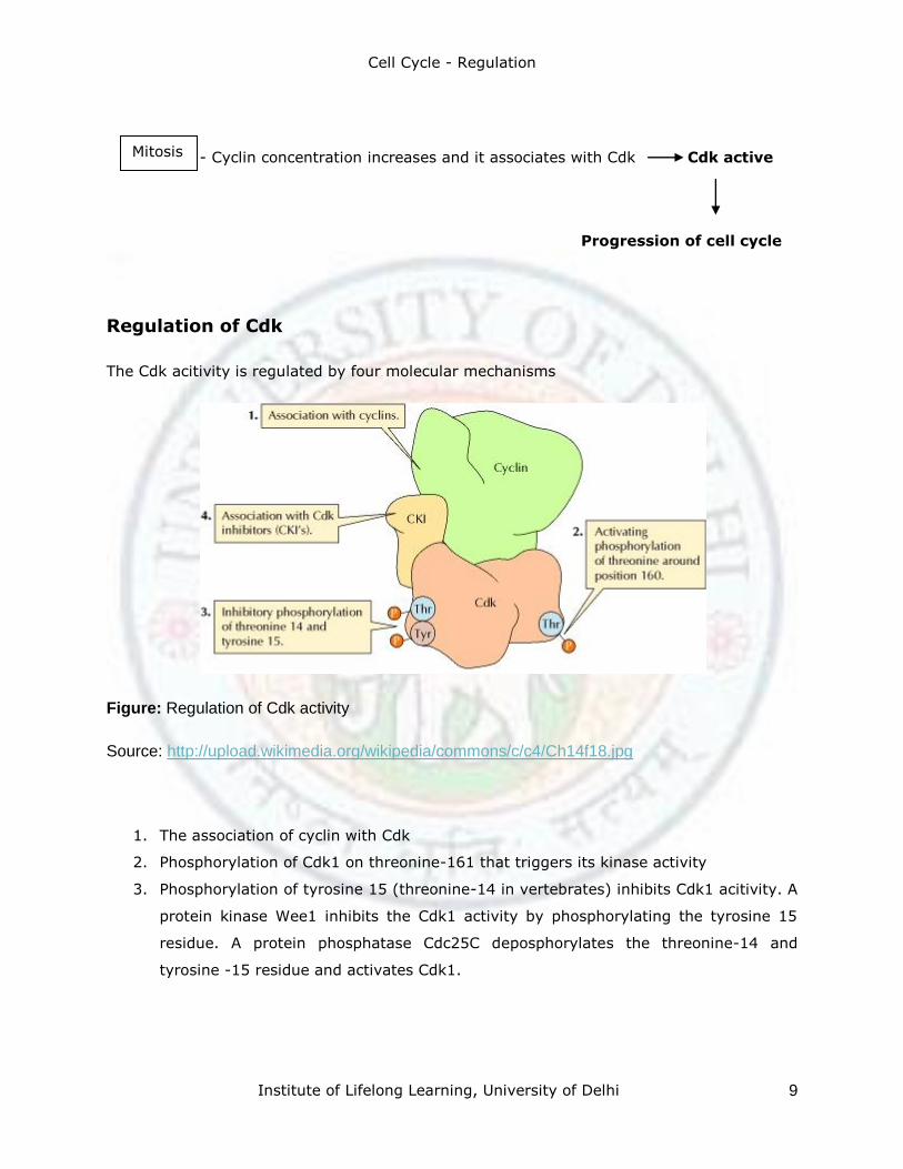
- Cyclin concentration increases and it associates with Cdk Cdk active
Progression of cell cycle
Regulation of Cdk
The Cdk acitivity is regulated by four molecular mechanisms
Figure: Regulation of Cdk activity
Source: http://upload.wikimedia.org/wikipedia/commons/c/c4/Ch14f18.jpg
1. The association of cyclin with Cdk
2. Phosphorylation of Cdk1 on threonine-161 that triggers its kinase activity
3. Phosphorylation of tyrosine 15 (threonine-14 in vertebrates) inhibits Cdk1 acitivity. A
protein kinase Wee1 inhibits the Cdk1 activity by phosphorylating the tyrosine 15
residue. A protein phosphatase Cdc25C deposphorylates the threonine-14 and
tyrosine -15 residue and activates Cdk1.
Mitosis
Cell Cycle - Regulation
Institute of Lifelong Learning, University of Delhi 10
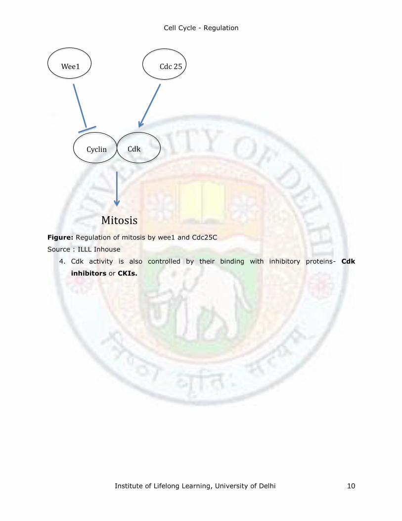
Figure: Regulation of mitosis by wee1 and Cdc25C
Source : ILLL Inhouse
4. Cdk activity is also controlled by their binding with inhibitory proteins- Cdk
inhibitors or CKIs.
Wee1 Cdc 25
Mitosis
Cyclin Cdk
Cell Cycle - Regulation
Institute of Lifelong Learning, University of Delhi 11
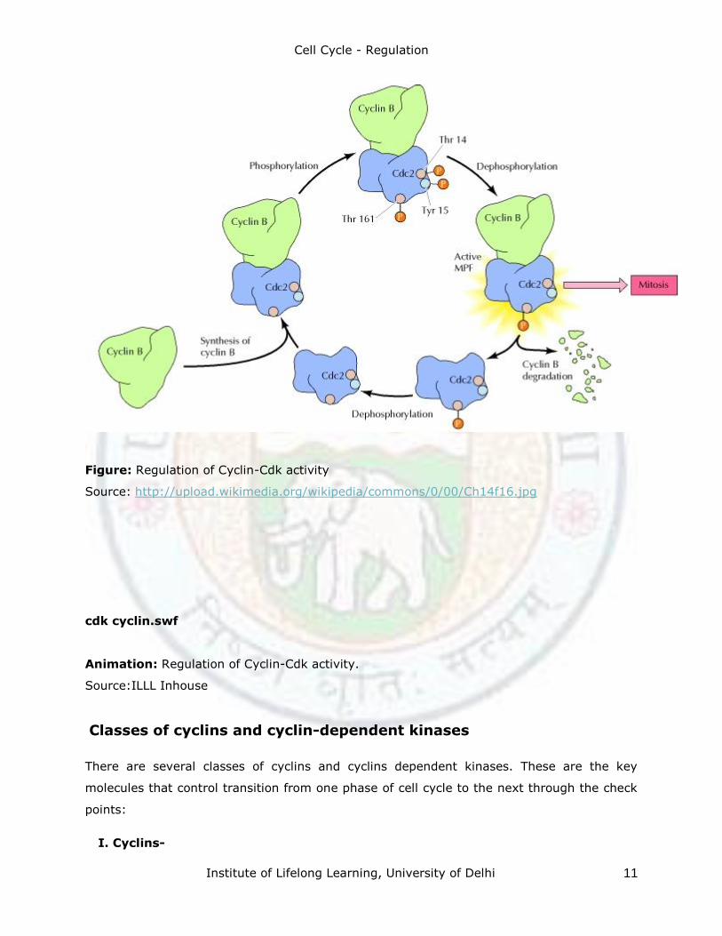
Figure: Regulation of Cyclin-Cdk activity
Source: http://upload.wikimedia.org/wikipedia/commons/0/00/Ch14f16.jpg
cdk cyclin.swf
Animation: Regulation of Cyclin-Cdk activity.
Source:ILLL Inhouse
Classes of cyclins and cyclin-dependent kinases
There are several classes of cyclins and cyclins dependent kinases. These are the key
molecules that control transition from one phase of cell cycle to the next through the check
points:
I. Cyclins-
Cell Cycle - Regulation
Institute of Lifelong Learning, University of Delhi 12
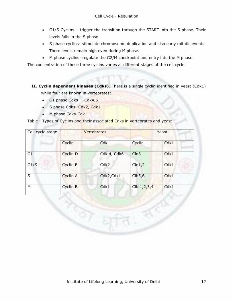
G1/S Cyclins – trigger the transition through the START into the S phase. Their
levels falls in the S phase.
S phase cyclins- stimulate chromosome duplication and also early mitotic events.
There levels remain high even during M phase.
M phase cyclins- regulate the G2/M checkpoint and entry into the M phase.
The concentration of these three cyclins varies at different stages of the cell cycle.
II. Cyclin dependent kinases (Cdks). There is a single cyclin identified in yeast (Cdk1)
while four are known in vertebrates:
G1 phase Cdks – Cdk4,6
S phase Cdks- Cdk2, Cdk1
M phase Cdks-Cdk1
Table : Types of Cyclins and their associated Cdks in vertebrates and yeast
Cell cycle stage Vertebrates Yeast
Cyclin Cdk Cyclin Cdk1
G1 Cyclin D Cdk 4, Cdk6 Cln3 Cdk1
G1/S Cyclin E Cdk2 Cln1,2 Cdk1
S Cyclin A Cdk2,Cdk1 Clb5,6 Cdk1
M Cyclin B Cdk1 Clb 1,2,3,4 Cdk1
Cell Cycle - Regulation
Institute of Lifelong Learning, University of Delhi 13
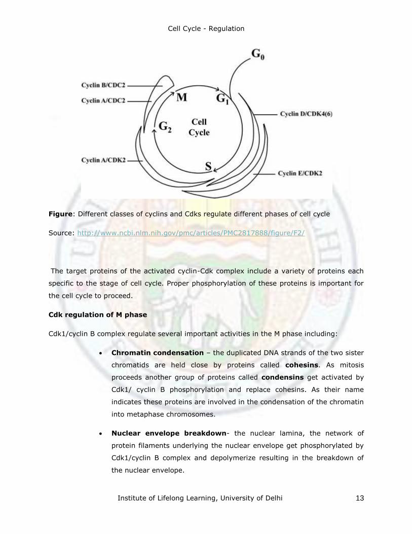
Figure: Different classes of cyclins and Cdks regulate different phases of cell cycle
Source: http://www.ncbi.nlm.nih.gov/pmc/articles/PMC2817888/figure/F2/
The target proteins of the activated cyclin-Cdk complex include a variety of proteins each
specific to the stage of cell cycle. Proper phosphorylation of these proteins is important for
the cell cycle to proceed.
Cdk regulation of M phase
Cdk1/cyclin B complex regulate several important activities in the M phase including:
Chromatin condensation – the duplicated DNA strands of the two sister
chromatids are held close by proteins called cohesins. As mitosis
proceeds another group of proteins called condensins get activated by
Cdk1/ cyclin B phosphorylation and replace cohesins. As their name
indicates these proteins are involved in the condensation of the chromatin
into metaphase chromosomes.
Nuclear envelope breakdown- the nuclear lamina, the network of
protein filaments underlying the nuclear envelope get phosphorylated by
Cdk1/cyclin B complex and depolymerize resulting in the breakdown of
the nuclear envelope.
Cell Cycle - Regulation
Institute of Lifelong Learning, University of Delhi 14
Breakdown of the golgi apparatus- The phosphorylation of the golgi
matrix proteins by the Cdk1/cyclin B complex results in the fragmentation
of the golgi apparatus into small vesicles.
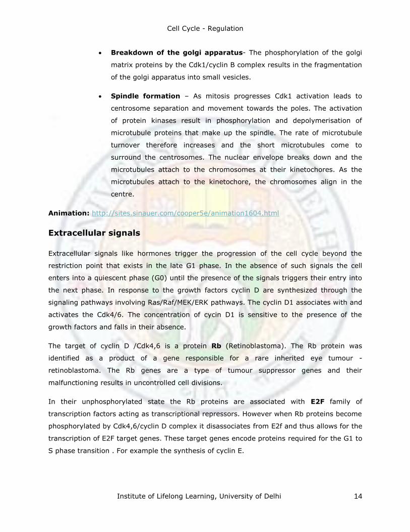
Spindle formation – As mitosis progresses Cdk1 activation leads to
centrosome separation and movement towards the poles. The activation
of protein kinases result in phosphorylation and depolymerisation of
microtubule proteins that make up the spindle. The rate of microtubule
turnover therefore increases and the short microtubules come to
surround the centrosomes. The nuclear envelope breaks down and the
microtubules attach to the chromosomes at their kinetochores. As the
microtubules attach to the kinetochore, the chromosomes align in the
centre.
Animation: http://sites.sinauer.com/cooper5e/animation1604.html
Extracellular signals
Extracellular signals like hormones trigger the progression of the cell cycle beyond the
restriction point that exists in the late G1 phase. In the absence of such signals the cell
enters into a quiescent phase (G0) until the presence of the signals triggers their entry into
the next phase. In response to the growth factors cyclin D are synthesized through the
signaling pathways involving Ras/Raf/MEK/ERK pathways. The cyclin D1 associates with and
activates the Cdk4/6. The concentration of cycin D1 is sensitive to the presence of the
growth factors and falls in their absence.
The target of cyclin D /Cdk4,6 is a protein Rb (Retinoblastoma). The Rb protein was
identified as a product of a gene responsible for a rare inherited eye tumour -
retinoblastoma. The Rb genes are a type of tumour suppressor genes and their
malfunctioning results in uncontrolled cell divisions.
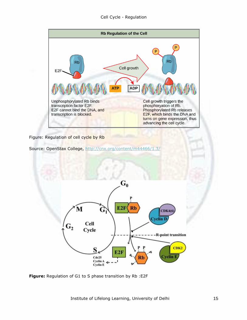
In their unphosphorylated state the Rb proteins are associated with E2F family of
transcription factors acting as transcriptional repressors. However when Rb proteins become
phosphorylated by Cdk4,6/cyclin D complex it disassociates from E2f and thus allows for the
transcription of E2F target genes. These target genes encode proteins required for the G1 to
S phase transition . For example the synthesis of cyclin E.
Cell Cycle - Regulation
Institute of Lifelong Learning, University of Delhi 15
Figure: Regulation of cell cycle by Rb
Source: OpenStax College, http://cnx.org/content/m44466/1.3/
Figure: Regulation of G1 to S phase transition by Rb :E2F
Cell Cycle - Regulation
Institute of Lifelong Learning, University of Delhi 16
Check point control
DNA damage induced checkpoints
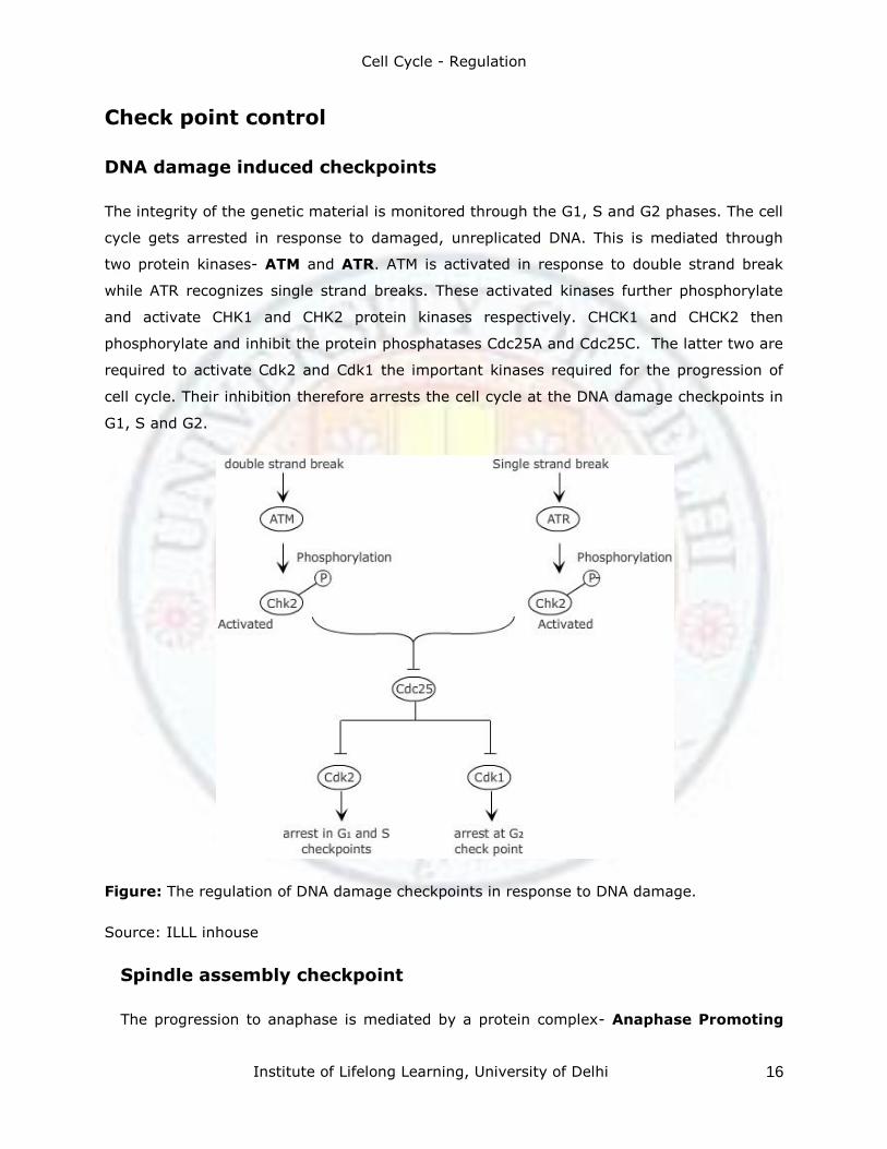
The integrity of the genetic material is monitored through the G1, S and G2 phases. The cell
cycle gets arrested in response to damaged, unreplicated DNA. This is mediated through
two protein kinases- ATM and ATR. ATM is activated in response to double strand break
while ATR recognizes single strand breaks. These activated kinases further phosphorylate
and activate CHK1 and CHK2 protein kinases respectively. CHCK1 and CHCK2 then
phosphorylate and inhibit the protein phosphatases Cdc25A and Cdc25C. The latter two are
required to activate Cdk2 and Cdk1 the important kinases required for the progression of
cell cycle. Their inhibition therefore arrests the cell cycle at the DNA damage checkpoints in
G1, S and G2.
Figure: The regulation of DNA damage checkpoints in response to DNA damage.
Source: ILLL inhouse
Spindle assembly checkpoint
The progression to anaphase is mediated by a protein complex- Anaphase Promoting
Cell Cycle - Regulation
Institute of Lifelong Learning, University of Delhi 17
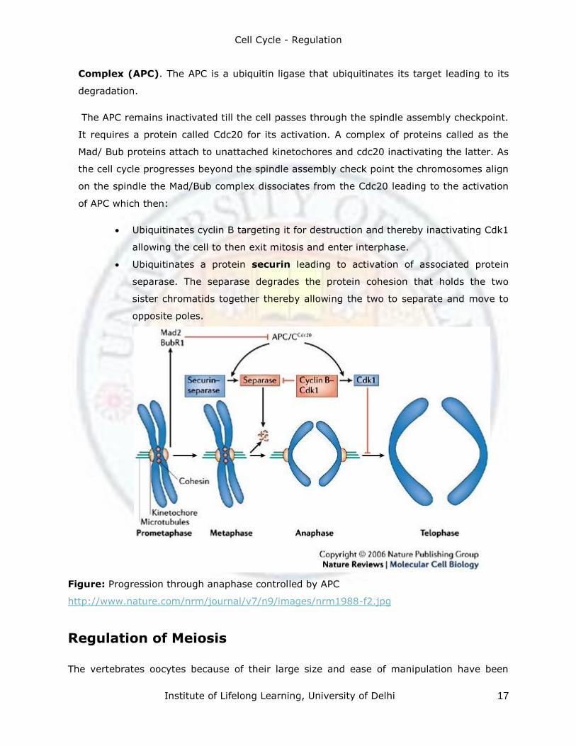
Complex (APC). The APC is a ubiquitin ligase that ubiquitinates its target leading to its
degradation.
The APC remains inactivated till the cell passes through the spindle assembly checkpoint.
It requires a protein called Cdc20 for its activation. A complex of proteins called as the
Mad/ Bub proteins attach to unattached kinetochores and cdc20 inactivating the latter. As
the cell cycle progresses beyond the spindle assembly check point the chromosomes align
on the spindle the Mad/Bub complex dissociates from the Cdc20 leading to the activation
of APC which then:
Ubiquitinates cyclin B targeting it for destruction and thereby inactivating Cdk1
allowing the cell to then exit mitosis and enter interphase.
Ubiquitinates a protein securin leading to activation of associated protein
separase. The separase degrades the protein cohesion that holds the two
sister chromatids together thereby allowing the two to separate and move to
opposite poles.
Figure: Progression through anaphase controlled by APC
http://www.nature.com/nrm/journal/v7/n9/images/nrm1988-f2.jpg
Regulation of Meiosis
The vertebrates oocytes because of their large size and ease of manipulation have been
Cell Cycle - Regulation
Institute of Lifelong Learning, University of Delhi 18
used as model systems to investigate meiosis.
The oocytes remain arrested for a long period in the diplotene stage where active DNA
synthesis takes place. In response to hormonal stimulation the cells proceed through and
enter meiosis I. This division is asymmetric resulting in a small polar body and a large cell-
oocyte. This involves the activation of Cdk1 and similar accompanying cellular changes as
seen in mitosis. The oocyte then arrests again in meiosis II at the metaphase II stage and
proceeds further upon hormonal stimulation again.
Interestingly the levels of Cdk1 gets reduced but not completely as the oocyte proceeds
from meiosis I to meiosis II with the chromatin remaining condensed and the nuclear
envelope remaining disintegrated. In 1971 in a experiment carried out by Yoshio Masui
and Clement Markert it was seen that the upon injection of cytoplasm of an oocyte
arrested in meiosis II into a early embryonic cell undergoing mitosis, arrested the
divisions in the embryonic cell. This indicated the presence of a cytostatic factor in the
cytoplasm since it could arrest a dividing cell. Recent experiments have shown that a
component of the cytostatic factor to be a protein serine/threonine kinase Mos.
Mos maintains the Cdk1/cyclin B activity in two ways:
Stimulating cyclin B synthesis
Inhibiting the degradation of cyclin B by APC
The levels of Cdk1/cyclin B therefore remains high arresting the progression of the cell
cycle. The arrested oocyte would then remain so till fertilization.
Fertilization
Upon fertilization of the oocyte several important changes are seen:
The Ca+2 level increase in the cytoplasm due to the hydrolysis of
phosphatidylinositol4,5-bisphosphate (PIP2).
The raised Ca+2 levels prevent other sperms from entering the egg due to
alterations in the wall.
The arrested oocyte resumes the cell cycle and proceeds beyond metaphase II.
This involves the Ca+2 dependent activation of the APC.
The oocyte meiosis completes and the fertilized egg now contains two haploid
nuclei. Each of these nuclei replicate their DNA and the zygote enters into the M
phase producing two diploid cells that divide further to give rise to the
Cell Cycle - Regulation
Institute of Lifelong Learning, University of Delhi 19
multicelled embryo.
Summary
The cell cycle is divided into interphase (G1, S, G2) and the divisional M phase. There are
several checkpoints that regulate the transition from one phase to the other. The key
component of the control system that coordinates the progress of the cell cycle are the
cyclin dependent protein kinases. The activity of Cdks is regulated primarily by their
association with proteins-cyclins. The concentration of cyclin fluctuates during the various
phases of cell cycle while that of Cdks remains constant. When the concentration of cyclin is
low, it is not associated with Cdk and therefore the latter is inactive. Phosphorylation and
association with inhibitors (CkIs) are some of the mechanisms that control the activity of
Cdks.
Extracellular signals like hormones stimulate the synthesis of D type cyclins and the
activation of Cdk4/6 that control the movement of the cell cycle beyond the G1/S
checkpoint. The transition to metaphase is mediated by Cdk1/cyclin B complex that
phosphorylates and activates proteins involved in the M phase. These include the proteins
involved in breakdown of the nuclear membrane, condensation of chromatin, spindle
assembly etc.
The progression into anaphase is regulated by the spindle assembly checkpoint. The
assembly and the alignment of the chromosomes triggers the anaphase promoting complex.
This promotes the ubiqutination and degradation of proteins like cohesions that results in
separation of the sister chromatids and also the degradation of cyclinB promoting to
transition to anaphase.
Glossary
Anaphase: Stage during mitosis during which the sister chromatids separate and move
towards the poles. Composed of Anaphase A – chromosomes move towards the spindle
poles and Anaphase B- Spindle poles move apart.
APC: Anaphase promoting complex which is a ubiquitin ligase that catalyzes the
ubiquitylation and destruction of proteins –securin and M and S cyclins resulting in the
separation of chromatids and transition from metaphase to anaphase.
Cdk: Cyclin dependent protein kinases. These trigger the progression of cel cycle by
Cell Cycle - Regulation
Institute of Lifelong Learning, University of Delhi 20
phosphorylating their target molecules.
Cell cycle: Reproductive cycle of a cell involving the orderly sequence of events that
includes the duplication of the genetic material and division into two cells.
Checkpoints: the mechanism that controls the progression of the cell cycle in response to
(1) availability of growth hormones/ appropriate size (2) DNA damage (3) proper spindle
assembly etc.
Chiasma (plural Chiasmata) : The X shaped connections between the paired homologous
chromosomes during the prophase stage of meiosis. These represent the sites where
crossing over (genetic recombination has occurred).
Cdk: Cyclin dependent protein kinases which are active when associated with cyclins. These
trigger the different steps of the cell cycle by phosphorylating their targets.
CKI: Cdk inhibitor proteins that bind to and inhibit the Cyclin-Cdk complexes involved in the
control of G1 and S phase.
Condensins: Complex of proteins involved in condensation of chromosomes on the onset
of mitosis.
Cyclin: a family of enzymes which vary in their concentrations in particular phase of cell
cycle. Cyclins activate important protein kinases called as cyclin dependent protein kinases
that trigger the progression of the cell cycle.
E2F protein: Gene regulatory protein that switches on many genes that encode proteins
required for entry into the S phase of the cell cycle.
Kinases: Enzymes that phosphorylate their targets.
MPF: Maturation promoting complex. Has two components Cdk1/Cyclin B and controls the
G2 and M checkpoint.
PIP2: Phosphatidyl inositol phosphate.
Protein Kinase: Enzyme that transfers the terminal phosphate group of ATP to one or
more specific amino acids for e.g. serine, threonine or tyrosine of a target protein.
Retinoblastoma protein (Rb): Tumour suppressor protein involved in the regulation of
cell cycle. Its main function is to bind and inhibit the activity of E2F genes thus binding
blocking the progression of DNA replication and cell cycle. Rb protein is mutated in the
retinoblastoma cancer.
Ubiquitin: Small conserved eukaryotic protein that becomes covalently attached to lysines
of other proteins and acts as a tag for intracellular proteolytic destruction.
Cell Cycle - Regulation
Institute of Lifelong Learning, University of Delhi 21
Ubiquitin ligase: Enzymes that catalyze the addition of ubiquitin molecules to a protein
targeting them for destructution.
Exercises
1. Identify the different stages of cell cycle.
2. What is the difference between the cells in G0 and G1 phases?
3. What is the importance of DNA replication in cell cycle.
4. What is the effect on fusing cells :
In G1 phase with one in S phase
In G2 phase with one in M phase.
5. Briefly mention the four mechanisms which regulate Cdk activity.
6. Discuss the role of Anaphase Promoting Complex in separation of sister chromatids
during anaphase.
References
1. Campbell, N.A. and Reece, J. B. (2008) Biology 8th edition, Pearson Benjamin
Cummings, San Francisco.
2. Raven, P.H et al (2006) Biology 7th edition Tata McGrawHill Publications, New Delhi
3. Karp, G. 2010 Cell and Molecular Biology: Concepts and Experiments. 6th edition. John
Wiley & Sons. Inc.
4. De Robertis, E.D.P. and De Robertis, E.M.F. 2006 Cell and Molecular Biology. 8th edition.
5. Cooper, G.M. and Hausman, R.E. 2009 The Cell: A Molecular Approach. 5th edition. ASM
Lippincott Williams and Wilkins, Philadelphia.Press & Sunderland, Washington, D.C.; Sinauer
Associates, MA.
6. Becker, W.M., Kleinsmith, L.J., Hardin. J. and Bertoni, G. P. 2009 The World of the Cell.
7th edition. Pearson Benjamin Cummings Publishing, San Francisco.
Suggested Readings
1. Enders,G.H. (2010) Cell Division 5:12.Gauchos and ochos: a Wee1-Cdk tango regulating
mitotic entry.
2. Yang et al. 2006. Cell Division 1: 32 Variations in cyclin D1 levels through the cell cycle
determine the proliferative fate of a cell.
Web Links
1. http://www.nature.com/scitable/ebooks/essentials-of-cell-biology-
14749010/14759908
Cell Cycle - Regulation
Institute of Lifelong Learning, University of Delhi 22
2. http://www.sciencedirect.com/science/article/pii/S1357272596001781#
3. http://www.google.co.in/url?sa=t&rct=j&q=&esrc=s&source=web&cd=9&cad=rja&ved=0
CG8QFjAI&url=http%3A%2F%2Fbiology.buffalo.edu%2Fcourses%2Fbio404%2Fberezn
ey%2Farticles%2FLodish_Chapter_13_p495_533.pdf&ei=PXVnUp7YBsWRiQeo5oD4B
g&usg=AFQjCNGP_YL30vp98ksnPCzdvKwe_yLK1A