Embed Size (px)
Citation preview
MD0961 3-1
LESSON ASSIGNMENT LESSON 3 Positioning Terminology. LESSON ASSIGNMENT Paragraphs 3-1 through 3-23. LESSON OBJECTIVES After completing this lesson, you should be able to: 3-1. Identify body part terminology. 3-2. Identify definitions of the anteroposterior (AP), posteroanterior (PA), lateral, and oblique views. 3-3. Identify position names based the point of entry of the central ray (CR). 3-4. Identify definitions of the supine, prone recumbent, lateral, and decubitus positions. 3-5. Identify definitions of suspended breathing,
suspended inspiration, suspended expiration, and normal breathing.
SUGGESTION After reading and studying the assignment, complete the exercises at the end of this lesson. These exercises will help you to achieve the lesson objectives.
MD0961 3-2
LESSON 3
POSITIONING TERMINOLOGY
Section I. GENERAL TERMS AND BODY PART TERMS 3-1. INTRODUCTION a. The Importance of Terms. In order to function effectively as a radiologic technologist, you must have a working knowledge of the terms commonly used in performing radiographic examinations. If your knowledge of these terms is inaccurate or incomplete, you may well end up positioning the patient for the wrong view. You might conceivably perform an incomplete examination or produce a blurry image. Thus, you would fail to provide the radiologist with the radiographic information needed to make a prompt and accurate diagnosis. You would also run the risk of exposing the patient to unnecessary repeat exposures. This lesson is intended to present the terms that you will need to know and use in positioning your patient for radiographs. Where possible, the terms are used in context and an illustration is provided to help you master the material and provide greater clarity. b. Where Terms are Introduced. As you may have noticed, by now, this is not the only lesson in which new terms have been introduced. Other terms have been presented, in context, where they are logically needed to present a concept. Learning to use the terms in context is, in fact, a better way to learn new terms than by simply memorizing definitions in isolation. The terms covered in this lesson are a catch-all for remaining terms that have not cropped up in the two previous lessons. As for the previous lessons, new terms and their definitions are separated out of the text visually (boxed) for ease of reference. In addition, all the new terms in the subcourse are compiled in a glossary presented in the Appendix to facilitate subsequent review and mastery. 3-2. THE TERMS, X-RAY FILM AND RADIOGRAPH a. X-ray Film. The terms "X-ray film" and "radiograph" are quite often used interchangeably. But, in fact, they have distinct and separate meanings. The X-ray film refers more specifically to the physical piece of material on which the radiograph is taken. X-ray film: emulsion composed of silver bromide crystals suspended in a gelatin substance and spread evenly upon a transparent, blue-tinted, polyester support base. b. Radiograph. The radiograph, on the other hand, refers to the X-ray film once it has an image on it. In other words, the end result used by the radiologist for diagnosis of the patient's condition.

MD0961 3-3
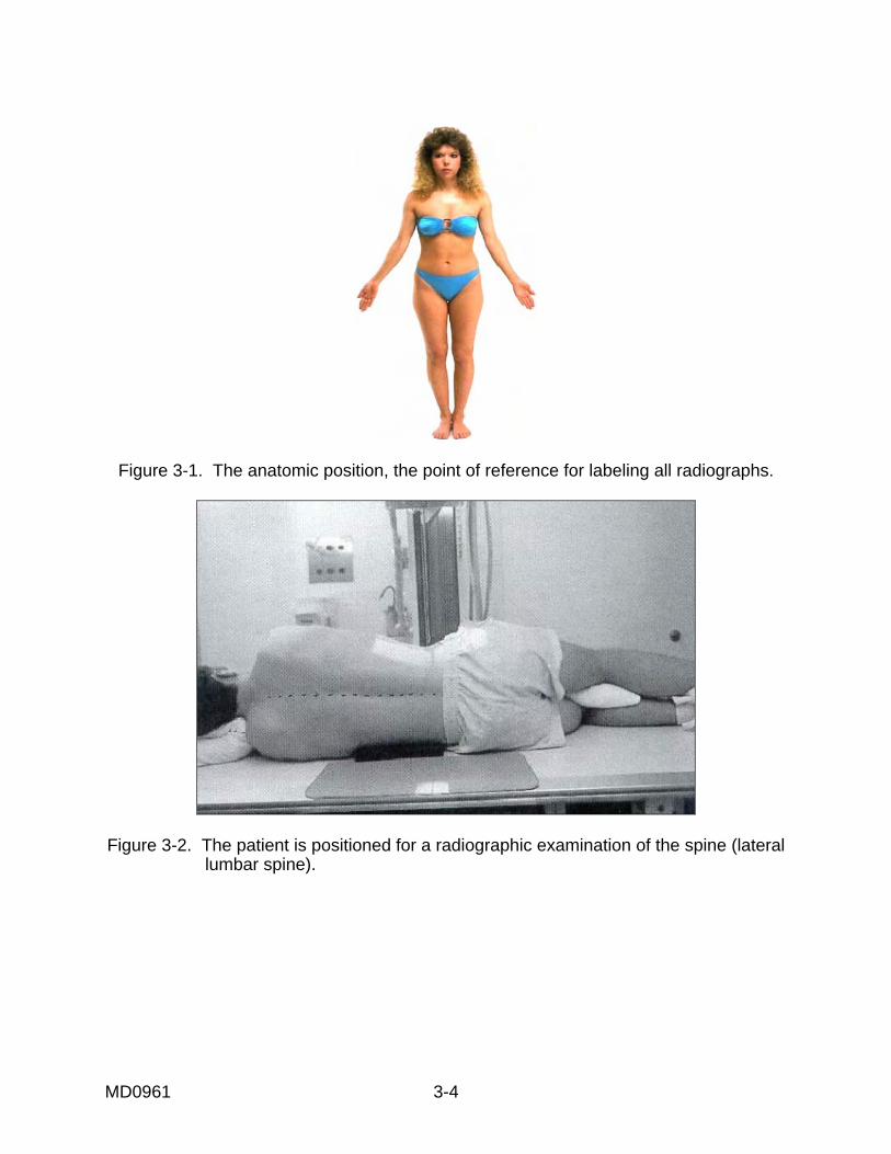
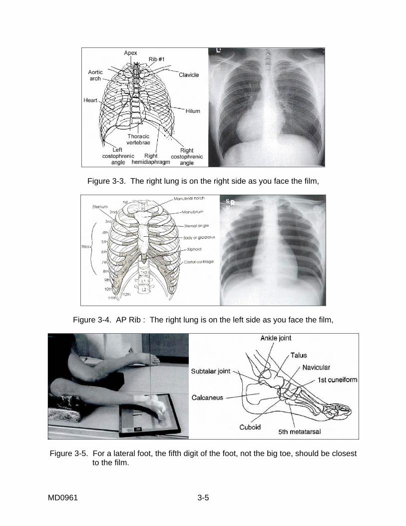
Radiograph: an X-ray film containing an image of an anatomical structure. 3-3. RADIOGRAPHIC EXAMINATION A radiographic examination involves everything you do so as to produce the desired end-product--a diagnostic radiograph. This includes positioning the anatomical structure, making the exposure, having the film processed, and so forth radiographic exam: encompasses positioning the body part, exposing the film, and processing the film. 3-4. ANATOMIC POSITION The anatomic position is a well established point of reference for describing the relationship of one body part to another. In the anatomic position, the patient is upright, facing straight ahead, body erect, feet together, arms at sides, and palms turned forward (figure 3-1). No matter what the actual position of the patient, such as figure 3-2, one must think in terms of the person standing erect in the anatomic position even when describing parts of a patient who is lying down. If you fail to think in terms of the anatomic position, you may incorrectly describe or label a radiograph. Consider figure 3-3, a posteroanterior (PA) chest in which both lungs are demonstrated. Because it is a PA projection, the right side is on the right side of the film. Thinking in terms of the anatomical position will orient you correctly. In an AP position, the right lung would be demonstrated on the left side of the film (figure 3-4). Thinking in terms of the anatomic position is especially important for properly placing the patient for positions involving the extremities, such as the foot or hand. Suppose you want to take a lateral view of the foot (figure 3-5). To do this, you must first determine which side to place closest to the film. For a lateral foot, the side to be placed closest to the film would correspond with the fifth digit of the foot, not the big toe.
MD0961 3-4
Figure 3-1. The anatomic position, the point of reference for labeling all radiographs.
Figure 3-2. The patient is positioned for a radiographic examination of the spine (lateral
lumbar spine).
MD0961 3-5
Figure 3-3. The right lung is on the right side as you face the film,
Figure 3-4. AP Rib : The right lung is on the left side as you face the film,
Figure 3-5. For a lateral foot, the fifth digit of the foot, not the big toe, should be closest to the film.
MD0961 3-6
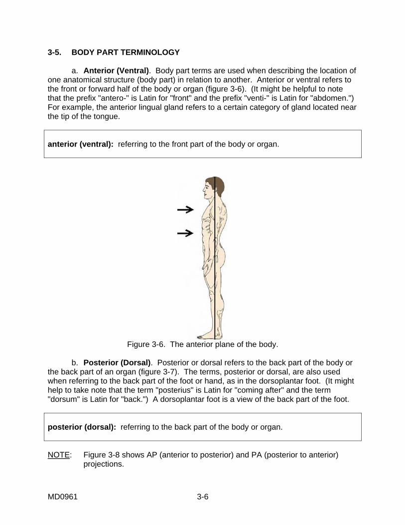
3-5. BODY PART TERMINOLOGY a. Anterior (Ventral). Body part terms are used when describing the location of one anatomical structure (body part) in relation to another. Anterior or ventral refers to the front or forward half of the body or organ (figure 3-6). (It might be helpful to note that the prefix "antero-" is Latin for "front" and the prefix "venti-" is Latin for "abdomen.") For example, the anterior lingual gland refers to a certain category of gland located near the tip of the tongue. anterior (ventral): referring to the front part of the body or organ.
Figure 3-6. The anterior plane of the body.
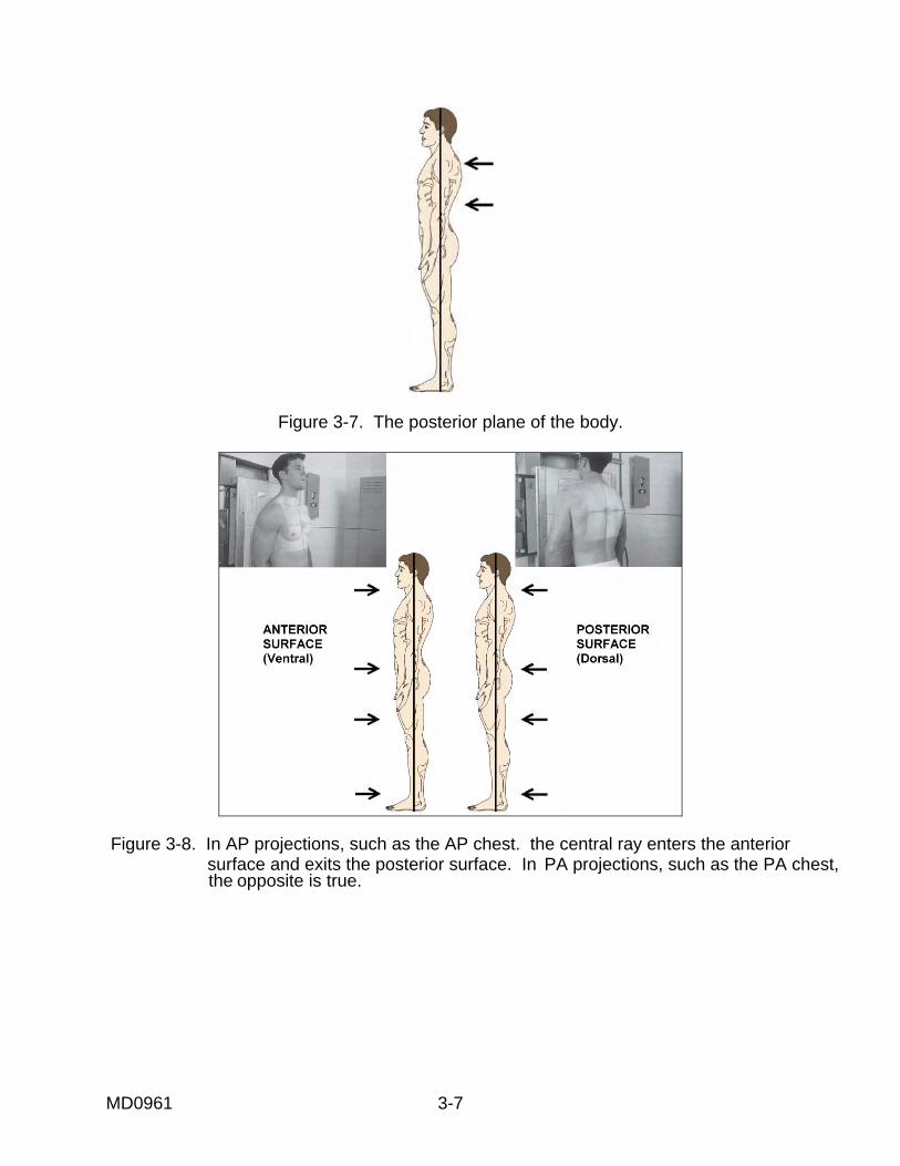
b. Posterior (Dorsal). Posterior or dorsal refers to the back part of the body or
the back part of an organ (figure 3-7). The terms, posterior or dorsal, are also used when referring to the back part of the foot or hand, as in the dorsoplantar foot. (It might help to take note that the term "posterius" is Latin for "coming after" and the term "dorsum" is Latin for "back.") A dorsoplantar foot is a view of the back part of the foot. posterior (dorsal): referring to the back part of the body or organ. NOTE: Figure 3-8 shows AP (anterior to posterior) and PA (posterior to anterior) projections.
MD0961 3-7
Figure 3-7. The posterior plane of the body.
Figure 3-8. In AP projections, such as the AP chest. the central ray enters the anterior surface and exits the posterior surface. In PA projections, such as the PA chest,
the opposite is true.
MD0961 3-8
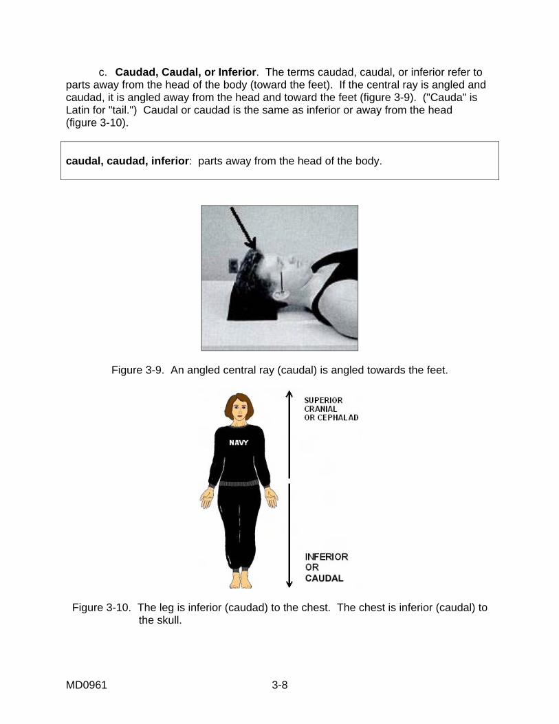
c. Caudad, Caudal, or Inferior. The terms caudad, caudal, or inferior refer to parts away from the head of the body (toward the feet). If the central ray is angled and caudad, it is angled away from the head and toward the feet (figure 3-9). ("Cauda" is Latin for "tail.") Caudal or caudad is the same as inferior or away from the head (figure 3-10). caudal, caudad, inferior: parts away from the head of the body.
Figure 3-9. An angled central ray (caudal) is angled towards the feet.
Figure 3-10. The leg is inferior (caudad) to the chest. The chest is inferior (caudal) to the skull.
MD0961 3-9
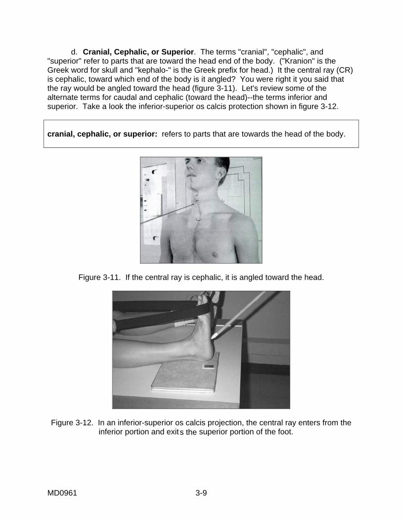
d. Cranial, Cephalic, or Superior. The terms "cranial", "cephalic", and "superior" refer to parts that are toward the head end of the body. ("Kranion" is the Greek word for skull and "kephalo-" is the Greek prefix for head.) It the central ray (CR) is cephalic, toward which end of the body is it angled? You were right it you said that the ray would be angled toward the head (figure 3-11). Let's review some of the alternate terms for caudal and cephalic (toward the head)--the terms inferior and superior. Take a look the inferior-superior os calcis protection shown in figure 3-12. cranial, cephalic, or superior: refers to parts that are towards the head of the body.
Figure 3-11. If the central ray is cephalic, it is angled toward the head.
Figure 3-12. In an inferior-superior os calcis projection, the central ray enters from the
inferior portion and exits the superior portion of the foot.
MD0961 3-10
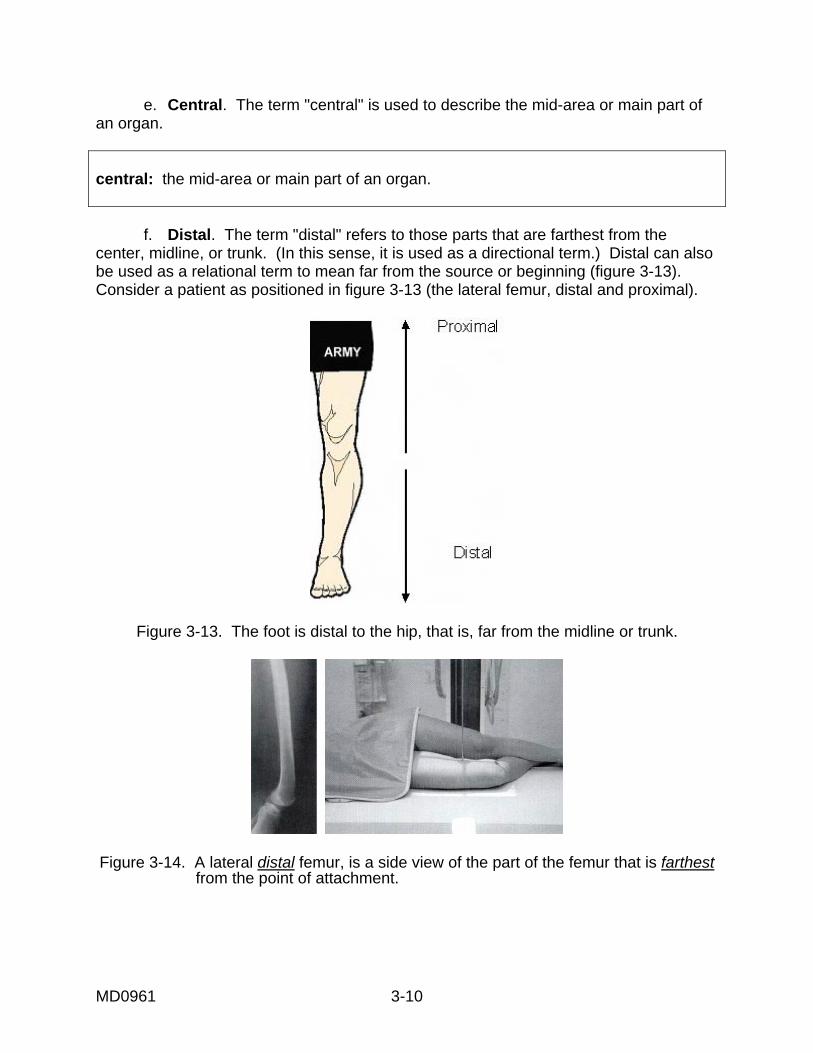
e. Central. The term "central" is used to describe the mid-area or main part of an organ. central: the mid-area or main part of an organ. f. Distal. The term "distal" refers to those parts that are farthest from the center, midline, or trunk. (In this sense, it is used as a directional term.) Distal can also be used as a relational term to mean far from the source or beginning (figure 3-13). Consider a patient as positioned in figure 3-13 (the lateral femur, distal and proximal).
Figure 3-13. The foot is distal to the hip, that is, far from the midline or trunk.
Figure 3-14. A lateral distal femur, is a side view of the part of the femur that is farthest
from the point of attachment.
MD0961 3-11
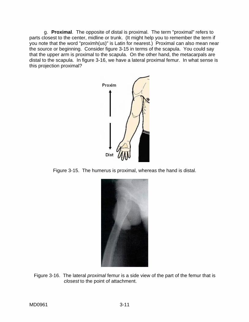
g. Proximal. The opposite of distal is proximal. The term "proximal" refers to parts closest to the center, midline or trunk. (It might help you to remember the term if you note that the word "proximh(us)" is Latin for nearest.) Proximal can also mean near the source or beginning. Consider figure 3-15 in terms of the scapula. You could say that the upper arm is proximal to the scapula. On the other hand, the metacarpals are distal to the scapula. In figure 3-16, we have a lateral proximal femur. In what sense is this projection proximal?
Figure 3-15. The humerus is proximal, whereas the hand is distal.
Figure 3-16. The lateral proximal femur is a side view of the part of the femur that is closest to the point of attachment.
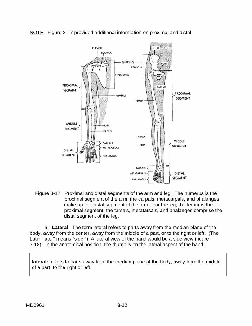
NOTE: Figure 3-17 provided additional information on proximal and distal.
Figure 3-17. Proximal and distal segments of the arm and leg. The humerus is the proximal segment of the arm; the carpals, metacarpals, and phalanges
make up the distal segment of the arm. For the leg, the femur is the proximal segment; the tarsals, metatarsals, and phalanges comprise the distal segment of the leg.
h. Lateral. The term lateral refers to parts away from the median plane of the body, away from the center, away from the middle of a part, or to the right or left. (The Latin "later" means "side.") A lateral view of the hand would be a side view (figure 3-18). In the anatomical position, the thumb is on the lateral aspect of the hand.
lateral: refers to parts away from the median plane of the body, away from the middle of a part, to the right or left.
MD0961 3-12
MD0961 3-13
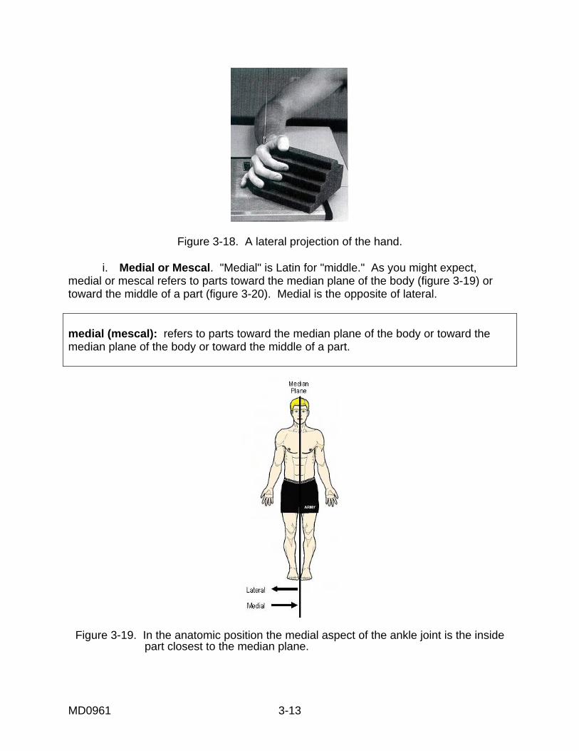
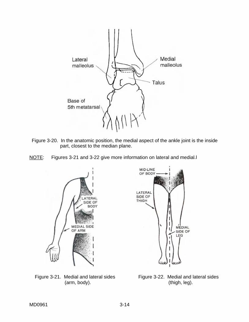
Figure 3-18. A lateral projection of the hand. i. Medial or Mescal. "Medial" is Latin for "middle." As you might expect, medial or mescal refers to parts toward the median plane of the body (figure 3-19) or toward the middle of a part (figure 3-20). Medial is the opposite of lateral. medial (mescal): refers to parts toward the median plane of the body or toward the median plane of the body or toward the middle of a part.
Figure 3-19. In the anatomic position the medial aspect of the ankle joint is the inside part closest to the median plane.
MD0961 3-14
Figure 3-20. In the anatomic position, the medial aspect of the ankle joint is the inside
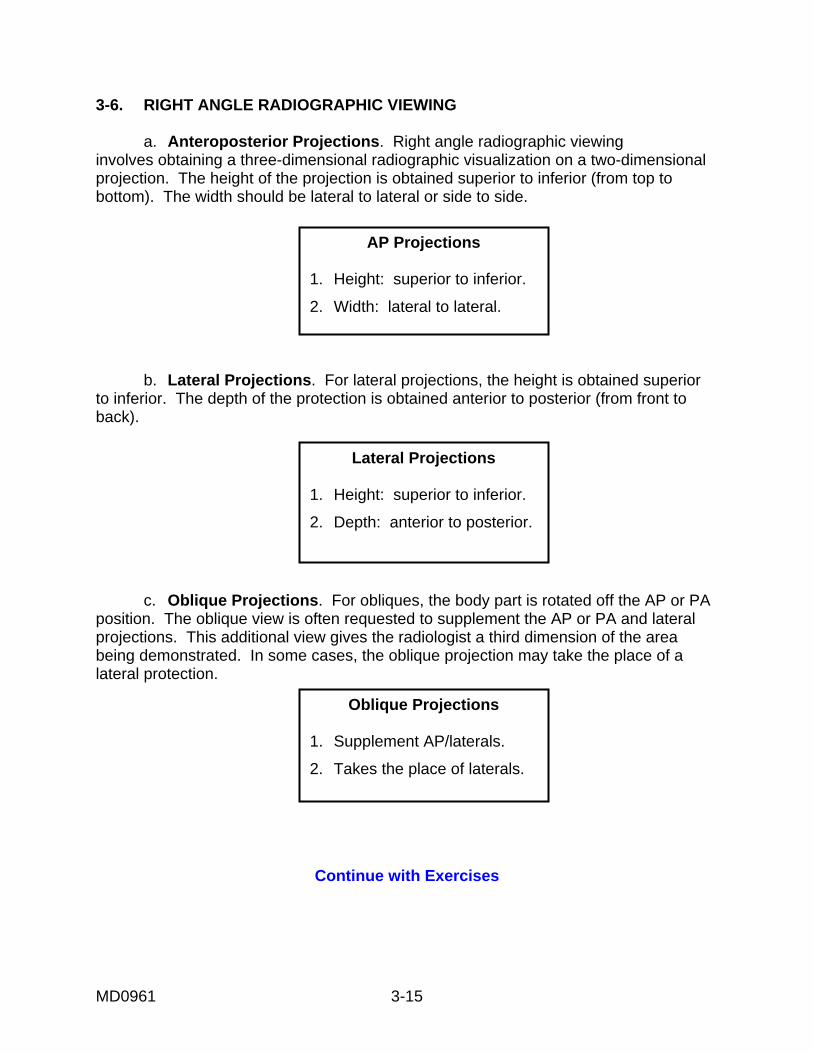
part, closest to the median plane. NOTE: Figures 3-21 and 3-22 give more information on lateral and medial.l
Figure 3-21. Medial and lateral sides Figure 3-22. Medial and lateral sides (arm, body). (thigh, leg).
MD0961 3-15
AP Projections
1. Height: superior to inferior.
2. Width: lateral to lateral.
Lateral Projections
1. Height: superior to inferior.
2. Depth: anterior to posterior.
Oblique Projections
1. Supplement AP/laterals.
2. Takes the place of laterals.
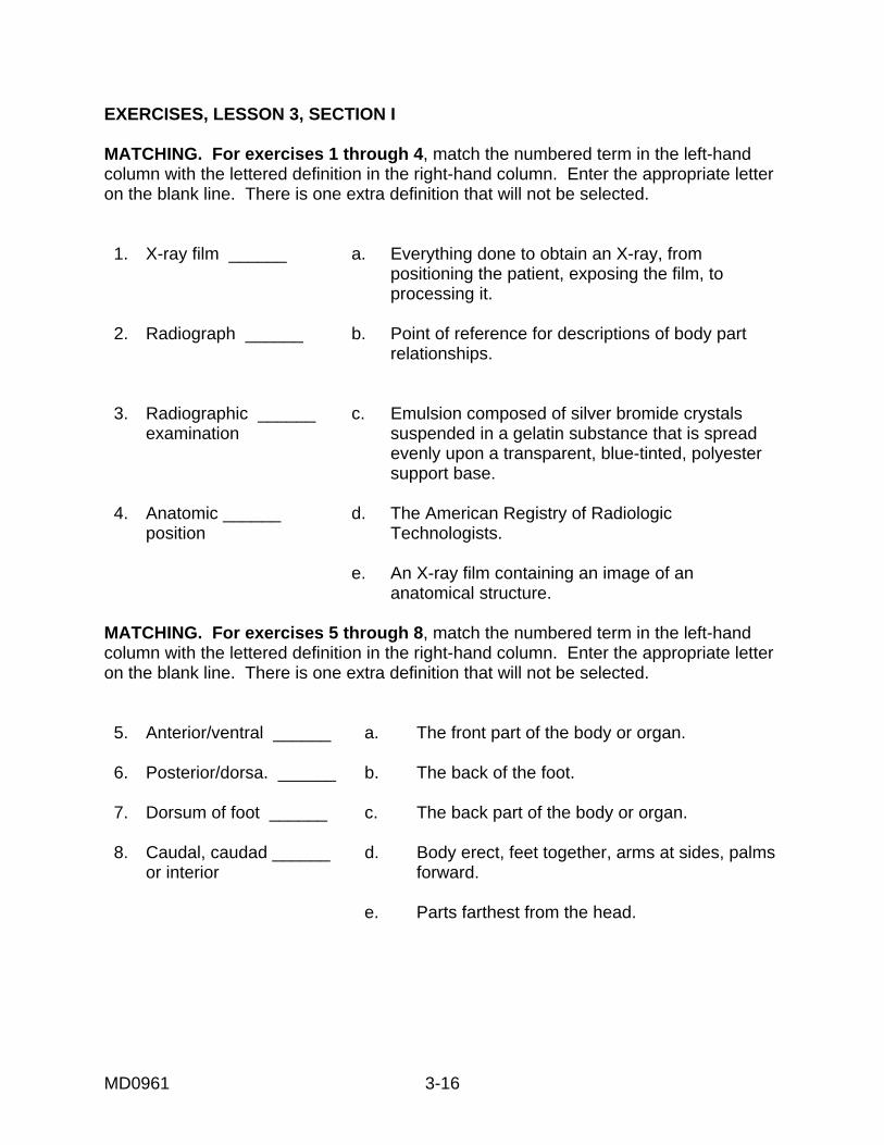
3-6. RIGHT ANGLE RADIOGRAPHIC VIEWING a. Anteroposterior Projections. Right angle radiographic viewing involves obtaining a three-dimensional radiographic visualization on a two-dimensional projection. The height of the projection is obtained superior to inferior (from top to bottom). The width should be lateral to lateral or side to side.
b. Lateral Projections. For lateral projections, the height is obtained superior to inferior. The depth of the protection is obtained anterior to posterior (from front to back). c. Oblique Projections. For obliques, the body part is rotated off the AP or PA position. The oblique view is often requested to supplement the AP or PA and lateral projections. This additional view gives the radiologist a third dimension of the area being demonstrated. In some cases, the oblique projection may take the place of a lateral protection.
Continue with Exercises
MD0961 3-16
EXERCISES, LESSON 3, SECTION I MATCHING. For exercises 1 through 4, match the numbered term in the left-hand column with the lettered definition in the right-hand column. Enter the appropriate letter on the blank line. There is one extra definition that will not be selected. 1. X-ray film ______ a. Everything done to obtain an X-ray, from positioning the patient, exposing the film, to processing it. 2. Radiograph ______ b. Point of reference for descriptions of body part relationships. 3. Radiographic ______ c. Emulsion composed of silver bromide crystals examination suspended in a gelatin substance that is spread evenly upon a transparent, blue-tinted, polyester support base. 4. Anatomic ______ d. The American Registry of Radiologic position Technologists. e. An X-ray film containing an image of an anatomical structure. MATCHING. For exercises 5 through 8, match the numbered term in the left-hand column with the lettered definition in the right-hand column. Enter the appropriate letter on the blank line. There is one extra definition that will not be selected. 5. Anterior/ventral ______ a. The front part of the body or organ. 6. Posterior/dorsa. ______ b. The back of the foot. 7. Dorsum of foot ______ c. The back part of the body or organ. 8. Caudal, caudad ______ d. Body erect, feet together, arms at sides, palms or interior forward. e. Parts farthest from the head.
MD0961 3-17
MATCHING. For exercises 9 through 14, match the numbered term in the left-hand column with the lettered definition in the right-hand column. Enter the appropriate letter on the blank line. There is one extra definition that will not be selected. 9. Cranial, cephalic ______ a. Nearest the center, midline, or trunk. or superior 10. Proximal ______ b. Toward the head. 11. Lateral ______ c. Mid-area or main part of an organ. 12. Medial/mesial ______ d. Away from the head. 13. Distal ______ e. Away from the median plane or middle part: to the right or left. 14. Central ______ f. Toward the median plane; toward the middle of a part. g. Farthest from the center, midline, or trunk. IDENTIFICATION, For exercises 15 through 29, correctly label the figures above the question by entering the appropriate word(s) in the space provided.
15. This is known as the _________________ position.
16. This is known as the____________________ or ____________________ surface.
MD0961 3-18
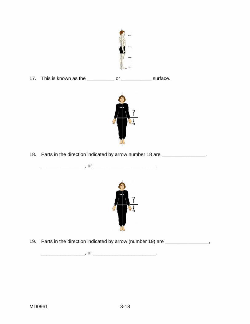
17. This is known as the __________ or ___________ surface.
18. Parts in the direction indicated by arrow number 18 are ________________, ________________, or _______________________.
19. Parts in the direction indicated by arrow (number 19) are ________________, ________________, or _______________________.
MD0961 3-19
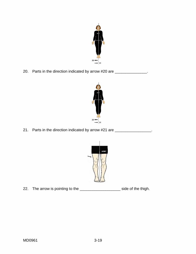
20. Parts in the direction indicated by arrow #20 are _______________.
21. Parts in the direction indicated by arrow #21 are _________________.
22. The arrow is pointing to the ___________________ side of the thigh.
MD0961 3-20
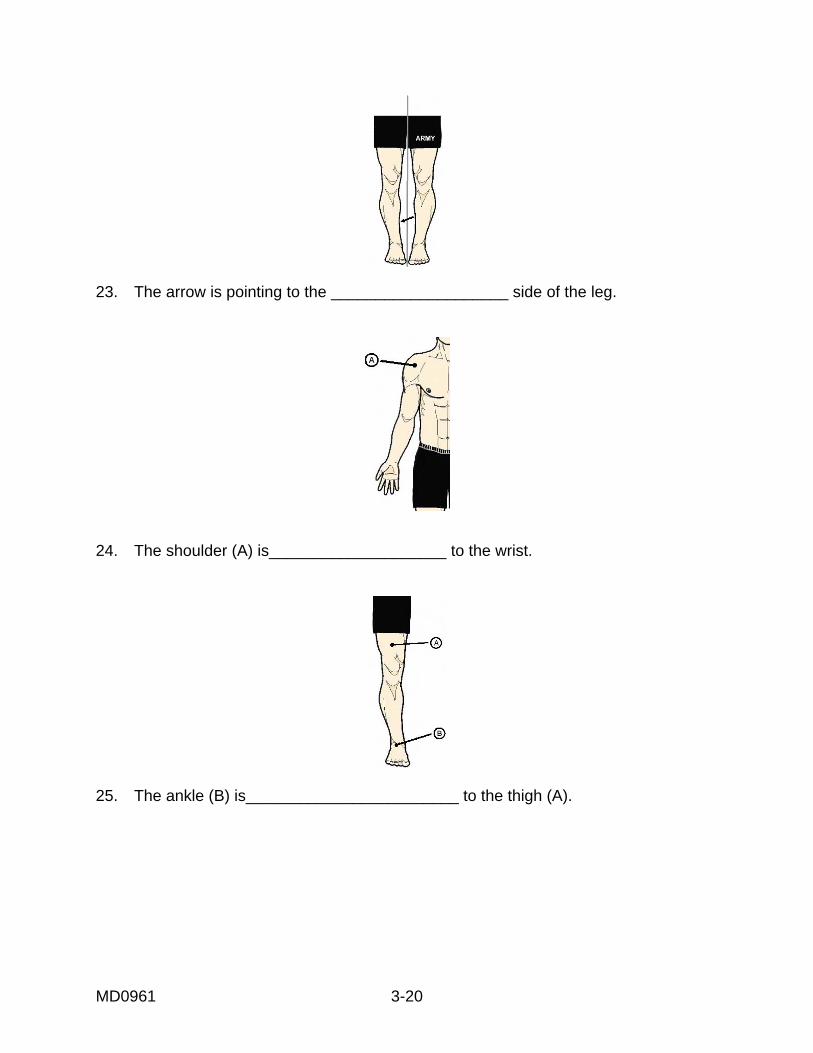
23. The arrow is pointing to the ____________________ side of the leg.
24. The shoulder (A) is____________________ to the wrist.
25. The ankle (B) is________________________ to the thigh (A).
MD0961 3-21
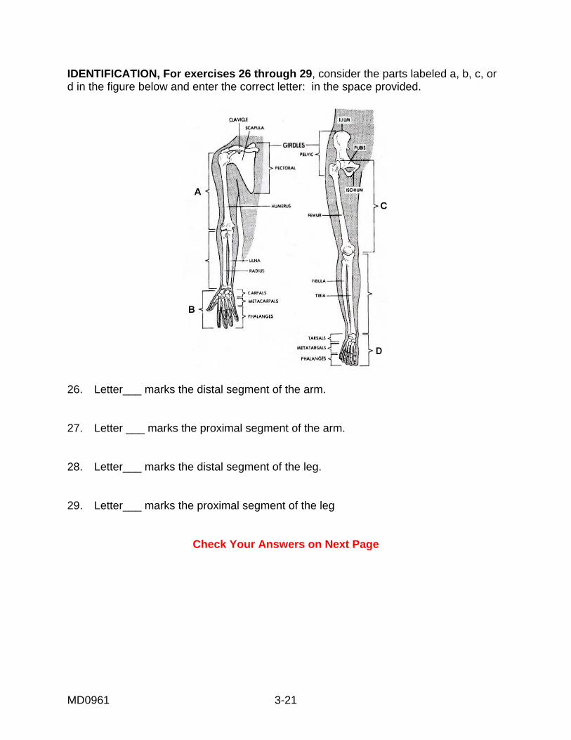
IDENTIFICATION, For exercises 26 through 29, consider the parts labeled a, b, c, or d in the figure below and enter the correct letter: in the space provided.
26. Letter___ marks the distal segment of the arm. 27. Letter ___ marks the proximal segment of the arm. 28. Letter___ marks the distal segment of the leg. 29. Letter___ marks the proximal segment of the leg
Check Your Answers on Next Page
MD0961 3-22
SOLUTIONS, LESSON 3, SECTION I Be sure to re-read and study the paragraph(s) pertaining to any exercises you might have answered incorrectly. The relevant paragraph(s) is (are) listed after each of the answers below. 1. c (para 3-2a) 2. e (para 3-2b) 3. a (para 3-3) 4. b (para 3-4) 5. a (para 3-5a0 6. c (para 3-5b) 7. b (para 3-5b) 8. e (para 3-5c 9. b (para 3-5d) 10. a (para 3-5g) 11. e (para 3-5h) 12. f (para 3-5i) 13. g (para 3-5f) 14. c (para 3-5e) 15. Anatomic (figure 3-1, para 3-4) 16. Posterior or dorsal (figure 3-8, para 3-5b) 17. Anterior or ventral (figure 3-8, para 3-5a) 18. Superior, cranial, or cephalic (figure 3-10, para 3-5d) 19. Inferior, caudad, or caudal (figure 3-10, para 3-5c) 20. Lateral (figure 3-22) 21. Medial (figure 3-22) 22. Lateral (figure 3-22) 23. Medial (figure 3-22) 24. Proximal (figure 3-15) 25. Distal (figure 3-13) 26. B (figure 3-17) 27. A (figure 3-17) 28. D (figure 3-17) 29. C (figure 3-17)
MD0961 3-23
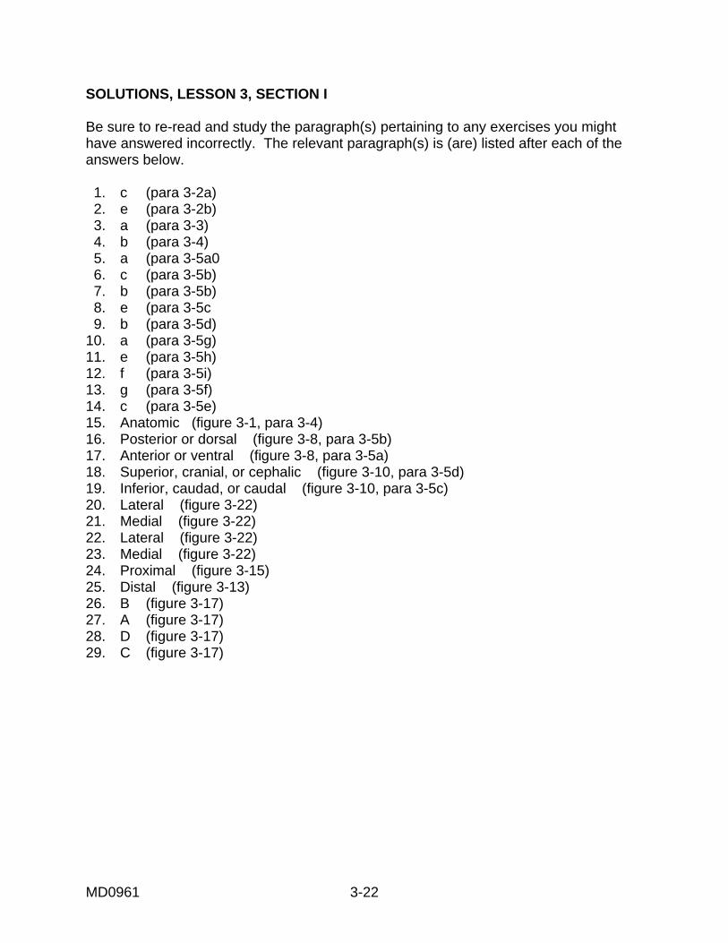
Section II. POSITIONING AND PROJECTION TERMINOLOGY 3-7. PROJECTION The process of recording a body part on an image receptor (film) is referred to as projection. The projection describes the path of the central ray (CR) from the X-ray tube through the patient to the image receptor. projection: process of recording a body part on an image receptor (film); path of central ray from the X-ray tube to film 3-8. POSTEROANTERIOR PROJECTION Here, the terms posterior and anterior (posteroanterior (PA)) are combined to describe the protection or direction in which the X-ray beam travels. In a PA projection, the X-ray beam enters a posterior surface and exits an anterior surface (figure 3-23). As stated earlier, in a PA projection the height of the film is superior to inferior and the width is lateral to lateral.
Figure 3-23. In this PA projection of the chest, the CR enters a posterior surface and exits an anterior surface.
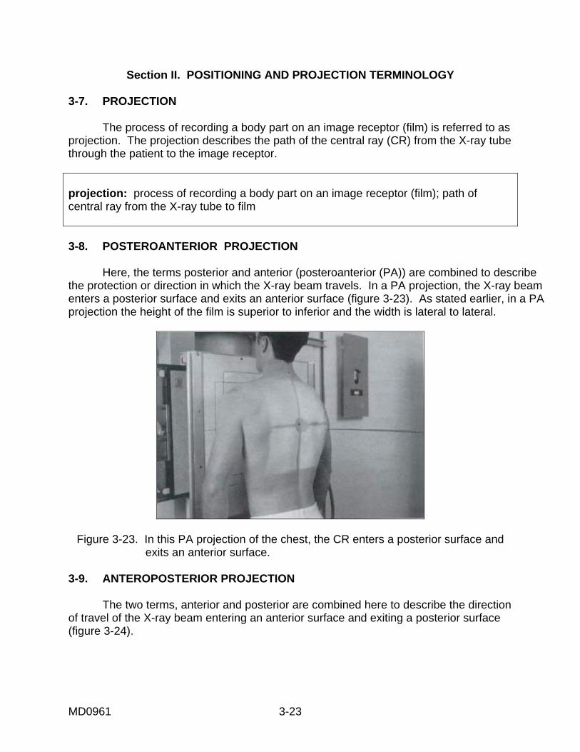
3-9. ANTEROPOSTERIOR PROJECTION The two terms, anterior and posterior are combined here to describe the direction of travel of the X-ray beam entering an anterior surface and exiting a posterior surface (figure 3-24).
MD0961 3-24
anteroposterior projection: a projection of the X-my beam human anterior surface to a posterior surface.
Figure 3-24. Anteroposterior chest projection with central ray entering anterior surface and traveling to posterior surface
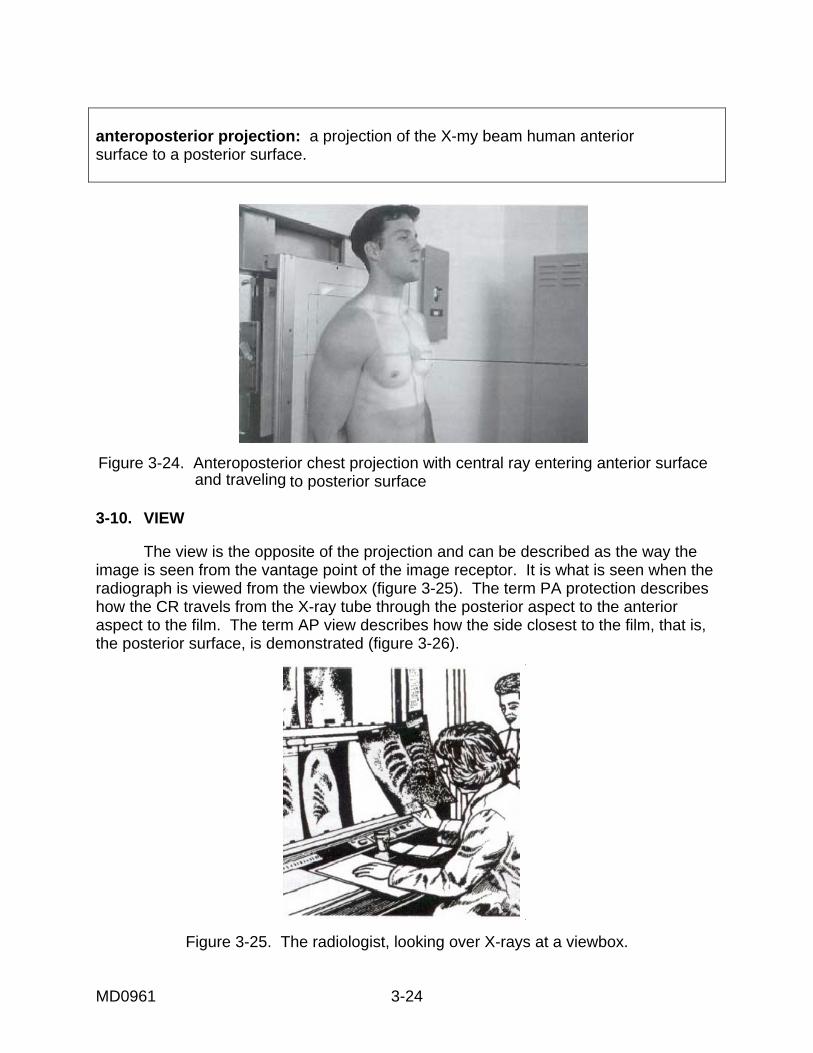
3-10. VIEW The view is the opposite of the projection and can be described as the way the image is seen from the vantage point of the image receptor. It is what is seen when the radiograph is viewed from the viewbox (figure 3-25). The term PA protection describes how the CR travels from the X-ray tube through the posterior aspect to the anterior aspect to the film. The term AP view describes how the side closest to the film, that is, the posterior surface, is demonstrated (figure 3-26).
Figure 3-25. The radiologist, looking over X-rays at a viewbox.
MD0961 3-25
view: the image as seen from the vantage of the image receptor, the way the film “sees” the part.
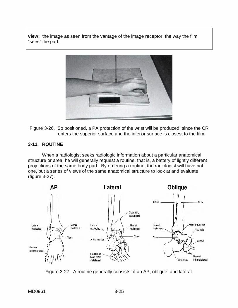
Figure 3-26. So positioned, a PA protection of the wrist will be produced, since the CR
enters the superior surface and the inferior surface is closest to the film. 3-11. ROUTINE When a radiologist seeks radiologic information about a particular anatomical structure or area, he will generally request a routine, that is, a battery of lightly different projections of the same body part. By ordering a routine, the radiologist will have not one, but a series of views of the same anatomical structure to look at and evaluate (figure 3-27).
Figure 3-27. A routine generally consists of an AP, oblique, and lateral.
MD0961 3-26
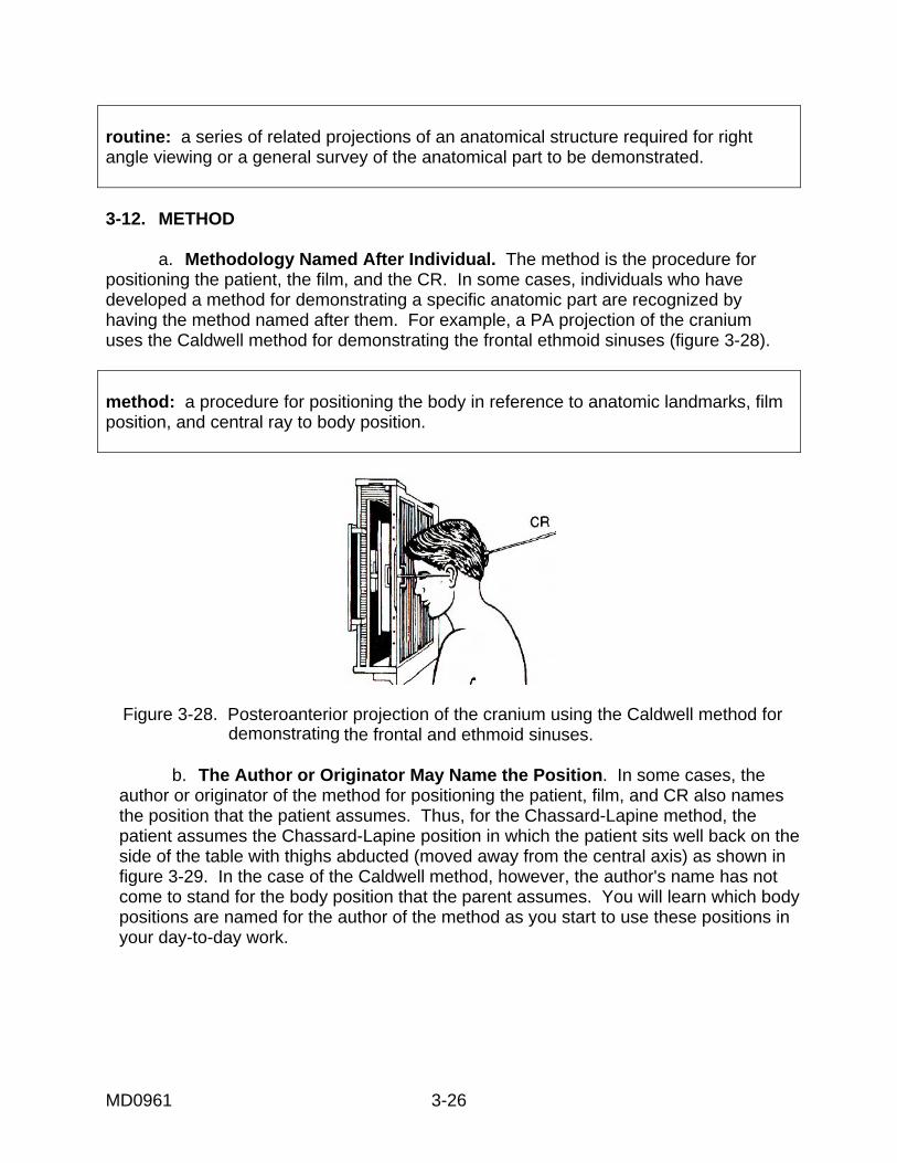
routine: a series of related projections of an anatomical structure required for right angle viewing or a general survey of the anatomical part to be demonstrated. 3-12. METHOD a. Methodology Named After Individual. The method is the procedure for positioning the patient, the film, and the CR. In some cases, individuals who have developed a method for demonstrating a specific anatomic part are recognized by having the method named after them. For example, a PA projection of the cranium uses the Caldwell method for demonstrating the frontal ethmoid sinuses (figure 3-28). method: a procedure for positioning the body in reference to anatomic landmarks, film position, and central ray to body position.
Figure 3-28. Posteroanterior projection of the cranium using the Caldwell method for
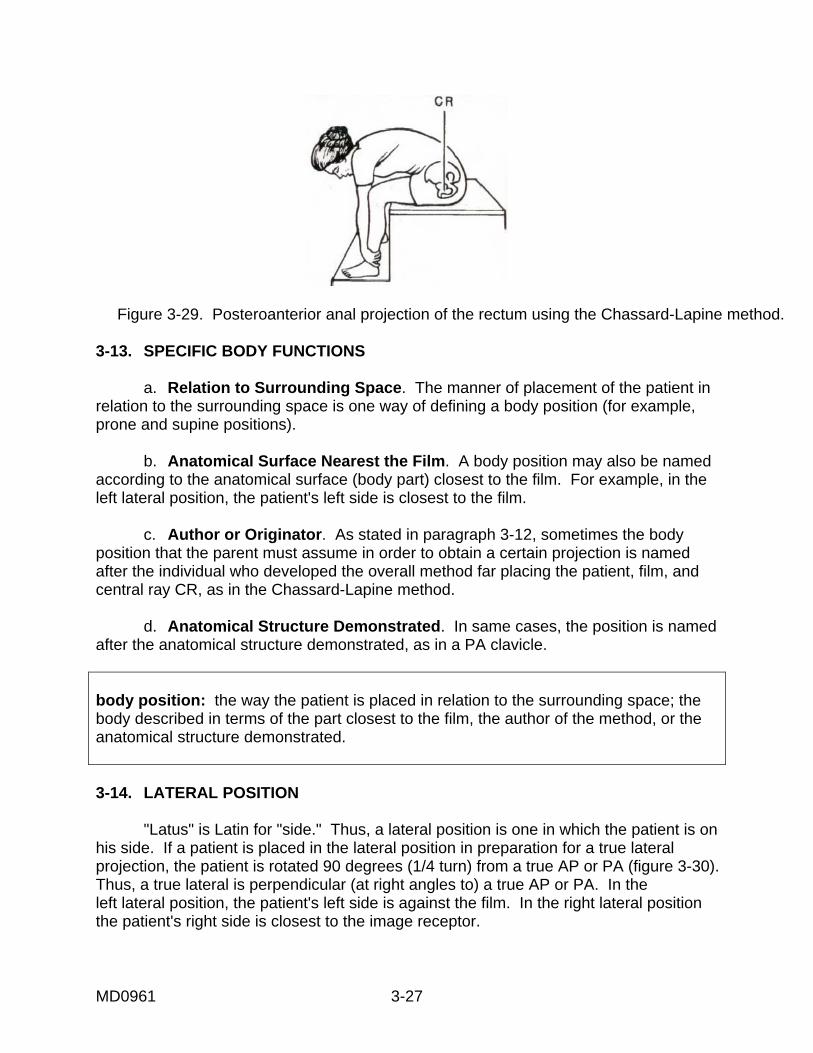
demonstrating the frontal and ethmoid sinuses. b. The Author or Originator May Name the Position. In some cases, the author or originator of the method for positioning the patient, film, and CR also names the position that the patient assumes. Thus, for the Chassard-Lapine method, the patient assumes the Chassard-Lapine position in which the patient sits well back on the side of the table with thighs abducted (moved away from the central axis) as shown in figure 3-29. In the case of the Caldwell method, however, the author's name has not come to stand for the body position that the parent assumes. You will learn which body positions are named for the author of the method as you start to use these positions in your day-to-day work.
MD0961 3-27
Figure 3-29. Posteroanterior anal projection of the rectum using the Chassard-Lapine method. 3-13. SPECIFIC BODY FUNCTIONS a. Relation to Surrounding Space. The manner of placement of the patient in relation to the surrounding space is one way of defining a body position (for example, prone and supine positions). b. Anatomical Surface Nearest the Film. A body position may also be named according to the anatomical surface (body part) closest to the film. For example, in the left lateral position, the patient's left side is closest to the film. c. Author or Originator. As stated in paragraph 3-12, sometimes the body position that the parent must assume in order to obtain a certain projection is named after the individual who developed the overall method far placing the patient, film, and central ray CR, as in the Chassard-Lapine method. d. Anatomical Structure Demonstrated. In same cases, the position is named after the anatomical structure demonstrated, as in a PA clavicle. body position: the way the patient is placed in relation to the surrounding space; the body described in terms of the part closest to the film, the author of the method, or the anatomical structure demonstrated. 3-14. LATERAL POSITION "Latus" is Latin for "side." Thus, a lateral position is one in which the patient is on his side. If a patient is placed in the lateral position in preparation for a true lateral projection, the patient is rotated 90 degrees (1/4 turn) from a true AP or PA (figure 3-30). Thus, a true lateral is perpendicular (at right angles to) a true AP or PA. In the left lateral position, the patient's left side is against the film. In the right lateral position the patient's right side is closest to the image receptor.
MD0961 3-28
Figure 3-30. A true lateral is rotated 90 degree or 1/4 turn from a PA or AP projection.
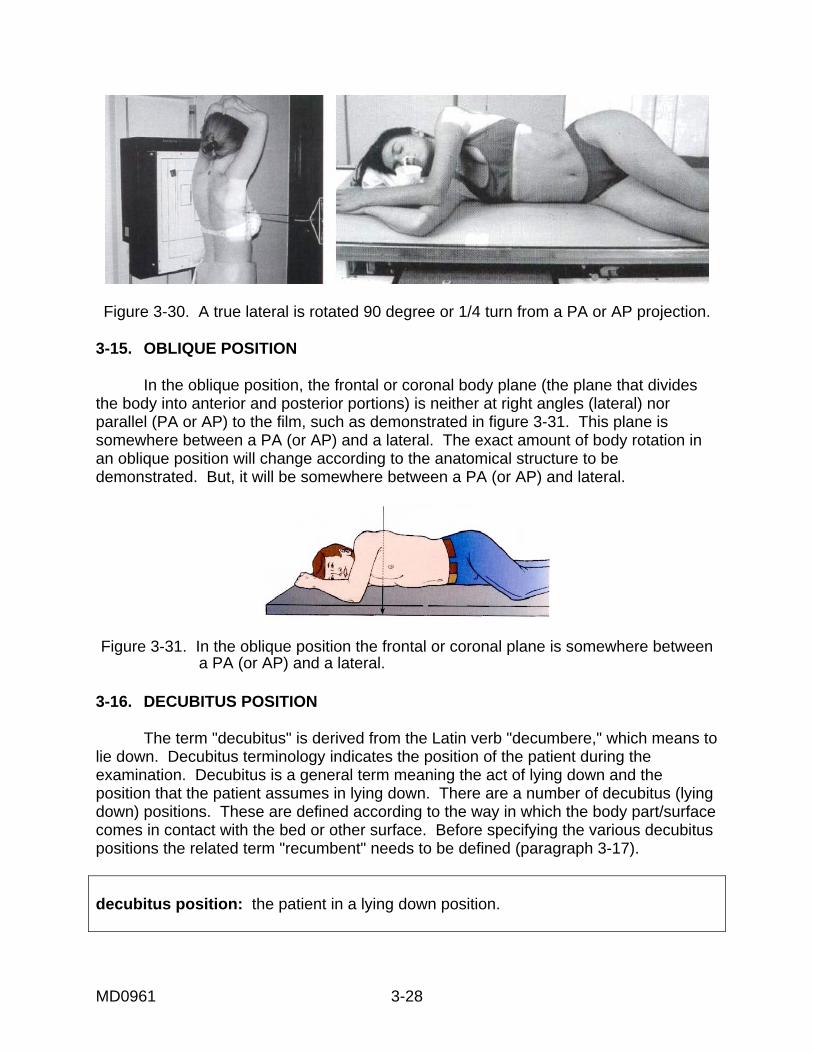
3-15. OBLIQUE POSITION In the oblique position, the frontal or coronal body plane (the plane that divides the body into anterior and posterior portions) is neither at right angles (lateral) nor parallel (PA or AP) to the film, such as demonstrated in figure 3-31. This plane is somewhere between a PA (or AP) and a lateral. The exact amount of body rotation in an oblique position will change according to the anatomical structure to be demonstrated. But, it will be somewhere between a PA (or AP) and lateral.
Figure 3-31. In the oblique position the frontal or coronal plane is somewhere between
a PA (or AP) and a lateral. 3-16. DECUBITUS POSITION The term "decubitus" is derived from the Latin verb "decumbere," which means to lie down. Decubitus terminology indicates the position of the patient during the examination. Decubitus is a general term meaning the act of lying down and the position that the patient assumes in lying down. There are a number of decubitus (lying down) positions. These are defined according to the way in which the body part/surface comes in contact with the bed or other surface. Before specifying the various decubitus positions the related term "recumbent" needs to be defined (paragraph 3-17). decubitus position: the patient in a lying down position.
MD0961 3-29
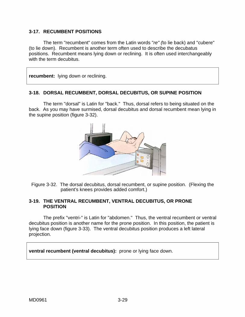
3-17. RECUMBENT POSITIONS The term "recumbent" comes from the Latin words "re" (to lie back) and "cubere" (to lie down). Recumbent is another term often used to describe the decubatus positions. Recumbent means lying down or reclining. It is often used interchangeably with the term decubitus. recumbent: lying down or reclining. 3-18. DORSAL RECUMBENT, DORSAL DECUBITUS, OR SUPINE POSITION The term "dorsal" is Latin for "back." Thus, dorsal refers to being situated on the back. As you may have surmised, dorsal decubitus and dorsal recumbent mean lying in the supine position (figure 3-32).
Figure 3-32. The dorsal decubitus, dorsal recumbent, or supine position. (Flexing the patient's knees provides added comfort.)
3-19. THE VENTRAL RECUMBENT, VENTRAL DECUBITUS, OR PRONE POSITION The prefix "ventri-" is Latin for "abdomen." Thus, the ventral recumbent or ventral decubitus position is another name for the prone position. In this position, the patient is lying face down (figure 3-33). The ventral decubitus position produces a left lateral projection. ventral recumbent (ventral decubitus): prone or lying face down.
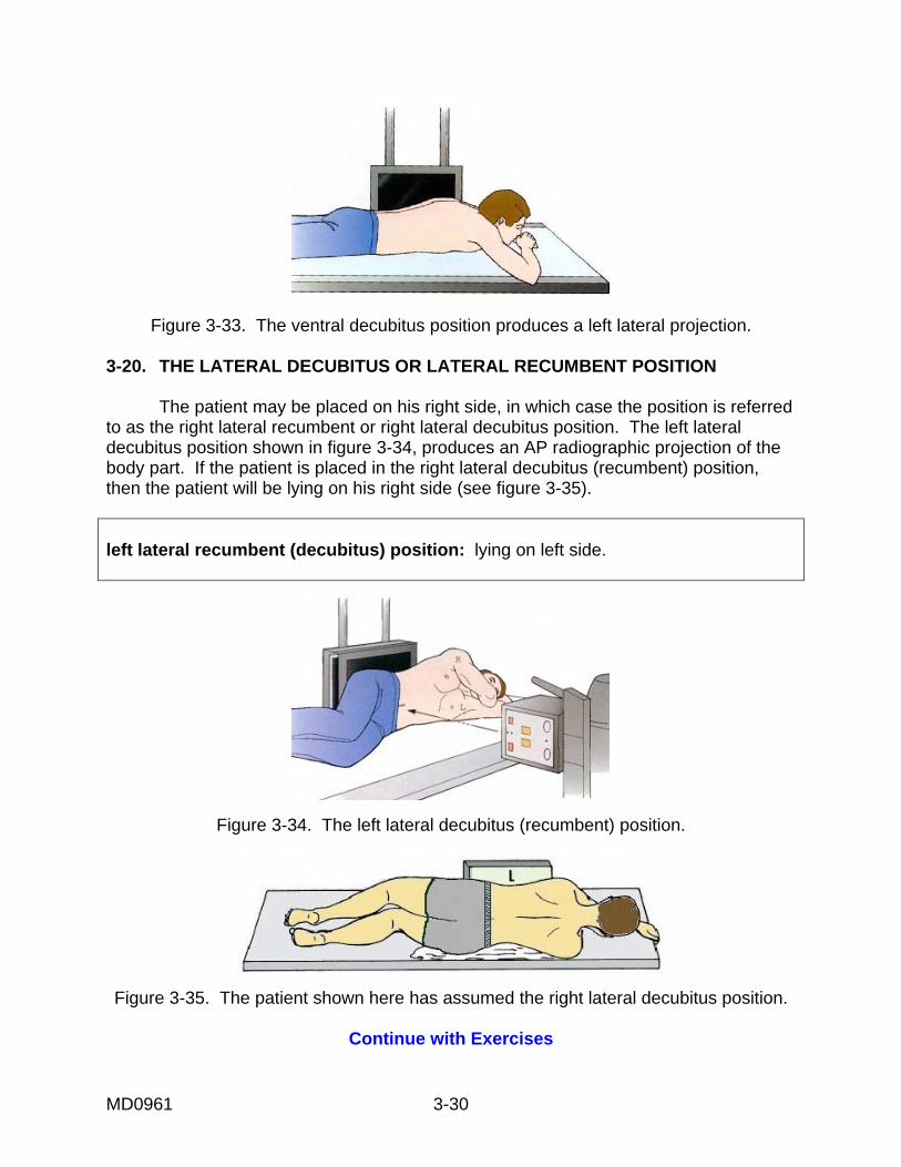
MD0961 3-30
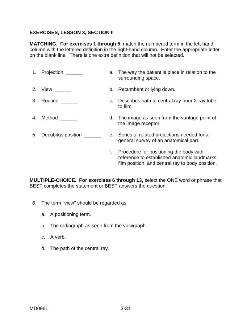
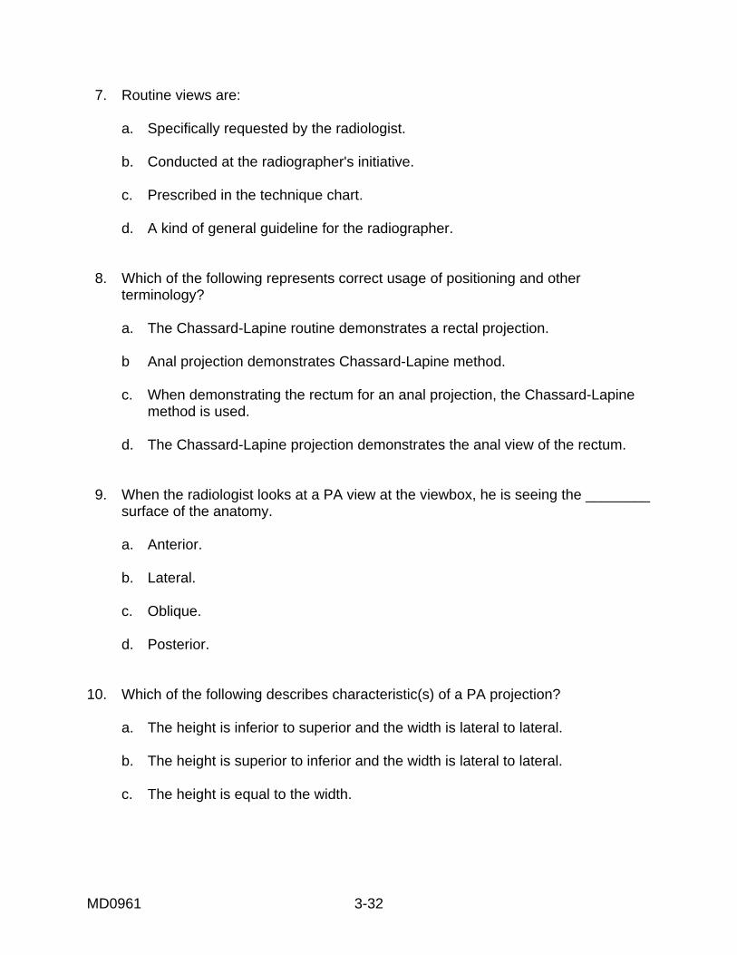
Figure 3-33. The ventral decubitus position produces a left lateral projection. 3-20. THE LATERAL DECUBITUS OR LATERAL RECUMBENT POSITION The patient may be placed on his right side, in which case the position is referred to as the right lateral recumbent or right lateral decubitus position. The left lateral decubitus position shown in figure 3-34, produces an AP radiographic projection of the body part. If the patient is placed in the right lateral decubitus (recumbent) position, then the patient will be lying on his right side (see figure 3-35). left lateral recumbent (decubitus) position: lying on left side.
Figure 3-34. The left lateral decubitus (recumbent) position.
Figure 3-35. The patient shown here has assumed the right lateral decubitus position.
Continue with Exercises
MD0961 3-31
EXERCISES, LESSON 3, SECTION II MATCHING. For exercises 1 through 5, match the numbered term in the left-hand column with the lettered definition in the right-hand column. Enter the appropriate letter on the blank line. There is one extra definition that will not be selected. 1. Projection ______ a. The way the patient is place in relation to the surrounding space. 2. View ______ b. Recumbent or lying down. 3. Routine ______ c. Describes path of central ray from X-ray tube to film. 4. Method ______ d. The image as seen from the vantage point of the image receptor. 5. Decubitus position ______ e. Series of related projections needed for a general survey of an anatomical part. f. Procedure for positioning the body with reference to established anatomic landmarks, film position, and central ray to body position. MULTIPLE-CHOICE. For exercises 6 through 13, select the ONE word or phrase that BEST completes the statement or BEST answers the question. 6. The term "view" should be regarded as: a. A positioning term. b. The radiograph as seen from the viewgraph. c. A verb. d. The path of the central ray.
MD0961 3-32
7. Routine views are: a. Specifically requested by the radiologist. b. Conducted at the radiographer's initiative. c. Prescribed in the technique chart. d. A kind of general guideline for the radiographer. 8. Which of the following represents correct usage of positioning and other terminology? a. The Chassard-Lapine routine demonstrates a rectal projection. b Anal projection demonstrates Chassard-Lapine method. c. When demonstrating the rectum for an anal projection, the Chassard-Lapine method is used. d. The Chassard-Lapine projection demonstrates the anal view of the rectum. 9. When the radiologist looks at a PA view at the viewbox, he is seeing the ________ surface of the anatomy. a. Anterior. b. Lateral. c. Oblique. d. Posterior. 10. Which of the following describes characteristic(s) of a PA projection? a. The height is inferior to superior and the width is lateral to lateral. b. The height is superior to inferior and the width is lateral to lateral. c. The height is equal to the width.
MD0961 3-33
11. The body position assumed by the patient is NOT named for: a. The path of the central ray. b. The anatomical surface nearest the film. c. The author of the overall radiographic method used to obtain the projection. d. The anatomical structure demonstrated. 12. In a true lateral position, the body is: a. Parallel to the film. b. Rotated 45 degrees from a true AP or PA projection. c. Perpendicular to a true AP or PA projection. d. Neither at right angles nor parallel to the image receptor. 13. In an oblique position, the body is: a. Parallel to the film. b. Rotated 90 degrees from a true AP or PA projection. c. Somewhere between the position required for an AP projection and a PA projection. d. Neither parallel to nor at right angles to the film.
MD0961 3-34
IDENTIFICATION, For exercises 14 through 22, correctly label the figures above the question by entering the appropriate word (or letter) in the space provided.
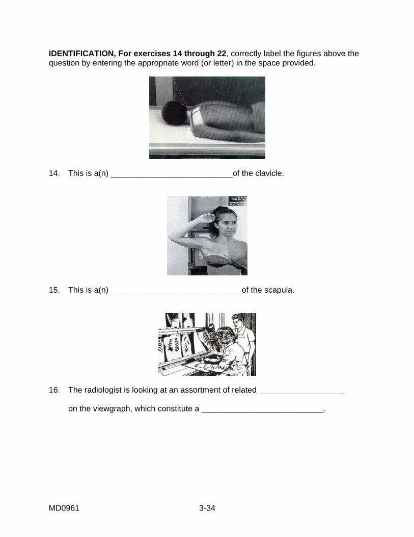
14. This is a(n) ___________________________of the clavicle.
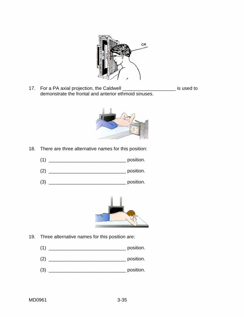
15. This is a(n) _____________________________of the scapula.
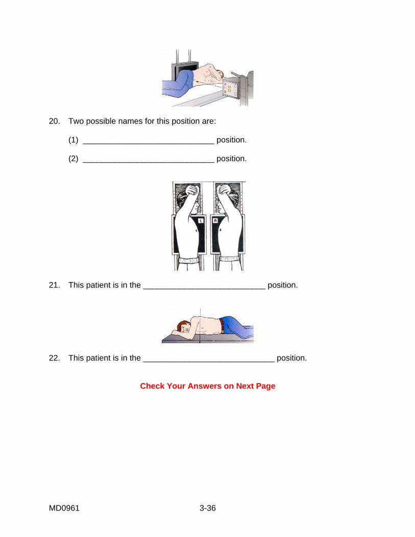
16. The radiologist is looking at an assortment of related ___________________ on the viewgraph, which constitute a ___________________________.
MD0961 3-35
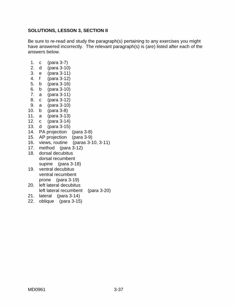
17. For a PA axial projection, the Caldwell ____________________ is used to demonstrate the frontal and anterior ethmoid sinuses.
18. There are three alternative names for this position: (1) _____________________________ position. (2) _____________________________ position. (3) _____________________________ position.
19. Three alternative names for this position are: (1) _____________________________ position. (2) _____________________________ position. (3) _____________________________ position.
MD0961 3-36
20. Two possible names for this position are: (1) _____________________________ position. (2) _____________________________ position.
21. This patient is in the ___________________________ position.
22. This patient is in the _____________________________ position.
Check Your Answers on Next Page
MD0961 3-37
SOLUTIONS, LESSON 3, SECTION II Be sure to re-read and study the paragraph(s) pertaining to any exercises you might have answered incorrectly. The relevant paragraph(s) is (are) listed after each of the answers below. 1. c (para 3-7) 2. d (para 3-10) 3. e (para 3-11) 4. f (para 3-12) 5. b (para 3-16) 6. b (para 3-10) 7. a (para 3-11) 8. c (para 3-12) 9. a (para 3-10) 10. b (para 3-8) 11. a (para 3-13) 12. c (para 3-14) 13. d (para 3-15) 14. PA projection (para 3-8) 15. AP projection (para 3-9) 16. views, routine (paras 3-10, 3-11) 17. method (para 3-12) 18. dorsal decubitus dorsal recumbent supine (para 3-18) 19. ventral decubitus ventral recumbent prone (para 3-19) 20. left lateral decubitus left lateral recumbent (para 3-20) 21. lateral (para 3-14) 22. oblique (para 3-15)
MD0961 3-38
Section III. PROJECTION, CENTRAL RAY, AND BREATHING TECHNIQUE TERMINOLOGY
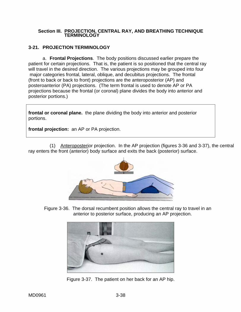
3-21. PROJECTION TERMINOLOGY a. Frontal Projections. The body positions discussed earlier prepare the patient for certain projections. That is, the patient is so positioned that the central raywill travel in the desired direction. The various projections may be grouped into four major categories frontal, lateral, oblique, and decubitus projections. The frontal (front to back or back to front) projections are the anteroposterior (AP) and posteroanterior (PA) projections. (The term frontal is used to denote AP or PA projections because the frontal (or coronal) plane divides the body into anterior and posterior portions.) frontal or coronal plane. the plane dividing the body into anterior and posterior portions. frontal projection: an AP or PA projection. (1) Anteroposterior projection. In the AP projection (figures 3-36 and 3-37), the central ray enters the front (anterior) body surface and exits the back (posterior) surface.
Figure 3-36. The dorsal recumbent position allows the central ray to travel in an anterior to posterior surface, producing an AP projection.
Figure 3-37. The patient on her back for an AP hip.
MD0961 3-39
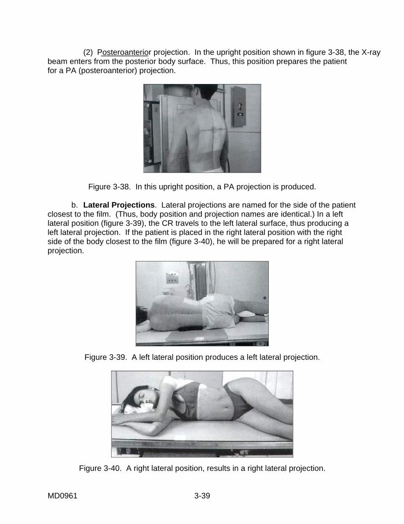
(2) Posteroanterior projection. In the upright position shown in figure 3-38, the X-ray beam enters from the posterior body surface. Thus, this position prepares the patient for a PA (posteroanterior) projection.
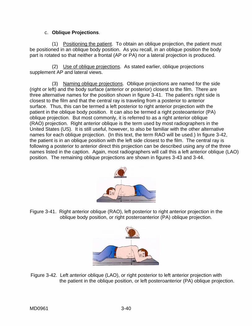
Figure 3-38. In this upright position, a PA projection is produced. b. Lateral Projections. Lateral projections are named for the side of the patient closest to the film. (Thus, body position and projection names are identical.) In a left lateral position (figure 3-39), the CR travels to the left lateral surface, thus producing a left lateral projection. If the patient is placed in the right lateral position with the right side of the body closest to the film (figure 3-40), he will be prepared for a right lateral projection.
Figure 3-39. A left lateral position produces a left lateral projection.
Figure 3-40. A right lateral position, results in a right lateral projection.
MD0961 3-40
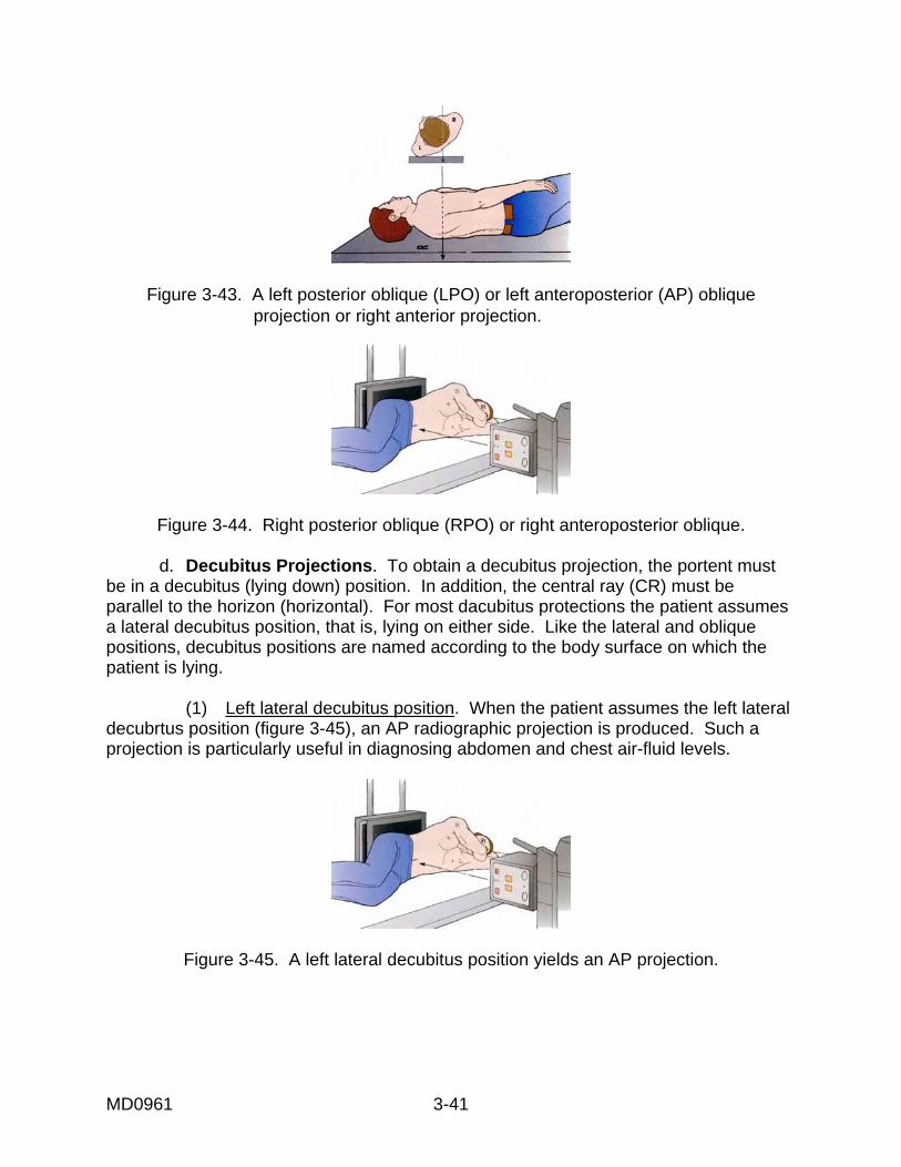
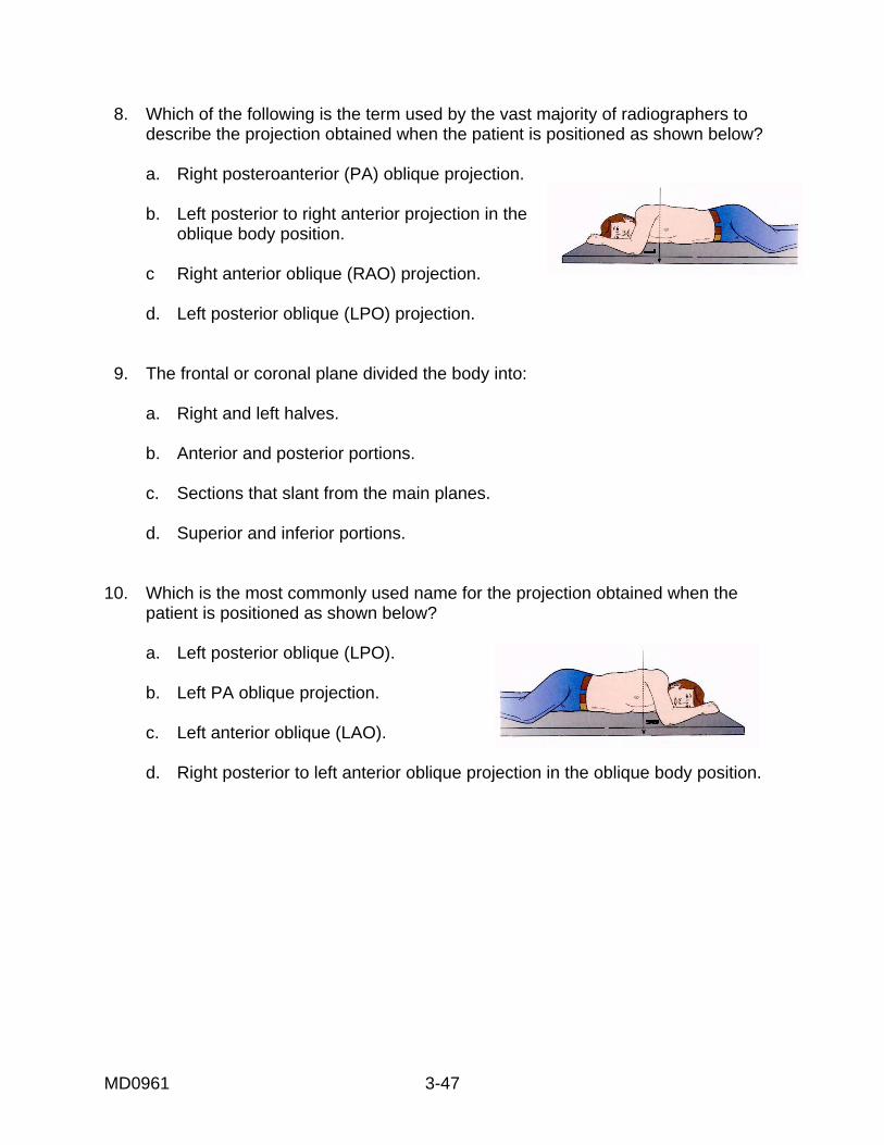
c. Oblique Projections. (1) Positioning the patient. To obtain an oblique projection, the patient must be positioned in an oblique body position. As you recall, in an oblique position the body part is rotated so that neither a frontal (AP or PA) nor a lateral projection is produced. (2) Use of oblique projections. As stated earlier, oblique projections supplement AP and lateral views. (3) Naming oblique projections. Oblique projections are named for the side (right or left) and the body surface (anterior or posterior) closest to the film. There are three alternative names for the position shown in figure 3-41. The patient's right side is closest to the film and that the central ray is traveling from a posterior to anterior surface. Thus, this can be termed a left posterior to right anterior projection with the patient in the oblique body position. It can also be termed a right posteoanterior (PA) oblique projection. But most commonly, it is referred to as a right anterior oblique (RAO) projection. Right anterior oblique is the term used by most radiographers in the United States (US). It is still useful, however, to also be familiar with the other alternative names for each oblique projection. (In this text, the term RAO will be used.) In figure 3-42, the patient is in an oblique position with the left side closest to the film. The central ray is following a posterior to anterior direct this projection can be described using any of the three names listed in the caption. Again, most radiographers will call this a left anterior oblique (LAO) position. The remaining oblique projections are shown in figures 3-43 and 3-44.
Figure 3-41. Right anterior oblique (RAO), left posterior to right anterior projection in the
oblique body position, or right posteroanterior (PA) oblique projection.
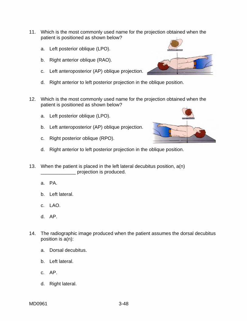
Figure 3-42. Left anterior oblique (LAO), or right posterior to left anterior projection with
the patient in the oblique position, or left posteroanterior (PA) oblique projection.
MD0961 3-41
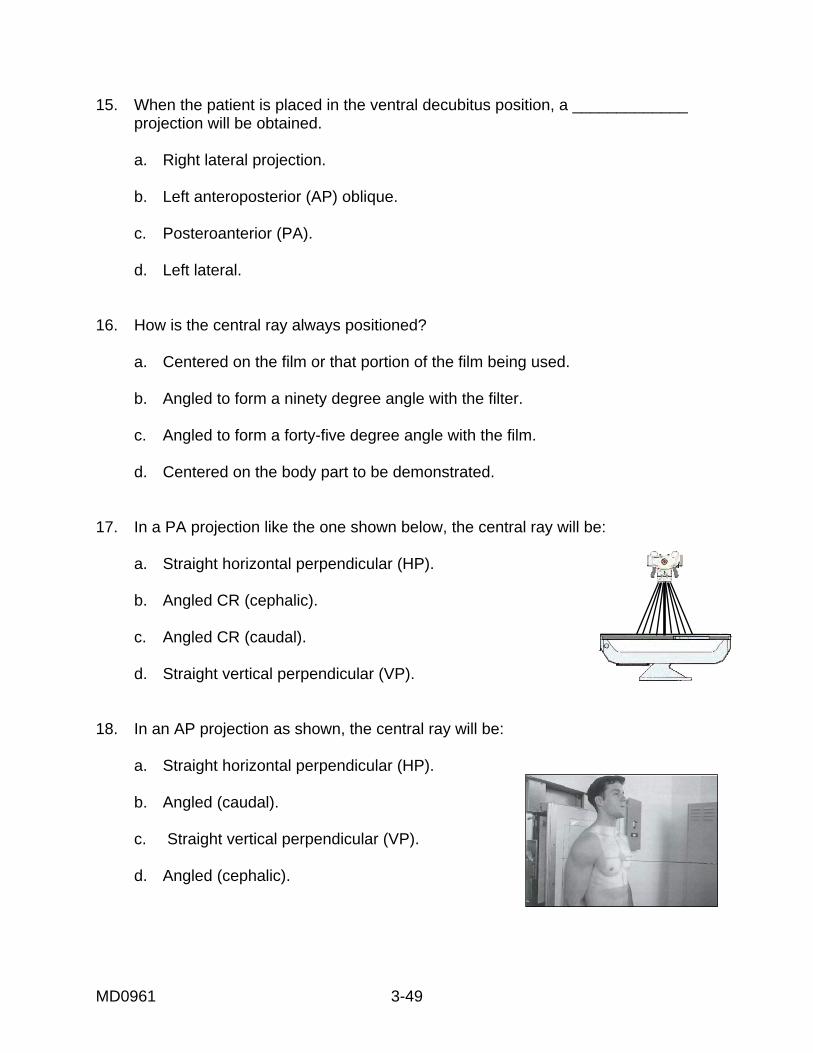
Figure 3-43. A left posterior oblique (LPO) or left anteroposterior (AP) oblique projection or right anterior projection.
Figure 3-44. Right posterior oblique (RPO) or right anteroposterior oblique. d. Decubitus Projections. To obtain a decubitus projection, the portent must be in a decubitus (lying down) position. In addition, the central ray (CR) must be parallel to the horizon (horizontal). For most dacubitus protections the patient assumes a lateral decubitus position, that is, lying on either side. Like the lateral and oblique positions, decubitus positions are named according to the body surface on which the patient is lying. (1) Left lateral decubitus position. When the patient assumes the left lateral decubrtus position (figure 3-45), an AP radiographic projection is produced. Such a projection is particularly useful in diagnosing abdomen and chest air-fluid levels.
Figure 3-45. A left lateral decubitus position yields an AP projection.
MD0961 3-42
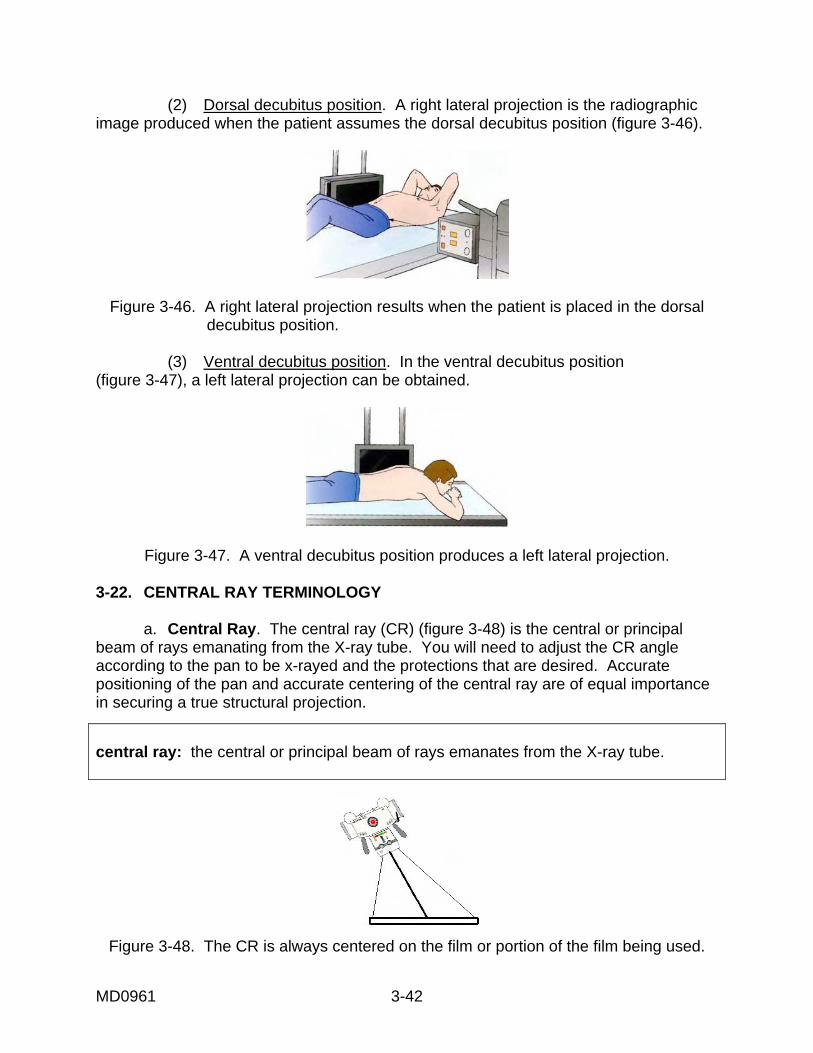
(2) Dorsal decubitus position. A right lateral projection is the radiographic image produced when the patient assumes the dorsal decubitus position (figure 3-46).
Figure 3-46. A right lateral projection results when the patient is placed in the dorsal decubitus position.
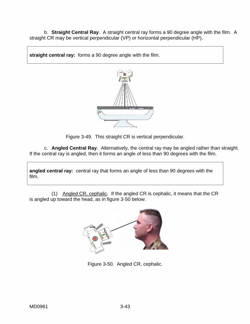
(3) Ventral decubitus position. In the ventral decubitus position (figure 3-47), a left lateral projection can be obtained.
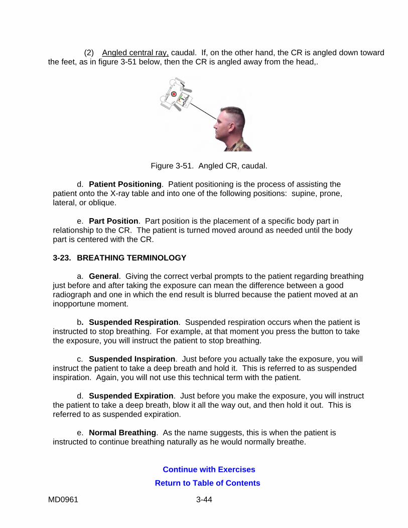
Figure 3-47. A ventral decubitus position produces a left lateral projection. 3-22. CENTRAL RAY TERMINOLOGY a. Central Ray. The central ray (CR) (figure 3-48) is the central or principal beam of rays emanating from the X-ray tube. You will need to adjust the CR angle according to the pan to be x-rayed and the protections that are desired. Accurate positioning of the pan and accurate centering of the central ray are of equal importance in securing a true structural projection.
central ray: the central or principal beam of rays emanates from the X-ray tube.
Figure 3-48. The CR is always centered on the film or portion of the film being used.
MD0961 3-43
b. Straight Central Ray. A straight central ray forms a 90 degree angle with the film. A straight CR may be vertical perpendicular (VP) or horizontal perpendicular (HP). straight central ray: forms a 90 degree angle with the film.
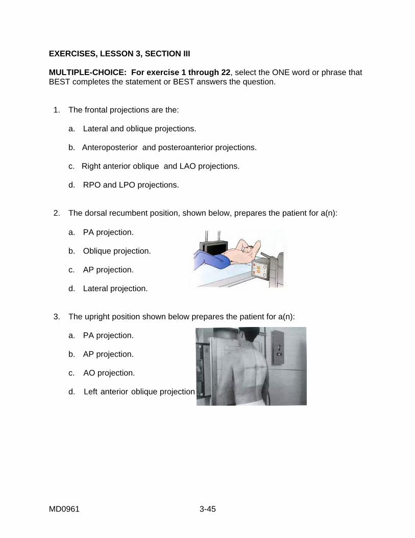
Figure 3-49. This straight CR is vertical perpendicular. c. Angled Central Ray. Alternatively, the central ray may be angled rather than straight. If the central ray is angled, then it forms an angle of less than 90 degrees with the film. angled central ray: central ray that forms an angle of less than 90 degrees with the film. (1) Angled CR, cephalic. If the angled CR is cephalic, it means that the CR is angled up toward the head, as in figure 3-50 below.
Figure 3-50. Angled CR, cephalic.
MD0961 3-44
(2) Angled central ray, caudal. If, on the other hand, the CR is angled down toward the feet, as in figure 3-51 below, then the CR is angled away from the head,.
Figure 3-51. Angled CR, caudal. d. Patient Positioning. Patient positioning is the process of assisting the patient onto the X-ray table and into one of the following positions: supine, prone, lateral, or oblique. e. Part Position. Part position is the placement of a specific body part in relationship to the CR. The patient is turned moved around as needed until the body part is centered with the CR. 3-23. BREATHING TERMINOLOGY a. General. Giving the correct verbal prompts to the patient regarding breathing just before and after taking the exposure can mean the difference between a good radiograph and one in which the end result is blurred because the patient moved at an inopportune moment. b. Suspended Respiration. Suspended respiration occurs when the patient is instructed to stop breathing. For example, at that moment you press the button to take the exposure, you will instruct the patient to stop breathing. c. Suspended Inspiration. Just before you actually take the exposure, you will instruct the patient to take a deep breath and hold it. This is referred to as suspended inspiration. Again, you will not use this technical term with the patient. d. Suspended Expiration. Just before you make the exposure, you will instruct the patient to take a deep breath, blow it all the way out, and then hold it out. This is referred to as suspended expiration. e. Normal Breathing. As the name suggests, this is when the patient is instructed to continue breathing naturally as he would normally breathe.
Continue with Exercises
Return to Table of Contents
MD0961 3-45
EXERCISES, LESSON 3, SECTION III MULTIPLE-CHOICE: For exercise 1 through 22, select the ONE word or phrase that BEST completes the statement or BEST answers the question. 1. The frontal projections are the: a. Lateral and oblique projections. b. Anteroposterior and posteroanterior projections. c. Right anterior oblique and LAO projections. d. RPO and LPO projections. 2. The dorsal recumbent position, shown below, prepares the patient for a(n): a. PA projection. b. Oblique projection. c. AP projection. d. Lateral projection. 3. The upright position shown below prepares the patient for a(n): a. PA projection. b. AP projection. c. AO projection. d. Left anterior oblique projection.
MD0961 3-46
4. Lateral projections are named for: a. The author or originator of the method. b. The relationship between the patient's body to the surrounding space. c. The anatomical structure demonstrated. d. The side of the patient closest to the film. 5. If the patient is placed in the right lateral position, he is being prepared for a: a. Left lateral projection. b. Left anterior oblique projection. c. Right posterior oblique projection. d. Right lateral projection. 6. Which of the following supplement AP and lateral projections? a. PA projections. b. Routines. c. Projections that are angled CR. d. Oblique projections. 7. Which category of projections is named for the side and the body surface closest to the film? a. Frontal projections. b. Lateral projections. c. Oblique projections. d. Decubitus projections.
MD0961 3-47
8. Which of the following is the term used by the vast majority of radiographers to describe the projection obtained when the patient is positioned as shown below? a. Right posteroanterior (PA) oblique projection. b. Left posterior to right anterior projection in the oblique body position. c Right anterior oblique (RAO) projection. d. Left posterior oblique (LPO) projection. 9. The frontal or coronal plane divided the body into: a. Right and left halves. b. Anterior and posterior portions. c. Sections that slant from the main planes. d. Superior and inferior portions. 10. Which is the most commonly used name for the projection obtained when the patient is positioned as shown below? a. Left posterior oblique (LPO). b. Left PA oblique projection. c. Left anterior oblique (LAO). d. Right posterior to left anterior oblique projection in the oblique body position.
MD0961 3-48
11. Which is the most commonly used name for the projection obtained when the patient is positioned as shown below? a. Left posterior oblique (LPO). b. Right anterior oblique (RAO). c. Left anteroposterior (AP) oblique projection. d. Right anterior to left posterior projection in the oblique position. 12. Which is the most commonly used name for the projection obtained when the patient is positioned as shown below? a. Left posterior oblique (LPO). b. Left anteroposterior (AP) oblique projection. c. Right posterior oblique (RPO). d. Right anterior to left posterior projection in the oblique position. 13. When the patient is placed in the left lateral decubitus position, a(n) _____________ projection is produced. a. PA. b. Left lateral. c. LAO. d. AP. 14. The radiographic image produced when the patient assumes the dorsal decubitus position is a(n): a. Dorsal decubitus. b. Left lateral. c. AP. d. Right lateral.
MD0961 3-49
15. When the patient is placed in the ventral decubitus position, a _____________ projection will be obtained. a. Right lateral projection. b. Left anteroposterior (AP) oblique. c. Posteroanterior (PA). d. Left lateral. 16. How is the central ray always positioned? a. Centered on the film or that portion of the film being used. b. Angled to form a ninety degree angle with the filter. c. Angled to form a forty-five degree angle with the film. d. Centered on the body part to be demonstrated. 17. In a PA projection like the one shown below, the central ray will be: a. Straight horizontal perpendicular (HP). b. Angled CR (cephalic). c. Angled CR (caudal). d. Straight vertical perpendicular (VP). 18. In an AP projection as shown, the central ray will be: a. Straight horizontal perpendicular (HP). b. Angled (caudal). c. Straight vertical perpendicular (VP). d. Angled (cephalic).
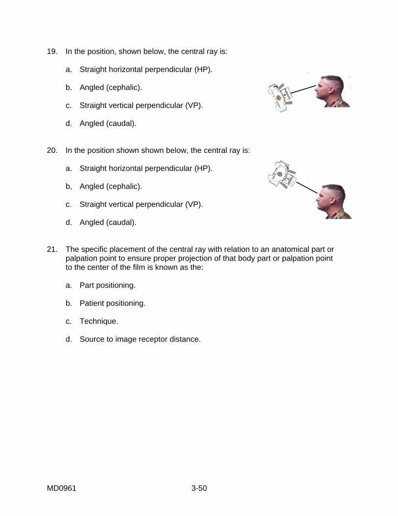
MD0961 3-50
19. In the position, shown below, the central ray is: a. Straight horizontal perpendicular (HP). b. Angled (cephalic). c. Straight vertical perpendicular (VP). d. Angled (caudal). 20. In the position shown shown below, the central ray is: a. Straight horizontal perpendicular (HP). b, Angled (cephalic). c. Straight vertical perpendicular (VP). d. Angled (caudal). 21. The specific placement of the central ray with relation to an anatomical part or palpation point to ensure proper projection of that body part or palpation point to the center of the film is known as the: a. Part positioning. b. Patient positioning. c. Technique. d. Source to image receptor distance.
MD0961 3-51
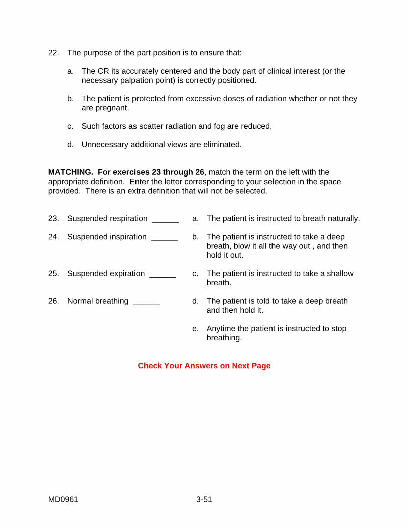
22. The purpose of the part position is to ensure that: a. The CR its accurately centered and the body part of clinical interest (or the necessary palpation point) is correctly positioned. b. The patient is protected from excessive doses of radiation whether or not they are pregnant. c. Such factors as scatter radiation and fog are reduced, d. Unnecessary additional views are eliminated. MATCHING. For exercises 23 through 26, match the term on the left with the appropriate definition. Enter the letter corresponding to your selection in the space provided. There is an extra definition that will not be selected. 23. Suspended respiration ______ a. The patient is instructed to breath naturally. 24. Suspended inspiration ______ b. The patient is instructed to take a deep breath, blow it all the way out , and then hold it out. 25. Suspended expiration ______ c. The patient is instructed to take a shallow breath. 26. Normal breathing ______ d. The patient is told to take a deep breath and then hold it. e. Anytime the patient is instructed to stop breathing.
Check Your Answers on Next Page

MD0961 3-52
SOLUTIONS, LESSON 3, SECTION III Be sure to re-read and study the paragraph(s) pertaining to any exercises you might have answered incorrectly. The relevant paragraph(s) is (are) listed after each of the answers below. 1. b (para 3-21a) 2. d (para 3-21d(2)) 3. a (para 3-21a(2)) 4. d (para 3-21b) 5. d (para 3-21b) 6. d (para 3-21c(2)) 7. c (para 3-21c(3)) 8. c (para 3-21c(3)) 9. b (para 3-21a) 10. c (para 3-21c(3)) 11. a (para 3-21c(3)) 12. c (para 3-21c(3)) 13. d (para 3-21d(1)) 14 d (para 3-21d(2)) 15. d (para 3-21d(3)) 16. a (para 3-22a) 17. d (para 3-22b) 18. a (para 3-22b) 19. b (para 3-22c(1)) 20. d (para 3-22c(2)) 21. a (para 3-22e) 22. a (para 3-22e) 23. e (para 3-23b) 24. d (para 3-23c) 25. b (para 3-23d) 26. a (para 3-23e)
Return to Table of Contents