Embed Size (px)
Citation preview
Behavioral/Systems/Cognitive
The Prefrontal Cortex Communicates with the Amygdalato Impair Learning after Acute Stress in Females but Notin Males
Lisa Y. Maeng, Jaylyn Waddell, and Tracey J. ShorsDepartment of Psychology and Center for Collaborative Neuroscience, Rutgers University, Piscataway, New Jersey 08854
Acute stress exposure enhances classical eyeblink conditioning in male rats, whereas exposure to the same event dramatically impairsperformance in females (Wood and Shors, 1998; Wood et al., 2001). We hypothesized that stress affects learning differently in males andfemales because different brain regions and circuits are being activated. In the first experiment, we determined that neuronal activitywithin the medial prefrontal cortex (mPFC) during the stressful event is necessary to disrupt learning in females. In both males andfemales, the mPFC was bilaterally inactivated with GABA agonist muscimol before the stressor. Inactivation prevented only the impairedperformance in females; it had no consequence for performance in males. However, in the second experiment, excitation of the mPFCalone with GABA antagonist picrotoxin was insufficient to elicit the stress effect that was prevented through the inactivation of this regionin females. Therefore, we hypothesized that the mPFC communicates with the basolateral amygdala to disrupt learning in females afterthe stressor. To test this hypothesis, these structures were disconnected from each other with unilateral excitotoxic (NMDA) lesions oneither the same or opposite sides of the brain. Females with contralateral lesions, which disrupt the connections on both sides of the brain,were able to learn after the stressful event, whereas those with ipsilateral lesions, which disrupt only one connection, did not learn afterthe stressor. Together, these data indicate that the mPFC is critically involved in females during stress to impair subsequent learning anddoes so via communication with the amygdala.
IntroductionTraumatic life experiences tend to be more debilitating inwomen, rendering them twice as likely as men to developstress and anxiety-related disorders (10.4 vs 5.0%; Kessler etal., 1995; Carter-Snell and Hegadoren, 2003; Foa and Street,2001; Tolin and Foa, 2006). This vulnerability in women mayrelate to sex differences in the stress response as reported inlaboratory animals. For example, stressors such as inescapableswim stress or brief stimulations to the tail enhance a type ofassociative learning, classical eyeblink conditioning, in malerats and mice, whereas the same stressors elicit profoundlearning deficits in females (Wood and Shors, 1998; Wood etal., 2001). These behavioral differences in response to stress aremediated by sex differences in neural and hormonal processeswithin specific brain regions. The hippocampus and basolateralamygdala (BLA) are critically involved in both males and females tomodify learning after stress (Bangasser and Shors, 2007; Waddell etal., 2008). However, the bed nucleus of the stria terminalis (BNST) isnecessary only in males (Bangasser et al., 2005; Bangasser and Shors,2008).
Another likely participant is the medial prefrontal cortex(mPFC), which is activated during the stress response and inter-connects with the hippocampus, BNST, and BLA (Diorio et al.,1993; Vertes, 2004, 2006; Cerqueira et al., 2008). Stress-relateddisorders are associated with differences in both structure andfunction of the mPFC (Bremner et al., 1999; Drevets, 2000;Rajkowska, 2000; Luine, 2002). Furthermore, the femalemPFC is especially sensitive to stress (Garrett and Wellman,2009; Ter Horst et al., 2009) as well as to fluctuating estrogenconcentrations (Gerrits et al., 2006). One study found thatmPFC-mediated learning is more sensitive to stress in femalesthan in males (Shansky et al., 2006). Therefore, we first hypoth-esized that the mPFC would be critically involved in females, butless so in males, to modify learning after a stressful experience. Totest this hypothesis, the mPFC was either inactivated or activatedduring the stressor. One day later, both sexes were trained to learnthe classically conditioned eyeblink response.
The mPFC and amygdala interconnect to affect emotionalresponses to stress, presumably via anatomical connections be-tween them (Heidbreder and Groenewegen, 2003; Vertes, 2004).For example, lesions to the prefrontal cortex reduce the extinc-tion of a fear response, which depends on the amygdala to learn(Morgan and LeDoux, 1995). Based on these interactions, wehypothesized that the mPFC and amygdala communicate witheach other to reduce learning after stress, specifically in females.To test our hypothesis, both structures were excitotoxically le-sioned on either the same (ipsilateral) or opposite (contralateral)
Received May 3, 2010; revised Sept. 22, 2010; accepted Sept. 30, 2010.This work was supported by National Institutes of Health Grants 59970 and ARRA-59970-10S1 and National
Science Foundation Grants IOB-044364 and IOS-0914386 (to T.J.S.).Correspondence should be addressed to Tracey J. Shors, Department of Psychology and Center for Collaborative
Neuroscience, Rutgers University, 152 Frelinghuysen Road, Piscataway, NJ 08854. E-mail: [email protected]:10.1523/JNEUROSCI.2265-10.2010
Copyright © 2010 the authors 0270-6474/10/3016188-09$15.00/0
16188 • The Journal of Neuroscience, December 1, 2010 • 30(48):16188 –16196
sides of the brain. Those with ipsilateral lesions would have oneintact connection, whereas those with contralateral lesions of thebrain would have neither connection intact. As before, animalswere either stressed or not and then trained 24 h later to learn theclassically conditioned eyeblink response.
Materials and MethodsExperiments 1 and 2: mPFC inactivation and activationSubjects. Male and cycling female Sprague Dawley rats between 90 and120 d of age were obtained from a breeding facility at Rutgers University.Rats were housed in groups of 3– 4 until surgery. Following surgery, ratswere housed alone in standard plastic “shoebox” cages (44.5 cm long,21.59 cm wide, and 23.32 cm high). Rats were maintained on ad libitumaccess to rat chow and water on a 12 h light/dark cycle. All experiments wereconducted with full compliance to the rules and regulations specified by thePublic Health Service (PHS) Policy on Humane Care and Use of LaboratoryAnimals and the Guide for the Care and Use of Laboratory Animals.
Vaginal cytology. To monitor the four phases of the estrus cycle, sam-ples of loose vaginal cells were taken with cotton-tipped swabs soaked insterile 0.9% saline and rolled onto slides (lavage). The slides were thenstained with 1% toluidine blue, rinsed, and dehydrated with 95% ETOHfor estrus phase assessment under a light microscope. Proestrus is char-acterized by purple staining of epithelial cell nuclei, estrus by masses ofaggregated dark blue cornified cells, diestrus 1 by dark leukocytes andscattered epithelial cells, and diestrus 2 by similar, but more sparse, celltypes. It has been determined that the stress effect is most pronounced infemales when they are stressed in diestrus 2 and trained 24 h later inproestrus (Shors et al., 1998). Therefore, females used in this study werelavaged daily after a 1 week recovery period following surgery, stressed indiestrus, and trained in proestrus, when estrogen concentrations wereincreasing (Shors et al., 1998; Wood et al., 2001). Animals that failed toexhibit a normal estrus cycle were eliminated from the study.
Surgery. All rats were anesthetized with sodium pentobarbital (50mg/kg for males and 40 mg/kg for females). After being placed in thestereotaxic instrument, the scalp was cleaned with Betadine, and an inci-sion was made. Guide cannulas (23 gauge, Plastics One) were implantedinto the mPFC bilaterally aimed at the junction of the prelimbic andinfralimbic cortex. Cannulas were implanted at a 15° angle at the follow-ing coordinates relative to bregma: (AP: �3.1 mm; ML: �1.6 mm; DV:�3.3 mm from dura). Following cannulation, both cannulas and head-stages were fitted onto the skull with dental cement and anchored by skullscrews. The headstages were attached to four electrodes; two deliveredthe unconditioned stimulus (US) of periorbital stimulation, and tworecorded electromyographic (EMG) activity as a measure of blinks. Theelectrodes (insulated stainless steel wire with a diameter of 0.005 inches)were implanted through the upper eyelid muscle. The insulation wasremoved from a section of each electrode to make contact with the mus-cle. Each of the electrode wires was coiled securely in place.
Infusions. During microinfusions, stylets were replaced with infusioncannulas protruding 1 mm past the guide cannula. Infusion cannulas wereattached to a microinfusion pump via polyethylene tubes connected to 10 �lHamilton syringes. The syringe and tubes were filled with water, and a smallair bubble separated the water from the vehicle or drug solution. In experi-ment 1, the mPFC was either infused with artificial CSF (aCSF) vehicle orbilaterally inactivated via infusions of 0.5 �g of GABA-A receptor agonistmuscimol (1 �g/�l) into each hemisphere. In experiment 2, the mPFC wasbilaterally activated via infusions of 0.5 �l of either 100 ng of picrotoxin(Sigma) or received microinjections of saline vehicle. All infusions weregiven at a rate of 0.125 �l/min over 4 min for a total of 0.5 �l.
Stress procedure. At least 7 d after the surgery, rats were acclimated tothe conditioning chamber (60 min) and spontaneous blinks were re-corded. They were then transported to a separate context and infusedwith aCSF or muscimol. Half of this group was transferred into anothercontext (different from those in which the infusions and training oc-curred) and placed into a dark soundproof chamber. In this chamber,they were loosely restrained and exposed to 30 low-intensity (1 mA, 60Hz, 1 s) stimulations to the tail. This is the amount of stress reported to besufficient to induce the opposite effects of stress on classical eyeblink
conditioning (Shors and Servatius, 1997; Shors, 2004). Moreover, it isalso important to note that the sex difference in the stress-induced effectson learning is not due to differential activation of pain sensitivity or alter-ations in the unconditioned response after the stressor (Wood and Shors,1998; Bangasser and Shors, 2004). In experiment 2, following infusions,unstressed animals were also taken into the context in which stressed animalswere exposed to the stressor but were only loosely restrained for 30 min in thechambers before being returned to their home cages.
Classical conditioning. Twenty-four hours later, rats were returned tothe conditioning chamber and exposed to 10 white noise stimuli alone(250 ms, 80 dB, ITI 25 � 5 s) before the first session. This procedure isused to assess potential effects of the stressor or infusion on sensitizedresponses to the CS (blinks during first 100 ms of the CS) (Servatius andShors, 1994). The rats were then trained with 400 trials (100 trials/d) ofpaired stimuli using a 80 dB, 850 ms burst of white noise CS overlappingwith a 100 ms, 0.5 mA periorbital stimulation of the eyelid (US). Thesestudies were conducted using a delay conditioning procedure in whichthe CS and the US overlap in time and coterminate. Eyeblinks were assessedby significant changes in the magnitude of the EMG response recorded fromthe eyelid muscles. Activity that lasted 10 ms and exceeded 0.3 mV and 4 SDabove baseline activity recorded during a 250 ms pre-CS period was consid-ered indicative of an eyeblink.
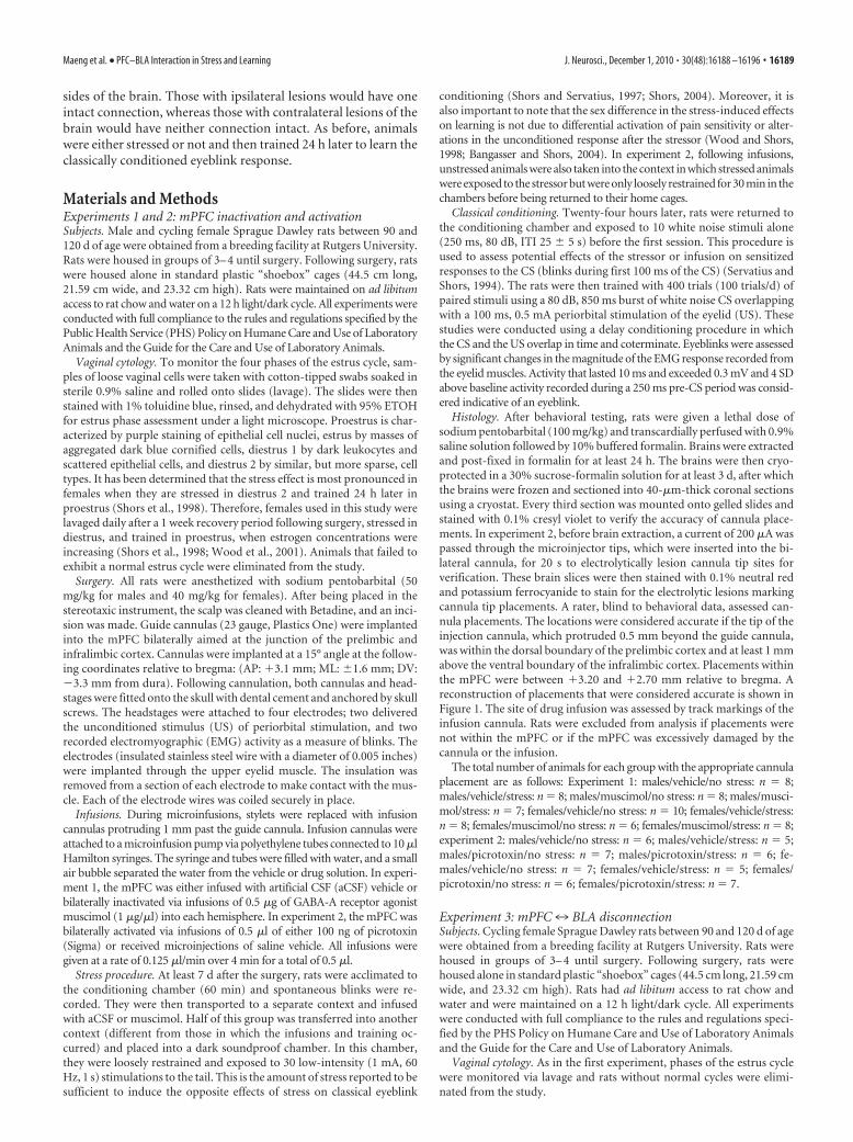
Histology. After behavioral testing, rats were given a lethal dose ofsodium pentobarbital (100 mg/kg) and transcardially perfused with 0.9%saline solution followed by 10% buffered formalin. Brains were extractedand post-fixed in formalin for at least 24 h. The brains were then cryo-protected in a 30% sucrose-formalin solution for at least 3 d, after whichthe brains were frozen and sectioned into 40-�m-thick coronal sectionsusing a cryostat. Every third section was mounted onto gelled slides andstained with 0.1% cresyl violet to verify the accuracy of cannula place-ments. In experiment 2, before brain extraction, a current of 200 �A waspassed through the microinjector tips, which were inserted into the bi-lateral cannula, for 20 s to electrolytically lesion cannula tip sites forverification. These brain slices were then stained with 0.1% neutral redand potassium ferrocyanide to stain for the electrolytic lesions markingcannula tip placements. A rater, blind to behavioral data, assessed can-nula placements. The locations were considered accurate if the tip of theinjection cannula, which protruded 0.5 mm beyond the guide cannula,was within the dorsal boundary of the prelimbic cortex and at least 1 mmabove the ventral boundary of the infralimbic cortex. Placements withinthe mPFC were between �3.20 and �2.70 mm relative to bregma. Areconstruction of placements that were considered accurate is shown inFigure 1. The site of drug infusion was assessed by track markings of theinfusion cannula. Rats were excluded from analysis if placements werenot within the mPFC or if the mPFC was excessively damaged by thecannula or the infusion.
The total number of animals for each group with the appropriate cannulaplacement are as follows: Experiment 1: males/vehicle/no stress: n � 8;males/vehicle/stress: n � 8; males/muscimol/no stress: n � 8; males/musci-mol/stress: n � 7; females/vehicle/no stress: n � 10; females/vehicle/stress:n � 8; females/muscimol/no stress: n � 6; females/muscimol/stress: n � 8;experiment 2: males/vehicle/no stress: n � 6; males/vehicle/stress: n � 5;males/picrotoxin/no stress: n � 7; males/picrotoxin/stress: n � 6; fe-males/vehicle/no stress: n � 7; females/vehicle/stress: n � 5; females/picrotoxin/no stress: n � 6; females/picrotoxin/stress: n � 7.
Experiment 3: mPFC7 BLA disconnectionSubjects. Cycling female Sprague Dawley rats between 90 and 120 d of agewere obtained from a breeding facility at Rutgers University. Rats werehoused in groups of 3– 4 until surgery. Following surgery, rats werehoused alone in standard plastic “shoebox” cages (44.5 cm long, 21.59 cmwide, and 23.32 cm high). Rats had ad libitum access to rat chow andwater and were maintained on a 12 h light/dark cycle. All experimentswere conducted with full compliance to the rules and regulations speci-fied by the PHS Policy on Humane Care and Use of Laboratory Animalsand the Guide for the Care and Use of Laboratory Animals.
Vaginal cytology. As in the first experiment, phases of the estrus cyclewere monitored via lavage and rats without normal cycles were elimi-nated from the study.
Maeng et al. • PFC–BLA Interaction in Stress and Learning J. Neurosci., December 1, 2010 • 30(48):16188 –16196 • 16189
Surgery. Female rats were anesthetized withsodium pentobarbital (40 mg/kg for females).After being placed in the stereotaxic instru-ment, the scalp was cleaned with Betadine, andan incision was made. Excitotoxic lesion siteswere infused with NMDA via a 10 �l Hamiltonsyringe attached to a microinfusion pump. Forlesions of the medial prefrontal cortex, the sy-ringe tip was aimed at the prelimbic/infralim-bic junction (AP: �3.0/�2.5 mm; ML: �0.7mm; DV: �4.5 mm from skull), and 0.1 �l of10 mg/ml NMDA was infused at a rate of 0.1�l/min. Coordinates for lesions of the basolat-eral amygdala were as follows: AP: �2.8 mm;ML: �4.8 mm; DV: �8.5/�8.3 mm from skull(20 mg/ml NMDA; volume: 0.25 �l/0.15 �l;rate: 0.1 �l/min). All contralateral and ipsilat-eral lesions (diagrammed in Fig. 2) were coun-terbalanced and subsequently assessed forpossible lateralization effects. Following infu-sions of excitotoxin, headstages were fittedonto the skull with dental cement and an-chored by skull screws. The headstages wereattached to four electrodes; two delivered theUS of periorbital stimulation and two recordedEMG activity as a measure of blinks. The elec-trodes were implanted as described in experi-ment 1.
Stress procedure. At least 7 d were allowed forrecovery time after surgery. Cycling rats indiestrus 2 were placed into the conditioningchamber for an acclimation period. Rats to bestressed were then transferred to a separateroom (different from that in which condition-ing occurred) into an enclosed soundproof boxand underwent brief stress exposure as de-scribed in experiment 1.
Classical conditioning. Before either stressexposure or none, the rats were placed into theconditioning boxes for a habituation period inwhich they acclimated for 1 h while spontane-ous blinks were recorded and then 24 h later,were returned to the chamber. As in experi-ment 1, rats were observed for a sensitizationperiod and then began training with delay eye-blink conditioning.
Histology. After behavioral testing, rats wereadministered a lethal dose of sodium pento-barbital (100 mg/kg) and transcardially per-fused with 0.9% saline solution followed by10% buffered formalin. Brains were extractedand post-fixed in formalin for at least 24 h. Thebrains were then cryoprotected in a 30%sucrose-formalin solution for at least 3 d, afterwhich the brains were frozen and sectioned into 50-�m-thick coronalsections using a cryostat. Every third section was mounted onto gelledslides and stained with the cresyl violet to verify lesion size. A rater, blindto the behavioral data, assessed lesion placements. Rats were excluded fromthe study if lesions were misplaced or incomplete. Lesions were identified bythe location of the needle track, absence of nerve cell bodies, and gliosis, orthe presence of darkly stained astrocytes (Bangasser et al., 2005). The extentof the smallest and largest lesion is presented in Figure 3. The number ofremaining animals were as follows: ipsilateral/no stress: n � 10; ipsilateral/stress: n � 10; contralateral/no stress: n � 10; contralateral/stress: n � 10.
ResultsExperiments 1 and 2: mPFC activity in males versus femalesIn the first experiment, the mPFC was inactivated during thestressor in males and females. All animals were trained with delay
conditioning 24 h later. Anticipatory conditioned responses(CRs) before the US were counted and averaged across blocks of100 trials (Fig. 4). Males were analyzed separately from females.Four groups of males were trained: one group whose mPFC wasinactivated during the stressor, one with mPFC inactivation and nostressor exposure, another stressed and injected with aCSF, and avehicle group that was not stressed. The independent measures werestress versus no stress and inactivation with muscimol versus aCSFvehicle infusion.
To assess acquisition of the CR across trials of training inmales, a 2 � 2 repeated-measures ANOVA across the four ses-sions of trials was conducted on each group. The analysis revealedan effect of session as the number of CRs increased as trainingprogressed (F(3,81) � 19.99; p � 0.01). There was an effect of stress
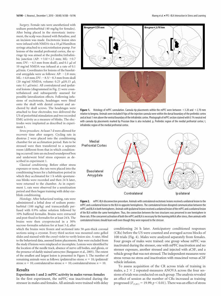
Figure 2. mPFC-BLA disconnection procedure. Animals with contralateral excitotoxic lesions received a unilateral lesion to themPFC and a unilateral lesion to the BLA in opposite hemispheres. The contralateral lesion disrupted communication between themPFC and BLA in both hemispheres. Animals with ipsilateral lesions received a unilateral lesion of the mPFC and a unilateral lesionof the BLA within the same hemisphere. Thus, the connection between the two structures was preserved in one hemisphere inthese rats. If the concurrent activation of both the mPFC and BLA is necessary for the learning deficit after stress, then animals withcontralateral lesions should learn well even though they were exposed to the stressor.
Figure 1. Histology of mPFC cannulation. Cannula tip placements within the mPFC were between �3.20 and �2.70 mmrelative to bregma. Animals were included if tips of the injection cannula were within the dorsal boundary of the prelimbic cortexand at least 1 mm above the ventral boundary of the infralimbic cortex. Photograph of mPFC section stained with 0.1% neutral redwith cannula tip placements marked by Prussian blue is also included. p, Prelimbic region of the medial prefrontal cortex; I,infralimbic region of the medial prefrontal cortex.
16190 • J. Neurosci., December 1, 2010 • 30(48):16188 –16196 Maeng et al. • PFC–BLA Interaction in Stress and Learning
(F(1,27) � 19.67; p � 0.01) but no effect of muscimol (F(1,27) �0.15; p � 0.05). Furthermore, there was no interaction betweenmuscimol treatment and stress (F(1,27) � 0.35; p � 0.05). Malesthat were exposed to the stressor emitted more CRs than thosethat were not exposed to the stressor regardless of drug treatment( p � 0.01) (Fig. 4A). The increase in responding occurred re-gardless of whether or not the mPFC was inactivated with mus-cimol during the stressor. Thus, muscimol infusion before stressexposure did not abolish the subsequent facilitation of eyeblinkconditioning elicited by stress in males.
To evaluate early acquisition, the first five blocks of 20 trialswere analyzed with a 2 � 2 repeated-measures ANOVA. Again,there was no effect of muscimol (F(1,27) � 0.77; p � 0.05), norwas there an interaction between muscimol and stress (F(1,27)
� 0.29; p � 0.05). However, there was a main effect of blocks oftrials (F(4,108) � 12.90; p � 0.01) as males increased their condi-tioned responding across trial blocks. There was also a main effectof stress (F(1,27) � 6.70; p � 0.05). Thus, there was no effect ofmuscimol inactivation but an effect of stress on both early acqui-sition and later conditioned responding in males.
Four groups of females were also trained: one group whosemPFC was inactivated during the stressor, one with mPFC inac-tivation without exposure to the stressor, another stressed andinjected with aCSF, and an unstressed vehicle group (Fig. 4B). To
assess acquisition across training in females,a 2 � 2 repeated-measures ANOVA acrossthe four sessions of trials was conducted oneach group. The analysis revealed an effectof session, as conditioned responding in-creased across training days (F(3,81) �37.28; p � 0.01). However, stressed fe-males with vehicle infusions did not ex-press such an increase (F(3,21) � 2.96; p �0.05) contrary to the remaining 3 groupsthat did increase. Using the percentage ofCRs across trials of training as the depen-dent measure, there was a main effect ofmuscimol (F(1,27) � 8.06; p � 0.01) andstress (F(1,27) � 7.63; p � 0.05). More crit-ically, there was an interaction betweenthe muscimol inactivation and stress ex-posure (F(1,27) � 9.12; p � 0.01). A New-man–Keuls post hoc test confirmed thatthe females that were injected with vehiclein the mPFC before the stressor emittedfewer responses than those that were notstressed and received vehicle ( p � 0.01).Furthermore, females that were injectedwith muscimol and stressed emitted moreCRs than those that were injected with ve-hicle and stressed ( p � 0.01). Also, fe-males that were only injected withmuscimol emitted similar numbers ofCRs when compared with females thatwere injected with saline 24 h before train-ing ( p � 0.01). Thus, muscimol alone didnot alter responding 24 h later. This resultindicates that the stress-induced impair-ment was prevented in muscimol-infusedfemales because the mPFC was inacti-vated and not because muscimol alonewas altering or enhancing the response.Collectively, these data indicate that neu-
ronal activity within the mPFC during a stressor is necessary toimpair performance of the CR in females.
To assess the effects of stress or muscimol infusion on earlyacquisition, a 2 � 2 repeated-measures ANOVA was con-ducted on five 20 trial blocks for the first 100 trials. There wasno effect of muscimol (F(1,27) � 2.29; p � 0.05) or stress (F(1,27) �1.24; p � 0.05) and no drug � stress interaction (F(1,27) � 1.84;p � 0.05). However, there was an effect of blocks (F(4,108) � 11.31;p � 0.01), as the animals learned and increased responding asblocks of trials proceeded. However, a one-way repeated-measures ANOVA revealed again that this increase in respondingacross blocks as the animals learned did not occur in stressedfemales with vehicle infusions (F(4,28) � 1.34; p � 0.05), whichpersisted throughout the remaining three sessions. Therefore,stress or drug infusion did not differentially influence early re-sponding but rather altered performance in females later in thetraining sessions.
In experiment 2, the mPFC was activated with GABA-A receptorantagonist picrotoxin during and in the absence of the stressor inmales and females. As in the first experiment, all animals weretrained with delay conditioning 24 h later, and the same CR mea-surements were assessed (Fig. 5). Again, males were analyzed sepa-rately from females. Four groups of males were trained: one groupwhose mPFC was activated during the stressor, one with mPFC ac-
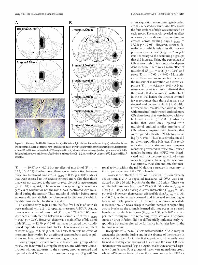
Figure 3. Histology of mPFC-BLA disconnection. A, mPFC lesions. B, BLA lesions. Largest lesions (in gray) and smallest lesions(in black) of rats included are depicted here. The unilateral images are representative of lesions in both hemispheres. Brain sectionsof the mPFC and BLA were stained with 0.1% cresyl violet to verify sites of excitotoxic damage (marked by arrowheads). Note thedarkly stained astrocytes and absence of cell bodies in lesioned tissue (4�). C, Intact mPFC. D, Lesioned mPFC. E, Lesioned BLA. F,Intact BLA.
Maeng et al. • PFC–BLA Interaction in Stress and Learning J. Neurosci., December 1, 2010 • 30(48):16188 –16196 • 16191
tivation without tail shock stressor exposure, another stressed andinjected with saline, and a saline group that was not tail shockstressed. The independent measures were stress versus no stress andactivation with picrotoxin versus saline vehicle infusion.
To assess acquisition of the CR across trials of training inmales, a 2 � 2 repeated-measures ANOVA across the four ses-sions of trials was conducted on each group. All four groupsincreased the number of CRs as training progressed (F(3,60) �7.18; p � 0.01). There was no effect of picrotoxin (F(1,20) � 0.23;p � 0.05) and no interaction between picrotoxin treatment andstress (F(1,20) � 0.21; p � 0.05). However, there was an effect ofstress (F(1,20) � 9.62; p � 0.01). Males injected with saline beforethe stressor emitted more CRs than males that were unstressedand injected with saline ( p � 0.01). Furthermore, unstressedmales treated with picrotoxin performed similarly to thosetreated with the vehicle ( p � 0.05). Most importantly, males thatwere exposed to the stressor emitted more CRs than those that
were not exposed to the stressor ( p � 0.01) regardless of thepresence of picrotoxin (Fig. 5A). The increase in responding oc-curred regardless of whether or not the mPFC was activated withpicrotoxin during the stressor. Thus, picrotoxin infusion beforestress exposure did not alter the subsequent facilitation of eye-blink conditioning elicited by stress in males.
The first five blocks of 20 trials were analyzed with a 2 � 2repeated-measures ANOVA. There was no effect of picrotoxin(F(1,20) � 0.00; p � 0.05), stress (F(1,20) � 0.27; p � 0.05), with nointeraction between picrotoxin treatment and stress (F(1,20) �0.01; p � 0.05). However, there was a main effect of blocks oftrials (F(4,80) � 9.04; p � 0.01), as males increased their condi-tioned responding across trial blocks. Thus, there was no effect ofpicrotoxin treatment or stress on early learning with changesemerging later in conditioning.
Four groups of females were also trained: one group whosemPFC was activated during the stressor, one with mPFC activation
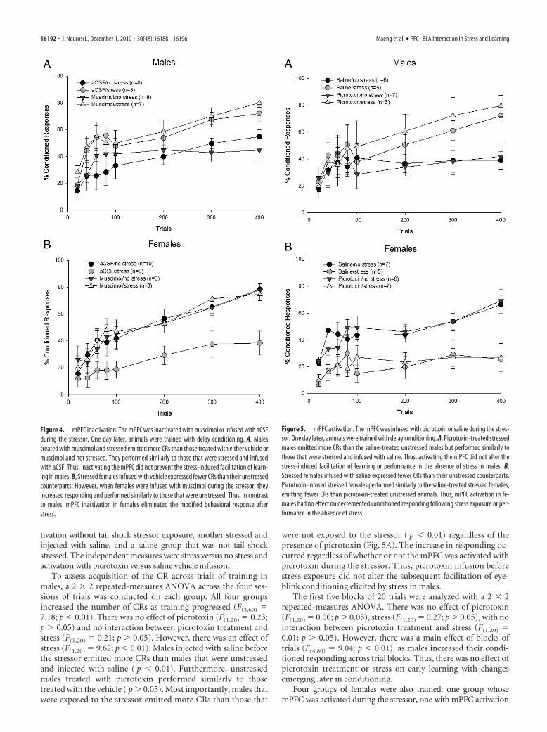
Figure 4. mPFC inactivation. The mPFC was inactivated with muscimol or infused with aCSFduring the stressor. One day later, animals were trained with delay conditioning. A, Malestreated with muscimol and stressed emitted more CRs than those treated with either vehicle ormuscimol and not stressed. They performed similarly to those that were stressed and infusedwith aCSF. Thus, inactivating the mPFC did not prevent the stress-induced facilitation of learn-ing in males. B, Stressed females infused with vehicle expressed fewer CRs than their unstressedcounterparts. However, when females were infused with muscimol during the stressor, theyincreased responding and performed similarly to those that were unstressed. Thus, in contrastto males, mPFC inactivation in females eliminated the modified behavioral response afterstress.
Figure 5. mPFC activation. The mPFC was infused with picrotoxin or saline during the stres-sor. One day later, animals were trained with delay conditioning. A, Picrotoxin-treated stressedmales emitted more CRs than the saline-treated unstressed males but performed similarly tothose that were stressed and infused with saline. Thus, activating the mPFC did not alter thestress-induced facilitation of learning or performance in the absence of stress in males. B,Stressed females infused with saline expressed fewer CRs than their unstressed counterparts.Picrotoxin-infused stressed females performed similarly to the saline-treated stressed females,emitting fewer CRs than picrotoxin-treated unstressed animals. Thus, mPFC activation in fe-males had no effect on decremented conditioned responding following stress exposure or per-formance in the absence of stress.
16192 • J. Neurosci., December 1, 2010 • 30(48):16188 –16196 Maeng et al. • PFC–BLA Interaction in Stress and Learning
without exposure to the stressor, another stressed and injected withsaline, and an unstressed saline group (Fig. 5B). To assess acquisitionacross training in females, a 2 � 2 repeated-measures ANOVAacross the four sessions of trials was conducted on each group. Theanalysis revealed an effect of session, as conditioned respondingincreased across training days (F(3,63) � 8.19; p � 0.01). However,a one-way repeated-measures ANOVA revealed that stressed fe-males did not show an increase in responding across sessions withsaline (F(3,12) � 0.47; p � 0.05) or even picrotoxin infusions(F(3,18) � 0.94; p � 0.05), whereas unstressed animals did. Therewas no effect of picrotoxin (F(1,21) � 0.02; p � 0.05) and nointeraction between picrotoxin treatment and stress (F(1,21) �0.00; p � 0.05). Using the percentage of CRs across trials of train-ing as the dependent measure, there was a main effect of stress(F(1,21) � 27.77; p � 0.01). Females that were injected with avehicle in the mPFC before the stressor emitted fewer responsesthan those that were not stressed and received vehicle or picro-toxin infusions ( p � 0.01). Moreover, females that were stressedwhile their mPFC was activated also did not learn well, emittingfewer CRs than those that were unstressed and microinjectedwith the vehicle or picrotoxin ( p � 0.01) and performing simi-larly to the vehicle-treated stressed females ( p � 0.05). Together,these data suggest that stress impairs subsequent learning vianeuronal activity within the mPFC during the stressor in females,but activation of this brain region alone may not be sufficient.
To assess the effects of stress or picrotoxin infusion on earlyacquisition, a 2 � 2 repeated-measures ANOVA was run onfive 20 trial blocks for the first 100 trials. There was no effect ofdrug (F(1,21) � 0.02; p � 0.05) and no drug � stress interaction(F(1,21) � 0.03; p � 0.05). However, there was an effect of stress(F(1,21) � 14.85; p � 0.01) and blocks (F(4,84) � 10.57; p �0.01), as the animals learned and increased responding as blocksof trials proceeded. However, a one-way repeated-measuresANOVA revealed that this increase in responding across blocksdid not occur in stressed females with vehicle (F(4,16) � 2.54; p �0.05) as well as picrotoxin infusions (F(4,24) � 2.35; p � 0.05).Thus, there was no effect of picrotoxin but an effect of stress onearly acquisition as well as later conditioned responding.
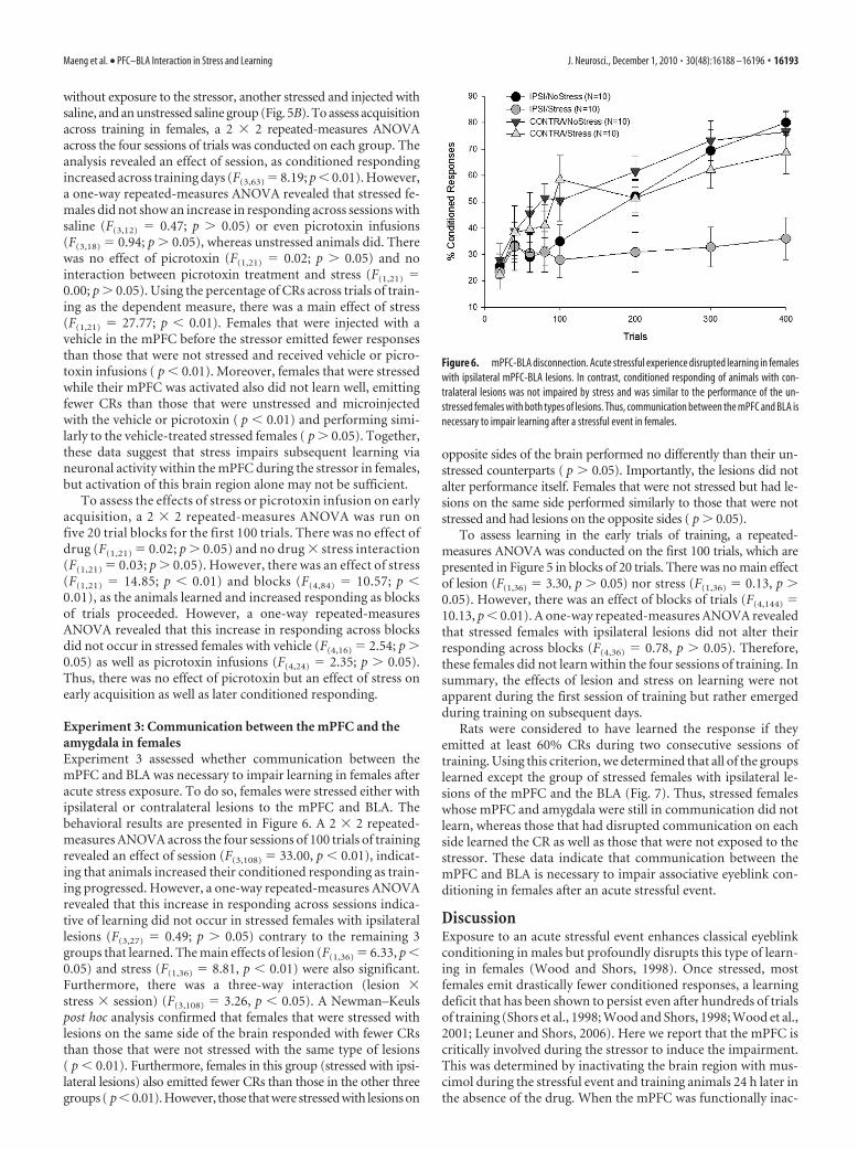
Experiment 3: Communication between the mPFC and theamygdala in femalesExperiment 3 assessed whether communication between themPFC and BLA was necessary to impair learning in females afteracute stress exposure. To do so, females were stressed either withipsilateral or contralateral lesions to the mPFC and BLA. Thebehavioral results are presented in Figure 6. A 2 � 2 repeated-measures ANOVA across the four sessions of 100 trials of trainingrevealed an effect of session (F(3,108) � 33.00, p � 0.01), indicat-ing that animals increased their conditioned responding as train-ing progressed. However, a one-way repeated-measures ANOVArevealed that this increase in responding across sessions indica-tive of learning did not occur in stressed females with ipsilaterallesions (F(3,27) � 0.49; p � 0.05) contrary to the remaining 3groups that learned. The main effects of lesion (F(1,36) � 6.33, p �0.05) and stress (F(1,36) � 8.81, p � 0.01) were also significant.Furthermore, there was a three-way interaction (lesion �stress � session) (F(3,108) � 3.26, p � 0.05). A Newman–Keulspost hoc analysis confirmed that females that were stressed withlesions on the same side of the brain responded with fewer CRsthan those that were not stressed with the same type of lesions( p � 0.01). Furthermore, females in this group (stressed with ipsi-lateral lesions) also emitted fewer CRs than those in the other threegroups ( p�0.01). However, those that were stressed with lesions on
opposite sides of the brain performed no differently than their un-stressed counterparts ( p � 0.05). Importantly, the lesions did notalter performance itself. Females that were not stressed but had le-sions on the same side performed similarly to those that were notstressed and had lesions on the opposite sides ( p � 0.05).
To assess learning in the early trials of training, a repeated-measures ANOVA was conducted on the first 100 trials, which arepresented in Figure 5 in blocks of 20 trials. There was no main effectof lesion (F(1,36) � 3.30, p � 0.05) nor stress (F(1,36) � 0.13, p �0.05). However, there was an effect of blocks of trials (F(4,144) �10.13, p � 0.01). A one-way repeated-measures ANOVA revealedthat stressed females with ipsilateral lesions did not alter theirresponding across blocks (F(4,36) � 0.78, p � 0.05). Therefore,these females did not learn within the four sessions of training. Insummary, the effects of lesion and stress on learning were notapparent during the first session of training but rather emergedduring training on subsequent days.
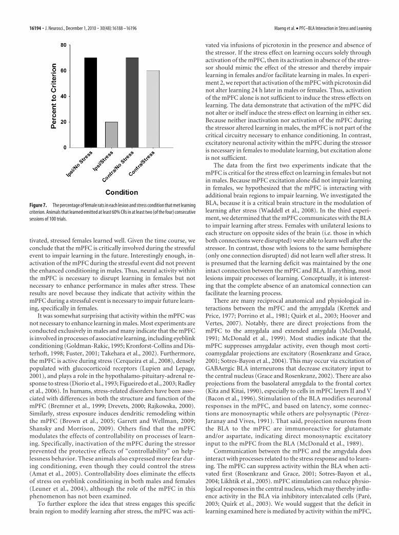
Rats were considered to have learned the response if theyemitted at least 60% CRs during two consecutive sessions oftraining. Using this criterion, we determined that all of the groupslearned except the group of stressed females with ipsilateral le-sions of the mPFC and the BLA (Fig. 7). Thus, stressed femaleswhose mPFC and amygdala were still in communication did notlearn, whereas those that had disrupted communication on eachside learned the CR as well as those that were not exposed to thestressor. These data indicate that communication between themPFC and BLA is necessary to impair associative eyeblink con-ditioning in females after an acute stressful event.
DiscussionExposure to an acute stressful event enhances classical eyeblinkconditioning in males but profoundly disrupts this type of learn-ing in females (Wood and Shors, 1998). Once stressed, mostfemales emit drastically fewer conditioned responses, a learningdeficit that has been shown to persist even after hundreds of trialsof training (Shors et al., 1998; Wood and Shors, 1998; Wood et al.,2001; Leuner and Shors, 2006). Here we report that the mPFC iscritically involved during the stressor to induce the impairment.This was determined by inactivating the brain region with mus-cimol during the stressful event and training animals 24 h later inthe absence of the drug. When the mPFC was functionally inac-
Figure 6. mPFC-BLA disconnection. Acute stressful experience disrupted learning in femaleswith ipsilateral mPFC-BLA lesions. In contrast, conditioned responding of animals with con-tralateral lesions was not impaired by stress and was similar to the performance of the un-stressed females with both types of lesions. Thus, communication between the mPFC and BLA isnecessary to impair learning after a stressful event in females.
Maeng et al. • PFC–BLA Interaction in Stress and Learning J. Neurosci., December 1, 2010 • 30(48):16188 –16196 • 16193
tivated, stressed females learned well. Given the time course, weconclude that the mPFC is critically involved during the stressfulevent to impair learning in the future. Interestingly enough, in-activation of the mPFC during the stressful event did not preventthe enhanced conditioning in males. Thus, neural activity withinthe mPFC is necessary to disrupt learning in females but notnecessary to enhance performance in males after stress. Theseresults are novel because they indicate that activity within themPFC during a stressful event is necessary to impair future learn-ing, specifically in females.
It was somewhat surprising that activity within the mPFC wasnot necessary to enhance learning in males. Most experiments areconducted exclusively in males and many indicate that the mPFCis involved in processes of associative learning, including eyeblinkconditioning (Goldman-Rakic, 1995; Kronforst-Collins and Dis-terhoft, 1998; Fuster, 2001; Takehara et al., 2002). Furthermore,the mPFC is active during stress (Cerqueira et al., 2008), denselypopulated with glucocorticoid receptors (Lupien and Lepage,2001), and plays a role in the hypothalamo-pituitary-adrenal re-sponse to stress (Diorio et al., 1993; Figueiredo et al., 2003; Radleyet al., 2006). In humans, stress-related disorders have been asso-ciated with differences in both the structure and function of themPFC (Bremner et al., 1999; Drevets, 2000; Rajkowska, 2000).Similarly, stress exposure induces dendritic remodeling withinthe mPFC (Brown et al., 2005; Garrett and Wellman, 2009;Shansky and Morrison, 2009). Others find that the mPFCmodulates the effects of controllability on processes of learn-ing. Specifically, inactivation of the mPFC during the stressorprevented the protective effects of “controllability” on help-lessness behavior. These animals also expressed more fear dur-ing conditioning, even though they could control the stress(Amat et al., 2005). Controllability does eliminate the effectsof stress on eyeblink conditioning in both males and females(Leuner et al., 2004), although the role of the mPFC in thisphenomenon has not been examined.
To further explore the idea that stress engages this specificbrain region to modify learning after stress, the mPFC was acti-
vated via infusions of picrotoxin in the presence and absence ofthe stressor. If the stress effect on learning occurs solely throughactivation of the mPFC, then its activation in absence of the stres-sor should mimic the effect of the stressor and thereby impairlearning in females and/or facilitate learning in males. In experi-ment 2, we report that activation of the mPFC with picrotoxin didnot alter learning 24 h later in males or females. Thus, activationof the mPFC alone is not sufficient to induce the stress effects onlearning. The data demonstrate that activation of the mPFC didnot alter or itself induce the stress effect on learning in either sex.Because neither inactivation nor activation of the mPFC duringthe stressor altered learning in males, the mPFC is not part of thecritical circuitry necessary to enhance conditioning. In contrast,excitatory neuronal activity within the mPFC during the stressoris necessary in females to modulate learning, but excitation aloneis not sufficient.
The data from the first two experiments indicate that themPFC is critical for the stress effect on learning in females but notin males. Because mPFC excitation alone did not impair learningin females, we hypothesized that the mPFC is interacting withadditional brain regions to impair learning. We investigated theBLA, because it is a critical brain structure in the modulation oflearning after stress (Waddell et al., 2008). In the third experi-ment, we determined that the mPFC communicates with the BLAto impair learning after stress. Females with unilateral lesions toeach structure on opposite sides of the brain (i.e. those in whichboth connections were disrupted) were able to learn well after thestressor. In contrast, those with lesions to the same hemisphere(only one connection disrupted) did not learn well after stress. Itis presumed that the learning deficit was maintained by the oneintact connection between the mPFC and BLA. If anything, mostlesions impair processes of learning. Conceptually, it is interest-ing that the complete absence of an anatomical connection canfacilitate the learning process.
There are many reciprocal anatomical and physiological in-teractions between the mPFC and the amygdala (Krettek andPrice, 1977; Porrino et al., 1981; Quirk et al., 2003; Hoover andVertes, 2007). Notably, there are direct projections from themPFC to the amygdala and extended amygdala (McDonald,1991; McDonald et al., 1999). Most studies indicate that themPFC suppresses amygdalar activity, even though most corti-coamygdalar projections are excitatory (Rosenkranz and Grace,2001; Sotres-Bayon et al., 2004). This may occur via excitation ofGABAergic BLA interneurons that decrease excitatory input tothe central nucleus (Grace and Rosenkranz, 2002). There are alsoprojections from the basolateral amygdala to the frontal cortex(Kita and Kitai, 1990), especially to cells in mPFC layers II and V(Bacon et al., 1996). Stimulation of the BLA modifies neuronalresponses in the mPFC, and based on latency, some connec-tions are monosynaptic while others are polysynaptic (Perez-Jaranay and Vives, 1991). That said, projection neurons fromthe BLA to the mPFC are immunoreactive for glutamateand/or aspartate, indicating direct monosynaptic excitatoryinput to the mPFC from the BLA (McDonald et al., 1989).
Communication between the mPFC and the amgydala doesinteract with processes related to the stress response and to learn-ing. The mPFC can suppress activity within the BLA when acti-vated first (Rosenkranz and Grace, 2001; Sotres-Bayon et al.,2004; Likhtik et al., 2005). mPFC stimulation can reduce physio-logical responses in the central nucleus, which may thereby influ-ence activity in the BLA via inhibitory intercalated cells (Pare,2003; Quirk et al., 2003). We would suggest that the deficit inlearning examined here is mediated by activity within the mPFC,
Figure 7. The percentage of female rats in each lesion and stress condition that met learningcriterion. Animals that learned emitted at least 60% CRs in at least two (of the four) consecutivesessions of 100 trials.
16194 • J. Neurosci., December 1, 2010 • 30(48):16188 –16196 Maeng et al. • PFC–BLA Interaction in Stress and Learning
which then modulates the expression of fear during aversivelearning via output from the amygdala. Indeed, many studiesreport inhibitory control of the amygdala by the mPFC duringemotional learning, such as during extinction (Morgan et al.,1993; Morgan and LeDoux, 1995; Quirk et al., 2000; Sotres-Bayon et al., 2004). Alternatively, the amygdala may modulateactivity in the mPFC to impair learning. Acute stress exposureprevents the induction of LTP in vivo within the mPFC in re-sponse to stimulation of the BLA-mPFC pathway (Maroun andRichter-Levin, 2003). Others found that BLA stimulation modu-lates neuronal activity in the mPFC (Perez-Jaranay and Vives,1991). Furthermore, fear conditioning, which relies on the amyg-dala, can inhibit activity of prefrontal cortical neurons (Garcia etal., 1999), again pointing to amygdalar regulation of the mPFC.Alternatively, it could be that concurrent activity within the BLAand the mPFC is necessary to impair learning after stress. Bothstructures can modulate performance of the conditioned eye-blink response (Powell et al., 1996; Kronforst-Collins and Dister-hoft, 1998; Lee and Kim, 2004). They project not just to eachother but to brain structures involved in the limbic-hypothalamic-pituitary-adrenal stress circuit (Lopez et al., 1999).Previously, we found that the hippocampus is involved inthese effects of stress on learning (Bangasser et al., 2007).Thus, activity within the mPFC may communicate with theamygdala by way of the hippocampus.
The vast majority of studies about the mPFC and BLA havebeen conducted exclusively in males, but a few studies do reportsex differences. In ovariectomized females, stress and estrogentogether induce dendritic arborization in neurons that projectfrom the mPFC to the BLA when compared with the same mea-sures in males (Shansky and Morrison, 2009). The learning defi-cit after stress in females depends on the presence of estrogen(Wood and Shors, 1998), and thus it seems likely that estrogen isacting within one and/or the other structure to modulate learn-ing. Regardless of the exact mechanism, the present data indicatethat the mPFC and the amygdala interact with each other toimpair associative learning, specifically in females. Minimally,they indicate that males and females are using different brainregions and circuits to modify learning after stress. More gener-ally, they may provide clues as to why women are so much morevulnerable than men are to stress-related mental illness, such aspost-traumatic stress disorder and depression.
ReferencesAmat J, Baratta MV, Paul E, Bland ST, Watkins LR, Maier SF (2005) Medial
prefrontal cortex determines how stressor controllability affects behaviorand dorsal raphe nucleus. Nat Neurosci 8:365–371.
Bacon SJ, Headlam AJ, Gabbott PL, Smith AD (1996) Amygdala input tomedial prefrontal cortex (mPFC) in the rat: a light and electron micro-scope study. Brain Res 720:211–219.
Bangasser DA, Shors TJ (2004) Acute stress impairs trace eyeblink condi-tioning in females without altering the unconditioned response. Neuro-biol Learn Mem 82:57– 60.
Bangasser DA, Shors TJ (2007) The hippocampus is necessary for enhance-ments and impairments of learning following stress. Nat Neurosci10:1401–1403.
Bangasser DA, Shors TJ (2008) The bed nucleus of the stria terminalis mod-ulates learning after stress in masculinized but not cycling females. J Neu-rosci 28:6383– 6387.
Bangasser DA, Santollo J, Shors TJ (2005) The bed nucleus of the stria ter-minalis is involved in the persistent increase in associative learning afterstress. Behav Neurosci 119:1459 –1466.
Bremner JD, Staib LH, Kaloupek D, Southwick SM, Soufer R, Charney DS(1999) Neural correlates of exposure to traumatic pictures and sound inVietnam combat veterans with and without posttraumatic stress disorder:a positron emission tomography study. Biol Psychiatry 45:806 – 816.
Brown SM, Henning S, Wellman CL (2005) Mild, short-term stress altersdendritic morphology in rat medial prefrontal cortex. Cereb Cortex15:1714 –1722.
Carter-Snell C, Hegadoren K (2003) Stress disorders and gender: implica-tions for theory and research. Can J Nurs Res 35:34 –55.
Cerqueira JJ, Almeida OF, Sousa N (2008) The stressed prefrontal cortex.Left? Right! Brain Behav Immun 22:630 – 638.
Diorio D, Viau V, Meaney MJ (1993) The role of the medial prefrontalcortex (cingulate gyrus) in the regulation of hypothalamic-pituitary-adrenal responses to stress. J Neurosci 13:3839 –3847.
Drevets WC (2000) Neuroimaging studies of mood disorders. Biol Psychi-atry 48:813– 829.
Figueiredo HF, Bruestle A, Bodie B, Dolgas CM, Herman JP (2003) Themedial prefrontal cortex differentially regulates stress-induced c-fos ex-pression in the forebrain depending on type of stressor. Eur J Neurosci18:2357–2364.
Foa EB, Street GP (2001) Women and traumatic events. J Clin Psychiatry 62[Suppl 17]:29 –34.
Fuster JM (2001) The prefrontal cortex—an update: time is of the essence.Neuron 30:319 –333.
Garcia R, Vouimba RM, Baudry M, Thompson RF (1999) The amygdalamodulates prefrontal cortex activity relative to conditioned fear. Nature402:294 –296.
Garrett JE, Wellman CL (2009) Chronic stress effects on dendritic morphol-ogy in medial prefrontal cortex: sex differences and estrogen dependence.Neuroscience 162:195–207.
Gerrits M, Bakker PL, Koch T, Ter Horst GJ (2006) Stress-induced sensiti-zation of the limbic system in ovariectomized rats is partly restored bycyclic 17�-estradiol administration. Eur J Neurosci 23:1747–1756.
Goldman-Rakic PS (1995) Cellular basis of working memory. Neuron 14:477– 485.
Grace AA, Rosenkranz JA (2002) Regulation of conditioned responses ofbasolateral amygdala neurons. Physiol Behav 77:489 – 493.
Heidbreder CA, Groenewegen HJ (2003) The medial prefrontal cortex inthe rat: evidence for a dorso-ventral distinction based upon functionaland anatomical characteristics. Neurosci Biobehav Rev 27:555–579.
Hoover WB, Vertes RP (2007) Anatomical analysis of afferent projections tothe medial prefrontal cortex in rat. Brain Struct Funct 212:149 –179.
Kessler RC, Sonnega A, Bromet E, Hughes M, Nelson CB (1995) Posttrau-matic stress disorder in the National Comorbidity Survey. Arch Gen Psy-chiatry 52:1048 –1060.
Kita H, Kitai ST (1990) Amygdaloid projections to the frontal cortex and thestriatum in the rat. J Comp Neurol 298:40 – 49.
Krettek JE, Price JL (1977) Projections from the amygdaloid complex tothe cerebral cortex and thalamus in the rat and cat. J Comp Neurol172:687–722.
Kronforst-Collins MA, Disterhoft JF (1998) Lesions of the caudal area ofrabbit medial prefrontal cortex impair trace eyeblink conditioning. Neu-robiol Learn Mem 69:147–162.
Lee T, Kim JJ (2004) Differential effects of cerebellar, amygdalar and hip-pocampal lesions on classical eyeblink conditioning in rats. J Neurosci24:3242–3250.
Leuner B, Shors TJ (2006) Learning during motherhood: a resistance tostress. Horm Behav 50:38 –51.
Leuner B, Mendolia-Loffredo S, Shors TJ (2004) Males and females responddifferently to controllability and antidepressant treatment. Biol Psychia-try 56:964 –970.
Likhtik E, Pelletier JG, Paz R, Pare D (2005) Prefrontal control of the amyg-dala. J Neurosci 25:7429 –7437.
Lopez JF, Akil H, Watson SJ (1999) Neural circuits mediating stress. BiolPsychiatry 46:1461–1471.
Luine V (2002) Sex differences in chronic stress effects on memory in rats.Stress 5:205–216.
Lupien SJ, Lepage M (2001) Stress, memory, and the hippocampus: can’tlive with it, can’t live without it. Behav Brain Res 127:137–158.
Maroun M, Richter-Levin G (2003) Exposure to acute stress blocks the in-duction of long-term potentiation of the amygdala—prefrontal cortexpathway in vivo. J Neurosci 23:4406 – 4409.
McDonald AJ (1991) Organization of amygdaloid projections to the pre-frontal cortex and associated striatum in the rat. Neuroscience 44:1–14.
McDonald AJ, Beitz AJ, Larson AA, Kuriyama R, Sellitto C, Madl JE (1989)Co-localisation of glutamate and tubulin in putative excitatory neurons of
Maeng et al. • PFC–BLA Interaction in Stress and Learning J. Neurosci., December 1, 2010 • 30(48):16188 –16196 • 16195
the hippocampus and amygdala: an immunohistochemical study usingmonoclonal antibodies. Neuroscience 30:405– 421.
McDonald AJ, Shammah-Lagnado SJ, Shi C, Davis M (1999) Cortical affer-ents to the extended amygdala. Ann N Y Acad Sci 877:309 –338.
Morgan MA, LeDoux JE (1995) Differential contribution of dorsal and ven-tral medial prefrontal cortex to the acquisition and extinction of condi-tioned fear in rats. Behav Neurosci 109:681– 688.
Morgan MA, Romanski LM, LeDoux JE (1993) Extinction of emotionallearning: contribution of medial prefrontal cortex. Neurosci Lett163:109 –113.
Pare D (2003) Role of the basolateral amygdala in memory consolidation.Prog Neurobiol 70:409 – 420.
Perez-Jaranay JM, Vives F (1991) Electrophysiological study of the responseof medial prefrontal cortex neurons to stimulation of the basolateral nu-cleus of the amygdala in the rat. Brain Res 564:97–101.
Porrino LJ, Crane AM, Goldman-Rakic PS (1981) Direct and indirect path-ways from the amygdala to the frontal lobe in rhesus monkeys. J CompNeurol 198:121–136.
Powell DA, Maxwell B, Penney J (1996) Neuronal activity in the medialprefrontal cortex during Pavlovian eyeblink and nictitating membraneconditioning. J Neurosci 16:6296 – 6306.
Quirk GJ, Russo GK, Barron JL, Lebron K (2000) The role of ventromedialprefrontal cortex in the recovery of extinguished fear. J Neurosci20:6225– 6231.
Quirk GJ, Likhtik E, Pelletier JG, Pare D (2003) Stimulation of medial pre-frontal cortex decreases the responsiveness of central amygdala outputneurons. J Neurosci 23:8800 – 8807.
Radley JJ, Arias CM, Sawchenko PE (2006) Regional differentiation of themedial prefrontal cortex in regulating adaptive responses to acute emo-tional stress. J Neurosci 26:12967–12976.
Rajkowska G (2000) Postmortem studies in mood disorders indicate alterednumbers of neurons and glial cells. Biol Psychiatry 48:766 –777.
Rosenkranz JA, Grace AA (2001) Dopamine attenuates prefrontal corticalsuppression of sensory inputs to the basolateral amygdala of rats. J Neu-rosci 21:4090 – 4103.
Servatius RJ, Shors TJ (1994) Exposure to inescapable stress persistently fa-cilitates associative and nonassociative learning in rats. Behav Neurosci108:1101–1106.
Shansky RM, Morrison JH (2009) Stress-induced dendritic remodeling in
the medial prefrontal cortex: effects of circuit, hormones and rest. BrainRes 1293:108 –113.
Shansky RM, Rubinow K, Brennan A, Arnsten AF (2006) The effects of sexand hormonal status on restraint-stress-induced working memory im-pairment. Behav Brain Funct 2:8.
Shors TJ (2004) Learning during stressful times. Learn Mem 11:137–144.Shors TJ, Servatius RJ (1997) The contribution of stressor intensity, dura-
tion, and context to the stress-induced facilitation of associative learning.Neurobiol Learn Mem 68:92–96.
Shors TJ, Lewczyk C, Pacynski M, Mathew PR, Pickett J (1998) Stages ofestrous mediate the stress-induced impairment of associative learning inthe female rat. Neuroreport 9:419 – 423.
Sotres-Bayon F, Bush DE, LeDoux JE (2004) Emotional perseveration: anupdate on prefrontal-amygdala interactions in fear extinction. LearnMem 11:525–535.
Takehara K, Kawahara S, Takatsuki K, Kirino Y (2002) Time-limited role ofthe hippocampus in the memory for trace eyeblink conditioning in mice.Brain Res 951:183–190.
Ter Horst GJ, Wichmann R, Gerrits M, Westenbroek C, Lin Y (2009) Sexdifferences in stress responses: focus on ovarian hormones. Physiol Behav97:239 –249.
Tolin DF, Foa EB (2006) Sex differences in trauma and posttraumatic stressdisorder: a quantitative review of 25 years of research. Psychol Bull132:959 –992.
Vertes RP (2004) Differential projections of the infralimbic and prelimbiccortex in the rat. Synapse 51:32–58.
Vertes RP (2006) Interactions among the medial prefrontal cortex, hip-pocampus and midline thalamus in emotional and cognitive processingin the rat. Neuroscience 142:1–20.
Waddell J, Bangasser DA, Shors TJ (2008) The basolateral nucleus of theamygdala is necessary to induce the opposing effects of stressful experi-ence on learning in males and females. J Neurosci 28:5290 –5294.
Wood GE, Shors TJ (1998) Stress facilitates classical conditioning in malesbut impairs conditioning in females through activational influences ofovarian hormones. Proc Natl Acad Sci U S A 95:4066 – 4071.
Wood GE, Beylin AV, Shors TJ (2001) The contribution of adrenal andreproductive hormones to the opposing effects of stress on trace condi-tioning in males versus females. Behav Neurosci 115:175–187.
16196 • J. Neurosci., December 1, 2010 • 30(48):16188 –16196 Maeng et al. • PFC–BLA Interaction in Stress and Learning