Embed Size (px)
Citation preview

Leptosomatides brevicaudatus n. sp. and a Redescriptionof Leptosomatides marinae Platonova, 1967
(Enoplida: Leptosomatidae) 1
Kenji Kito2 and W. Duane Hope3
Abstract: The free-living marine nematodes Leptosomatides brevicaudatus n. sp. and L. marinae weredescribed and redescribed, respectively, from material collected in the northwest Pacific. Leptosomatidesbrevicaudatus n. sp. from Simushir Island differs from L. marinae in the ratio c8 (body length divided bytail length measured on the chord) and the length of the spicules. Leptosomatides marinae is redescribedfrom light microscopy (LM) observations of the type specimens and LM and scanning electron micros-copy (SEM) observations of specimens from Hokkaido, Japan. It appears to be impossible to distinguishamong some species of Leptosomatides because they are either insufficiently described or known only fromfemales. Secondary sexual characters of males are essential for purposes of identification.
Key words: Leptosomatides brevicaudatus, Leptosomatides marinae, marine nematode, nematode, new spe-cies, SEM observation, subventral supplement, taxonomy.
A small population of large-bodied ma-rine nematodes of the genus LeptosomatidesFilipjev, 1918 was found on the Pacific coastof Hokkaido, Japan. The nematodes mostclosely resemble Leptosomatides marinae Pla-tonova, 1976, whose known distribution isthe northwest Pacific (Belogurov and Fadee-va, 1985; Platonova, 1976, 1978, 1981) andEast Siberian Sea (Platonova and Ku-langieva, 1995) (Fig. 1). The genus Leptoso-matides was recently revised by Bongers(1984) with 14 nominal species, and L. ma-rinae was redescribed. Also, after examiningthe type specimens of L. acutipapillosus Pla-tonova, 1976 and L. brevisetosus Platonova,1976, Bongers (1984) followed the opinionof Platonova by designating both species asjunior subjective synonyms of L. marinae.However, identification of our specimenswas uncertain because the original descrip-tion of L. marinae was inadequate andBongers (1984) redescription and synony-mizations introduced diagnostic inconsis-
tencies. To resolve this difficulty, we exam-ined the specimens from Hokkaido, which isclose to the type locality Iturup Island, withlight microscopy (LM) and scanning elec-tron microscopy (SEM) and compared themwith paratype specimens of L. marinae and L.brevisetosus.
Materials and Methods
The nematodes were collected on 23 Au-gust 1992 from sponges, Halichondria sp.,growing on rocks in the littoral zone nearthe Akkeshi Marine Biological Station ofHokkaido University in Akkeshi Bay, Hok-kaido, Japan (Fig. 2). The specimens werefixed in 4% formalin in seawater and exam-ined with LM and SEM. For LM, 5 males and5 females were mounted in anhydrous glyc-erin between two coverslips on Cobb alumi-num frames, H-S slides, or photographicslide mounts (Abe et al., 1993). Two malesand 1 female were dissected for examinationof the ventromedian supplement, spiculesand gubernaculum, and the vulval region.Five males and 2 females selected for SEMwere examined by the method described inHope (1982).
Three male and 2 female paratypes of L.marinae collected at the type locality werecompared with the Japanese specimens.They were mounted in glycerin-jelly on glassslides. Three paratype females of L. marinaeand 3 paratype males of L. brevisetosus usedin the redescription of L. marinae by
Received for publication 13 January 1999.1 Supported in part by a grant for the first author from the
Smithsonian Institution.2 Department of Biology, School of Medicine, Sapporo Medi-
cal University, Sapporo 060-8556, Japan.3 Department of Invertebrate Zoology, National Museum of
Natural History, Smithsonian Institution, Washington, DC,20560.
E-mail: [email protected] authors thank Abbie Yorkoff and Susann Braden for
their technical assistance. The authors also thank the followingpersons for the loan of type specimens of Leptosomatides marinaeand L. brevisetosus: Tom Bongers, Valentina V. Galt’sova, D.Hirst, Chittima Aryuthaka, and Yoshihisa Shirayama.
This paper was edited by E. C. Bernard.
Journal of Nematology 31(4):460–474. 1999.
460

Bongers (1984) were also examined. Thesespecimens were remounted in glycerin onaluminum slide frames.
Morphometric data were obtained fromcamera lucida drawings. Data regarding bi-laterally paired structures are generallygiven as ‘‘right;left’’ except for same figureor as a combined range in the text andtables. If paratypes were flattened, correctedbody diameters were calculated using thesecond formula in Geraert (1961). In thisstudy tail length on chord (t8) was measuredas a distance from anus (cloaca) to tail tipand de Man’s c was replaced by c8.
Besides standard abbreviations, the fol-lowing are used in this paper: abd = bodydiameter at level of the cloacal (anal) open-ing; cbd = corresponding body diameter;mbd = maximum body diameter; SA/t8 =spicule length measured on arc divided byt8; SC/t8 = spicule length measured onchord divided by t8; t = tail length; V = posi-tion of vulva as a percentage of the body
length from the anterior body end. Theterm papilla is used as defined by Bongers(1984).
Systematics
Family Leptosomatidae Filipjev, 1918Leptosomatides marinae Platonova, 1976
(Figs. 3–29, Tables 1–3)
Description (emended)
Leptosomatides marinae Platonova, 1976: 70,77–79, Fig. 29; Platonova, 1978: 498, Fig. 2-1;1981: 60, Fig. 4 (morphological study onhead); Bongers, 1984: 24–25, Figs. 3-C,4-A,B, Appendix (partim; text, figures, andmeasurements for females only); Belogurovand Fadeeva, 1985: 184–185, Fig. 1 (mor-phological study on head); Platonova andKulangieva, 1995: 175.
Leptosomatides acutipapillosus Platonova,1976: 70, 75–76, Fig. 27 (op. Platonova inBongers, 1984: 25).
Figs. 1, 2. Leptosomatides spp. collected in the vicinity of Japan and eastern part of Russia. 1) Distribution of theleptosomatid species previously reported: A = L. acutipapillosus, B = L. brevisetosus, F = L. filiformis, G = L. grebnickii,M = L. marinae, S = L. steineri. 2) Localities where leptosomatid specimens are known in the vicinity of Hokkaido.
New, Known Leptosomatid Nematodes: Kito, Hope 461

Leptosomatides brevisetosus Platonova, 1976:70, 76–77, Fig. 28 (op. Platonova in Bongers,1984: 25).
nec. Leptosomatides marinae: Bongers,1984: 13–14, 24–25, Figs. 3A-B, Appendix(partim; text, figures, and measurements formales only. See description of Leptosomatidesbrevicaudatus n. sp. below.)
Males (n = 5): Body 12.0–14.1 mm long,gradually tapering to anterior extremity(Fig. 3), abruptly tapering from level ofcloaca to blunt end of curved tail (Fig. 7).Diameter of body at level of cephalic sensilla47–49 µm, at nerve ring 108–119 µm, at pos-terior end of esophagus 126–149 µm, at mid-body 159–176 µm, at level between midbodyand cloacal vent (mbd) 164–183, and atlevel of cloacal vent (abd) 131–141 µm.Transverse striae observed with SEM incephalic (Figs. 12–14) and caudal regions.Somatic papilliform sensilla observed in sub-median position (Figs. 12,13,16,18). Ortho-metanemes and type I and II loxomet-anemes present in lateral chord; type IIloxometanemes scarce.
Head end (Figs. 4,12–13) rounded, with 6+ 10 sensilla. Inner labial sensilla papilli-form. Distance from head end to circle ofouter labial and cephalic sensilla 18–22 µm;outer labial and cephalic sensilla papilli-form, 3.2–3.8 µm long. Cervical sensilla pa-pilliform, about 3 µm long (Figs. 3,4,13).Cephalic capsule tapered anteriorly, margincrenate posteriorly, 11–15 µm long. Dis-tance from head end to amphidal aperture19–30 µm; fovea circular to transversely oval,7.6–10.9 µm wide; external aperture trans-versely elliptical, 5.1–7.0 µm wide. Distancefrom head end to ocellar lens 83–106 µm or1.1–1.3 cbd; diameter of lens and pigmentspots 6.2–7.6 and 12–17 µm, respectively.Oral aperture triradiate, embraced by onedorsal and two subventral inner (microla-bia) and outer (mandibular ridges) ele-ments (see Hope, 1982) (Figs. 12,14). Dor-sal microlabium narrower than subventralmicrolabium (Fig. 14). Mandibular ridgeswithout odontia. Esophagus 1.7–2.0 mmlong, slightly clavate; width of esophagus atanterior margin of nerve ring 28–33 µm and
Figs. 3–6. Leptosomatides marinae. 3, 4) Male (ZIHU 1243). 3) Anterior region, left lateral view. 4) Head, leftlateral view. 5, 6) Female (5, ZIHU 1252; 6, ZIHU 1253). 5) Vulval region, left lateral view. 6) Vulval region, ventralview. Scale = 200 µm in Fig. 3, 50 µm in Fig. 4, 100 µm in Figs. 5, 6.
462 Journal of Nematology, Volume 31, No. 4, December 1999

55–68 µm at posterior end of esophagus.Distance from head end to nerve ring 434–482 µm or 24.1–26.1% of esophageal length.Renette cell and pore not observed.
Reproductive system diorchic; testes op-posed with anterior testis on right and pos-terior on left of gut in two males, and bothtestes on left in three males. Spicules (Figs.7,8) paired, robust; proximal third straight,midregion dorsoventrally expanded, distalthird curved and tapered to tip. Spicules(right; left) 134–142;129–142 µm long onarc, 128–134;122–136 µm long on chord,or 0.9–1.1 (on arc) and 0.9–1.0 (on chord)abd. Spicule length in relation to taillength: SA/t8 = 0.9–1.1, SC/t8 = 0.9–1.0. Gu-bernaculum with right and left corporajoined medially by cuticularized bridgeacross posterior margin of spicula; each cor-pus comprised of parallel anterior and pos-terior ribs, fused together dorsally to formflattened cuneus; cuneus enveloped by in-sertions of protractor and retractor muscles;
anterior rib of corpus with anteriorly di-rected, triangular crus hooked mesad to em-brace adjacent spicule; distal end of corpuswith lateral bulge 8–15 µm by 5–8 µm (Figs.7,9,28). Length of gubernaculum from ven-tral end to dorsal edge of cuneus, 81–102;80–104 µm or 60–74% of spicule lengthon arc.
Ventromedian supplement (Figs. 7,17–18,20–22,28) raised, 8–10 µm long, with cy-lindrical sensory process in shallow depres-sion; sensory process with central pore; dis-tance from center of supplement to cloacalopening 108–161 µm or 0.8–1.2 abd. Ven-tromedian supplement provided with ante-rior and posterior alae of fine, intra-cuticular punctations (Fig. 22); anterior andposterior alae 17–22 and 15–18 µm long, re-spectively. Subventral supplements (Figs.16–19) arranged in 16–20 pairs in two lon-gitudinal rows; pairs sometimes irregular inarrangement and missing a supplement;7–10 supplements anterior to ventromedian
Figs. 7–11. Leptosomatides marinae. 7–9) Male (7, ZIHU 1243; 8, 9, ZIHU 1245). 7) Posterior region, right lateralview. 8) Spicule, right. 9) Gubernaculum, right. 10, 11) Female (10, ZIHU 1252; 11, ZIHU 1253). 10) Tail, rightlateral view. 11) Tail, ventral view. Scale = 100 µm in Figs. 7, 10, 11; 20 µm in Figs. 8, 9.
New, Known Leptosomatid Nematodes: Kito, Hope 463
supplement, 6–8 between ventromediansupplement and cloacal vent, 1–3 on tail.Anterior 5–9 supplements well-developed,each bearing single seta between two longi-tudinal, crescent-like elevations of cuticle;posterior supplements small, round, pro-vided with papilla or short seta. Distancefrom cloacal vent to anteriormost subventralsupplement 2.3–3.7 abd or 2.4–4.2% of body
length (Fig. 7). Posteriomost supplementsconverging toward ventromedian plane, 30–53% of tail length on chord from cloaca(Figs. 17,19).
Tail short, conical, with taper mostly ondorsal side, length 143–161 µm or 1.0–1.2abd; t8 = 131–149 µm. Papillae and (or)minute setae present on tail, especially neartail terminus. Caudal gland cells three, cell
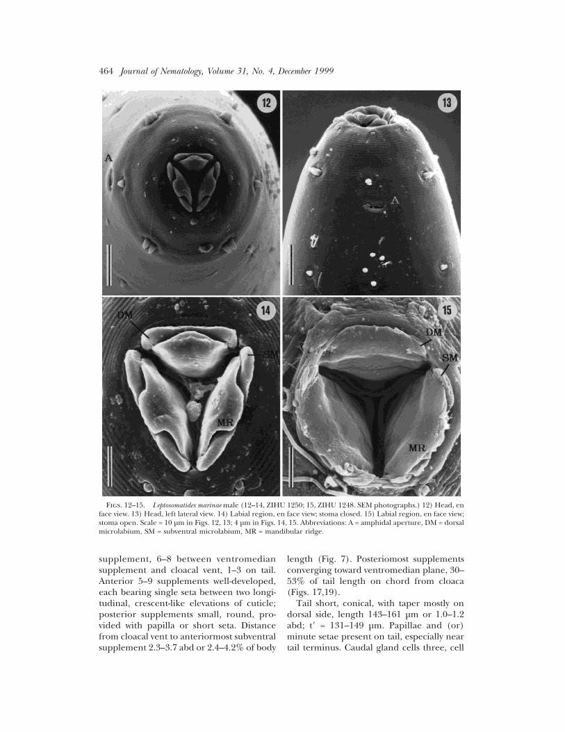
Figs. 12–15. Leptosomatides marinae male (12–14, ZIHU 1250; 15, ZIHU 1248. SEM photographs.) 12) Head, enface view. 13) Head, left lateral view. 14) Labial region, en face view; stoma closed. 15) Labial region, en face view;stoma open. Scale = 10 µm in Figs. 12, 13; 4 µm in Figs. 14, 15. Abbreviations: A = amphidal aperture, DM = dorsalmicrolabium, SM = subventral microlabium, MR = mandibular ridge.
464 Journal of Nematology, Volume 31, No. 4, December 1999

bodies extending anterior to cloaca; dis-tance from anterior margin of anteriormostcell to tail tip 8.3–14.2% of body length. Cu-
ticle of tail terminus with crescent-shapedlamella around spinneret.
Females (n = 5): Similar to males in general
Figs. 16–19. Leptosomatides marinae male (ZIHU 1247), SEM photographs. 16) Posterior region, right lateralview. 17) Posterior region, ventral view. 18) Subventral cloacal supplements and ventromedian precloacal supple-ment, ventral view. 19) Cloacal vent and subventral cloacal supplements on the tail, ventral view. Scale = 100 µmin Figs. 16, 17; 40 µm in Figs. 18, 19. Abbreviations: LS = large subventral supplements, SS = small subventralsupplement, VS = ventromedian supplement.
New, Known Leptosomatid Nematodes: Kito, Hope 465

characteristics. Body 12.2–15.3 mm long,posterior portion weakly curved. Diameterof body at level of cephalic sensilla 47–52µm, at level of nerve ring 118–126 µm, atposterior end of esophagus 153–168 µm, atlevel of vulva 178–198 µm, and at level ofanal opening 127–138 µm. Maximum bodydiameter near vulva 188–215 µm.
Distance from head end to cephalic sen-silla 18–22 µm; cephalic sensilla 2.9–3.8 µmlong. Cephalic capsule 13–14 µm long. Dis-tance from head end to amphidal aperture20–30 µm; fovea 7.6–8.9 µm wide, externalaperture 4.9–7.0 µm wide. Distance fromhead end to ocellar lens 92–118 µm or 1.1–1.4 cbd; diameter of lens and pigment spots6.4–7.6 and 11–16 µm, respectively. Esopha-gus 1.7–2.1 mm long. Distance from headend to nerve ring 449–511 µm or 24.0–26.4% of esophageal length.
Reproductive system amphidelphic; ova-ries opposed, antidromous and both left ofgut. Vulva 8.3–9.9 mm from head end; dis-tance from vulva to flexure of anterior andposterior ovaries 20.0–23.4% and 19.3–27.1% of body length, respectively. Vulvatransversely slit-like, 82–88 µm wide (n = 2),anterior and posterior margins with cres-cent-shaped zone bearing intra-cuticular pil-lar-like granules (Figs. 23–25). Right and leftlateral chords at level of vulva with 3–6 large
glands, each gland with external aperture attip of minute papilla (Figs. 5,6,26,27). Manysmall cell bodies located around vulva.
Tail short, bluntly conoid (Figs. 10,11);146–153 µm long or 1.1–1.2 abd; t8 = 144–160 µm. Distance from anterior margin ofanteriormost caudal gland cell to tail tip5.6–13.5% of body length.
Distribution and habitats
Western North Pacific: Iturup Island (Ka-satka Bay; type locality), Kuril Islands, depth4–5 m, sponge (Bongers, 1984; Platonova,1976); Kunashir Island (Izmena Bay), KurilIslands, depth 4–19 m, silty sand among al-gae and in sponge (Platonova, 1976); Hok-kaido (Akkeshi Bay), littoral, sponge Hali-chondria sp. (present study). Sea of Okhotsk:no locality given, depth 25 m, muddy bot-tom (= L. acutipapillosus; Platonova, 1976);Sakhalin (Aniva Bay), littoral, sponge (Pla-tonova, 1978). Sea of Japan: Primorski Krai(Posjet Bay), sponge Halichondria paniceaand others (Belogurov and Fadeeva, 1985).East Siberian Sea: Chaunskaya Bay, depth 3m, sand and shingle (Platonova and Ku-langieva, 1995).
Specimens examined
Twelve males and 8 females, collectedfrom Akkeshi Bay, Hokkaido (23-VIII-1992,
Figs. 20–22. Leptosomatides marinae male (20, 21, ZIHU 1247; 22, ZIHU 1246). 20) SEM of ventromedianprecloacal supplement, right lateral view. 21) SEM of ventromedian precloacal supplement, ventral view. 22)Ventromedian precloacal supplement with alae, ventral view. Scale = 4 µm in Figs. 20, 21; 20 µm in Fig. 22.Abbreviations: AA = anterior ala of ventromedian supplement, PA = posterior ala of ventromedian supplement.
466 Journal of Nematology, Volume 31, No. 4, December 1999

coll. K. Kito). Slide nos. ZIHU 1242-1257,deposited in the Zoological Institute of Fac-ulty of Sciences, Hokkaido University, Sap-poro; the other specimens deposited in the
National Museum of Natural History, Smith-sonian Institution, Washington, DC, and theNematology Department, Wageningen Agri-cultural University, Wageningen, The Neth-
Figs. 23–27. Leptosomatides marinae. Female (23, ZIHU 1255; 24–26, ZIHU 1257; 27, paratype with L = 15.6 mmon slide N 7887 M). 23) Vulva, ventral view. 24) SEM of vulva, ventral view. 25) SEM of vulva, left lateral view. 26)SEM of vulva and orifices of the lateral glands, ventral view. 27) Vulval region showing 6 lateral glands and theirorifices, left ventrolateral view. Scales = 40, 30, 15, 50, and 100 µm in Figs. 23–27, respectively. Abbreviations: LG= lateral gland in vulval region, P = papilla associated with lateral gland, V = vulva.
New, Known Leptosomatid Nematodes: Kito, Hope 467
erlands. Three paratype males and 2 para-type females, collected from Kasatka Bay,Iturup Island (6-VIII-1969, coll. Golikova).Slide nos. N7882M and N7887M depositedin the Zoological Institute of the RussianAcademy of Sciences, St. Petersburg. Threeparatype females, collected from KunashirIsland (Izmena Bay, according to the geo-graphic distribution of L. marinae in Pla-tonova 1976; 13-VII-1969, coll. Golikova).Slide nos. USNM76111, WT2352, and V3932deposited in the South Australian Museum,SA, Australia.
Diagnosis
Body length of male and female 10.0–18.6mm and 12.0–17.6 mm, respectively; ratios cand c8 62–124, 81–104 and 70–117, 77–104,respectively. Cephalic sensilla papilliform3–4 µm long. Cephalic capsule 11–17 µmlong. Diameter of amphidal fovea 7–11 µm;diameter of ocellar lens 6–9 µm. Spiculesrobust with expansion at mid-length; SA/t8 =0.9–1.1, SC/t8 = 0.9–1.1. Gubernaculum
with ventral bulges. Alae of ventromediansupplement finely punctate. Subventralsupplements 16–26 in number with posteri-ormost subventral supplement situatednearest sagittal plane of tail. Lateral glands2–6 in number on each side of vulva. In-tracuticular granules numerous aroundvulva. t(t8) = 143–176 (131–159) µm formales and 133–171 (136–176) µm for fe-males.
Remarks
The morphometric data for male speci-mens from Hokkaido and for the male para-types agree in body length (12.0–14.1 vs.12.8–13.1 mm), tail length (t8) (131–149 vs.149–159 µm), c(75–90 vs. 74–78), c8(81–104vs. 81–88), SA/t8 (0.9–1.1 vs. 1.0), and SC/t8(0.9–1.0 for both populations) (see Table1). The variation observed in the number ofsubventral supplements (16–20 vs. 21–26),in the size (129–142 vs. 144–147 µm on arcand 122–136 vs. 140–166 µm on chord) andshape of spicules, and in the size (8–15 µm
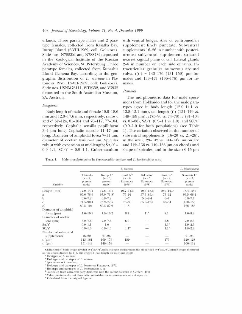
TABLE 1. Male morphometrics in Leptosomatides marinae and L. brevicaudatus n. sp.
Variable
L. marinae L. brevicaudatus
Hokkaido(n = 5;presentstudy)
Iturup I.a
(n = 3;presentstudy)
Kuril Is.b
(n = 6;Platonova,
1976)
Sakhalinc
(n = 5;Platonova,
1978)
Kuril Is.d
(n = 9;Platonova,
1976)
Simushir I.e
(n = 3;presentstudy)
Length (mm) 12.0–14.1 12.8–13.1 10.7–14.5 10.5–18.6 10.0–12.0 18.4–19.7a 65.6–78.9 67.0–71.8f 75–94 57.3–85.4 73–92 63.5–68.4b 6.6–7.2 6.9–7.2 6–7 5.6–9.4 6–7 6.8–7.7c 74.5–90.4 73.9–77.5 73–80 65.0–124 62–84 150–156c8 80.5–104 80.5–87.9 —g — — 166–186Diameter of amphidal
fovea (µm) 7.6–10.9 7.9–10.2 8.4 11h 8.1 7.6–8.9Diameter of ocellar
lens (µm) 6.2–7.6 7.0–7.6 6.0 — 5.8 7.0–8.3SA/t8 0.9–1.1 1.0 — — 1.1h 1.9–2.3SC/t8 0.9–1.0 0.9–1.0 1.1h — 1.1h 1.8–2.2Number of subventral
supplements 16–20 21–26 — — — 21–24t (µm) 143–161 169–176 159 — 175 120–128t8 (µm) 131–149 149–159 — — — 106–112
Characters: c8, body length divided by t8; SA/t8, spicule length measured on the arc divided by t8; SC/t8, spicule length measuredon the chord divided by t8; t, tail length; t8, tail length on its chord length.
a Paratypes of L. marinae.b Holotype and paratypes of L. marinae.c Specimens as L. marinae.d Holotype and paratypes of L. brevisetosus Platonova, 1976.e Holotype and paratypes of L. brevicaudatus n. sp.f Calculated from corrected body diameters with the second formula in Geraert (1961).g Value questionable, not observable, unsuitable for measurements, or not reported.h Calculated from the original figures.
468 Journal of Nematology, Volume 31, No. 4, December 1999

by 5–8 µm vs. 18–22 µm by 14–23 µm) andshape of the lateral bulge at the distal end ofthe gubernaculum is so small that we con-clude that the specimens from Hokkaidoand Iturup are conspecific and in agree-ment with the original descriptions of L. ma-rinae and L. brevisetosus (Platonova, 1976)(Figs. 7–9,28,29; Table 1). Our observationsof type and newly collected specimens re-vealed that the caudal ventromedian supple-ment (see Platonova, 1976 and Bongers,1984) is actually paired and one of the pos-teriormost subventral supplements.
From the foregoing observations of males
we conclude that the specimens from Hok-kaido are L. marinae, and we concur that L.brevisetosus is synonymous with it. Con-versely, the male specimens from Simushirupon which Bongers (1984) based his re-description of L. marinae differ from thatspecies in body length (18.4–19.7 mm), taillength t8 (106–112 µm), c8 (166–186), SA/t8(1.9–2.3), and SC/t8 (1.8–2.2) (Table 1).The Simushir specimens differ from allother known species of Leptosomatides and,thus, represent a new species.
The females collected in Hokkaido alsoclosely resemble the paratype specimens
Figs. 28, 29. Leptosomatides marinae. Male (28, ZIHU 1243; 29, paratype with L = 13.0 mm on slide N7882 M).28) Posterior region, left lateral view. 29) Posterior region, left ventrolateral view. Scales = 100 µm. Figs. 30, 31.Leptosomatides brevicaudatus n. sp., holotype. 30) Head, right lateral view. 31) Posterior region, left lateral view.Scale = 50 µm in Fig. 30; 100 µm in Fig. 31.
New, Known Leptosomatid Nematodes: Kito, Hope 469
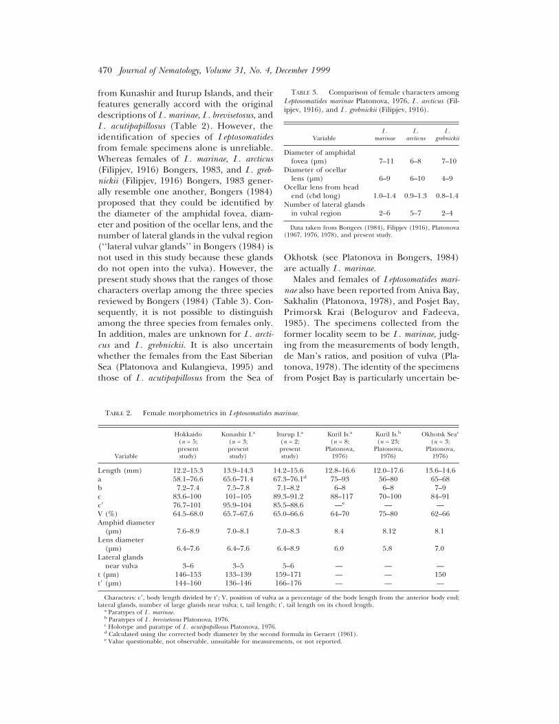
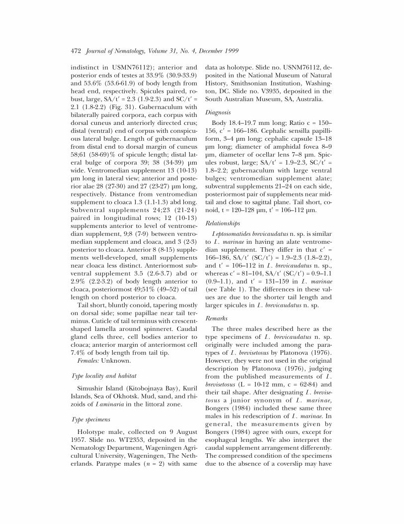
from Kunashir and Iturup Islands, and theirfeatures generally accord with the originaldescriptions of L. marinae, L. brevisetosus, andL. acutipapillosus (Table 2). However, theidentification of species of Leptosomatidesfrom female specimens alone is unreliable.Whereas females of L. marinae, L. arcticus(Filipjev, 1916) Bongers, 1983, and L. greb-nickii (Filipjev, 1916) Bongers, 1983 gener-ally resemble one another, Bongers (1984)proposed that they could be identified bythe diameter of the amphidal fovea, diam-eter and position of the ocellar lens, and thenumber of lateral glands in the vulval region(‘‘lateral vulvar glands’’ in Bongers (1984) isnot used in this study because these glandsdo not open into the vulva). However, thepresent study shows that the ranges of thosecharacters overlap among the three speciesreviewed by Bongers (1984) (Table 3). Con-sequently, it is not possible to distinguishamong the three species from females only.In addition, males are unknown for L. arcti-cus and L. grebnickii. It is also uncertainwhether the females from the East SiberianSea (Platonova and Kulangieva, 1995) andthose of L. acutipapillosus from the Sea of
Okhotsk (see Platonova in Bongers, 1984)are actually L. marinae.
Males and females of Leptosomatides mari-nae also have been reported from Aniva Bay,Sakhalin (Platonova, 1978), and Posjet Bay,Primorsk Krai (Belogurov and Fadeeva,1985). The specimens collected from theformer locality seem to be L. marinae, judg-ing from the measurements of body length,de Man’s ratios, and position of vulva (Pla-tonova, 1978). The identity of the specimensfrom Posjet Bay is particularly uncertain be-
TABLE 2. Female morphometrics in Leptosomatides marinae.
Variable
Hokkaido(n = 5;presentstudy)
Kunashir I.a
(n = 3;presentstudy)
Iturup I.a
(n = 2;presentstudy)
Kuril Is.a
(n = 8;Platonova,
1976)
Kuril Is.b
(n = 23;Platonova,
1976)
Okhotsk Seac
(n = 3;Platonova,
1976)
Length (mm) 12.2–15.3 13.9–14.3 14.2–15.6 12.8–16.6 12.0–17.6 13.6–14.6a 58.1–76.6 65.6–71.4 67.3–76.1d 75–93 56–80 65–68b 7.2–7.4 7.5–7.8 7.1–8.2 6–8 6–8 7–9c 83.6–100 101–105 89.3–91.2 88–117 70–100 84–91c8 76.7–101 95.9–104 85.5–88.6 —e — —V (%) 64.5–68.0 65.7–67.6 65.0–66.6 64–70 75–80 62–66Amphid diameter
(µm) 7.6–8.9 7.0–8.1 7.0–8.3 8.4 8.12 8.1Lens diameter
(µm) 6.4–7.6 6.4–7.6 6.4–8.9 6.0 5.8 7.0Lateral glands
near vulva 3–6 3–5 5–6 — — —t (µm) 146–153 133–139 159–171 — — 150t8 (µm) 144–160 136–146 166–176 — — —
Characters: c8, body length divided by t8; V, position of vulva as a percentage of the body length from the anterior body end;lateral glands, number of large glands near vulva; t, tail length; t8, tail length on its chord length.
a Paratypes of L. marinae.b Paratypes of L. brevisetosus Platonova, 1976.c Holotype and paratype of L. acutipapillosus Platonova, 1976.d Calculated using the corrected body diameter by the second formula in Geraert (1961).e Value questionable, not observable, unsuitable for measurements, or not reported.
TABLE 3. Comparison of female characters amongLeptosomatides marinae Platonova, 1976, L. arcticus (Fil-ipjev, 1916), and L. grebnickii (Filipjev, 1916).
VariableL.
marinaeL.
arcticusL.
grebnickii
Diameter of amphidalfovea (µm) 7–11 6–8 7–10
Diameter of ocellarlens (µm) 6–9 6–10 4–9
Ocellar lens from headend (cbd long) 1.0–1.4 0.9–1.3 0.8–1.4
Number of lateral glandsin vulval region 2–6 5–7 2–4
Data taken from Bongers (1984), Filipjev (1916), Platonova(1967, 1976, 1978), and present study.
470 Journal of Nematology, Volume 31, No. 4, December 1999
cause those specimens were the subject of ahead morphology study (Belogurov andFadeeva, 1985) before their specific identitywas ascertained.
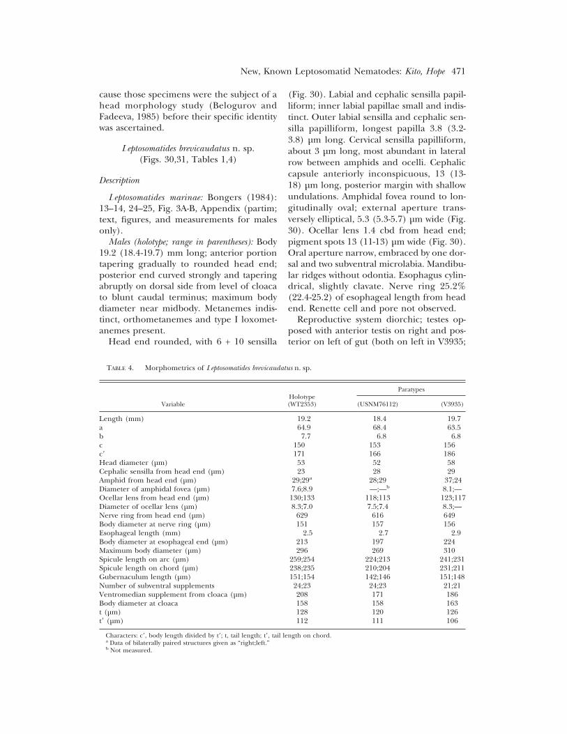
Leptosomatides brevicaudatus n. sp.(Figs. 30,31, Tables 1,4)
Description
Leptosomatides marinae: Bongers (1984):13–14, 24–25, Fig. 3A-B, Appendix (partim;text, figures, and measurements for malesonly).
Males (holotype; range in parentheses): Body19.2 (18.4-19.7) mm long; anterior portiontapering gradually to rounded head end;posterior end curved strongly and taperingabruptly on dorsal side from level of cloacato blunt caudal terminus; maximum bodydiameter near midbody. Metanemes indis-tinct, orthometanemes and type I loxomet-anemes present.
Head end rounded, with 6 + 10 sensilla
(Fig. 30). Labial and cephalic sensilla papil-liform; inner labial papillae small and indis-tinct. Outer labial sensilla and cephalic sen-silla papilliform, longest papilla 3.8 (3.2-3.8) µm long. Cervical sensilla papilliform,about 3 µm long, most abundant in lateralrow between amphids and ocelli. Cephaliccapsule anteriorly inconspicuous, 13 (13-18) µm long, posterior margin with shallowundulations. Amphidal fovea round to lon-gitudinally oval; external aperture trans-versely elliptical, 5.3 (5.3-5.7) µm wide (Fig.30). Ocellar lens 1.4 cbd from head end;pigment spots 13 (11-13) µm wide (Fig. 30).Oral aperture narrow, embraced by one dor-sal and two subventral microlabia. Mandibu-lar ridges without odontia. Esophagus cylin-drical, slightly clavate. Nerve ring 25.2%(22.4-25.2) of esophageal length from headend. Renette cell and pore not observed.
Reproductive system diorchic; testes op-posed with anterior testis on right and pos-terior on left of gut (both on left in V3935;
TABLE 4. Morphometrics of Leptosomatides brevicaudatus n. sp.
VariableHolotype(WT2353)
Paratypes
(USNM76112) (V3935)
Length (mm) 19.2 18.4 19.7a 64.9 68.4 63.5b 7.7 6.8 6.8c 150 153 156c8 171 166 186Head diameter (µm) 53 52 58Cephalic sensilla from head end (µm) 23 28 29Amphid from head end (µm) 29;29a 28;29 37;24Diameter of amphidal fovea (µm) 7.6;8.9 —;—b 8.1;—Ocellar lens from head end (µm) 130;133 118;113 123;117Diameter of ocellar lens (µm) 8.3;7.0 7.5;7.4 8.3;—Nerve ring from head end (µm) 629 616 649Body diameter at nerve ring (µm) 151 157 156Esophageal length (mm) 2.5 2.7 2.9Body diameter at esophageal end (µm) 213 197 224Maximum body diameter (µm) 296 269 310Spicule length on arc (µm) 259;254 224;213 241;231Spicule length on chord (µm) 238;235 210;204 231;211Gubernaculum length (µm) 151;154 142;146 151;148Number of subventral supplements 24;23 24;23 21;21Ventromedian supplement from cloaca (µm) 208 171 186Body diameter at cloaca 158 158 163t (µm) 128 120 126t8 (µm) 112 111 106
Characters: c8, body length divided by t8; t, tail length; t8, tail length on chord.a Data of bilaterally paired structures given as “right;left.”b Not measured.
New, Known Leptosomatid Nematodes: Kito, Hope 471
indistinct in USMN76112); anterior andposterior ends of testes at 33.9% (30.9-33.9)and 53.6% (53.6-61.9) of body length fromhead end, respectively. Spicules paired, ro-bust, large, SA/t8 = 2.3 (1.9-2.3) and SC/t8 =2.1 (1.8-2.2) (Fig. 31). Gubernaculum withbilaterally paired corpora, each corpus withdorsal cuneus and anteriorly directed crus;distal (ventral) end of corpus with conspicu-ous lateral bulge. Length of gubernaculumfrom distal end to dorsal margin of cuneus58;61 (58-69)% of spicule length; distal lat-eral bulge of corpora 39; 38 (34-39) µmwide. Ventromedian supplement 13 (10-13)µm long in lateral view; anterior and poste-rior alae 28 (27-30) and 27 (23-27) µm long,respectively. Distance from ventromediansupplement to cloaca 1.3 (1.1-1.3) abd long.Subventral supplements 24;23 (21-24)paired in longitudinal rows; 12 (10-13)supplements anterior to level of ventrome-dian supplement, 9;8 (7-9) between ventro-median supplement and cloaca, and 3 (2-3)posterior to cloaca. Anterior 8 (8-15) supple-ments well-developed, small supplementsnear cloaca less distinct. Anteriormost sub-ventral supplement 3.5 (2.6-3.7) abd or2.9% (2.2-3.2) of body length anterior tocloaca, posteriormost 49;51% (49–52) of taillength on chord posterior to cloaca.
Tail short, bluntly conoid, tapering mostlyon dorsal side; some papillae near tail ter-minus. Cuticle of tail terminus with crescent-shaped lamella around spinneret. Caudalgland cells three, cell bodies anterior tocloaca; anterior margin of anteriormost cell7.4% of body length from tail tip.
Females: Unknown.
Type locality and habitat
Simushir Island (Kitobojnaya Bay), KurilIslands, Sea of Okhotsk. Mud, sand, and rhi-zoids of Laminaria in the littoral zone.
Type specimens
Holotype male, collected on 9 August1957. Slide no. WT2353, deposited in theNematology Department, Wageningen Agri-cultural University, Wageningen, The Neth-erlands. Paratype males (n = 2) with same
data as holotype. Slide no. USNM76112, de-posited in the National Museum of NaturalHistory, Smithsonian Institution, Washing-ton, DC. Slide no. V3935, deposited in theSouth Australian Museum, SA, Australia.
Diagnosis
Body 18.4–19.7 mm long; Ratio c = 150–156, c8 = 166–186. Cephalic sensilla papilli-form, 3–4 µm long; cephalic capsule 13–18µm long; diameter of amphidal fovea 8–9µm, diameter of ocellar lens 7–8 µm. Spic-ules robust, large; SA/t8 = 1.9–2.3, SC/t8 =1.8–2.2; gubernaculum with large ventralbulges; ventromedian supplement alate;subventral supplements 21–24 on each side,posteriormost pair of supplements near mid-tail and close to sagittal plane. Tail short, co-noid, t = 120–128 µm, t8 = 106–112 µm.
Relationships
Leptosomatides brevicaudatus n. sp. is similarto L. marinae in having an alate ventrome-dian supplement. They differ in that c8 =166–186, SA/t8 (SC/t8) = 1.9–2.3 (1.8–2.2),and t8 = 106–112 in L. brevicaudatus n. sp.,whereas c8 = 81–104, SA/t8 (SC/t8) = 0.9–1.1(0.9–1.1), and t8 = 131–159 in L. marinae(see Table 1). The differences in these val-ues are due to the shorter tail length andlarger spicules in L. brevicaudatus n. sp.
Remarks
The three males described here as thetype specimens of L. brevicaudatus n. sp.originally were included among the para-types of L. brevisetosus by Platonova (1976).However, they were not used in the originaldescription by Platonova (1976), judgingfrom the published measurements of L.brevisetosus (L = 10-12 mm, c = 62-84) andtheir tail shape. After designating L. brevise-tosus a junior synonym of L. marinae,Bongers (1984) included these same threemales in his redescription of L. marinae. Ingeneral, the measurements given byBongers (1984) agree with ours, except foresophageal lengths. We also interpret thecaudal supplement arrangement differently.The compressed condition of the specimensdue to the absence of a coverslip may have
472 Journal of Nematology, Volume 31, No. 4, December 1999
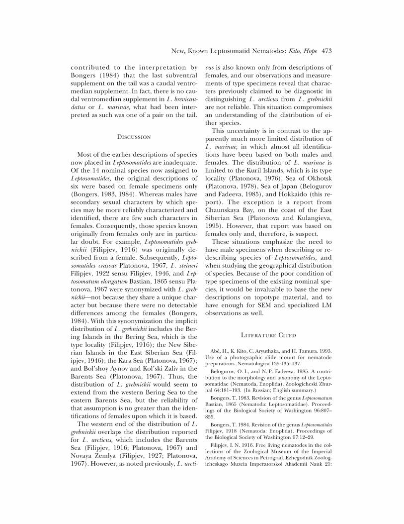
contributed to the interpretation byBongers (1984) that the last subventralsupplement on the tail was a caudal ventro-median supplement. In fact, there is no cau-dal ventromedian supplement in L. brevicau-datus or L. marinae, what had been inter-preted as such was one of a pair on the tail.
Discussion
Most of the earlier descriptions of speciesnow placed in Leptosomatides are inadequate.Of the 14 nominal species now assigned toLeptosomatides, the original descriptions ofsix were based on female specimens only(Bongers, 1983, 1984). Whereas males havesecondary sexual characters by which spe-cies may be more reliably characterized andidentified, there are few such characters infemales. Consequently, those species knownoriginally from females only are in particu-lar doubt. For example, Leptosomatides greb-nickii (Filipjev, 1916) was originally de-scribed from a female. Subsequently, Lepto-somatides crassus Platonova, 1967, L. steineriFilipjev, 1922 sensu Filipjev, 1946, and Lep-tosomatum elongatum Bastian, 1865 sensu Pla-tonova, 1967 were synonymized with L. greb-nickii—not because they share a unique char-acter but because there were no detectabledifferences among the females (Bongers,1984). With this synonymization the implicitdistribution of L. grebnickii includes the Ber-ing Islands in the Bering Sea, which is thetype locality (Filipjev, 1916); the New Sibe-rian Islands in the East Siberian Sea (Fil-ipjev, 1946); the Kara Sea (Platonova, 1967);and Bol’shoy Aynov and Kol’ski Zaliv in theBarents Sea (Platonova, 1967). Thus, thedistribution of L. grebnickii would seem toextend from the western Bering Sea to theeastern Barents Sea, but the reliability ofthat assumption is no greater than the iden-tifications of females upon which it is based.
The western end of the distribution of L.grebnickii overlaps the distribution reportedfor L. arcticus, which includes the BarentsSea (Filipjev, 1916; Platonova, 1967) andNovaya Zemlya (Filipjev, 1927; Platonova,1967). However, as noted previously, L. arcti-
cus is also known only from descriptions offemales, and our observations and measure-ments of type specimens reveal that charac-ters previously claimed to be diagnostic indistinguishing L. arcticus from L. grebnickiiare not reliable. This situation compromisesan understanding of the distribution of ei-ther species.
This uncertainty is in contrast to the ap-parently much more limited distribution ofL. marinae, in which almost all identifica-tions have been based on both males andfemales. The distribution of L. marinae islimited to the Kuril Islands, which is its typelocality (Platonova, 1976), Sea of Okhotsk(Platonova, 1978), Sea of Japan (Belogurovand Fadeeva, 1985), and Hokkaido (this re-port). The exception is a report fromChaunskaya Bay, on the coast of the EastSiberian Sea (Platonova and Kulangieva,1995). However, that report was based onfemales only and, therefore, is suspect.
These situations emphasize the need tohave male specimens when describing or re-describing species of Leptosomatides, andwhen studying the geographical distributionof species. Because of the poor condition oftype specimens of the existing nominal spe-cies, it would be invaluable to base the newdescriptions on topotype material, and tohave enough for SEM and specialized LMobservations as well.
Literature Cited
Abe, H., K. Kito, C. Aryuthaka, and H. Tamura. 1993.Use of a photographic slide mount for nematodepreparations. Nematologica 135:135–137.
Belogurov, O. I., and N. P. Fadeeva. 1985. A contri-bution to the morphology and taxonomy of the Lepto-somatidae (Nematoda, Enoplida). Zoologicheski Zhur-nal 64:181–193. (In Russian; English summary.)
Bongers, T. 1983. Revision of the genus LeptosomatumBastian, 1865 (Nematoda: Leptosomatidae). Proceed-ings of the Biological Society of Washington 96:807–855.
Bongers, T. 1984. Revision of the genus LeptosomatidesFilipjev, 1918 (Nematoda: Enoplida). Proceedings ofthe Biological Society of Washington 97:12–29.
Filipjev, I. N. 1916. Free living nematodes in the col-lections of the Zoological Museum of the ImperialAcademy of Sciences in Petrograd. Ezhegodnik Zoolog-icheskago Muzeia Imperatorskoi Akademii Nauk 21:
New, Known Leptosomatid Nematodes: Kito, Hope 473

59–116. (In Russian: English translation by M. M.Haque, 1973: New Delhi: Amerind Publishing).
Filipjev, I. N. 1918/1921. Free-living marine nema-todes of the Sevastopol area. Trudy Osoboi Zoolog-icheskoi Laboratorii i Sebastopol’skoi BiologicheskoiStantsii, Rossiskoi Akademii Nauk, Ser. II 4:1–350(1918), 351–614 (1921). (In Russian: English transla-tion by M. Raveh, Issue 1, 1968, Issue 2, 1970. Jerusa-lem: Israel Program for Scientific Translations.)
Filipjev, I. N. 1927. Les Nematodes libres mers sep-tentrionales appartenant a la famille des Enoplidae. Ar-chiv fur Naturgeschichte 91(A)(6):1–216.
Filipjev, I. N. 1946. Nematodes libres du bassin po-laire. Dreifuiushchaia Ekspeditsiia Glavsevmorputi NaLedokol’nom Purokhode ‘‘G. Sedov.’’ 1937–1940.Trudy 3:158–184. (In Russian and French.)
Geraert, E. 1961. Corrections of measurements ofnematode diameters. Nematologica 6:258–259.
Hope, W. D. 1967. Free-living marine nematodes ofthe genera Pseudocella Filipjev, 1927, ThoracostomaMarion, 1870, and Deontostoma Filipjev, 1916 (Nema-toda: Leptosomatidae) from the west coast of NorthAmerica. Transactions of the American MicroscopicalSociety 86:307–334.
Hope, W. D. 1982. Structure of head and stoma inthe marine nematode genus Deontostoma (Enoplida:
Leptosomatidae). Smithsonian Contributions to Zool-ogy 353:1–22.
Platonova, T. A. 1967. Free-living marine nematodesof the family Leptosomatidae from the European Arc-tic. Zoologicheski Zhurnal 46:828–839. (In Russian; En-glish summary.)
Platonova, T. A. 1976. Lower Enoplida (free-livingmarine nematodes) of the seas of the USSR. Pp. 3–164in Nematodes and their role in the meiobenthos. Issle-dovanie Fauny Morei 15 (23). (In Russian: Englishtranslation by S. Sharma, 1985. New Delhi: AmerindPublishing Co.)
Platonova, T. A. 1978. Marine nematodes of the or-der Enoplida from the coastal waters of the South Sa-khalin. Zoologicheski Zhurnal 57:495–504. (In Russian;English summary.)
Platonova, T. A. 1981. Evolution of skeleton structureof head in the family Leptosomatidae (Enoplida). Pp.57–64 in T. A. Platonova and S. Y. Tsalolikhin, eds. Evo-lution, taxonomy, morphology, and ecology of free-living nematodes. Leningrad: Akademiia Nauk SSSR.(In Russian.)
Platonova, T. A. and L. V. Kulangieva. 1995. MarineEnoplida from the East Siberian Sea (Nematoda). Zoo-systematica Rossica 3:175–180.
474 Journal of Nematology, Volume 31, No. 4, December 1999