Embed Size (px)
Citation preview

Takara Bio USA, Inc.
1290 Terra Bella Avenue, Mountain View, CA 94043, USA
U.S. Technical Support: [email protected]
United States/Canada
800.662.2566
Asia Pacific
+1.650.919.7300
Europe
+33.(0)1.3904.6880
Japan
+81.(0)77.565.6999
Page 1 of 30
Takara Bio USA, Inc.
Lenti-X™ ProteoTuner™ Shield Systems User Manual
Cat. Nos. 631074, 631748, 631751, 631753, 632173 & 632175
(110416)

Lenti-X ProteoTuner Shield Systems User Manual
(110416) takarabio.com Takara Bio USA, Inc.
Page 2 of 30
Table of Contents I. Introduction ..................................................................................................................................................................... 4
II. List of Components ......................................................................................................................................................... 8
III. Additional Materials Required .................................................................................................................................... 9
IV. General Considerations ............................................................................................................................................. 11
A. General Cell Culture ................................................................................................................................................. 11
B. Safety Guidelines for Working with Lentiviruses .................................................................................................... 11
V. Creating Vector Constructs Encoding DD-Tagged Proteins of Interest ....................................................................... 13
A. Protocol: Creating ProteoTuner Vector Constructs using In-Fusion HD.................................................................. 13
VI. Pilot Expression Testing of Your Construct ............................................................................................................. 13
A. Protocol: Transient Transfection of Lenti-X ProteoTuner Constructs ...................................................................... 13
VII. Producing Lentivirus from the Lenti-X Vectors ....................................................................................................... 15
A. General Considerations ............................................................................................................................................. 15
B. Protocol: Transfecting Lentiviral Vectors into Lenti-X 293T Packaging Cells ........................................................ 16
VIII. Lentivirus Titration ................................................................................................................................................... 17
A. Summary ................................................................................................................................................................... 17
B. Protocol: Determining Viral Titer by Colony Formation.......................................................................................... 17
IX. Transducing Target Cells with a Lenti-X ProteoTuner Lentivirus ........................................................................... 18
A. Protocol: Transducing Target Cells with Lenti-X ProteoTuner Lentiviruses ........................................................... 18
X. Protein Stabilization & Destabilization Using Lenti-X ProteoTuner Cell Lines .......................................................... 19
A. Protocol: Optimizing Shield1 Concentration and Incubation Time of Transduced Cells ......................................... 19
B. Protocol: DD-Protein Stabilization of Transduced Cells .......................................................................................... 20
C. Protocol: DD-Protein Destabilization ....................................................................................................................... 21
D. Protocol: Working with Stable Cell Lines Expressing a DD-Tagged Protein of Interest ......................................... 22
E. Protocol: In Vivo Use of Shield1 .............................................................................................................................. 22
XI. References for ProteoTuner ...................................................................................................................................... 23
Appendix A. Troubleshooting Guide .................................................................................................................................... 24
Appendix B. Lenti-X ProteoTuner Vector Maps .................................................................................................................. 26
Appendix C: Preparing and Handling Cell Line Stocks ....................................................................................................... 28
A. Protocol: Freezing Cell Line Stocks ......................................................................................................................... 28
B. Protocol: Thawing Cell Line Frozen Stocks ............................................................................................................. 29
Lenti-X ProteoTuner Shield Systems User Manual
(110416) takarabio.com Takara Bio USA, Inc.
Page 3 of 30
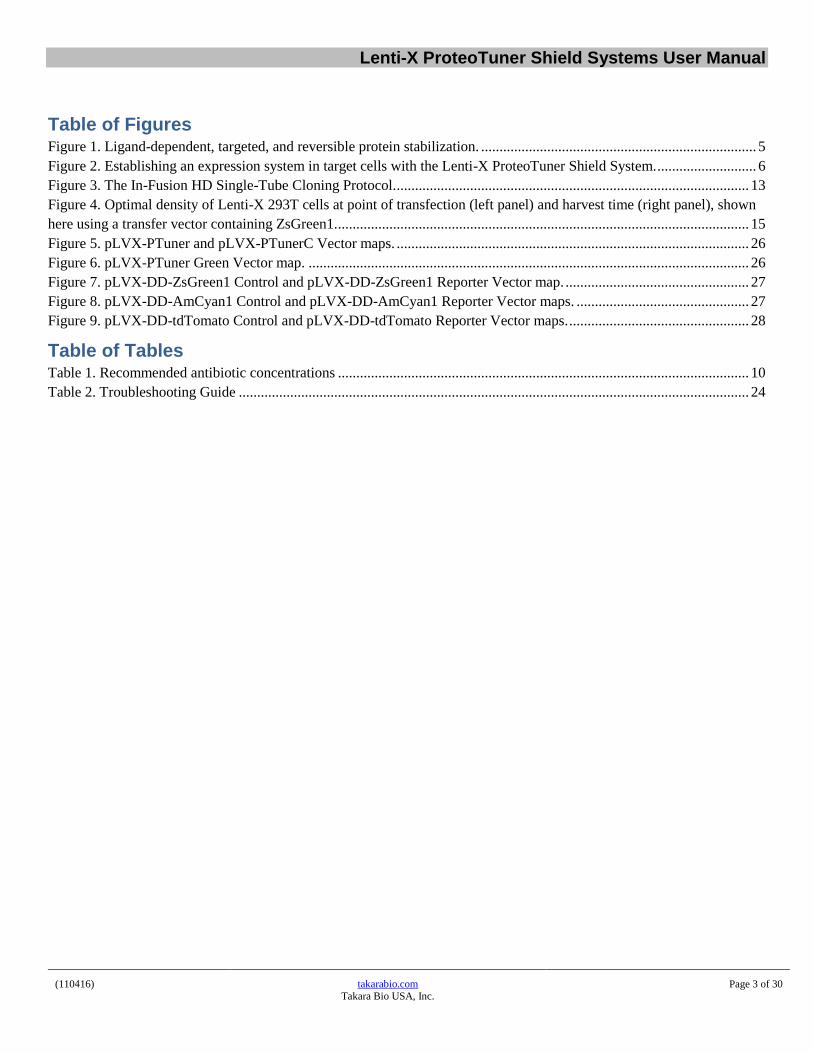
Table of Figures Figure 1. Ligand-dependent, targeted, and reversible protein stabilization. ........................................................................... 5
Figure 2. Establishing an expression system in target cells with the Lenti-X ProteoTuner Shield System. ........................... 6
Figure 3. The In-Fusion HD Single-Tube Cloning Protocol. ................................................................................................ 13
Figure 4. Optimal density of Lenti-X 293T cells at point of transfection (left panel) and harvest time (right panel), shown
here using a transfer vector containing ZsGreen1. ................................................................................................................ 15
Figure 5. pLVX-PTuner and pLVX-PTunerC Vector maps. ................................................................................................ 26
Figure 6. pLVX-PTuner Green Vector map. ........................................................................................................................ 26
Figure 7. pLVX-DD-ZsGreen1 Control and pLVX-DD-ZsGreen1 Reporter Vector map. .................................................. 27
Figure 8. pLVX-DD-AmCyan1 Control and pLVX-DD-AmCyan1 Reporter Vector maps. ............................................... 27
Figure 9. pLVX-DD-tdTomato Control and pLVX-DD-tdTomato Reporter Vector maps. ................................................. 28
Table of Tables Table 1. Recommended antibiotic concentrations ................................................................................................................ 10
Table 2. Troubleshooting Guide ........................................................................................................................................... 24

Lenti-X ProteoTuner Shield Systems User Manual
(110416) takarabio.com Takara Bio USA, Inc.
Page 4 of 30
I. Introduction
A. Summary
Analyzing protein function is a key focus in discovery-based cell biology research. ProteoTuner
technology allows you to directly investigate the function of a specific protein of interest—by directly
manipulating the level of the protein itself. This fast regulation occurs directly at the protein level, rather
than at the mRNA or promoter induction level, and enables you to control the quantity of a specific
protein in the cell, in as little as 15 to 30 minutes.
This revolutionary method takes advantage of ligand-dependent, tunable stabilization/destabilization of
the protein of interest. It is based on a 12 kDa mutant of the FKBP protein (the destabilization domain, or
DD) that can be expressed as a tag on your protein of interest. In the presence of the small (750 Da),
membrane-permeant, stabilizing ligand Shield1, the DD-tagged protein of interest is stabilized (protected
from proteasomal degradation) and accumulates inside the cell (Figure 1). Ligand-dependent stabilization
occurs very quickly: DD fusion proteins have been shown to accumulate to detectable levels just 15–30
minutes after the addition of Shield1 (Banaszynski et al., 2006).
The ProteoTuner method is not restricted to protein stabilization—it can also be used to destabilize the
DD-tagged protein when you culture your cells in medium without Shield1, allowing proteasomal
degradation of the DD-protein (Figure 1). This makes it possible to “tune” the amount of stabilized DD-
tagged protein present in the cell by titrating the amount of Shield1 in the culture medium, and to
repeatedly stabilize and destabilize the protein of interest using the same set of cells.
NOTE: To be degraded effectively, the DD fusion protein must have access to proteasomes within the
cell. Cell regions that lack such access (e.g., the ER lumen) will not allow DD-tagged protein degradation.
A variety of ProteoTuner shield systems are available:
Your choices include N- or C-terminal DD fusions, conventional plasmid or viral delivery, and systems
with or without a Living Colors® Fluorescent Protein marker for transfection. One system contains a tag
for ProLabel quantitation. ProteoTuner technology also plays an important role in the On-Demand
Fluorescent Reporter Systems. This manual describes the Lenti-X ProteoTuner Shield Systems, which
provide lentiviral delivery of DD-fusion proteins (via transduction) to your target cells. You can learn
about all of our ProteoTuner Shield Systems at takarabio.com

Lenti-X ProteoTuner Shield Systems User Manual
(110416) takarabio.com Takara Bio USA, Inc.
Page 5 of 30
Figure 1. Ligand-dependent, targeted, and reversible protein stabilization. A small destabilization domain (DD; grey) is fused to a
target protein of interest. The small membrane-permeable ligand Shield1 (black) binds to the DD and protects it from proteasomal
degradation. Removal of Shield1, however, causes rapid degradation of the entire fusion protein. The default pathway for the ProteoTuner
Shield Systems is the degradation of the DD-tagged protein, unless Shield1 is present to stabilize it.
B. Protocol Overview: Creating a Lentiviral ProteoTuner Expression System
The following steps are required to create a ligand-dependent, tunable stabilization/destabilization system
for your protein of interest using lentivirus. (Steps 2 and 3 are illustrated in Figure 2.)
1. Create and test Lenti-X ProteoTuner constructs containing your gene of interest (GOI).
a. Clone your gene of interest into a Lenti-X ProteoTuner fusion vector (such as a pLVX-PTuner or
pLVX-PTunerC) or a Lenti-X ProteoTuner reporter vector (such as pLVX-DD-ZsGreen1, pLVX-
DD-Am Cyan1, or pLVX-DD-td Tomato) using fast, easy In-Fusion® HD cloning (Section V),
or a standard ligation method.
NOTE: All the Lenti-X ProteoTuner systems and their components are listed in Section II.
Additional required materials are listed in Section III.
b. Pilot test Shield1 protein stabilization of your protein of interest using your Lenti-X ProteoTuner-
GOI constructs (Section VI).
2. Create Lenti-X ProteoTuner cell lines expressing your protein of interest.
a. Produce supernatants containing lentiviral particles that express your protein of interest by
transfecting your Lenti-X ProteoTuner-GOI constructs from Step 1 into the Lenti-X 293T Cell
Line using the Lenti-X Packaging Single Shots (VSV-G) (Section VII).
b. Determine the titer of your lentiviral supernatants (Section VIII).
c. Infect (transduce) your target cells with your titered lentivirus (Section IX).
3. Perform protein stabilization and destabilization experiments using the Lenti-X ProteoTuner cell lines
you created in Step 2 (Section X). See Section I.C for an overview of these experiments.

Lenti-X ProteoTuner Shield Systems User Manual
(110416) takarabio.com Takara Bio USA, Inc.
Page 6 of 30
Figure 2. Establishing an expression system in target cells with Lenti-X ProteoTuner Shield Systems. The Lenti-X Packaging Single Shots
(VSV-G) and 293T cells are used to generate a high-titer lentiviral supernatant from a Lenti-X ProteoTuner fusion vector or a Lenti-X ProteoTuner
reporter vector which contains your gene or promoter of interest. Target cells are then transduced with these packaged lentiviral particles and your
protein of interest can be stabilized or destabilized by adding or removing Shield1 from the culture medium.

Lenti-X ProteoTuner Shield Systems User Manual
(110416) takarabio.com Takara Bio USA, Inc.
Page 7 of 30
C. Protocol Overview: The ProteoTuner Assay
After you have established a Lenti-X ProteoTuner Shield System in your target cells (see Section I.B for
an overview), you can perform a protein stabilization protocol to observe the effects of stabilizing your
protein of interest, and a protein destabilization protocol to observe the effects of the loss of protein of
interest (Figure 2). Both protocols are based on Shield1’s ability to reversibly stabilize DD-tagged fusion
proteins (Figure 1).
1. Protein Stabilization (Protein “On” Experiment)
In order to stabilize your protein of interest, you need to add the stabilizing ligand, Shield1, to one
of two parallel cell cultures which were previously untreated with Shield1 (Figure 2). The other
culture will be continuously cultured in the absence of Shield1 as a negative control.
The added Shield1 will protect your DD-tagged protein of interest from proteasomal
degradation, causing a dramatic increase in its level in the cell. Stabilization has been
reported in as little as 15–30 minutes (Banaszynski et al., 2006) but we recommend
performing a timecourse experiment in order to determine the Shield1-based stabilization
rate for your protein of interest as well as testing different Shield1 concentrations (50–
1,000 nM).
At different time points, analyze the treated and control cells using your method of choice
(e.g., Western blot or phenotypic analysis), depending on your experimental goals.
2. Protein Destabilization (Protein “Off” Experiment)
The default pathway of the ProteoTuner shield systems in the absence of the ligand Shield1 is
rapid destabilization and degradation of the DD-tagged protein. In order to destabilize/degrade a
protein of interest that has been stabilized with Shield 1, split the cells expressing the stabilized
protein into two parallel cell cultures (Figure 2). One culture will continue to be maintained in the
presence of Shield1 as a positive control, and the second (experimental) culture will be
maintained without the stabilizing ligand, Shield1.
In the absence of Shield1, the DD-tagged protein of interest will be rapidly degraded.
Degradation half-lives of one to two hours have been reported (Banaszynski et al., 2006),
but we recommend performing a time-course assay in order to assess the rate of
degradation of your protein of interest.
At different time points, analyze the treated and control cells using your method of choice
(e.g., Western blot or phenotypic analysis), depending on your experimental goals.

Lenti-X ProteoTuner Shield Systems User Manual
(110416) takarabio.com Takara Bio USA, Inc.
Page 8 of 30
II. List of ComponentsStore all components at –20°C.
A. Expression Systems
Lenti-X ProteoTuner Shield System N (Cat. No. 632173)
pLVX-PTuner Vector (20 µg) (Cat. No. 632174; not sold separately)
Shield1 (500 µl) (also sold separately as Cat. No. 632189)
Lenti-X ProteoTuner Shield System C (Cat. No. 631074)
pLVX-PTunerC Vector (20 µg) (Cat. No. 631075; not sold separately)
Shield1 (500 µl) (also sold separately as Cat. No. 632189)
Lenti-X ProteoTuner Shield System N (w/ ZsGreen1) (Cat. No. 632175)
pLVX-PTuner Green Vector (20 µg) (Cat. No. 632176; not sold separately)
Shield1 (500 µl) (also sold separately as Cat. No. 632189)
B. On Demand Reporter Systems
Lenti-X DD Green Reporter System (Cat. No. 631751)
Lenti-X DD-ZsGreen1 Vector Set (Cat. No. 631752; not sold separately)
o pLVX-DD-ZsGreen1 Reporter Vector (20 µg)
o pLVX-DD-ZsGreen1 Control Vector (20 µg)
Lenti-X Packaging Single Shots (VSV-G) (16 rxns) (Cat. No. 631275)
Shield1 (500 µl) (also sold separately as Cat. No. 632189)
Lenti-X GoStix™ (Sample) (3 tests) (Cat. No. 631242; not sold separately)
Lenti-X DD Cyan Reporter System (Cat. No. 631748)
Lenti-X DD- AmCyan1 Vector Set (Cat. No. 631749; not sold separately)
o pLVX-DD-AmCyan1 Reporter Vector (20 µg)
o pLVX-DD-AmCyan1 Control Vector (20 µg)
Lenti-X Packaging Single Shots (VSV-G) (16 rxns) (Cat. No. 631275)
Shield1 (500 µl) (also sold separately as Cat. No. 632189)
Lenti-X GoStix (Sample) (3 tests) (Cat. No. 631242; not sold separately)
Lenti-X DD Red Reporter System (Cat. No. 631753)
• Lenti-X DD-tdTomato Vector Set (Cat. No. 631754; not sold separately)
o pLVX-DD-tdTomato1 Reporter Vector (20 µg)
o pLVX-DD-tdTomato1 Control Vector (20 µg)
• Lenti-X Packaging Single Shots (VSV-G) (16 rxns) (Cat. No. 631275)
• Shield1 (500 µl) (also sold separately as Cat. No. 632189)
• Lenti-X GoStix (Sample) (3 tests) (Cat. No. 631242; not sold separately)

Lenti-X ProteoTuner Shield Systems User Manual
(110416) takarabio.com Takara Bio USA, Inc.
Page 9 of 30
III. Additional Materials Required
A. Shield1
Each Lenti-X ProteoTuner Shield System includes 500 μl of Shield1 (0.5 mM; see Section II). Additional
Shield1 can also be purchased separately in the following sizes:
Cat. No. Product Name Size
632189 Shield1 (0.5 mM) 500 µl
632188 Shield1* 5 mg
* Designed for in vivo use; supplied in a dry-down format.
B. ProteoTuner Accessory Products
Cat. No. Product Name Size
631073 DD Monoclonal Antibody 50 µl
C. Mammalian Cell Culture Supplies
Medium for Lenti-X 293T Cells:
90% Dulbecco's Modified Eagle's Medium (DMEM) with high glucose (4.5 g/L), 4 mM L-glutamine, and
sodium bicarbonate (Sigma-Aldrich, D5796); 10% Fetal Bovine Serum (FBS); 100 units/ml penicillin G
sodium & 100 μg/ml streptomycin sulfate.
Culture medium, supplies, and additives specific for your target cells
Trypsin/EDTA (e.g., Sigma, Cat. No. T4049)
Cloning cylinders or discs for isolating colonies of adherent cell lines (Sigma, Cat. No. C1059)
Cell Freezing Medium, with or without DMSO (Sigma, Cat. Nos. C6164 or C6039), for freezing
ProteoTuner cell lines.
6-well, 12-well, and 24-well cell culture plates; 10 cm cell culture dishes
D. Lenti-X Packaging Single Shots (VSV-G)
This 4th generation lentiviral packaging system can generate lentiviral titers that are superior to most other
commercially available lentiviral packaging systems. The concerted effects of multiple components in an
optimized five-vector plasmid mix allow Lenti-X 293T Cells (Section III.E) to produce the highest amounts
of safe, replication-incompetent lentivirus (see takarabio.com).
Cat. No. Packaging System Size
631275 Lenti-X Packaging Single Shots (VSV-G) 16 rxns

Lenti-X ProteoTuner Shield Systems User Manual
(110416) takarabio.com Takara Bio USA, Inc.
Page 10 of 30
E. Lenti-X 293T Cell Line
Getting the most from any lentiviral packaging system requires a host 293T cell line that transfects easily and
supports high-level expression of viral proteins. Our Lenti-X 293T Cell Line was clonally selected to meet
these requirements, allowing you to produce the highest possible lentiviral titers when combined with the
Lenti-X Packaging Single Shots (VSV-G).
Cat. No. Cell Line Size
632180 Lenti-X 293T Cell Line 1 ml
F. Antibiotics for Selecting Stable Cell Lines
Table 1. Recommended antibiotic concentrations
Recommended Concentration (µg/ml)
Cat. No. Antibiotic Selecting Colonies* Maintenance
631306 Puromycin (100 mg) 0.25–10 0.25
631305 Puromycin (25 mg) * The appropriate dose must be determined empirically for your specific cell line.
G. Lentiviral Titer Determination
For accurate and consistent transductions, we highly recommend titrating your lentiviral stocks.
Various technologies are available from Takara Bio; visit takarabio.com for details.
Cat. No. Lentiviral Titration Technology Size
632200 Lenti-X p24 Rapid Titer Kit 96 rxns
631235 Lenti-X qRT-PCR Titration Kit 200 rxns
631243 Lenti-X GoStix 20 tests
H. Lentivirus Concentration
Use Lenti-X Concentrator to easily increase your available titer up to 100-fold without ultracentrifugation—
see takarabio.com for details.
Cat. No. Concentrator Size 631231 Lenti-X Concentrator 100 ml 631232 Lenti-X Concentrator 500 ml
I. Transduction Enhancers
Use Polybrene (hexadimethrine bromide; Sigma-Aldrich, No. H9268), Lenti-X Accelerator (see below), orRetroNectin® (see below).
Lenti-X Accelerator is a magnetic bead-based technology designed to accelerate lentiviral and
retroviral transduction experiments; visit takarabio.com for details.
RetroNectin is a multivalent molecule that simultaneously binds virus particles and cell surface
proteins, maximizing cell-virus contact. RetroNectin, in particular, is recommended for increasing the
transduction efficiency of suspension cells and stem cells; see takarabio.com for details.
Cat. No. Transduction Enhancer Size 631256 Lenti-X Accelerator 400 µl 631257 Lenti-X Accelerator 1,000 µl 631254 Lenti-X Accelerator Starter Kit Each T110A RetroNectin Precoated Dish 10 dishes T100B RetroNectin Recombinant Human Fibronectin Fragment 2.5 mg T100A RetroNectin Recombinant Human Fibronectin Fragment 0.5 mg

Lenti-X ProteoTuner Shield Systems User Manual
(110416) takarabio.com Takara Bio USA, Inc.
Page 11 of 30
J. Xfect™ Transfection Reagent
Xfect Transfection Reagent provides high transfection efficiency and low cytotoxicity for most commonly
used cell types, including 293T cells.
Cat. No. Transfection Reagent Size
631317 Xfect Transfection Reagent 100 rxns
631318 Xfect Transfection Reagent 300 rxns
K. In-Fusion® HD Cloning System
In-Fusion Cloning technology is a revolutionary technology that permits highly efficient, seamless, and
directional cloning.
For more information, visit www.clontech.com/infusion
Cat. No. In-Fusion Cloning Kit Size
639645 In-Fusion HD Cloning System 10 rxns
639646 In-Fusion HD Cloning System 50 rxns
639647 In-Fusion HD Cloning System 100 rxns
L. Stellar™ Competent Cells
Stellar Competent Cells are recommended by Takara Bio for cloning of lentiviral and retroviral vectors.
Propagation of vectors containing repeat sequences such as viral LTRs using other strains of E. coli may
result in plasmid rearrangements. Stellar Competent Cells are sold separately and provided with all In-Fusion
HD Cloning Systems.
Cat. No. Competent Cells Size 636763 Stellar Competent Cells 10 x 100 µl 636766 Stellar Competent Cells 50 x 100 µl
IV. General Considerations
A. General Cell Culture
This user manual provides only general guidelines for mammalian cell culture techniques. For users
requiring more information on mammalian cell culture, transfection, and creating stable cell lines, we
recommend the following general reference:
Freshney, R.I. (2005). Culture of Animal Cells: A Manual of Basic Technique, 5th Edition (Wiley-Liss,
Hoboken, NJ).
B. Safety Guidelines for Working with LentivirusesThe protocols in this User Manual require the production, handling, and storage of infectious lentivirus. It
is imperative to fully understand the potential hazards of, and necessary precautions for, the laboratory
use of lentiviruses.
The National Institute of Health and Center for Disease Control have designated recombinant lentiviruses
as Level 2 organisms. This requires the maintenance of a Biosafety Level 2 facility for work involving
this virus and others like it. The VSV-G pseudotyped lentiviruses packaged from the HIV-1-based vectors
described here are capable of infecting human cells. The viral supernatants produced by these lentiviral
systems could, depending on your insert, contain potentially hazardous recombinant virus. Similar vectors
have been approved for human gene therapy trials, attesting to their potential ability to express genes in
vivo.

Lenti-X ProteoTuner Shield Systems User Manual
(110416) takarabio.com Takara Bio USA, Inc.
Page 12 of 30
IMPORTANT: For these reasons, due caution must be exercised in the production and handling of any
recombinant lentivirus. The user is strongly advised not to create VSV-G pseudotyped lentiviruses
capable of expressing known oncogenes.
For more information on Biosafety Level 2 agents and practices, download the following reference:
Biosafety in Microbiological and Biomedical Laboratories. Public Heal. Serv. Centers Dis. Control Prev.
Natl. Institutes Heal. HHS Publ. No. 21–1112
Available on the web at http://www.cdc.gov/od/ohs/biosfty/bmbl5/bmbl5toc.htm
Biosafety Level 2: The following information is a brief description of Biosafety Level 2. It is neither
detailed nor complete. Details of the practices, safety equipment, and facilities that combine to produce a
Biosafety Level 2 are available in the above publication. If possible, observe and learn the practices
described below from someone who has experience working with lentiviruses.
Summary of Biosafety Level 2:
1. Practices:
Standard microbiological practices
Limited access to work area
Biohazard warning signs posted
Minimize production of aerosols
Decontaminate potentially infectious wastes before disposal
Use precautions with sharps (e.g., syringes, blades)
Biosafety manual defining any needed waste decontamination or medical surveillance policies
2. Safety equipment:
Biological Safety Cabinet, preferably a Class II BSC/laminar flow hood (with a HEPA
microfilter) used for all manipulations of agents that cause splashes or aerosols of infectious
materials; exhaust air is unrecirculated
PPE: protective laboratory coats, gloves, face protection as needed
3. Facilities:
Autoclave available for waste decontamination
Chemical disinfectants available for spills
Lenti-X ProteoTuner Shield Systems User Manual
(110416) takarabio.com Takara Bio USA, Inc.
Page 13 of 30
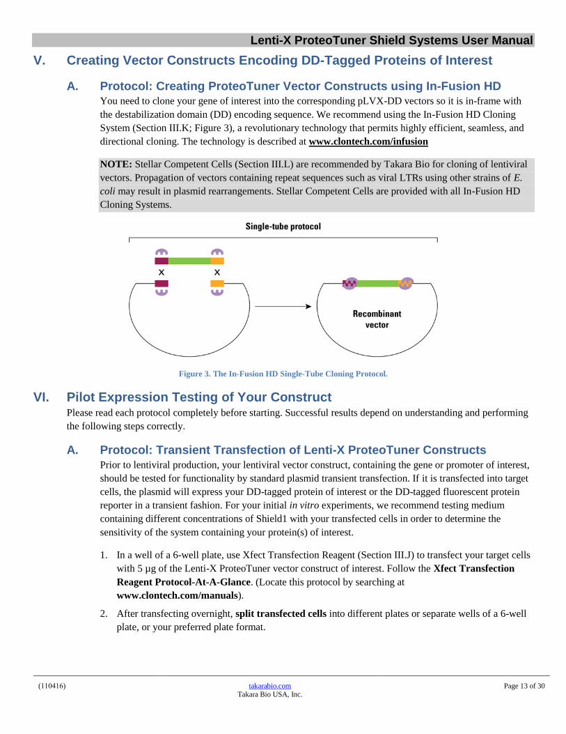
V. Creating Vector Constructs Encoding DD-Tagged Proteins of Interest
A. Protocol: Creating ProteoTuner Vector Constructs using In-Fusion HDYou need to clone your gene of interest into the corresponding pLVX-DD vectors so it is in-frame with
the destabilization domain (DD) encoding sequence. We recommend using the In-Fusion HD Cloning
System (Section III.K; Figure 3), a revolutionary technology that permits highly efficient, seamless, and
directional cloning. The technology is described at www.clontech.com/infusion
NOTE: Stellar Competent Cells (Section III.L) are recommended by Takara Bio for cloning of lentiviral
vectors. Propagation of vectors containing repeat sequences such as viral LTRs using other strains of E.
coli may result in plasmid rearrangements. Stellar Competent Cells are provided with all In-Fusion HD
Cloning Systems.
Figure 3. The In-Fusion HD Single-Tube Cloning Protocol.
VI. Pilot Expression Testing of Your ConstructPlease read each protocol completely before starting. Successful results depend on understanding and performing
the following steps correctly.
A. Protocol: Transient Transfection of Lenti-X ProteoTuner Constructs
Prior to lentiviral production, your lentiviral vector construct, containing the gene or promoter of interest,
should be tested for functionality by standard plasmid transient transfection. If it is transfected into target
cells, the plasmid will express your DD-tagged protein of interest or the DD-tagged fluorescent protein
reporter in a transient fashion. For your initial in vitro experiments, we recommend testing medium
containing different concentrations of Shield1 with your transfected cells in order to determine the
sensitivity of the system containing your protein(s) of interest.
1. In a well of a 6-well plate, use Xfect Transfection Reagent (Section III.J) to transfect your target cells
with 5 µg of the Lenti-X ProteoTuner vector construct of interest. Follow the Xfect Transfection
Reagent Protocol-At-A-Glance. (Locate this protocol by searching at
www.clontech.com/manuals).
2. After transfecting overnight, split transfected cells into different plates or separate wells of a 6-well
plate, or your preferred plate format.

Lenti-X ProteoTuner Shield Systems User Manual
(110416) takarabio.com Takara Bio USA, Inc.
Page 14 of 30
3. Incubate the transfected cells with Shield1 at specific time intervals and concentrations. Replace the
medium in the plates holding the transfected cells with medium containing the appropriate amount of
Shield1, diluted as described below. Maintain at least one culture in medium containing no Shield1 as
a negative control.
NOTE: In the case of adherent cells, let the cells reattach after the split before removing medium.
a. Recommended Shield1 Concentrations and Time Points
Try Shield1 concentrations between 50 nM and 1,000 nM for different lengths of time
(30 minutes to 12+ hours) to determine the best experimental conditions.
b. General Guidelines for Preparing Medium Containing Shield1
Dilute the supplied Shield1 stock solution (0.5 mM, supplied in ethanol) in tissue culture
media to the final concentration(s) needed in your experiment.
EXAMPLE: Preparation of 10 ml of medium containing 500 nM of Shield1: Dilute 10
µl of Shield1 stock solution (500 µM) in 10 ml of medium.
Working concentrations of Shield1 can be obtained by adding it directly from ethanol
stocks, or by diluting it serially in culture medium just before use.
Dilute the Shield1 stock solution using one of the two following types of culture medium:
1) Culture medium that has already been used to culture the cells: Collect the
media supernatant from your cell culture into a clean and sterile container and
add the appropriate amount of Shield1 to reach the appropriate final
concentration. After mixing, add the medium containing Shield1 back into the
plate.
2) Fresh culture medium: Warm up the appropriate volume of fresh culture media
needed for your experiment to ~37°C. Then add the appropriate volume of
Shield1 stock solution, to obtain the final concentration of Shield1 to be used in
the experiment.
If you are making serial dilutions of Shield1 into culture medium, we recommend that the
highest concentration not exceed 5 μM, to ensure complete solubility in the (aqueous)
culture medium.
In either case, the final concentration of ethanol in the medium added to mammalian cells
should be kept below 0.5% (a 200-fold dilution of a 100% ethanol solution) to prevent
this solvent from having a detrimental effect on the cells.
4. After adding the medium containing Shield1 at the appropriate concentration and for the appropriate
length of time, the effect and extent of protein stabilization can be analyzed with an assay that is
appropriate for your experiment, e.g., Western blot.

Lenti-X ProteoTuner Shield Systems User Manual
(110416) takarabio.com Takara Bio USA, Inc.
Page 15 of 30
VII. Producing Lentivirus from the Lenti-X Vectors
A. General Considerations
1. Optimizing Lentiviral Titer
To obtain the highest titers from the Lenti-X Packaging Single Shots (VSV-G), use the Lenti-X 293T
Cell Line (Section III.E) and adhere strictly to the following protocol, especially with respect to:
Culture size and volume
DNA amounts and transfection-grade quality
Tetracycline-free serum in Lenti-X 293T growth media
Incubation times
2. Required Materials & Precautions
All Xfect transfection reagents, volumes, and conditions are optimized for use with Lenti-X Vectors,
the Lenti-X Packaging Single Shots (VSV-G), and Lenti-X 293T cells. For optimal results, it is also
necessary to use:
Tetracycline-free FBS
10-cm culture plates
Transfection-grade DNA
Be sure to use Tetracycline-free FBS*, both in the transfection medium (Step 1) and in the
medium used to collect the virus (Step 9).
* Tetracycline-contaminated serum is detrimental to the expression of essential packaging components in the Lenti-X
Packaging System.
IMPORTANT: Perform all steps in a sterile tissue culture hood. Lentivirus requires the use of a
Biosafety Level 2 facility. Recombinant pseudotyped lentiviruses packaged from HIV-1-based
vectors are capable of infecting human cells. Know and use appropriate safety precautions (see
Section IV.B).
3. Optimal Cell Densities at Transfection & Harvest
Figure 4. Optimal density of Lenti-X 293T cells at point of transfection (left panel) and harvest time (right panel),
shown here using a transfer vector containing ZsGreen1.

Lenti-X ProteoTuner Shield Systems User Manual
(110416) takarabio.com Takara Bio USA, Inc.
Page 16 of 30
B. Protocol: Transfecting Lentiviral Vectors into Lenti-X 293T Packaging CellsNOTE: To achieve the highest titers, it is critical to pay close attention to the transfection. You may want
to perform a cotransfection with a lentiviral vector that contains a fluorescent protein. You should be able
to achieve transfection efficiencies of greater than 90%.
1. Approximately 24 hr before transfection, seed 4–5 x 10⁶ Lenti-X 293T cells/10-cm plate, in 8 ml of
growth medium. Make sure that the cells are plated evenly. Incubate at 37°C, 5% CO2 overnight.
Continue to incubate the cells until you are ready to add the transfection mixture in Step 5. The cells
should be 80–90% confluent at the time of transfection.
2. In a sterile microfuge tube, dilute 7.0 μg of your lentiviral vector plasmid DNA with sterile water to a
final volume of 600 μl. Mix thoroughly by vortexing.
NOTE: Always dilute your DNA in water prior to adding it to a Lenti-X Packaging Single Shot. Do
not add water and DNA separately (since undiluted DNA should not be mixed with Xfect
Transfection Reagent).
3. Add the 600 μl of diluted DNA to a tube of Lenti-X Packaging Single Shots, replace the cap, and
vortex well at a high speed for 20 seconds. The pellet should dissolve completely.
NOTE: In some cases some insoluble material may be visible after vortexing. This material does not
have a negative effect on transfection efficiency or virus yields.
4. Incubate the samples for 10 min at room temperature to allow nanoparticle complexes to form. After
the 10 min incubation, centrifuge the tube for 2 seconds to bring the sample to the bottom of the tube.
NOTE: Sample tubes can be inserted into 1.5-ml microfuge tubes for a brief centrifugation.
5. Add the entire 600 μl of nanoparticle complex solution dropwise to the 8 ml of cell culture prepared
in Step 1. Rock the plate gently back and forth to mix.
NOTE: It is normal for the medium to change color slightly upon addition of nanoparticle complex
solution.
6. Incubate the cells at 37°C supplied with 5% CO2.
NOTE: A 4-hr incubation with Xfect-DNA nanoparticles is sufficient for optimal transfection.
Incubation may be continued overnight for convenience but does not generally increase transfection
efficiency or titer.
7. After 4 hr to overnight, add an additional 6 ml of fresh complete growth medium and incubate at
37°C, 5% CO2 for an additional 24–48 hr. Virus titers will generally be highest 48 hr after the start of
transfection.
8. Harvest the lentiviral supernatants and pool similar stocks, if desired (a 48-hr sample may be stored at
4°C until a 72-hr sample is harvested).
CAUTION: Supernatants contain infectious lentivirus.
Centrifuge briefly (500g for 10 min) or filter through a 0.45-μm filter to remove cellular debris.
NOTE: The filter used should be made of cellulose acetate, or polysulfone (low protein binding),
instead of nitrocellulose. Nitrocellulose binds proteins present in the membrane of lentivirus and
destroys the virus.
9. Verify virus production using Lenti-X GoStix (Section III.G; see Lenti-X GoStix (50 & 200)
Protocol-At-A-Glance at www.clontech.com/manuals for details). Alternatively, titrate the virus
stock, then use the virus to transduce target cells, or store at –80°C.
NOTE: Titers can drop as much as 2–4 fold with each freeze-thaw cycle.
10. For protocols describing how to transduce your target cells or create frozen stocks, see the Lenti-X
Lentiviral Expression Systems User Manual at www.clontech.com/manuals

Lenti-X ProteoTuner Shield Systems User Manual
(110416) takarabio.com Takara Bio USA, Inc.
Page 17 of 30
VIII. Lentivirus Titration
A. Summary
1. Instant Qualitative Titer Test
You can assess the quality of your lentivirus stock in 30 seconds with Takara Bio’s Lenti-X GoStix
(Cat. Nos. 631241, 631243 & 631244). The GoStix detect lentiviral p24 in only 20 μl, and can be
used to determine whether virus production is within a usable range or for selecting the best time to
harvest your virus. A 3-prep sample is supplied for free with the Lenti-X Packaging Single Shots
(VSV-G) (Section III.D).
2. Quantitative Titer Test
a. Determining the viral titer is necessary to obtain the following information:
Confirmation that viral stocks are viable
The proper transduction conditions for your particular cell type by adjusting the MOI for the
desired transduction efficiency. MOI = No. of infectious virus particles per target cell at the
time of infection.
The maximum number of target cells that can be transduced by a given virus volume.
b. To transduce using a known multiplicity of infection (MOI), it is necessary to titrate your
lentiviral stocks. We recommend the Lenti-X qRT-PCR Titration Kit (Cat. No. 631235) or
Lenti-X p24 Rapid Titer Kit (Cat. No. 632200) for very rapid quantitative titrations of virus
stocks (~4 hr), or a standard method that relies on infection.
c. The standard viral titration protocol consists of infecting cells with serial dilutions of the stock,
selecting for stable transductants with antibiotic, and counting the resulting cell colonies (Section
VIII.B).
Freshly harvested virus can be titrated immediately, or frozen in aliquots at –80°C and then
titrated. Note that each freeze-thaw cycle can reduce the functional titers of infectious virus
by up to 2–4 fold.
Absolute titers will depend heavily on the cell type used for titration, and there may be
significant differences between the titer values determined in cells typically used for lentiviral
titration (i.e., HT-1080) and the number of target cells transduced by the titrated virus.
However, titrations serve to determine the relative virus content of different viral stocks
prepared from different vectors.
B. Protocol: Determining Viral Titer by Colony Formation
NOTE: This protocol can be completed in 7–14 days.
1. Plate HT-1080 cells (or other) in 6-well plates the day before performing the titration infections. Plate
2 x 10⁵ cells/well, in 2 ml of medium. Allow at least one well to be used as a “no infection” control.
2. Prepare 20 ml of complete medium and add 60 µl of 4 mg/ml Polybrene. This will be diluted 3 fold
for a final Polybrene concentration of 4 µg/ml.
NOTE: Polybrene is a polycation that reduces charge repulsion between the virus and the cellular
membrane. The optimum final concentration of Polybrene may be determined empirically but
generally falls within 2–12 µg/ml. Excessive exposure to Polybrene (>24 hr) can be toxic to cells.

Lenti-X ProteoTuner Shield Systems User Manual
(110416) takarabio.com Takara Bio USA, Inc.
Page 18 of 30
3. Prepare filtered viral supernatant from packaging cells (Section VII). This is the virus stock.
4. Prepare six 10-fold serial dilutions of the virus stock as follows:
a. Add 1.35 ml of medium containing Polybrene (Step 2) to each of six sterile and numbered 1.5 ml
microfuge tubes.
b. Add 150 µl of the virus stock (Step 3) to the tube 1. Mix.
c. Transfer 150 µl tube 1 to tube 2 and mix. Continue making serial dilutions by transferring 150 µl
from each successive dilution into the next prepared tube.
5. Infect the HT-1080 cells by adding 1 ml of each viral dilution (Step 4) to each appropriate well. The
final Polybrene concentration will be 4 µg/ml in ~3 ml. Centrifuge the cultures to improve infection
efficiency*.
* NOTE: CULTURE CENTRIFUGATION INCREASES INFECTION EFFICIENCY. Centrifuging
the plate at 1,200g for 60–90 min at 32°C can significantly increase infection efficiency. A room
temperature centrifuge is acceptable if a 32°C unit is not available.
6. After infecting for 8–24 hours, remove supernatants and subject the cells to puromycin selection
using the selection concentrations that are optimal for your cell line (Section III.F).
7. Allow colonies to form for 7–14 days. Stain the colonies with 1% crystal violet solution (in 10%
ethanol) and count.
8. The titer of virus corresponds to the number of colonies generated by the highest dilution, multiplied
by the dilution factor. For example, the presence of 4 colonies in the 106 dilution would represent a
viral titer of 4 x 10⁶ colony forming units.
IX. Transducing Target Cells with a Lenti-X ProteoTuner Lentivirus
A. Protocol: Transducing Target Cells with Lenti-X ProteoTuner Lentiviruses
NOTE: This protocol can be completed in 2–3 days.
1. Plate target cells in complete growth medium 12–18 hr before transduction.
2. Thaw aliquots of your Lenti-X ProteoTuner lentiviral stocks, or use filtered virus stocks freshly
prepared from packaging cells (Section VII). Mix gently, but do not vortex.
3. Add Polybrene to the cell cultures to obtain the desired final concentration during the transduction
step (e.g., 4 μg/ml).
NOTE: Lenti-X Accelerator and RetroNectin (Section III.I) may be used as transduction enhancers
instead of Polybrene.
4. In general, we find that an MOI of 5–20 works best. If titer values are unknown, use serial dilutions
of the virus supernatant, such that the total volume of supernatant used makes up no more than 1/3 the
final volume of culture medium used in the transduction. Centrifuge the cultures to improve
transduction efficiency (see Section VIII.B).
5. Transduce the cells for 8–24 hr. If you are concerned that exposure to either the Polybrene or to the
viral supernatant (which contains medium conditioned by the packaging cells) may adversely affect
your target cells, limit the transduction to 6–8 hr.

Lenti-X ProteoTuner Shield Systems User Manual
(110416) takarabio.com Takara Bio USA, Inc.
Page 19 of 30
6. Remove and discard the virus-containing medium and replace it with fresh growth medium.
Use the transduced cells to optimize Shield 1 concentration and incubation time (Section X.A) in
preparation for protein stabilization and destabilization experiments using Shield1 (Sections X.B
& X.C).
Alternatively, passage the cultures and subject the cells to selection using puromycin to establish
a stable cell population or cell line. (Instructions for expansion and freezing of cell line stocks are
provided in Appendix B.)
X. Protein Stabilization & Destabilization Using Lenti-X ProteoTuner Cell Lines
A. Protocol: Optimizing Shield1 Concentration and Incubation Time of
Transduced Cells1. Split the transduced cells from Section IX.A, Step 6 into different plates or separate wells of a 6-well
plate, or your preferred plate format.
To begin incubation of the transduced cells with Shield1 at predetermined time intervals and
concentrations (these can be determined using transient transfection—see Section VI), replace the
medium in the plates containing the transduced cells with medium containing the appropriate amount
of Shield1, diluted as described below. Maintain at least one culture in medium containing no Shield1
as a negative control.
NOTE: In the case of adherent cells, let the cells reattach after the split before removing the medium.
a. Recommended Shield1 Concentrations and Time Points
Try Shield1 concentrations between 0.1 nM and 1,000 nM for different lengths of
time (30 min to 12+ hours) to determine the best experimental conditions.
b. General Guidelines for Preparing Medium Containing Shield1
Dilute the supplied Shield1 stock solution (0.5 mM, supplied in ethanol) in tissue
culture media to the final concentration(s) needed in your experiment.
EXAMPLE: Preparation of 10 ml of medium containing 500 nM of Shield1: Dilute
10 µl of Shield1 stock solution (500 µM) in 10 ml of medium to yield a final
concentration of 500 nM.
Working concentrations of Shield1 can be obtained by adding it directly from ethanol
stocks, or by diluting it serially in culture medium just before use.
Dilute the Shield1 stock solution using one of the two following types of culture
medium:
1) Culture medium that has already been used to culture the cells: Collect the
media supernatant from your cell culture into a clean and sterile container and
add the appropriate amount of Shield1 to reach the appropriate final
concentration. After mixing, add the medium containing Shield1 back into the
plate.

Lenti-X ProteoTuner Shield Systems User Manual
(110416) takarabio.com Takara Bio USA, Inc.
Page 20 of 30
2) Fresh culture medium: Warm up the appropriate volume of fresh culture media
needed for your experiment to ~37°C. Then add the appropriate volume of
Shield1 stock solution, to obtain the final concentration of Shield1 to be used in
the experiment.
If you are making serial dilutions of Shield1 into culture medium, the highest
concentration should not exceed 5 μM, to ensure complete solubility in the
(aqueous) culture medium.
In either case, the final concentration of ethanol in the medium added to
mammalian cells should be kept below 0.5% (a 200-fold dilution of a 100%
ethanol solution) to prevent this solvent from having a detrimental effect on
the cells.
2. After adding the medium containing Shield1 at the appropriate concentration and for the appropriate
length of time, the effect of stabilizing your DD-tagged protein of interest can be analyzed with an
assay that is appropriate for your experiment, e.g., Western blot.
B. Protocol: DD-Protein Stabilization of Transduced CellsBefore you begin, transduce your DD construct of interest into your cells of interest (Section IX.A) and
determine the optimal Shield1 concentration and incubation time (see Section X.A).
Stabilizing a protein of interest in attached cells
1. 12–24 hr posttransduction, split the cells into at least two parallel cultures. (The number of plates
depends on the number of samples you would like to collect.)
2. Culture the cells (all plates) in medium without Shield1 until the cells are attached to each plate.
NOTE: Shield1 does not interfere with the attachment process. Therefore, Shield1 can be added
immediately after splitting if required for your experimental needs.
3. Dilute the Shield1 to the optimal concentration determined in Section X.A. We recommend final
concentrations of ~50–1,000 nM Shield1 in the cell culture medium.
4. Remove the culture medium and replace it with warm medium with or without Shield1. Shield1
added to the experimental plate(s) will protect the DD-tagged protein of interest from
proteasomal degradation, causing a rapid increase in its level in the cell.
5. Collect cells at specific time points (defined by your needs) in order to analyze and compare cells
with and without the stabilized DD fusion protein of interest using the assay appropriate for your
experiment, e.g., Western blot.
Stabilizing a protein of interest in cells grown in suspension
1. 12–24 hr posttransduction, divide the cell suspension evenly into at least two tubes. (The number
of tubes depends on the number of samples you would like to collect.)
2. Dilute Shield1 to the optimal concentration determined in Section X.A. We recommend final
concentrations of ~50–1,000 nM Shield1 in the cell culture medium.
3. Centrifuge the tubes (from Step 1) for 5 min at ≤1,000 rpm.

Lenti-X ProteoTuner Shield Systems User Manual
(110416) takarabio.com Takara Bio USA, Inc.
Page 21 of 30
4. Remove the culture medium and replace with warm media with or without Shield1 (prepared in
Step 2) as determined by your needs.
NOTE: The added Shield1 will protect your DD-tagged protein of interest from proteasomal
degradation, causing a rapid increase in its level in the cell.
5. Collect cells at specific time points (defined by your needs) in order to analyze and compare cells
with and without the stabilized DD fusion protein of interest using the assay appropriate for your
experiment, e.g., Western blot.
C. Protocol: DD-Protein Destabilization
Before you begin, transduce your DD construct of interest into your cells of interest (Section IX.A).
Culture your cells in medium containing Shield1 at the optimal concentration determined in Section X.A
to stabilize your protein of interest.
Destabilizing a protein of interest in attached cells
Method A
Requires splitting cells (for quickest destabilization)
1. After stabilizing the protein of interest for the desired length of time via Shield1, remove the
medium containing Shield1.
2. Rinse the cells with warm Dulbecco’s Phosphate Buffered Saline (TC grade) to ensure complete
removal of Shield1.
3. Detach the cells by your method of choice (trypsin, cell dissociation buffer, etc.) and split them
into at least two new cell culture plates (the number of plates depends on the number of samples
you would like to collect).
4. Culture the cells in one plate in medium containing Shield1 (positive control) and culture the cells
in the other plate(s) in medium without Shield1.
NOTE: Growing the cells in the absence of Shield1 causes the fast degradation of the previously
stabilized protein of interest.
5. Collect cells at specific time points (defined by your needs) in order to analyze and compare cells
with and without the stabilized DD fusion protein of interest using the assay appropriate for your
experiment, e.g., Western blot.
Method B
No splitting required (for slower destabilization)
1. After stabilizing the protein of interest for the desired length of time via Shield1, remove the
medium containing Shield1.
2. In order to destabilize the protein of interest, wash the cells in the plates by rinsing them three
times with warm culture medium without Shield1.
3. Culture the cells in culture medium without Shield1.
4. Collect cells at specific time points (defined by your needs) in order to analyze and compare cells
with and without the stabilized DD fusion protein of interest using the assay appropriate for your
experiment, e.g., Western blot.
Destabilizing a protein of interest in cells grown in suspension

Lenti-X ProteoTuner Shield Systems User Manual
(110416) takarabio.com Takara Bio USA, Inc.
Page 22 of 30
1. After stabilizing the protein of interest for the desired length of time via Shield1, distribute the
cell suspension evenly into at least two tubes (the number of tubes depends on the number of
samples you would like to collect).
2. Centrifuge the tubes for 5 min at ≤1,000 rpm and remove the culture medium.
3. Resuspend one pellet in culture medium with Shield1 at the appropriate concentration (positive
control) and resuspend the remaining pellet(s) in culture medium without Shield1.
4. Collect cells at specific time points (defined by your needs) in order to analyze and compare cells
with and without the stabilized DD fusion protein of interest using the assay appropriate for your
experiment, e.g., Western blot.
D. Protocol: Working with Stable Cell Lines Expressing a DD-Tagged Protein
of Interest1. After establishing a stable cell line, you can culture your cells either in the absence or the presence of
Shield1, depending on your experimental needs.
2. If you grow your cells in the absence of Shield1, your protein of interest will be destabilized and
expressed only at a very low level in your stable cell line. Then Shield1 can be added to rapidly
increase the amount of your protein of interest (Section X.B).
3. Maintenance in, or addition of Shield1 to a stable cell line will stabilize your protein of interest and
quickly increase its level in the cell (Section X.C).
E. Protocol: In Vivo Use of Shield1
General Methods
Because Shield1 is suitable for use in vivo, studies can also be performed in a whole animal context via
one of the following commonly used methods:
Generate transgenic mice in which expression of the DD-tagged fusion protein is restricted to a
tissue of interest by a tissue-specific promoter.
Subcutaneously xenograft cells into nude mice.
Preparation and Injection of Shield1
1. Preparing solutions for injection
Shield1 can be reconstituted into DMA or ethanol at various concentrations up to 10
mg/ml. This stock solution may be kept for several months at −20 °C.
Make up a fresh solution of 9:1 PEG 400:Tween 80 before each injection.
2. Injecting Shield1 into mice
Inject Shield1 at a concentration of 3–10 mg/kg body weight using a 1:1 mixture of the
appropriate amount of Shield1 stock solution and the fresh 9:1 PEG 400:Tween 80
solution (Step 1). Control mice should be injected with a 1:1 mixture of DMA (without
any Shield1 added) and the PEG 400:Tween 80 solution.

Lenti-X ProteoTuner Shield Systems User Manual
(110416) takarabio.com Takara Bio USA, Inc.
Page 23 of 30
Shield-1 may be injected intravenously; however, intraperitoneal injections often produce
more reliable results.
The injection regiment can be repeated every 48 hr in order to maintain strong
stabilization of the DD-tagged protein of interest.
Effects of Shield1 In Vivo
Nude mice were injected with Shield1 every 48 hr for 2 months and showed no signs of toxicity (e.g.,
changes in feeding behavior, grooming, or activity levels) (Sellmyer et al., 2009).
XI. References for ProteoTunerArmstrong, C. M. & Goldberg, D. E. An FKBP destabilization domain modulates protein levels in Plasmodium
falciparum. Nat. Methods 4, 1007–1009 (2007).
Banaszynski, L. A., Chen, L. chun, Maynard-Smith, L. A., Ooi, A. G. L. & Wandless, T. J. A Rapid, Reversible, and Tunable Method to Regulate Protein Function in Living Cells Using Synthetic Small Molecules. Cell 126, 995–1004 (2006).
Banaszynski, L. a, Sellmyer, M. a, Contag, C. H., Wandless, T. J. & Thorne, S. H. Chemical control of protein stability and function in living mice. Nat. Med. 14, 1123–1127 (2008).
Banaszynski, L. a & Wandless, T. J. Conditional control of protein function. Chem. Biol. 13, 11–21 (2006).
Berdeaux, R. et al. SIK1 is a class IIHDAC kinase that promotes survival of skeletal myocytes. Nat. Med. 13, 597–603 (2007).
Grimley, J. S., Chen, D. A., Banaszynski, L. A. & Wandless, T. J. Synthesis and analysis of stabilizing ligands for FKBP-derived destabilizing domains. Bioorganic Med. Chem. Lett. 18, 759–761 (2008).
Herm-Götz, A. et al. Rapid control of protein level in the apicomplexan Toxoplasma gondii. Nat. Methods 4, 1003–1005 (2007).
Maynard-Smith, L. A., Chen, L. C., Banaszynski, L. A., Ooi, A. G. L. & Wandless, T. J. A directed approach for engineering conditional protein stability using biologically silent small molecules. J. Biol. Chem. 282, 24866–24872 (2007).
Schoeber, J. P. H. et al. Conditional fast expression and function of multimeric TRPV5 channels using Shield-1. Am. J. Physiol. Renal Physiol. 296, F204–F211 (2009).
Sellmyer, M. A., Thorne, S. H., Banaszynski, L. A., Contag, C. H. & Wandless, T. J. A general method for conditional
regulation of protein stability in living animals. Cold Spring Harb. Protoc. 2009, pdb.prot5173 (2009).

Lenti-X ProteoTuner Shield Systems User Manual
(110416) takarabio.com Takara Bio USA, Inc.
Page 24 of 30
Appendix A. Troubleshooting Guide Table 2. Troubleshooting Guide
Problem Possible Explanation Solution
A. Vector Cloning
Plasmid is difficult to grow or clone
Some viral vectors may undergo rearrangements between the 5’ and 3’ LTRs when propagated in less-than-optimal E. coli host strains
Use Stellar Competent Cells (Cat. No. 636763) to produce high DNA yields and to minimize the potential for DNA rearrangements.
B. Lenti-X 293T Packaging Cells
Poor viability upon thawing
Improper thawing techniques Use thawing procedure in Appendix B, and/or consult the Lenti-X 293T Cell Line Protocol-at-a-Glance
Incorrect culture medium Use DMEM with additives listed in Section III.C.
Improper tissue culture plasticware Use collagen I-coated plates to aid cell adherence during initial seeding.
Slow growth Incorrect culture medium Use DMEM with additives listed in Section III.C.
Cells do not attach to plate Improper tissue culture plasticware Use collagen I-coated plates to aid cell adherence during initial seeding.
Cells appear morphologically different Passage of cell culture is too high (old cells)
Thaw/purchase new aliquot of Lenti-X 293T cells.
C. Virus Production
Poor transfection efficiency (as determined by GOI or marker expression in the Lenti-X 293T cell line)
Cells plated too densely Plate 4–5 x 10⁶ cells/100-mm plate, or fewer if the cells divide rapidly. Use at 50–80% confluency. See Section VII.
Transfection is toxic to cells Use the optimized conditions provided in Section VII.
Cells harvested or analyzed too soon after transfection
Wait 48 hr after transfection for maximal expression of GOI or marker to determine efficiency.
Low titers (<105 cfu/ml)
Virus was harvested too early Harvest virus 48–72 hr after the start of transfection.
Vector is too large The limit for efficient packaging function is 9.7 kb from the end of the 5’-LTR to the end of the 3’-LTR
Polybrene is missing or at suboptimal concentration
Add Polybrene (4 µg/ml) during transduction or optimize the concentration (2–12 µg/ml)
Virus was exposed to multiple freeze-thaw cycles
Each cycle reduces titer by approximately 2–4 fold. Limit the number of freeze-thaws.
Suboptimal selection procedure during titration
Perform an antibiotic kill curve on the cell line prior to using it for titration.

Lenti-X ProteoTuner Shield Systems User Manual
(110416) takarabio.com Takara Bio USA, Inc.
Page 25 of 30
Problem Possible Explanation Solution
D. Transduction of Target Cells
Poor transduction efficiency
Low titer
See Section C or use the Lenti-X Concentrator (Section III.H) to increase your available titer up to 100-fold without ultracentrifugation.
Poor transfection efficiency Follow the protocol in Section VII.B. Be sure to use 5 µg of transfection-grade plasmid.
Low viability of target cells during transduction
Optimize culture conditions for target cells prior to infection
Packaging cell line-conditioned media may affect cell growth; dilute viral supernatant or shorten exposure time to viral supernatant. Consider using RetroNectin reagent and the RetroNectin-Bound Virus transduction protocol or purify your virus prior to transduction using the Lenti-X Maxi Purification Kit (Cat. Nos. 631233 & 631234).
Excessive exposure to Polybrene: optimize amount (titrate) or shorten exposure time to viral supernatant
Viral supernatant contains transduction inhibitors
Use RetroNectin reagent or RetroNectin-coated plates in the RetroNectin-Bound Virus transduction protocol, which allows virions to bind the RetroNectin substratum and be washed free of inhibitors prior to target cell infection; or, purify your virus prior to transduction using the Lenti-X Maxi Purification Kit (Cat. Nos. 631233 &
631234).
E. Establishment of Stable Cell Lines
Untransduced cells do not die at the high antibiotic concentration established via titration in Section III.F
The cells have not been recently passaged, so they remain well-attached to the plate surface even when they are dead.
To determine the appropriate antibiotic concentration, use cells that have been split within the last 2–3 days.
You have achieved 100% transduction efficiency.
There are no surviving cells after transduction followed by selection
The antibiotic concentration which caused massive cell death when determining the appropriate dose via titration could be too high.
Use a lower antibiotic concentration for selection of stably transfected cell clones.
Poor cell viability Cells were not properly frozen. See Appendix C, Section A.
Cells were not properly thawed. See Appendix C, Section B.

Lenti-X ProteoTuner Shield Systems User Manual
(110416) takarabio.com Takara Bio USA, Inc.
Page 26 of 30
Appendix B. Lenti-X ProteoTuner Vector Maps
Figure 5. pLVX-PTuner and pLVX-PTunerC Vector maps.
For more detailed vector information, see takarabio.com
Figure 6. pLVX-PTuner Green Vector map.
For more detailed vector information, see takarabio.com

Lenti-X ProteoTuner Shield Systems User Manual
(110416) takarabio.com Takara Bio USA, Inc.
Page 27 of 30
Figure 7. pLVX-DD-ZsGreen1 Control and pLVX-DD-ZsGreen1 Reporter Vector maps.
For more detailed vector information, see takarabio.com
Figure 8. pLVX-DD-AmCyan1 Control and pLVX-DD-AmCyan1 Reporter Vector maps.
For more detailed vector information, see takarabio.com

Lenti-X ProteoTuner Shield Systems User Manual
(110416) takarabio.com Takara Bio USA, Inc.
Page 28 of 30
Figure 9. pLVX-DD-tdTomato Control and pLVX-DD-tdTomato Reporter Vector maps.
For more detailed vector information, see takarabio.com
Appendix C: Preparing and Handling Cell Line Stocks
A. Protocol: Freezing Cell Line Stocks
Once you have created and tested your ProteoTuner cell line, you must prepare multiple frozen aliquots to
ensure a renewable source of cells, according to the following protocol:
1. Expand your cells to multiple 10-cm dishes or T75 flasks.
2. Trypsinize and pool all of the cells, then count the cells using a hemocytometer.
3. Centrifuge the cells at 100-g for 5 min. Aspirate the supernatant.
4. Resuspend the pellet at a density of at least 1–2 x 10⁶ cells/ml in freezing medium. Freezing medium
can be purchased from Sigma (Cat. Nos. C6164 & C6039), or use 70–90% FBS, 0–20% medium
(without selective antibiotics), and 10% DMSO.
5. Dispense 1 ml aliquots into sterile cryovials and freeze slowly (1°C per min). For this purpose, you
can place the vials in Nalgene cryo-containers (Nalgene, Cat. No. 5100-001) and freeze at –80°C
overnight. Alternatively, place vials in a thick-walled styrofoam container at –20°C for 1–2 hr.
Transfer to –80°C and freeze overnight.
6. The next day, remove the vials from the cryo-containers or styrofoam containers, and place in liquid
nitrogen storage or an ultra-low temperature freezer (–150°C) for storage.
7. Two or more weeks later, plate a vial of frozen cells to confirm viability.
Lenti-X ProteoTuner Shield Systems User Manual
(110416) takarabio.com Takara Bio USA, Inc.
Page 29 of 30
B. Protocol: Thawing Cell Line Frozen Stocks
To prevent osmotic shock and maximize cell survival, use the following procedure to start a new culture
from frozen cells:
1. Thaw the vial of cells rapidly in a 37°C water bath with gentle agitation. Immediately upon thawing,
wipe the outside of the vial with 70% ethanol. All of the operations from this point on should be
carried out in a laminar flow tissue culture hood under strict aseptic conditions.
2. Unscrew the top of the vial slowly and, using a pipet, transfer the contents of the vial to a 15-ml
conical centrifuge tube containing 1 ml of prewarmed medium (without selective antibiotics such as
puromycin). Mix gently.
3. Slowly add an additional 4 ml of fresh, prewarmed medium to the tube and mix gently.
4. Add an additional 5 ml of prewarmed medium to the tube and mix gently.
5. Centrifuge at 100g for 5 min, carefully aspirate the supernatant, and GENTLY resuspend the cells in
complete medium without selective antibiotics. (This method removes the cryopreservative and can
be beneficial when resuspending in small volumes. However, be sure to treat the cells gently to
prevent damaging fragile cell membranes.)
6. Mix the cell suspension thoroughly and add to a suitable culture vessel. Gently rock or swirl the
dish/flask to distribute the cells evenly over the growth surface and place in a 37°C humidified
incubator (5–10% CO2 as appropriate) for 24 hr.
NOTE: For some loosely adherent cells (e.g., HEK 293-based cell lines), we recommend using
collagen-coated plates to aid attachment after thawing. For suspension cultures, suspend cells at a
density of no less than 2 x 10⁵ cells/ml.
7. The next day, examine the cells under a microscope. If the cells are well-attached and confluent, they
can be passaged for use. If the majority of cells are not well-attached, continue culturing for another
24 hr.
NOTE: For some loosely adherent cell lines (e.g., HEK 293-based cell lines), complete attachment of
newly thawed cultures may require up to 48 hr.
8. Expand the culture as needed. The appropriate selective antibiotic(s) should be added to the medium
after 48–72 hr in culture. Maintain cell lines in complete culture medium containing a maintenance
concentration of puromycin, as appropriate (Section III.F).

Lenti-X ProteoTuner Shield Systems User Manual
(110416) takarabio.com Takara Bio USA, Inc.
Page 30 of 30
Contact Us
Customer Service/Ordering Technical Support
tel: 800.662.2566 (toll-free) tel: 800.662.2566 (toll-free)
fax: 800.424.1350 (toll-free) fax: 800.424.1350 (toll-free)
web: takarabio.com web: takarabio.com
e-mail: [email protected] e-mail: [email protected]
Notice to Purchaser
Our products are to be used for research purposes only. They may not be used for any other purpose, including, but not limited to, use in drugs, in vitro diagnostic
purposes, therapeutics, or in humans. Our products may not be transferred to third parties, resold, modified for resale, or used to manufacture commercial products or to provide a service to third parties without prior written approval of Takara Bio USA, Inc.
Your use of this product is also subject to compliance with any applicable licensing requirements described on the product’s web page at takarabio.com. It is your
responsibility to review, understand and adhere to any restrictions imposed by such statements.
©2016 Takara Bio Inc. All Rights Reserved.
All trademarks are the property of Takara Bio Inc. or its affiliate(s) in the U.S. and/or other countries or their respective owners. Certain trademarks may not be registered in all jurisdictions. Additional product, intellectual property, and restricted use information is available at takarabio.com.
This document has been reviewed and approved by the Quality Department.