Embed Size (px)
Citation preview

Lens fibers have a fully functional ubiquitin-proteasome pathway
Paulo Pereiraa,b, Fu Shanga,*, Marisa Hobbsa, Henrique Giraoa,b, Allen Taylora
aLaboratory for Nutrition and Vision Research, USDA HNRC at Tufts University, 711 Washington Street, Boston, MA 02111, USAbCentre of Ophthalmology, Biomedical Institute for Research in Light and Image (IBILI), Faculty of Medicine,
University of Coimbra, 3000 Coimbra, Portugal
Received 23 August 2002; accepted in revised form 12 December 2002
Abstract
We previously showed that lens epithelial cells have a fully functional ubiquitin-proteasome pathway (UPP) and that
ubiquitin-conjugating activity is up-regulated in response to oxidative stress. In this study we assessed the protein levels and activities of
different components of the UPP in lens fibers. Calf lenses were dissected into four different regions: epithelial layer, outer cortex, inner
cortex and nucleus. Relative levels of ubiquitin-activating enzyme (E1), ubiquitin-conjugating enzymes (E2s), endogenous ubiquitin
conjugates, 19S and 20S proteasome subunits were determined by Western blotting. The activities of E1 and E2 were determined by thiol
ester assays and the activities of the proteasome and isopeptidases were determined using ubiquitinated a-lactalbumin as a substrate.
This work demonstrates that lens fibers, including those in the nuclear region, contain most, if not all, of the components for the UPP.
Ubiquitin conjugation activity, proteasome activity and isopeptidase activity were also detected in all layers of the lens. The reduced
ubiquitin conjugation activity in the inner regions of the lens appeared to be due to a decline in levels of a specific family of E2s, Ubc4 or
Ubc5, which were shown to be the rate-limiting enzymes for the formation of high mass conjugates in the lens. Supplementation of Ubc4
or Ubc5 can partially restore the ubiquitin conjugation activity in the inner regions of the lens. Since Ubc4 and Ubc5 are involved in
selectively ubiquitinating damaged or abnormal proteins, the decline in levels and activities of these E2s may be responsible for the
accumulation of abnormal proteins in inner regions of the lens.
q 2003 Published by Elsevier Science Ltd.
Keywords: differentiation; development; lens; proteasome; ubiquitin
1. Introduction
The ubiquitin-proteasome pathway (UPP) is a major
cytosolic proteolytic pathway in most eukaryotic cells.
There are two stages in the UPP: substrate-recognition
by formation of ubiquitin-protein conjugates and the
subsequent degradation of the ubiquitin conjugates by
the 26S proteasome. In its simplest form, ubiquitination
involves the attachment of multiple molecules of ubiquitin
to protein substrates (Hershko and Ciechanover, 1998).
In order to initiate this process, ubiquitin is activated by the
formation of a high-energy thiol ester with ubiquitin-
activating enzyme (E1), the first enzyme in the ubiquitina-
tion pathway. The ubiquitin is then transferred to one of
many ubiquitin-conjugating enzymes (Ubc or E2s), also via
formation of a thiol ester. Subsequently, ubiquitin is
transferred directly to substrates or is transferred to
substrates via one of several ubiquitin ligases (E3s).
Multiple isoforms of E2s and E3 have been identified in
each species. The multiplicity of E2 and E3 enzymes is
responsible for the substrate specificity of the UPP.
Multiple ubiquitin moieties attach to substrate proteins to
form ubiquitin chains. Thus, most of the ubiquitin
conjugates attain high masses. These ubiquitin conjugates
are generally recognized and degraded by the 26S protea-
some at rates which are generally proportional to rates of
ubiquitin-protein conjugate formation (Chin et al., 1982;
Obin et al., 1994). The 26S proteasome consists of two large
subcomplexes, the 20S particle with a barrel-shaped
structure, which carries the catalytic activity, and the 19S
regulatory particle, which caps both ends of the barrel
(Baumeister et al., 1998). The 19S particle contains ATPase
activity and is presumably responsible for the recognition of
the ubiquitinated proteins (Tanaka and Tsurumi, 1997;
0014-4835/03/$ - see front matter q 2003 Published by Elsevier Science Ltd.
DOI:10.1016/S0014-4835(03)00020-4
Experimental Eye Research 76 (2003) 623–631
www.elsevier.com/locate/yexer
* Corresponding author. Dr Fu Shang, Laboratory for Nutrition and
Vision Research, USDA HNRC at Tufts University, 711 Washington
Street, Boston, MA 02111, USA.
E-mail address: [email protected] (F. Shang).
Lam et al., 2002). In some instances, ubiquitin-protein
conjugates are deconjugated by isopeptidases (Wilkinson
et al., 1995; Lam et al., 1997), rather than being degraded by
the 26S proteasome. Therefore, ubiquitination may
have other functions in addition to targeting proteins
for degradation. However, non-proteolytic functions for
ubiquitination remain to be elucidated.
The lens is composed of two types of cells: epithelial cells
and fiber cells. Epithelial cells cover the anterior surface of
the lens and fiber cells occupy almost the entire volume of the
lens. The fiber cells are differentiated from the epithelial
cells. During differentiation, lens epithelial cells exit from
cell cycle (Chamberlain and McAvoy, 1987) and undergo
significant morphological and biochemical changes that
result in the formation of fully differentiated fiber cells,
where virtually all organelles, including the nuclei, are
absent. This unique pattern of differentiation occurs in
the equatorial region of the lens, and fibers with increasingly
advanced stages of differentiation accumulate concentrically
at the interior of the lens (Kuszak, 1995). Proper execution of
the differentiation program and formation of mature fibers
seem to be required for lens transparency since abnormalities
that result in incomplete degradation of intracellular
organelles are associated with various forms of cataract
(Pan and Griep, 1994; Nakamura et al., 1995).
A fully functional UPP has been demonstrated in the lens
epithelial cell (Huang et al., 1993, 1995; Shang and Taylor,
1995; Shang et al., 1997a, 1999). The ubiquitin conjugating
activity and proteolytic activity in lens epithelial cells are
up-regulated during recovery from oxidative stress (Shang
et al., 1997b). One of the functions for the UPP is to
selectively degrade damaged proteins (Shang et al., 2001).
To test our hypothesis that the UPP plays a role in selective
removal of damaged proteins from the lens, it is necessary to
determine if the lens fibers also have a fully functional UPP,
because the majority of damaged proteins are in fiber cells.
We report here that the UPP components examined in this
work were detected in all layers of the lens, including fibers
in the nuclear region. However, the protein levels and
activities of these components for the UPP decreased in
inner regions of the lens. The ubiquitin conjugation activity
in the lens is rate limited by Ubc4/5, a family of E2s that are
involved in degradation of damaged or obsolete proteins
(Seufert and Jentsch, 1990). Supplementation of Ubc4 or
Ubc5 partially restored the conjugation activities in fibers in
the inner regions of the lens. Taken together, the data
demonstrate that lens fiber cells, including those in the
nucleus, have a fully functional UPP.
2. Materials and methods
2.1. Chemicals
Unless otherwise specified all chemicals used were from
Sigma (St Louis, MO, USA) or Fisher Scientific company
(Fairlawn, NJ, USA). Acrylamide N,N0-methylene-bis-
acrylamide, N,N,N0,N0-tetramethylenediamine, 2-mercap-
toethanol, sodium dodecyl sulfate (SDS), glycine and
protein molecular mass standards were obtained from
Bio-Rad (Richmond, CA, USA). Na125I and 125I-protein A
were obtained from Du Pont/NEN (Boston, MA, USA).
Ubiquitin aldehyde (Ubal), recombinant Ubc2, Ubc3, Ubc5,
Ubc7 and Ubc10 were purchased from Boston Biochem
(Cambridge, MA, USA). Recombinant Ubc1 was a gift from
Dr C.M. Pickart and recombinant Ubc5 was provided
by Dr S. Wing. MG132 (N-carbobenzoxyl-L-leucinyl-
L-leucinal) was obtained from Calbiochem (San Diego,
CA, USA). Sepharose-protein A was purchased from
Amersham Pharmacia Biotech (Uppsala, Sweden).
2.2. Antibodies
The monoclonal antibody to ubiquitin conjugates (FK2)
was a kind gift from Dr Takada and polyclonal antibodies to
ubiquitin were produced in New Zealand White rabbits
by injection of SDS-denatured ubiquitin conjugated to
g-globulin. Antibodies to Ubc2, Ubc5 and Ubc7 were
purchased from Boston Biochem (Cambridge, MA, USA).
Antibody to E1 was prepared by immunizing rabbit with
synthetic E1 peptide conjugated to ovalbumin (Shang et al.,
2001). Antibody to subunit 7 of the 19S proteasome particle
was a kind gift from Dr Tanaka, which was produced in
rabbit using recombinant MMS1 (S7) as the antigen.
The antibody to subunit a4 of the 20S proteasome was
purchased from Affiniti Research Products (Exeter, UK).
2.3. Lenses
Calf and cow eyes were obtained from the local
slaughterhouse and the lenses were extracted on ice.
The anterior capsule with attached epithelial cell layer
was carefully peeled off and designated as the epithelial
layer. The outer layers of the lens (about one third of the lens
volume) were carefully peeled off and designated as the
outer cortex. Then the middle layers (about one third of
the lens volume) were isolated in a similar way and
designated as the inner cortex. The remaining material
(about one third of the lens mass) was designated as the
nucleus. These lens regions were pooled from 8 to 10 lenses
and immediately stored at 2808C until use.
2.4. Protein electrophoresis and immunoblotting
Different layers of the lens were homogenized in 50 mM
Tris–HCl buffer, pH 7·6, containing 5 mM EDTA, 1% NP-
40, 0·1% SDS, 10 mM NEM and 2 mM AEBSF.
The homogenates were centrifuged at 12 000g at 48C for
10 min and protein concentrations in the supernatants were
determined by the Lowry method. For immunoblotting,
supernatants of different regions of the lens were mixed with
an equal volume of 2 £ Laemmli buffer, boiled for 5 min,
P. Pereira et al. / Experimental Eye Research 76 (2003) 623–631624
loaded on a 12% polyacrylamide gel and separated by
SDS–PAGE. Ten micrograms of epithelial layer proteins
and 100 mg of fiber proteins were loaded in respective lanes
of the gel. After electrophoretic separation, proteins were
transferred to nitrocellulose with a Bio-Rad sandwich
transfer system. The blots were probed with the appropriate
primary antibodies as described in the figure legends.
Specifically bound antibodies were detected by secondary
antibodies and visualized using the SuperSignal kit from
Pierce (Rockford, IL, USA).
2.5. Activity of ubiquitin-activating and
ubiquitin-conjugating enzymes
Activities of the ubiquitin-activating enzyme (E1) and
ubiquitin-conjugating enzymes (E2s) were determined by
their ability to form thiol esters with ubiquitin (Ciechanover
et al., 1982; Hershko et al., 1983; Shang et al., 1997a,b).
Different regions of the lens or harvested epithelial cells
were homogenized in 50 mM Tris–HCl, pH 7·6 and 1 mM
DTT on ice. Homogenates were then centrifuged at 12 000g
for 20 min at 48C. Thiol ester reactions contained
(final concentration) 50 mM Tris–HCl, pH 7·6, 5 mM
MgCl2, 1 mM DTT, 1 mM ATP, 10 mM creatine phosphate,
5 mg creatine phosphokinase, 2 mg 125I-ubiquitin
(,106 cpm) and 500 mg of lens fiber supernatant or 50 mg
of lens epithelial cell supernatant. The reaction mixture was
incubated for 5 min at 378C to allow formation of thiol
esters between 125I-ubiquitin and E1 and E2s. The reaction
was stopped by addition of an equal volume of either 2 £
Laemmli buffer (containing 2-mercaptoethanol) or thiol
ester assay buffer (50 mM Tris, 4% SDS, 8 M urea, 10%
glycerol, pH 6·8). After standing at room temperature for
20 min, 20 ml of sample (,50 000 cpm) were loaded on a
12% gel and proteins were separated by SDS–PAGE.
The gel was subsequently dried and exposed to film.
Activities of E1 and E2s were determined by quantifying the
densities of the respective thiol ester bands on the
autoradiogram.
2.6. Determination of ubiquitin conjugation activity
The ability of lens supernatants to form de novo125I-ubiquitin conjugates was determined as described
above for the thiol ester assays with the following
alterations: to inhibit the degradation and deconjugation of
the de novo formed ubiquitin conjugates, proteasome
activity was inhibited by addition of 80 mM MG132 and
the isopeptidase activity was inhibited by 8 mM Ubal.
The reaction proceeded for 20 min at 378C and was
terminated by incubating the reaction mixture with Laemmli
buffer at room temperature for 20 min. To test the effect of
different E2s on conjugation activity, 100 ng of the
respective E2 was added to the conjugation mixture. The
ubiquitin-conjugation activity was assessed by quantifying
the densities of the bands which represent the high mass
ubiquitin conjugates on the autoradiograms.
2.7. Immunoprecipitation
Twenty microliters of a 50% Sepharose-protein A slurry
was incubated with 20 ml of anti-Ubc4 or anti-Ubc2 serum
for 3 hr at 48C and the mixtures were briefly centrifuged at
12 000g. The supernatants were discarded and the pellets
were washed three times with PBS. The final pellets
containing the Sepharose-protein A-antibody complexes
were incubated with 500 mg lens supernatants at 48C
overnight. The mixtures were then briefly centrifuged at
12 000g and the supernatants were tested for conjugation
activity. A negative control was produced by carrying out
the same procedure with a pre-immune serum.
2.8. Activity of the proteasome
To detect the activities of the 26S proteasome,
a-lactalbumin was labeled with 125I and ubiquitin con-
jugates of the 125I-labeled substrate were formed in
proteasome-free fraction II of rabbit reticulocytes.
To obtain proteasome-free fraction II, the rabbit reticulocyte
fraction II was prepared as described (Huang et al., 1993)
and then the proteasome was removed from the fraction II
by centrifuging at 100 000g for 5 hr (Gaczynska et al.,
1996). The ubiquitin-conjugates of 125I-labeled a-lactalbu-
min were isolated chromatographically using a DE52
column and were used as substrates to determine the
proteasome activity in the supernatants of each layer of lens
fibers. The substrates were mixed with supernatants from
each layer of the calf lens in the presence or absence of
MG132 and incubated at 378C for 1 hr. Degradation rates
were determined as the percentage of the substrates
which became TCA-soluble after incubation with lens
supernatants. The portion of the degradation which was
inhibited by MG132 was designated as proteasome-
dependent degradation and the rest was designated as
proteasome-independent degradation.
2.9. Activity of isopeptidase
To determine isopeptidase activity, a-lactalbumin was
labeled with 125I, ubiquitinated and isolated as described
above. The isolated ubiquitin conjugates were incubated
with supernatants of lens fibers and incubated at 378C
for 15–60 min in the presence or absence of 4 mM Ubal.
For a negative control the isolated ubiqutin conjugates of125I-a-lactalbumin were incubated at 378C for 60 min alone
and then mixed with lens fiber supernatant just prior to
SDS–PAGE separation. The isopeptidase activity was
assessed by the amount of ubiquitin conjugates of125I-labeled a-lactalbumin which could be stabilized
by Ubal.
P. Pereira et al. / Experimental Eye Research 76 (2003) 623–631 625
3. Results
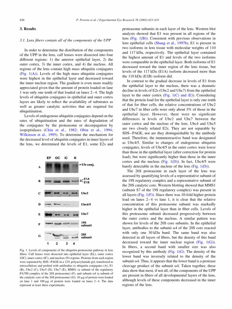
3.1. Lens fibers contain all of the components of the UPP
In order to determine the distribution of the components
of the UPP in the lens, calf lenses were dissected into four
different regions: 1) the anterior epithelial layer, 2) the
outer cortex, 3) the inner cortex, and 4) the nucleus. All
regions of the lens contain high mass ubiquitin conjugates
(Fig. 1(A)). Levels of the high mass ubiquitin conjugates
were highest in the epithelial layer and decreased toward
the inner nuclear region. The gradient is even more readily
appreciated given that the amount of protein loaded on lane
1 was only one tenth of that loaded on lanes 2–4. The high
levels of ubiquitin conjugates in epithelial and outer cortex
layers are likely to reflect the availability of substrates as
well as greater catalytic activities that are required for
ubiquitination.
Levels of endogenous ubiquitin conjugates depend on the
rates of ubiquitination and the rates of degradation of
the conjugates by the proteasome or deconjugation by
isopeptidases (Chin et al., 1982; Obin et al., 1994;
Wilkinson et al., 1995). To determine the mechanism for
the decreased level of ubiquitin conjugates in inner layers of
the lens, we determined the levels of E1, some E2s and
proteasome subunits in each layer of the lens. Western blot
analysis showed that E1 was present in all regions of the
lens (Fig. 1(B)). Consistent with previous observations in
lens epithelial cells (Shang et al., 1997b), E1 is present in
two isoforms in lens tissue with molecular weights of 110
and 117 kDa, respectively. The epithelial layer contained
the highest amount of E1 and levels of the two isoforms
were comparable in the epithelial layer. Both isoforms of E1
decreased toward the inner region of the lens tissue, but
levels of the 117 kDa (E1A) isoform decreased more than
the 110 kDa (E1B) isoform did.
In contrast to the gradual decrease in levels of E1 from
the epithelial layer to the nucleus, there was a dramatic
decline in levels of E2s (Ubc2 and Ubc7) from the epithelial
layer to the outer cortex (Fig. 1(C) and (E)). Considering
that the protein load for the epithelial layer is only one tenth
of that for fiber cells, the relative concentrations of Ubc2
and Ubc7 in fiber cells were only about 5% of those in the
epithelial layer. However, there were no significant
differences in levels of Ubc2 and Ubc7 between the
outer cortex and the nucleus of the lens. Ubc4 and Ubc5
are two closely related E2s. They are not separable by
SDS–PAGE, nor are they distinguishable by the antibody
used. Therefore, the immunoreactive band was designated
as Ubc4/5. Similar to changes of endogenous ubiquitin
conjugates, levels of Ubc4/5 in the outer cortex were lower
than those in the epithelial layer (after correction for protein
load), but were significantly higher than those in the inner
cortex and the nucleus (Fig. 1(D)). In fact, Ubc4/5 were
barely detectable in the nucleus of the lens (Fig. 1(D)).
The 26S proteasome in each layer of the lens was
assessed by quantifying levels of a representative subunit of
the 19S regulatory complex and a representative subunit of
the 20S catalytic core. Western blotting showed that MMS1
(subunit S7 of the 19S regulatory complex) was present in
all layers (Fig. 1(F)). Since there was 10-fold higher protein
load on lanes 2–4 vs lane 1, it is clear that the relative
concentration of this proteasome subunit was markedly
higher in the epithelial layer than in fiber cells. Levels of
this proteasome subunit decreased progressively between
the outer cortex and the nucleus. A similar pattern was
shown for levels of the 20S core subunits. In the epithelial
layer, antibodies to the subunit a4 of the 20S core reacted
with only one 30 kDa band. The same band was also
detected in all layers of fibers, but the density of this band
decreased toward the inner nuclear region (Fig. 1(G)).
In fibers, a second band with smaller size was also
recognized by this antibody (Fig. 1(G). The density of the
lower band was inversely related to the density of the
subunit a4. Thus, it appears that the lower band is a protease
cleavage product of the subunit a4. Taken together, these
data show that most, if not all, of the components of the UPP
are present in fibers of all developmental layers of the lens,
although levels of these components decreased in the inner
regions of the lens.
Fig. 1. Levels of components of the ubiquitin-proteasome pathway in lens
fibers. Calf lenses were dissected into epithelial layer (EL), outer cortex
(OC), inner cortex (IC), and nucleus (N) regions. Proteins from each region
were separated by SDS–PAGE in a 12% polyacrylamide gel, transferred to
nitrocellulose and probed with antibodies to ubiquitin conjugates (A), E1
(B), Ubc2 (C), Ubc5 (D), Ubc7 (E), MMS1 (a subunit of the regulatory
PA700 complex of the 26S proteasome) (F), and subunit a4 (a subunit of
the catalytic core of the 26S proteasome) (G). 10 mg of protein were loaded
on lane 1 and 100 mg of protein were loaded on lanes 2–4. The data
represent at least three experiments.
P. Pereira et al. / Experimental Eye Research 76 (2003) 623–631626
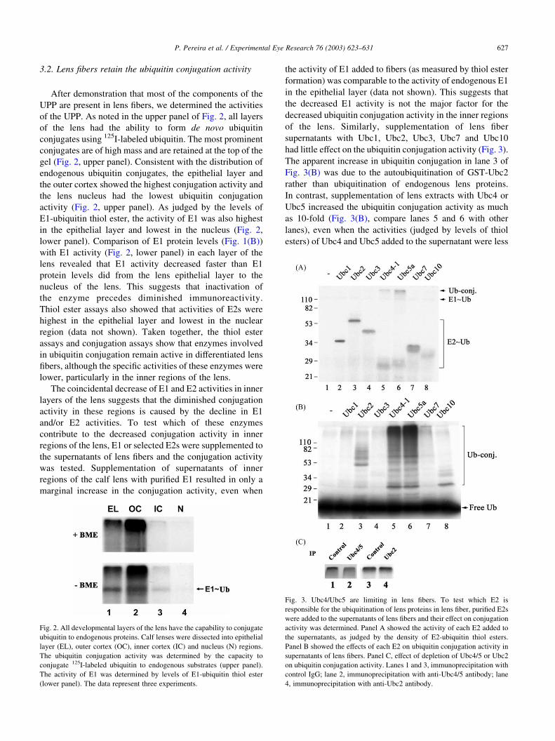
3.2. Lens fibers retain the ubiquitin conjugation activity
After demonstration that most of the components of the
UPP are present in lens fibers, we determined the activities
of the UPP. As noted in the upper panel of Fig. 2, all layers
of the lens had the ability to form de novo ubiquitin
conjugates using 125I-labeled ubiquitin. The most prominent
conjugates are of high mass and are retained at the top of the
gel (Fig. 2, upper panel). Consistent with the distribution of
endogenous ubiquitin conjugates, the epithelial layer and
the outer cortex showed the highest conjugation activity and
the lens nucleus had the lowest ubiquitin conjugation
activity (Fig. 2, upper panel). As judged by the levels of
E1-ubiquitin thiol ester, the activity of E1 was also highest
in the epithelial layer and lowest in the nucleus (Fig. 2,
lower panel). Comparison of E1 protein levels (Fig. 1(B))
with E1 activity (Fig. 2, lower panel) in each layer of the
lens revealed that E1 activity decreased faster than E1
protein levels did from the lens epithelial layer to the
nucleus of the lens. This suggests that inactivation of
the enzyme precedes diminished immunoreactivity.
Thiol ester assays also showed that activities of E2s were
highest in the epithelial layer and lowest in the nuclear
region (data not shown). Taken together, the thiol ester
assays and conjugation assays show that enzymes involved
in ubiquitin conjugation remain active in differentiated lens
fibers, although the specific activities of these enzymes were
lower, particularly in the inner regions of the lens.
The coincidental decrease of E1 and E2 activities in inner
layers of the lens suggests that the diminished conjugation
activity in these regions is caused by the decline in E1
and/or E2 activities. To test which of these enzymes
contribute to the decreased conjugation activity in inner
regions of the lens, E1 or selected E2s were supplemented to
the supernatants of lens fibers and the conjugation activity
was tested. Supplementation of supernatants of inner
regions of the calf lens with purified E1 resulted in only a
marginal increase in the conjugation activity, even when
the activity of E1 added to fibers (as measured by thiol ester
formation) was comparable to the activity of endogenous E1
in the epithelial layer (data not shown). This suggests that
the decreased E1 activity is not the major factor for the
decreased ubiquitin conjugation activity in the inner regions
of the lens. Similarly, supplementation of lens fiber
supernatants with Ubc1, Ubc2, Ubc3, Ubc7 and Ubc10
had little effect on the ubiquitin conjugation activity (Fig. 3).
The apparent increase in ubiquitin conjugation in lane 3 of
Fig. 3(B) was due to the autoubiquitination of GST-Ubc2
rather than ubiquitination of endogenous lens proteins.
In contrast, supplementation of lens extracts with Ubc4 or
Ubc5 increased the ubiquitin conjugation activity as much
as 10-fold (Fig. 3(B), compare lanes 5 and 6 with other
lanes), even when the activities (judged by levels of thiol
esters) of Ubc4 and Ubc5 added to the supernatant were less
Fig. 2. All developmental layers of the lens have the capability to conjugate
ubiquitin to endogenous proteins. Calf lenses were dissected into epithelial
layer (EL), outer cortex (OC), inner cortex (IC) and nucleus (N) regions.
The ubiquitin conjugation activity was determined by the capacity to
conjugate 125I-labeled ubiquitin to endogenous substrates (upper panel).
The activity of E1 was determined by levels of E1-ubiquitin thiol ester
(lower panel). The data represent three experiments.
Fig. 3. Ubc4/Ubc5 are limiting in lens fibers. To test which E2 is
responsible for the ubiquitination of lens proteins in lens fiber, purified E2s
were added to the supernatants of lens fibers and their effect on conjugation
activity was determined. Panel A showed the activity of each E2 added to
the supernatants, as judged by the density of E2-ubiquitin thiol esters.
Panel B showed the effects of each E2 on ubiquitin conjugation activity in
supernatants of lens fibers. Panel C, effect of depletion of Ubc4/5 or Ubc2
on ubiquitin conjugation activity. Lanes 1 and 3, immunoprecipitation with
control IgG; lane 2, immunoprecipitation with anti-Ubc4/5 antibody; lane
4, immunoprecipitation with anti-Ubc2 antibody.
P. Pereira et al. / Experimental Eye Research 76 (2003) 623–631 627
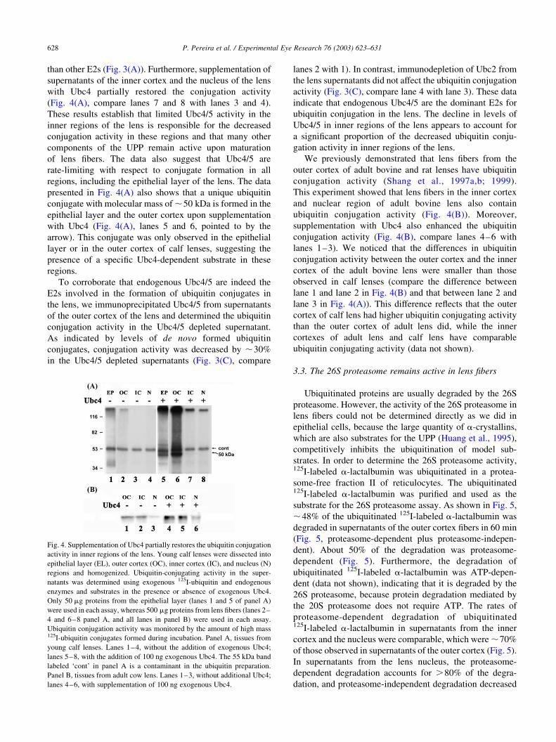
than other E2s (Fig. 3(A)). Furthermore, supplementation of
supernatants of the inner cortex and the nucleus of the lens
with Ubc4 partially restored the conjugation activity
(Fig. 4(A), compare lanes 7 and 8 with lanes 3 and 4).
These results establish that limited Ubc4/5 activity in the
inner regions of the lens is responsible for the decreased
conjugation activity in these regions and that many other
components of the UPP remain active upon maturation
of lens fibers. The data also suggest that Ubc4/5 are
rate-limiting with respect to conjugate formation in all
regions, including the epithelial layer of the lens. The data
presented in Fig. 4(A) also shows that a unique ubiquitin
conjugate with molecular mass of ,50 kDa is formed in the
epithelial layer and the outer cortex upon supplementation
with Ubc4 (Fig. 4(A), lanes 5 and 6, pointed to by the
arrow). This conjugate was only observed in the epithelial
layer or in the outer cortex of calf lenses, suggesting the
presence of a specific Ubc4-dependent substrate in these
regions.
To corroborate that endogenous Ubc4/5 are indeed the
E2s involved in the formation of ubiquitin conjugates in
the lens, we immunoprecipitated Ubc4/5 from supernatants
of the outer cortex of the lens and determined the ubiquitin
conjugation activity in the Ubc4/5 depleted supernatant.
As indicated by levels of de novo formed ubiquitin
conjugates, conjugation activity was decreased by ,30%
in the Ubc4/5 depleted supernatants (Fig. 3(C), compare
lanes 2 with 1). In contrast, immunodepletion of Ubc2 from
the lens supernatants did not affect the ubiquitin conjugation
activity (Fig. 3(C), compare lane 4 with lane 3). These data
indicate that endogenous Ubc4/5 are the dominant E2s for
ubiquitin conjugation in the lens. The decline in levels of
Ubc4/5 in inner regions of the lens appears to account for
a significant proportion of the decreased ubiquitin conju-
gation activity in inner regions of the lens.
We previously demonstrated that lens fibers from the
outer cortex of adult bovine and rat lenses have ubiquitin
conjugation activity (Shang et al., 1997a,b; 1999).
This experiment showed that lens fibers in the inner cortex
and nuclear region of adult bovine lens also contain
ubiquitin conjugation activity (Fig. 4(B)). Moreover,
supplementation with Ubc4 also enhanced the ubiquitin
conjugation activity (Fig. 4(B), compare lanes 4–6 with
lanes 1–3). We noticed that the differences in ubiquitin
conjugation activity between the outer cortex and the inner
cortex of the adult bovine lens were smaller than those
observed in calf lenses (compare the difference between
lane 1 and lane 2 in Fig. 4(B) and that between lane 2 and
lane 3 in Fig. 4(A)). This difference reflects that the outer
cortex of calf lens had higher ubiquitin conjugating activity
than the outer cortex of adult lens did, while the inner
cortexes of adult lens and calf lens have comparable
ubiquitin conjugating activity (data not shown).
3.3. The 26S proteasome remains active in lens fibers
Ubiquitinated proteins are usually degraded by the 26S
proteasome. However, the activity of the 26S proteasome in
lens fibers could not be determined directly as we did in
epithelial cells, because the large quantity of a-crystallins,
which are also substrates for the UPP (Huang et al., 1995),
competitively inhibits the ubiquitination of model sub-
strates. In order to determine the 26S proteasome activity,125I-labeled a-lactalbumin was ubiquitinated in a protea-
some-free fraction II of reticulocytes. The ubiquitinated125I-labeled a-lactalbumin was purified and used as the
substrate for the 26S proteasome assay. As shown in Fig. 5,
,48% of the ubiquitinated 125I-labeled a-lactalbumin was
degraded in supernatants of the outer cortex fibers in 60 min
(Fig. 5, proteasome-dependent plus proteasome-indepen-
dent). About 50% of the degradation was proteasome-
dependent (Fig. 5). Furthermore, the degradation of
ubiquitinated 125I-labeled a-lactalbumin was ATP-depen-
dent (data not shown), indicating that it is degraded by the
26S proteasome, because protein degradation mediated by
the 20S proteasome does not require ATP. The rates of
proteasome-dependent degradation of ubiquitinated125I-labeled a-lactalbumin in supernatants from the inner
cortex and the nucleus were comparable, which were ,70%
of those observed in supernatants of the outer cortex (Fig. 5).
In supernatants from the lens nucleus, the proteasome-
dependent degradation accounts for .80% of the degra-
dation, and proteasome-independent degradation decreased
Fig. 4. Supplementation of Ubc4 partially restores the ubiquitin conjugation
activity in inner regions of the lens. Young calf lenses were dissected into
epithelial layer (EL), outer cortex (OC), inner cortex (IC), and nucleus (N)
regions and homogenized. Ubiquitin-conjugating activity in the super-
natants was determined using exogenous 125I-ubiquitin and endogenous
enzymes and substrates in the presence or absence of exogenous Ubc4.
Only 50 mg proteins from the epithelial layer (lanes 1 and 5 of panel A)
were used in each assay, whereas 500 mg proteins from lens fibers (lanes 2–
4 and 6–8 panel A, and all lanes in panel B) were used in each assay.
Ubiquitin conjugation activity was monitored by the amount of high mass125I-ubiquitin conjugates formed during incubation. Panel A, tissues from
young calf lenses. Lanes 1–4, without the addition of exogenous Ubc4;
lanes 5–8, with the addition of 100 ng exogenous Ubc4. The 55 kDa band
labeled ‘cont’ in panel A is a contaminant in the ubiquitin preparation.
Panel B, tissues from adult cow lens. Lanes 1–3, without additional Ubc4;
lanes 4–6, with supplementation of 100 ng exogenous Ubc4.
P. Pereira et al. / Experimental Eye Research 76 (2003) 623–631628
further in this region (Fig. 5). These data show that lens
fibers, including those in the nuclear region, have the ability
to execute ubiquitin-dependent protein degradation.
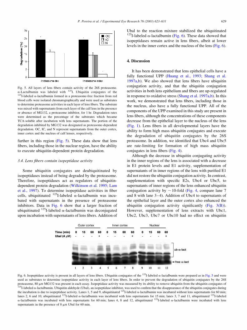
3.4. Lens fibers contain isopeptidase activity
Some ubiquitin conjugates are deubiquitinated by
isopeptidases instead of being degraded by the proteasome.
Therefore, isopeptidases act as regulators of ubiquitin-
dependent protein degradation (Wilkinson et al. 1995; Lam
et al., 1997). To determine isopeptidase activities in fiber
cells, ubiquitinated 125I-labeled a-lactalbumin was incu-
bated with supernatants in the presence of proteasome
inhibitors. Data in Fig. 6 show that a larger fraction of
ubiquitinated 125I-labeled a-lactalbumin was deconjugated
upon incubation with supernatants of lens fibers. Addition of
Ubal to the reaction mixture stabilized the ubiquitinated125I-labeled a-lactalbumin (Fig. 6). These data showed that
isopeptidases remain active in lens fibers, albeit at lower
levels in the inner cortex and the nucleus of the lens (Fig. 6).
4. Discussion
It has been demonstrated that lens epithelial cells have a
fully functional UPP (Huang et al., 1993; Shang et al.
1997a,b). We also showed that lens fibers have ubiquitin
conjugation activity, and that the ubiquitin conjugation
activities in both lens epithelium and fibers are up-regulated
in response to oxidative stress (Shang et al. 1997a,b). In this
work, we demonstrated that lens fibers, including those in
the nucleus, also have a fully functional UPP. All of the
components of the UPP examined in this study are present in
lens fibers, although the concentrations of these components
decrease from the epithelial layer to the nucleus of the lens
(Fig. 1). Lens fibers in all developmental layers have the
ability to form high mass ubiquitin conjugates and execute
the degradation of ubiquitin conjugates by the 26S
proteasome. In addition, we identified that Ubc4 and Ubc5
are rate-limiting for formation of high mass ubiquitin
conjugates in lens fibers (Fig. 4).
Although the decrease in ubiquitin conjugating activity
in the inner regions of the lens is associated with a decrease
in E1 protein levels and E1 activity, supplementation of
supernatants of in inner regions of the lens with purified E1
did not restore the ubiquitin conjugation activity. In contrast,
supplementation with specific E2s, Ubc4 or Ubc5, to
supernatants of inner regions of the lens enhanced ubiquitin
conjugation activity by ,10-fold (Fig. 4, compare lane 7
and 8 with lane 3–4). Addition of Ubc4 to supernatants of
the epithelial layer and the outer cortex also enhanced the
ubiquitin conjugation activity significantly (Fig. 3(B)).
However, supplementation of lens extracts with Ubc1,
Ubc2, Ubc3, Ubc7 or Ubc10 had no effect on ubiquitin
Fig. 5. All layers of lens fibers contain activity of the 26S proteasome.
a-Lactalbumin was labeled with 125I. Ubiquitin conjugates of the125I-labeled a-lactalbumin formed in a proteasome-free fraction from red
blood cells were isolated chromatographically and were used as substrates
to determine proteasome activities in each layer of lens fibers. The substrate
was mixed with supernatants from each layer of the calf lens in the presence
or absence of MG132, a proteasome inhibitor, for 1 hr. Degradation rates
were determined as the percentage of the substrates which became
TCA-soluble after incubation with lens supernatants. The portion of the
degradation inhibited by MG132 was designated as proteasome-dependent
degradation. OC, IC, and N represent supernatants from the outer cortex,
inner cortex and the nucleus of calf lenses, respectively.
Fig. 6. Isopeptidase activity is present in all layers of lens fibers. Ubiquitin conjugates of the 125I-labeled a-lactalbumin were prepared as in Fig. 5 and were
used as substrates to determine isopeptidase activity in each layer of lens fibers. In order to prevent the degradation of ubiquitin conjugates by the 26S
proteasome, 80 mM MG132 was present in each assay. Isopeptidase activity was measured by its ability to remove ubiquitin from the ubiquitin conjugates of125I-labeled a-lactalbumin. Ubiquitin aldehyde (Ubal), an isopeptidase inhibitor, was used to confirm that the disappearance of the ubiquitin conjugates during
the incubation is due to isopeptidase activity. Lanes 1, 5 and 9, ubiquitinated 125I-labeled a-lactalbumin was incubated without lens supernatants for 60 min;
lanes 2, 6 and 10, ubiquitinated 125I-labeled a-lactalbumin was incubated with lens supernatants for 15 min; lanes 3, 7 and 11, ubiquitinated 125I-labeled
a-lactalbumin was incubated with lens supernatants for 60 min; lanes 4, 8 and 12, ubiquitinated 125I-labeled a-lactalbumin were incubated with lens
supernatants in the presence of 8 mM Ubal for 60 min.
P. Pereira et al. / Experimental Eye Research 76 (2003) 623–631 629
conjugation activity. These data indicate that the decreased
E1 levels in the inner regions of the lens are probably not
causally related to the decreased conjugation activity in
these regions, but the decrease in levels of Ubc4 or Ubc5 is
responsible for the decline in ubiquitin conjugation activity
in the inner region of the lens. Moreover, Ubc4 and Ubc5
are rate-limiting in all regions of the lens. We previously
stated that E1 is rate-limiting with respect to conjugate
formation in cultured lens epithelial cells (Shang et al.,
1997b). That statement should be amended to indicate that
whereas E1 is limiting relative to most types of E2s and E3s,
Ubc4 and Ubc5 appear to be more limiting than E1 in lens
tissue.
In mammalian cells there are multiple isoforms of Ubc4
that share high sequence homology (Jensen et al., 1995;
Wing and Jain, 1995). The physiological functions of some
of these isoforms have been identified. For example, it has
been shown that Ubc4-1 is involved in postnatal rat testis
development, where it supports most of the increased
protein conjugation that accompanies the developmental
process (Rajapurohitam et al., 1999). Different isoforms are
expressed in different stages of testis development,
suggesting that they may have different roles during
spermatogenesis (Wing et al., 1996; Rajapurohitam et al.,
1999). It is tempting to speculate that a similar process may
also be involved in lens fiber differentiation, since depletion
of Ubc4/5 by immunoprecipitation reduces the conjugation
activity in the outer cortex, the region most likely to contain
differentiating fibers. In addition, formation of a unique
50 kDa ubiquitin conjugate in the outer cortex upon
supplementation with exogenous Ubc4 (Fig. 4(A), lanes
5–6) indicates a special role of Ubc4 in this region of the
lens.
Ubc4 and Ubc5 are thought to interact with specific E3s
that participate in recognition of specific types of substrates.
The fact that addition of Ubc4 or Ubc5 significantly restored
the conjugation activity of lens fibers in vitro shows that E1,
E3 and substrates for Ubc4/5 are available in all develop-
mental regions of the lens. Typical substrates for the Ubc4/5
in yeast are damaged or obsolete proteins (Seufert and
Jentsch, 1990). The substrates for Ubc4/5 in the lens may
involve modified proteins, since various modifications to
the long-lived lens proteins were observed in lens fibers
(Chen et al., 1997; Hanson et al., 2000; Takemoto, 2001).
The finding that a large portion of ubiquitin conjugation
activity is ascribed to Ubc4 or Ubc5 indicates that one of the
functions for the UPP in lens fibers is to degrade adversely
modified or obsolete proteins. We demonstrated that the
UPP was involved in degradation of oxidized proteins in
lens epithelial cells (Shang et al., 2001). If the UPP has
the same function in fibers as it does in the epithelial cells,
the decline in the UPP activity in inner regions of the lens
may contribute to the accumulation of modified or damaged
proteins in these regions. The finding that supplementation
of Ubc4 or Ubc5 partially restored the ubiquitin
conjugation activity and presumably ubiquitin-dependent
degradation capability prompts us to speculate that targeted
expression of Ubc4 or Ubc5 may enhance the degradation
of abnormal proteins in lens fibers and prolong the
transparence of the lens.
Acknowledgements
This work is supported partially by NIH grants EY11717
(to FS), the US Department of Agriculture, under agreement
No. 58-1950-9-001 (to A.T.), EY13250 (to A.T.) and
Fundacao para a Ciencia e Tecnologia (FCT) Project
PRAXIS/SAU/P/14197/98 (to P.P.). The authors thank
Drs C.M. Pickart, K. Tanaka, and S. Wing for providing
some of the precious reagents. The authors also thank
Mr M. Siegal for his help in preparation of this manuscript.
References
Baumeister, W., Walz, J., Zuhl, F., Seemuller, E., 1998. The
proteasome: paradigm of a self-compartmentalizing protease. Cell
92, 367–380.
Chamberlain, C.G., McAvoy, J.W., 1987. Evidence that fibroblast growth
factor promotes lens fibre differentiation. Curr. Eye Res. 6,
1165–1169.
Chen, Y.C., Reid, G.E., Simpson, R.J., Truscott, R.J., 1997. Molecular
evidence for the involvement of alpha crystallin in the colouration/
crosslinking of crystallins in age-related nuclear cataract. Exp. Eye Res.
65, 835–840.
Chin, D.T., Kuehl, L., Rechsteiner, M., 1982. Conjugation of ubiquitin to
denatured hemoglobin is proportional to the rate of hemoglobin
degradation in HeLa cells. Proc. Nat. Acad. Sci. U.S.A. 79, 5857–5861.
Ciechanover, A., Elias, S., Heller, H., Hershko, A., 1982. ‘Covalent affinity’
purification of ubiquitin-activating enzyme. J. Biol. Chem. 257,
2537–2542.
Gaczynska, M., Goldberg, A.L., Tanaka, K., Hendil, K.B., Rock, K.R.,
1996. Proteasome subunits X and Y alter peptidase activities in opposite
ways to the interferon-g-induced subunits LMP2 and LMP7. J. Biol.
Chem. 271, 17275–17280.
Hanson, S.R., Hasan, A., Smith, D.L., Smith, J.B., 2000. The major in vivo
modifications of the human water-insoluble lens crystallins are disulfide
bonds, deamidation, methionine oxidation and backbone cleavage. Exp.
Eye Res. 71, 195–207.
Hershko, A., Ciechanover, A., 1998. The ubiquitin system. Annu. Rev.
Biochem. 67, 425–479.
Hershko, A., Heller, H., Elias, S., Ciechanover, A., 1983. Components of
the ubiquitin-protein ligase system. Resolution, affinity purification, and
role in protein breakdown. J. Biol. Chem. 258, 8206–8214.
Huang, L.L., Jahngen-Hodge, J., Taylor, A., 1993. Bovine lens epithelial
cells have a ubiquitin-dependent proteolysis system. Biochim. Biophys.
Acta 1175, 181–187.
Huang, L.L., Shang, F., Nowell, T.R., Taylor, A., 1995. Degradation of
differentially oxidized a-crystallins in bovine lens epithelial cells. Exp.
Eye Res. 61, 45–54.
Jensen, J.P., Bates, P.W., Yang, M., Vierstra, R.D., Weissman, A.M., 1995.
Identification of a family of closely related human ubiquitin conjugating
enzymes. J. Biol. Chem. 270, 30408–30414.
Kuszak, J.R., 1995. The ultrastructure of epithelial and fiber cells in the
crystalline lens. Int. Rev. Cytol. 163, 305–350.
Lam, Y.A., Lawson, T.G., Velayutham, M., Zweier, J.L., Pickart, C.M.,
2002. A proteasomal ATPase subunit recognizes the polyubiquitin
degradation signal. Nature 416, 763–767.
P. Pereira et al. / Experimental Eye Research 76 (2003) 623–631630
Lam, Y.A., Xu, W., DeMartino, G.N., Cohen, R.E., 1997. Editing of
ubiquitin conjugates by an isopeptidase in the 26S proteasome. Nature
385, 737–740.
Nakamura, T., Pichel, J.G., Williams Simons, L., Westphal, H., 1995.
An apoptotic defect in lens differentiation caused by human p53 is
rescued by a mutant allele. Proc. Nat. Acad. Sci. U.S.A. 92 (13),
6142–6146.
Obin, M., Nowell, T., Taylor, A., 1994. The photoreceptor G-protein
transducin (Gt) is a substrate for ubiquitin dependent proteolysis.
Biochem. Biophys. Res. Commun. 200, 1169–1176.
Pan, H., Griep, A.E., 1994. Altered cell cycle regulation in the lens of
HPV-16 E6 or E7 transgenic mice: implications for tumor suppressor
gene function in development. Genes Dev. 8, 1285–1299.
Rajapurohitam, V., Morales, C.R., El-Alfy, M., Lefrancois, S., Bedard, N.,
Wing, S., 1999. Activation of UBC4-dependent pathway of ubiquitin
conjugation during postnatal development of the rat testis. Dev. Biol.
212, 217–228.
Seufert, W., Jentsch, S., 1990. Ubiquitin-conjugating enzymes UBC4 and
UBC5 mediate selective degradation of short-lived and abnormal
proteins. Embo J. 9, 543–550.
Shang, F., Deng, G., Obin, M., Wu, C.C., Gong, X., Smith, D., Laursen,
R.A., Andley, U.P., Reddan, J.R., Taylor, A., 2001. Ubiquitin-
activating enzyme (E1) isoforms in lens epithelial cells: origin of
translation, E2 specificity and cellular localization determined with
novel site-specific antibodies. Exp. Eye Res. 73, 827–836.
Shang, F., Gong, G., McAvoy, J.W., Chamberlain, C., Nowell, T.R.,
Taylor, A., 1999. Ubiquitin-dependent pathway is up-regulated in
differentiating lens cells. Exp. Eye Res. 68, 179–192.
Shang, F., Gong, X., Palmer, H.J., Nowell, T., Taylor, A., 1997a.
Age-related decline in ubiquitin conjugation in response to oxidative
stress in the lens. Exp. Eye Res. 64, 21–30.
Shang, F., Gong, X., Taylor, A., 1997b. Activity of ubiquitin dependent
pathway in response to oxidative stress: ubiquitin activating enzyme
(E1) is transiently upregulated. J. Biol. Chem. 272, 23086–23093.
Shang, F., Nowell Jr, T.R., Taylor, A., 2001. Removal of oxidatively
damaged proteins from lens cells by the ubiquitin-proteasome pathway.
Exp. Eye Res. 73, 229–238.
Shang, F., Taylor, A., 1995. Oxidative stress and recovery from
oxidative stress are associated with altered ubiquitin conjugating and
proteolytic activities in bovine lens epithelial cells. Biochem. J. 307,
297–303.
Takemoto, L., 2001. Deamidation of Asn-143 of gamma S crystallin from
protein aggregates of the human lens. Curr. Eye Res. 22, 148–153.
Tanaka, K., Tsurumi, C., 1997. The 26S proteasome: subunits and
functions. Mol. Biol. Rep. 24, 3–11.
Wilkinson, K.D., Tashayev, V.L., O’Connor, L.B., Larsen, C.N.,
Kasperek, E., Pickart, C.M., 1995. Metabolism of the polyubiquitin
degradation signal: structure, mechanism, and role of isopeptidase T.
Biochemistry 34, 14535–14546.
Wing, S.S., Bedard, N., Morales, C., Hingamp, P., Trasler, J., 1996. A novel
rat homologue of the S. Cerevisiae ubiquitin conjugating enzyme UBC4
with distinct biochemical features is induced during spermatogenesis.
Mol. Cell. Biol. 16, 4064–4072.
Wing, S., Jain, P., 1995. Molecular cloning, expression, and characteriz-
ation of a ubiquitin conjugation enzyme (E2 17 KB) highly expressed in
rat testis. Biochem. J. 305, 125–132.
P. Pereira et al. / Experimental Eye Research 76 (2003) 623–631 631