Embed Size (px)
Citation preview

LElBNlZ lNSTlTUTE of PHOTONlC TECHNOLOGY // ANNUAL REPORT 2018

» does the following scenario look familiar to you?
You have a great idea. For example: You want to
design and build the new bookshelf yourself, and
then you are standing desperately in front of a loose
wooden board, with a bleeding thumb and a hammer
in your hand? The idea of the perfect solution for
your book collection collapses – literally. Don‘t worry,
you‘re not alone with this experience. Leonardo da
Vinci seems to have been familiar with this phenome-
non, for he concluded: “Ingenious people begin great
works, diligent people complete them“.
At Leibniz IPHT we research procedures for applica-
tions in the fields of medicine, health, environment
and safety – from the idea to the final result. The
fact that our claim “From Ideas to Instruments“ is
anything but trivial becomes clear when considering
that for many people the construction of a bookshelf
is already a seemingly insurmountable challenge. Da
Vinci said that in order to start great works one needs
“brilliant people“ (a description that applies quite well
to our scientists). But in order for the many ideas,
which often have their roots in basic research, to
actually become applications suitable for everyday use,
diligence and a high degree of expertise are required.
And teamwork. Research teams at Leibniz IPHT set an
entire process chain in motion to achieve this. Some
will come up with a good idea, others will contribute
their technological expertise and the next one will
convince partners of the feasibility of the application.
Our research facility is characterized by this transfer
concept. In this annual report, we will tell you how
we succeed in working together successfully.
Enjoy reading!
Jürgen Popp
Scientific Director
Frank Sondermann
Administrative Director
Dear Readers,
Prof. Dr. Jürgen Popp © Sven Döring
Frank Sondermann © Sven Döring
3------
Thank you very much.
We would like to thank all our employees most cordially for their hard work and
high level of commitment on a daily basis. We would also like to thank the Free
State of Thuringia and the Federal Government, as well as all sponsors and part-
ners from politics, science and industry, for their many years of close and trus-
ting cooperation. We look forward to working successfully together in the future.
Research, development and selected events at Leibniz IPHT are supported by:

14
In Focus:
Sensitive Detection Methods – Trackers
Small quantities with big consequences:
How research teams find the lowest
concentrations of biomolecules in our
bodies and in our environment.
16
Tailor-made Treatment
Novel fibers help to determine the
drug level.
18
How much Antibiotics is in the River?
Mona Nissen detects drug residues in
water.
6
News from Leibniz IPHT
10
Crossing Borders
How Leibniz IPHT technologies interlock
12
A Portable Spectrometer
A cross-departmental team of
scientists is researching an analytical
laboratory that no longer needs a
laboratory: Raman2GO.
20
Well-prepared
How a research team optimizes chips
for optical analysis
22
In Focus:
High-throughput Methods – Thousands at one Blow
Optical methods detect cells and
pollen in high throughput.100 200 300 400 500 600 700 800
50
100
150
200
250
300
24
There‘s Something in the Air
Microfluidics meets Artificial Intelli-
gence: Andreas Kleiber analyzes pollen
in a rapid test run.
26
One Device for Several Applications
Tens of thousands of cells are auto-
matically analyzed for the first time.
28
“Be Persistent in What You Want”
Maria Chernysheva is one of the few
women to research ultrashort pulse
fiber lasers. She does not believe that it
is therefore necessary to protect her like
an endangered animal species though.
31
Female Experts in Photonics
Young female researchers established
relationships with Jena.
32
From the Worldto the Beutenberg
How Leibniz IPHT is living Welcome
Culture
34
Outstanding Personnel
35
Exchange of Ideas Across the Atlantic
JeDis Alliance researches new
approaches for the diagnosis of cancer
and infectious diseases.
36
“Anyone Can Build a Microscopy System”
Benedict Diederich and René Richter
researched a construction kit with
which researchers and hobbyists can
design creative optical solutions.
38
Outstanding Publications in 2018
40
Scientific Articles in the App
44
Key Figures of 2018
46
Organization Chart // Research Unit
47
Scientific Advisory Council 2018 //Board of Trustees 2018
48
Assembly of Members
49
Institute Finances 2018
50
Institute Staff 2018
51
Imprint
Contents
5------
4------

7------
6------
News from Leibniz IPHT
“Research that improves everyday life.” Federal Minister Visits Leibniz IPHT
“This is where research takes place that can im-
prove people‘s everyday lives,” said Anja Karliczek
during her visit to the Beutenberg Campus in
Jena. The Federal Minister of Education and
Research visited the Leibniz IPHT in June 2018
to gather information about research activities in
the field of optical health technologies.
Scientists and scholars explained how they use
light to detect infectious pathogens and their
resistances or to examine tissue for cancer
diagnosis. In laboratories and in the fiber
drawing plant, the Minister gained insight into
spectroscopic imaging methods and technolo-
gies for the production of optical fibers. “The
Beutenberg Campus is an example of how cut-
ting-edge research can advance our country,”
summarized Anja Karliczek. “I am particularly
pleased that the transfer of knowledge has
such a high value for researchers,” said the
Minister, emphasizing one aspect that is cen-
tral to Leibniz IPHT: the targeted implementa-
tion of research results in applicable solutions.
Detecting Diseases with Molecules: New European Network of Young Scientists
Molecular logic switches are chemical compounds that
function like electronic circuits in computers: They process
information into a logical response. An international team
of young scientists is investigating the properties of these
molecules and whether they can be used to diagnose
diseases in the future in the “Logic Lab – Molecular logic
lab-on-a-vesicle for intracellular diagnostics” project coordi-
nated by Leibniz IPHT. “Our goal is to adapt the molecular
logic switches for applications in biological environments
and cells,” explains network coordinator Benjamin Dietzek.
From April 2019 onwards, 14 PhD students will be working
at nine universities, research institutions and companies in
Germany, Ireland, the Netherlands, Poland and Slovakia in
the Innovative Training Network (ITN). The European Union
is funding “Logic Lab” with more than 3.5 million euros over
the next four years.
www.logiclab-itn.eu
Scientific Director Jürgen Popp with Federal MinisterAnja Karliczek. © Leibniz IPHT
Ying Zhang investigates light-induced processes in molecules. © Sven Döring
Progress in Medical Technology: New Innovation Center in Jena and Ilmenau
A new innovation centre for
medical technology is being
built in Thuringia. Teams of
scientists from Leibniz IPHT,
Jena University Hospital, and
Ilmenau Technical University
will work at the ThIMEDOP –
short for “Thuringia Innovation
Center for Medical Technology
Solutions (Diagnosis, Therapy,
Optimization through Optical
Solutions)”. There, they will
focus on stem cells, ageing and
oncology research, as well as
research in biomedical tech-
nology and microscopy. The
aim is to develop new optical,
spectroscopic and biotechno-
logical detection methods, to
have them certified as medical
devices as quickly as possible
and to accelerate the overall
translation of research re-
sults into economically viable
processes and products. The
centre, which is funded by the
state of Thuringia, will be lo-
cated in the CetraMed research
building, which will be built on
the premises of the University
Hospital from 2019 on.
Thuringia’s Minister of Science Wolfgang Tiefensee handed over the funding decision. © Leibniz IPHT
Planned innovation center ThIMEDOP in the CetraMed. © pbr

9------
8------
On a Space Mission: Sensors Explore the Secrets of the Planets
Measuring an asteroid, taking off for Mercury and landing
on Mars: Three space missions in 2018 had sensors from
Leibniz IPHT on board. The successful deployment on the
near-earth asteroid Ryugu in October marked the begin-
ning of the mission. The German-French measuring device
“MASCOT” collected data on the temperature, magnetic
properties and composition of Ryugu. Researchers want to
track down the origins of the solar system and investigate
whether the asteroid could pose a threat to Earth.
Shortly afterwards, the next Jena sensor launched into
space: with the BepiColombo space probe on Europe‘s
first mission to Mercury. Armed against extreme condi-
tions, the specially developed robust sensor will explore
temperature fluctuations of up to 430 degrees Celsius
during the day and down to minus 180 degrees Celsius
at night. The spacecraft is scheduled to reach Mercury by
the end of 2025.
“I‘m here, I‘m home”, announced the Twitter account
of InSight, the Mars lander, after grounding on the Red
Planet at the end of November. Using a parachute, the
thermoelectric sensors installed in a radiometer set off
with InSight on Mars to measure the heat radiation on
its surface. Researchers hope that the results will provide
them with a key to exploring the planet: They want to
find out whether Mars was created from the same ma-
terial as the Earth and the Moon and better understand
why it has developed differently from Earth over the past
4.5 billion years. “This is a highlight in my working life”,
said Frank Hänschke, head of the working group “Inte-
grated Thermo-Electrical and Micromechanical Technolo-
gies” in a television report by the MDR about the sensors
from Leibniz IPHT. Future joint space projects are already
being prepared by scientists from Leibniz IPHT and the
German Aerospace Center (Deutsches Zentrum für Luft-
und Raumfahrt; DLR).
For Future Technologies: European Team of Scientists Researches New Fiber Lasers
As the only network coordinated from Germany, a team of scientists
from the Department of Fiber Photonics at the Leibniz IPHT has been
awarded the funding for the top-class EU program “FET Open”. In the
interdisciplinary project “NCLas” scientists in Germany, Spain, Poland
and Great Britain are researching novel fiber lasers. “We want to incor-
porate nanocrystallites into a fiber in order to provide fiber lasers with
new wavelengths,” explaines the head of the “Active Fiber Modules”
working group, Matthias Jäger, who coordinates “NCLas”.
The research teams want to develop numerous new application pos-
sibilities in medicine and telecommunications. With the “FET Open”
programme, the European Union supports scientific and technological
research with the potential to develop new ideas for future technologies.
The EU will provide almost 3 million euros for “NCLas” over the next
four years, 900,000 of which for the Leibniz IPHT.
Excellent Research Partner in Spectroscopy and Imaging
The Jena research cluster “Balance of the
Microverse”, in which Leibniz IPHT also par-
ticipates, is one of 57 selected alliances that
will be funded in the Excellence Strategy of the
German federal and state governments over
the next seven years. The University of Jena
cooperates with its hospital and eight non-
university research institutions in the Cluster of
Excellence. The aim of the research network is
to explore the dynamic equilibrium of microbial
communities from a holistic perspective. This
has a stabilizing effect on living organisms and
the environment, such as the health of hu-
mans, animals and plants, the fertility of soils
or the quality of water bodies. Research teams
are developing new technologies to maintain
and restore these equilibria. Leibniz IPHT and
its partners from life sciences and medicine
are researching real-time imaging methods
with the highest spatial resolution to answer
biological and biomedical questions. For this
purpose, a “Microverse Imaging Center” will be
set up, where teams of scientists will develop
innovative microscopic and spectroscopic meth-
ods. “We provide state-of-the-art and visionary
microscopy platforms in biological security level
2 laboratories,” explains Christian Eggeling,
who heads the research department “Biophys-
ical Imaging” at Leibniz IPHT. “To this end,
we are accelerating the development of new
biophotonic technologies in order to identify
correlations between cause and effect.”
www.microverse-cluster.de
Producing Energy Following Nature‘s Example
The aim of the “CataLight” Sonderforschungsbereich (SFB, Collaborative
Research Centre) is to research sustainable energy converters modelled on
nature. Since July, Leibniz IPHT has been working on the project funded by
the German Research Foundation (DFG) together with the Universities of
Jena, Ulm and Vienna and the Max Planck Institute for Polymer Research
in Mainz. Over the next four years, teams of scientists from the fields of
chemistry, materials science and physics will work on using light to produce
high-energy chemicals and new materials for sustainable energy conversion.
Following the example of natural photosynthesis, they want to develop mo-
lecular catalyst systems for the light-controlled production of hydrogen and
oxygen from water. The goal is to stabilize them. “We look at how nature
does it and integrate the molecular components into soft matter,” explains
Benjamin Dietzek, head of the “Functional Interfaces” research department
and deputy speaker of the Collaborative Research Centre (SFB). “We want
to establish new concepts for photocatalytic water splitting.”
www.catalight.eu
Frank Hänschke with the thermosensor TS-72. Six of the sensors developed at Leibniz IPHT are used on the Mars mission InSight. © Leibniz IPHT
The Hayabusa 2 spacecraft approaches the asteroid Ryugu. © DLR
Rylene dyes, among others, are used as light collection units for photocatalytic water splitting. © Martin Schulz / FSU
The EU project NCLas is developing fiber lasers with new wavelengths. © Leibniz IPHT
Equipped for extreme conditions: the sensor for Mercury. © Leibniz IPHT
The Beutenberg Campus in Jena. © University Jena

The way we live has a lasting effect.
Half the pack of the generously
prescribed antibiotic against the mild
infection ends up in the toilet instead
of in the household waste. The meat
for lunch is inexpensive because it
comes from fattening farms, where
thousands of animals are kept and
given plenty of antibiotics. We are
outsourcing the production of active
ingredients to emerging markets.
Tourists import multi-resistant patho-
gens from overseas trips. All this
contributes to the fact that more and
more people are becoming infected
with germs against which available
antibiotics are no longer effective. Re-
sistant pathogens endanger people all
over the world. Diseases that can still
be easily treated today can become a
fatal danger again in the near future.
Infectious diseases are already among
the most frequent causes of death
worldwide. On the other hand, people
in industrialised countries are getting
older and older. More and more of
them have to be treated for cancer,
cardiovascular diseases or diabetes.
Longer hospital stays, in turn, increase
their risk of becoming infected with
resistant or even multi-resistant patho-
gens. This poses a major challenge for
healthcare systems worldwide.
Scientists at Leibniz IPHT are
researching methods for the social
challenges of our time. They are
researching photonic solutions for the
diagnosis and treatment of illnesses,
for pharmaceutics and process control
as well as for food and environmen-
tal safety. For example, to prove
how many drug residues pollute our
aquatic environment and drinking
water. Which pathogens have caused
an infection and which antibiotic they
are resistant to. Or whether a cancer
patient receives the therapy to which
the tumour responds best.
And just as the problems of our
globalised society are not detached
from each other, so the technologies
at Leibniz IPHT interlock to move from
basic research to application: From
Ideas to Instruments. Scientists at
Leibniz IPHT combine highly sensitive
and specific photonic and biophotonic
detection methods with technological
expertise in fiber technology and mi-
crofluidics, in micro- and nanotechnolo-
gies, quantum and systems technology.
In the clean room of Leibniz IPHT,
researchers produce tailor-made
substrates for surface-enhanced
vibrational spectroscopy in order to
detect infectious pathogens. They
combine highly specific optical detec-
tion methods with microfluidic sample
preparation and artificial intelligence:
for an analysis chip to identify drug
residues in water, pollen in the air
or cancer cells in the blood. And they
use microstructured special fibers
from the Institute’s drawing plant
to detect tiny amounts of antibiotics
in patients’ body fluids. They are
researching innovative processes and
integrated systems that are more
than the sum of their parts.
11------
10------
» Using light as a tool to explore future solutions: This is the goal of Leibniz IPHT. In this annual report, we tell you how physicists and microfluidicists, technologists, engineers, programmers and device developers are exchanging knowledge and ideas.
Crossing BordersHow Leibniz IPHT technologies interlock
Reinforced: The Transfer from Research to Practice
It takes a long time for techno-
logical solutions from research to
reach the patient: on average, it
takes around 14 years for them
to be applied in practice. One of
Leibniz IPHT’s main goals is to
accelerate this process of transla-
tion from idea to product. A team
headed by industrial researcher
Ralf Ehricht is working on new
bioinformatic methods for the
diagnosis of infectious diseases
and social health risks in the
research department ”Optical
Molecular Diagnostics and System
Technology“, which was found-
ed in December 2018. Ehricht,
previously project manager at the
Jena-based diagnostics compa-
ny Abbott (Alere Technologies
GmbH), wants to build a bridge
to industrial partners: ”To jointly
implement innovative products for
diagnostics and therapy“.
Engineer Henry John from the Sensor Systems group assembles the Raman2GO portable spectrometer.A cross-departmental team from Leibniz IPHT joined forces to design the device: from clean room to product design. © Sven Döring
© Sven Döring
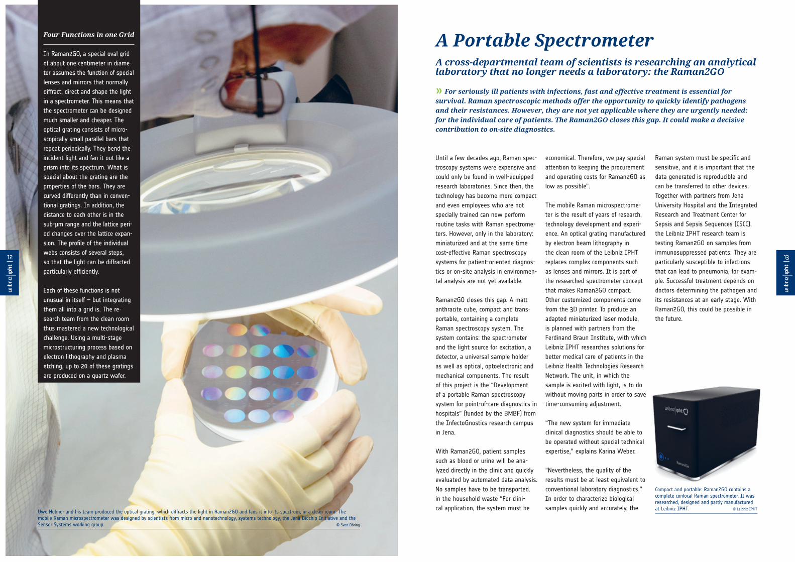
» For seriously ill patients with infections, fast and effective treatment is essential for survival. Raman spectroscopic methods offer the opportunity to quickly identify pathogens and their resistances. However, they are not yet applicable where they are urgently needed: for the individual care of patients. The Raman2GO closes this gap. It could make a decisive contribution to on-site diagnostics.
Until a few decades ago, Raman spec-
troscopy systems were expensive and
could only be found in well-equipped
research laboratories. Since then, the
technology has become more compact
and even employees who are not
specially trained can now perform
routine tasks with Raman spectrome-
ters. However, only in the laboratory:
miniaturized and at the same time
cost-effective Raman spectroscopy
systems for patient-oriented diagnos-
tics or on-site analysis in environmen-
tal analysis are not yet available.
Raman2GO closes this gap. A matt
anthracite cube, compact and trans-
portable, containing a complete
Raman spectroscopy system. The
system contains: the spectrometer
and the light source for excitation, a
detector, a universal sample holder
as well as optical, optoelectronic and
mechanical components. The result
of this project is the “Development
of a portable Raman spectroscopy
system for point-of-care diagnostics in
hospitals” (funded by the BMBF) from
the InfectoGnostics research campus
in Jena.
With Raman2GO, patient samples
such as blood or urine will be ana-
lyzed directly in the clinic and quickly
evaluated by automated data analysis.
No samples have to be transported.
in the household waste “For clini-
cal application, the system must be
economical. Therefore, we pay special
attention to keeping the procurement
and operating costs for Raman2GO as
low as possible”.
The mobile Raman microspectrome-
ter is the result of years of research,
technology development and experi-
ence. An optical grating manufactured
by electron beam lithography in
the clean room of the Leibniz IPHT
replaces complex components such
as lenses and mirrors. It is part of
the researched spectrometer concept
that makes Raman2GO compact.
Other customized components come
from the 3D printer. To produce an
adapted miniaturized laser module,
is planned with partners from the
Ferdinand Braun Institute, with which
Leibniz IPHT researches solutions for
better medical care of patients in the
Leibniz Health Technologies Research
Network. The unit, in which the
sample is excited with light, is to do
without moving parts in order to save
time-consuming adjustment.
“The new system for immediate
clinical diagnostics should be able to
be operated without special technical
expertise,” explains Karina Weber.
“Nevertheless, the quality of the
results must be at least equivalent to
conventional laboratory diagnostics.”
In order to characterize biological
samples quickly and accurately, the
Raman system must be specific and
sensitive, and it is important that the
data generated is reproducible and
can be transferred to other devices.
Together with partners from Jena
University Hospital and the Integrated
Research and Treatment Center for
Sepsis and Sepsis Sequences (CSCC),
the Leibniz IPHT research team is
testing Raman2GO on samples from
immunosuppressed patients. They are
particularly susceptible to infections
that can lead to pneumonia, for exam-
ple. Successful treatment depends on
doctors determining the pathogen and
its resistances at an early stage. With
Raman2GO, this could be possible in
the future.
13------
12------
A Portable SpectrometerA cross-departmental team of scientists is researching an analytical laboratory that no longer needs a laboratory: the Raman2GO
Four Functions in one Grid
In Raman2GO, a special oval grid
of about one centimeter in diame-
ter assumes the function of special
lenses and mirrors that normally
diffract, direct and shape the light
in a spectrometer. This means that
the spectrometer can be designed
much smaller and cheaper. The
optical grating consists of micro-
scopically small parallel bars that
repeat periodically. They bend the
incident light and fan it out like a
prism into its spectrum. What is
special about the grating are the
properties of the bars. They are
curved differently than in conven-
tional gratings. In addition, the
distance to each other is in the
sub-μm range and the lattice peri-
od changes over the lattice expan-
sion. The profile of the individual
webs consists of several steps,
so that the light can be diffracted
particularly efficiently.
Each of these functions is not
unusual in itself – but integrating
them all into a grid is. The re-
search team from the clean room
thus mastered a new technological
challenge. Using a multi-stage
microstructuring process based on
electron lithography and plasma
etching, up to 20 of these gratings
are produced on a quartz wafer.
Uwe Hübner and his team produced the optical grating, which diffracts the light in Raman2GO and fans it into its spectrum, in a clean room. The mobile Raman microspectrometer was designed by scientists from micro and nanotechnology, systems technology, the Jena Biochip Initiative and the Sensor Systems working group. © Sven Döring
Compact and portable: Raman2GO contains a complete confocal Raman spectrometer. It was researched, designed and partly manufactured at Leibniz IPHT. © Leibniz IPHT
15------
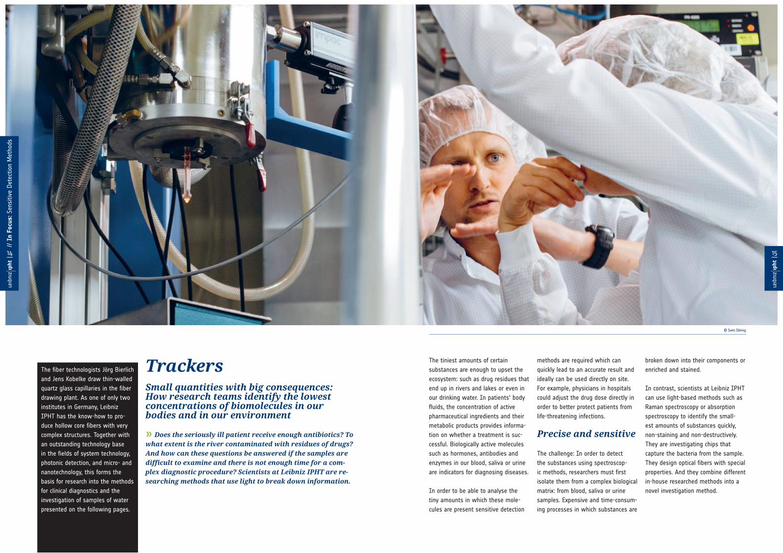
TrackersSmall quantities with big consequences: How research teams identify the lowest concentrations of biomolecules in our bodies and in our environment
» Does the seriously ill patient receive enough antibiotics? To what extent is the river contaminated with residues of drugs? And how can these questions be answered if the samples are difficult to examine and there is not enough time for a com-plex diagnostic procedure? Scientists at Leibniz IPHT are re-searching methods that use light to break down information.
The tiniest amounts of certain
substances are enough to upset the
ecosystem: such as drug residues that
end up in rivers and lakes or even in
our drinking water. In patients‘ body
fluids, the concentration of active
pharmaceutical ingredients and their
metabolic products provides informa-
tion on whether a treatment is suc-
cessful. Biologically active molecules
such as hormones, antibodies and
enzymes in our blood, saliva or urine
are indicators for diagnosing diseases.
In order to be able to analyse the
tiny amounts in which these mole-
cules are present sensitive detection
methods are required which can
quickly lead to an accurate result and
ideally can be used directly on site.
For example, physicians in hospitals
could adjust the drug dose directly in
order to better protect patients from
life-threatening infections.
Precise and sensitive
The challenge: In order to detect
the substances using spectroscop-
ic methods, researchers must first
isolate them from a complex biological
matrix: from blood, saliva or urine
samples. Expensive and time-consum-
ing processes in which substances are
broken down into their components or
enriched and stained.
In contrast, scientists at Leibniz IPHT
can use light-based methods such as
Raman spectroscopy or absorption
spectroscopy to identify the small-
est amounts of substances quickly,
non-staining and non-destructively.
They are investigating chips that
capture the bacteria from the sample.
They design optical fibers with special
properties. And they combine different
in-house researched methods into a
novel investigation method.
The fiber technologists Jörg Bierlich
and Jens Kobelke draw thin-walled
quartz glass capillaries in the fiber
drawing plant. As one of only two
institutes in Germany, Leibniz
IPHT has the know-how to pro-
duce hollow core fibers with very
complex structures. Together with
an outstanding technology base
in the fields of system technology,
photonic detection, and micro- and
nanotechnology, this forms the
basis for research into the methods
for clinical diagnostics and the
investigation of samples of water
presented on the following pages.
14------
// I
n Fo
cus:
Sen
sitiv
e Det
ectio
n M
etho
ds
© Sven Döring

» A network of ultrathin glass membranes surrounds the antibiotic solution in its hollow core with air holes: With the ingenious sensor fiber, even the smallest traces of antibiotics can be detected optically. A team of researchers developed it for patient-oriented diagnostics. In the future, physicians will be able to control which doses patients need.
Every hour that passes before a pa-
tient with an infection receives the
antibiotic, the patient’s risk of
dying of sepsis increases. Up to
now, it has been difficult to dose
this antibiotic exactly as required
by the patient. Dosage recom-
mendations result from studies
on healthy people or patients
with mild infections and cannot be
transferred to seriously ill patients.
Determining the drug level using
conventional methods such as chro-
matography and electrophoresis is
time-consuming and takes hours or
even days. Time that is not there:
In patients with sepsis, the way in
which their body absorbs drugs can
change within hours and cannot be
predicted. A rapid procedure to moni-
tor the concentration of antibiotics in
the patient’s body and adjust the dos-
age immediately is urgently needed.
Torsten Frosch and his team “Fiber
Spectroscopic Sensors” at Leibniz
IPHT are investigating a solution that
could enable patient-oriented labo-
ratory diagnostics in the future. In
cooperation with Mathias Pletz, who
is the head of the Institute for Infec-
tious Medicine and Hospital Hygiene
at the University Hospital of Jena,
they designed a complex fiber sensor
to detect the concentration of the
antibiotic cefuroxime in urine with
Raman spectroscopy.
The scientists are investigating
optical hollow fibers in order to
amplify the weak Raman scattering
of light at the antibiotic molecules.
They can also use the liquid-filled
hollow core fiber, which guides the
light with low loss, as a miniaturized
sample container. Together with fiber
technologists, they developed a new
type of hollow core fiber step by step.
“This sensor fiber far surpasses the
previous possibilities,” explains Torsten
Frosch, who heads the working group
“Fiber Spectroscopic Sensors”: “Both
in terms of its excellent optical prop-
erties, its efficiency and its broadband
light guidance, which enables a wide
range of wavelengths.”
The research team had already shown
that fiber-reinforced Raman spectrosco-
py can reliably detect very low concen-
trations of antibiotic active substances.
The new fiber significantly improved
the detection limit – and hereby the
chance that the method could in future
help patients with infections to receive
individualised treatment.
Tailor-made Treatment Novel fibers help to determine drug levels
17------
Torsten Frosch and Di Yan from the working group “Fiber Spectroscopic Sensor Technology” analyse the fiber-reinforced detection of antibiotics. © Leibniz IPHT
“The failure of antibiotic therapy has basically two causes. Either the pathogen is resistant or no effective an-tibiotic levels are reached at the site of infection because, for example, the dose is in-correct. The determination of antibiotic levels from urine at the point of care could, for example, help to identify the cause of treatment failure in urinary tract infections. The results are very promising and a great motivation for further work.”
Prof. Mathias W. Pletz //Head of the Institute for Infection Medicine and Hospital Hygiene at the University Hospital Jena
Microscope image of a hollow core fiber © Leibniz IPHT
Publications //Yan et al. (2018), „Fiber-Enhanced Raman Sensing of Cefuroxime in Humane Urine“, Analytical Chemistry, 90, 13243−13248, https://DOI: 10.1021/acs.analchem.8b01355 // Yan et al. (2018), „Fiber enhanced Raman Sensing of Levofloxacin by PCF Bandgap-shifting into the Visible Range“, Analytical Methods, 10, 586, https://DOI: 10.1039/C7AY02398G
16------
// I
n Fo
cus:
Sen
sitiv
e Det
ectio
n M
etho
ds

» Drugs are essential to our health. Yet they are becoming a problem for our environment. More and more drug residues end up in our waters. Simple and cost-effective procedures are needed to control water quality. Physicist Mona Nissen combined technologies from Leibniz IPHT into a new method.
Antibiotics, antihypertensives and
pain killers: In recent years, scientists
have discovered more than 150 active
pharmaceutical ingredients in German
rivers, streams and lakes, occasionally
even in drinking water. The residues,
which sewage treatment plants
cannot filter out, come from human
excreta, from drug residues that are
disposed of in drains and toilets or
reach the fields via liquid dung and
manure from treated farm animals.
Even in low concentrations, they
impair the fertility of fish or cause
antibiotic resistance to pathogenic
bacteria to spread further.
“In order to improve the quality of
our waters we need highly sensitive
methods that are inexpensive and
easy to use,” explains Mona Nissen,
PhD student in the Department of
Fiber Photonics at Leibniz IPHT. She
looked at what other research teams
at the institute were working on –
and combined in-house developed
technologies into a novel method
for detecting drug residues in water.
Using UV absorption spectroscopy, she
examined water samples for traces of
the antibiotic sulfamethoxazole, which
is supposed to help against urinary
tract infections, and for sodium
salicylate, a precursor of the headache
drug acetylsalicylic acid.
The spectroscopy method takes
advantage of the absorption charac-
teristics of many biological substances
at wavelengths below 300 nm to
determine the amount of a substance
contained in a liquid. The highlight:
Instead of the usual cuvette, Mona
Nissen used a 1 meter long fiber in
whose hollow core she filled the liquid.
The effect: The distance on which light
and matter interact is extended a hun-
dredfold – which in principle makes
it possible to detect substances at a
hundred times lower concentrations.
The solution for guiding the light inside
the waveguide filled with water comes
from Leibniz IPHT’s fiber drawing
plant: antiresonant hollow core fibers.
They are easy to manufacture, have
only a small optical loss and their
transmission windows are distributed
over a broad spectrum. In this case,
they also cover part of the UV range.
Because their core is relatively large at
about 30 micrometers, the water sam-
ples can also be exchanged quickly and
easily. But how can this be achieved
while at the same time light is coupled
into the fiber? – The researchers from
the Department of Microfluidics con-
tributed a tailor-made optofluidic chip.
Liquid can be pumped into the fiber
via the channels of the chip without
impairing the light guidance.
Mona Nissen was able to detect the
antibiotic sulfamethoxazole up to a
concentration of 0.1 μM. This corre-
sponds to ten granules of sugar in a
litre of water – pharmaceuticals in our
waters are up to ten times more con-
centrated. Her method is not yet ready
for application, classifies the young
scientist. However, it delivers accu-
rate results quickly, requires neither
time-consuming sample preparation
nor bulky equipment – and is thus a
building block for a future solution to
control the quality of our waters.
How Much Antibiotic is in the River?Mona Nissen detects drug residues in water
19------
“The recording of the UV Vis absorption spectra in a fiber is suitable for detecting impuri-ties in water samples on site.”
Dr. Dana Cialla-May //leads the Jena Biochip Initiative
UV spectroscopy with a water-filled antiresonant hollow fibre (red), a microfluidic chip and the delivery fiber (blue) into which the light is coupled. © Mona Nissen
Publications //Nissen et al. (2018), „UV Absorption Spectroscopy in Water-Filled Antiresonant Hollow Core Fibers for Pharmaceutical Detection“, Sensors, 18, 478, https://DOI: 10.3390/s18020478
One research department provided the fibers, the other one the chemical expertise: Mona Nissen (left) with Dana Cialla-May. For her research project, Leibniz IPHT awarded Mona Nissen the prize for the best master’s thesis. © Sven Döring
18------
// I
n Fo
cus:
Sen
sitiv
e Det
ectio
n M
etho
ds

21------
Well-preparedHow a research team optimizes chips for optical analysis
» In order for Raman spectroscopic methods to be used as rapid tests in hospitals and medical practices, simple strategies are required to prepare the samples. A research team from Leibniz IPHT found a new approach.
A patient‘s bodily fluids can reveal
the type of infection he is suffering
from. Susanne Pahlow can quickly
and precisely identify the pathogens
contained in saliva, blood or urine
using Raman spectroscopy. In order
to examine real samples with this
method, the bacteria must first be
isolated from the complex matrix of
this sample. The chemist captures the
bacteria on a chip whose surface she
has previously equipped with special
capture molecules. The bacteria are
immobilised, the chip is placed under
the spectrometer and the bacteria are
characterised by Raman spectroscopy.
In the clean room of Leibniz IPHT, the
silicon chips are coated with metals
such as nickel or aluminium that do
not interfere with the spectra of the
bacterial cells. Susanne Pahlow has
only been able to check whether the
biomolecules used to capture the
bacteria have actually been applied
to them during the measurement. In
order to be able to check the quality
during the production process, a suit-
able procedure was lacking.
Until, while exchanging ideas with
fellow scientist Thomas Mayerhöfer,
the following solution came up: Why
not use the same tool to monitor
substrate production for the investiga-
tion? So far, this has failed due to the
weak Raman signal of the biomole-
cules applied to the chip. In order to
reinforce this, the research team used
interference effects that occur when
the smooth, highly reflective alumin-
ium surface of the chip is modified.
They prepared them with thin layers
of aluminum oxide. The effect: The
Raman signal of the molecules can be
amplified or attenuated controllably.
For the first time, the scientists
succeeded in using interference
enhanced Raman scattering (IERS) to
detect biomolecules whose signals
would otherwise be too weak. To
date, IERS has mainly been used for
inorganic or organometallic materials.
In contrast to surface-enhanced Raman
spectroscopy (SERS) – the much more
common technique – to amplify the
weak Raman scattering – IERS sub-
strates allow a homogeneous but less
pronounced amplification of the signal
over the entire surface. “One can com-
pare the effect with the illumination
by different light sources. While with
SERS you can aim very bright lights at
certain points just like with a flash-
light, IERS provides a weaker but even
illumination, i. e. rather like a street
lamp”, explains Thomas Mayerhöfer.
IERS substrates are easier to produce
and remain stable for a long time.
Susanne Pahlow believes that there
is great potential to use this technol-
ogy in the future for quality control
in chip-based sample preparation for
Raman spectroscopy. She is current-
ly testing the method with other
molecules and bacteria. “The results
form a very good basis for our further
research work”.
B) Nachweis Funktionalisierung C) Chip-basierte Isolation der Erreger
A) Funktionalisierung Chipober�äche D) Raman-Spektroskopische Identi�zierung
OH
NH
N
CH3
O
O
NH
O
O
OO
N O
NH
NH
O
Fe3+
OH
NH
N
CH3
O
O
NH
O
O
OO
N O
NH
NH
O
Fe3+
OH
NH
N
CH3
O
O
NH
O
O
OO
N O
NH
NH
O
Fe3+
FerB FerB FerB FerB FerB FerB FerB FerB
Al
Al2O3
OH
O
NH
OH
O
NH
OH
O
NH
OH
O
NH
OH
O
NH
OH
O
NH
FerBFerBFerB FerBFerBFerB
3000 2800 1600 1400 1200 1000 800 600
Ram
an in
tens
ity
wavenumber / cm-1
K. pneumoniaeH. influenzae
P. aeruginosa
E. coli
FerB FerB FerB FerB FerB FerB FerB FerB
Whether the capture molecules have been successfully applied to the chip (Figure A) can be checked using interference-enhanced Raman spectroscopy (B). The pathogens are then immobilised on the chip and characterised by Raman spectroscopy (C and D). © Susanne Pahlow
In the clean room of Leibniz IPHT, employees produce tailor-made substrates for optical analysis. © Sven Döring
Depending on the thickness of the aluminum oxide layer, the chips shimmer in different colors. © Sven Döring
Publications //Pahlow et al. (2018), „Interference-Enhanced Raman Spectroscopy as a Promising Tool for the Detection of Biomolecules on Raman- Compatible Surfaces“, Analytical Chemistry, 90, 9025-9032, https://DOI: 10.1021/acs.analchem.8b01234
20------
// I
n Fo
cus:
Sen
sitiv
e Det
ectio
n M
etho
ds
100 200 300 400 500 600 700 800
50
100
150
200
250
300
23------
Thousands at One Blow Optical methods detect cells and pollen at high throughput rates
» The more doctors know about the state of health of their patients, the more individually they can respond to them in their treatment. This information must be precise and meaningful and the examination methods must be so sophisticated that they filter out what is important – even when it comes to detecting cells that are as rare as a needle in a haystack. In clinical routine, this is often impossible or can only be realized with great effort. Scientists at Leibniz IPHT are investigating solutions that can spare this time: with intelligent systems that open up high-throughput analysis for optical methods.
For physicians, precise observation
has always been the basis for making
a diagnosis. The more precise and the
earlier doctors make their diagnoses,
the more targeted their treatment can
be. Body fluids contain information.
They reveal, for example, whether a
tumor has already metastasized or
whether a cancer therapy is effective.
Circulating tumour cells in the blood
can be an indicator of this.
In this case, precise observation
means: to record more quickly than
laboratory staff can routinely detect.
Circulating tumour cells, for example,
are relatively rare in the blood. Previ-
ous methods yield too little, are not
specific enough or destroy the cells.
Research teams at Leibniz IPHT
are developing optical spectroscopic
methods with which they can quickly
and reliably analyse cells and bioparti-
cles in high throughput.
A microfluidic chip takes over the
function of a miniature laboratory, ar-
tificial neural networks automatically
evaluate captured data, and chemo-
metric methods extract the maximum
amount of information from them.
Simple and accurate
With the RamanCellAssay® platform,
Raman spectroscopy can be used for
the first time for high-throughput
analyses. This makes it possible to
identify tens of thousands of cells
using their molecular fingerprints,
including circulating tumor cells.
Another example: a microfluidic chip
automatically identifies thousands
of pollen in one go. This takes only
a few seconds. For comparison: For
pollen flight predictions, pollen is cur-
rently captured on adhesive foils and
counted under the microscope.
The optical detection methods do not
require time-consuming sample prepa-
ration. That makes them fast. Auto-
mated and digitized approaches make
them efficient and also help to avoid
sources of error and routine errors and
at the same time reduce costs.
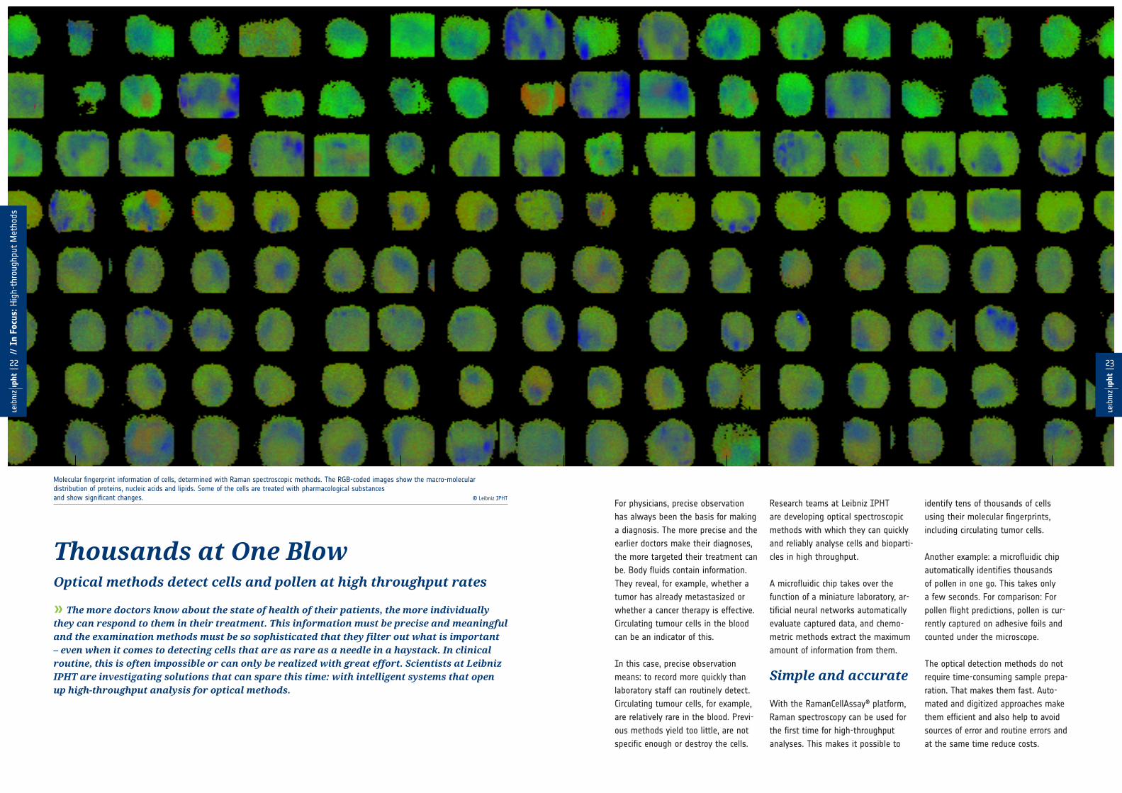
Molecular fingerprint information of cells, determined with Raman spectroscopic methods. The RGB-coded images show the macro-molecular distribution of proteins, nucleic acids and lipids. Some of the cells are treated with pharmacological substancesand show significant changes. © Leibniz IPHT
22------
// I
n Fo
cus:
Hig
h-th
roug
hput
Met
hods

25------
There‘s Something in the AirMicrofluidics meets Artificial Intelligence: Andreas Kleiber analyses pollen at high speed
» Birch, hazel or alder: As soon as the first pollen fly, people with allergies suffer. To protect themselves, they need to know what invisible pollen particles they are dealing with. Andreas Kleiber can answer this question: He uses a chip to capture several thousand pollen particles in high-resolution microscopic images. Neuronal networks process the images and classify the particles – a few seconds later, the result is there.
The grass under the apple tree glitters
in the late summer sun. All around,
wild flowers sprout, from which it
quietly hums. In the garden of the
German Bee Museum in Weimar,
bees find plenty of food. The carefully
inscribed labels read: wild mallow and
marigolds, columbine, chamomile and
lavender. Which of these end up in
honey is unknown to Klaudia Remus.
When the beekeeper wants to find out
what her honey consists of, she counts
pollen under the microscope. When
Andreas Kleiber wants to find out
which flowers provided the nectar for
Mrs. Remus‘ honey, he puts a stamp-
sized chip in front of a camera with a
microscope lens and opens his laptop.
A few moments later – and the
scientist knows what kind of pollen
his sample contains. Andreas Kleiber
is a doctoral student at Leibniz IPHT
and has designed a chip that works
like a miniature laboratory. It enables
high-resolution microscopic images of
several thousand pollen particles in
just a few seconds. Neural networks
identify which species they belong to.
The hit rate: almost 100 percent.
With this technology, Andreas Kleiber
can automatically analyse large quanti-
ties of bioparticles in the shortest pos-
sible time. Up to 100 pollen per second
flow past a viewing window on his
chip in a narrow channel. Each of the
tiny granules is captured by a digital
camera through a microscope lens. In
order to obtain sharp images for data
processing, Andreas Kleiber and the
Leibniz IPHT research team mastered
a technological challenge. The particles
must flow through the liquid channel
exactly in the focal plane of the objec-
tive. Which is not even a hundredth of
a millimeter narrow.
A chip as a miniature laboratory
The solution: an ingenious design that
Leibniz IPHT has already patented.
“As with a nozzle, we compress the
particle flow with two liquid streams
from the sides to form a vertical
lamella and rotate it 90 degrees into
a plane,” explains the scientist. The
research team can precisely control
how thick this layer is and where it
runs. They can arrange the particles in
such a way that they cross the image
field of the camera in a row – and let
them rotate in a controlled manner.
This provides 3D image information
about the outer shape and structure
of the pollen grain and makes identi-
fication more reliable. To evaluate the
images, Andreas Kleiber uses pro-
grams for particle tracking and feature
selection. A neural network, which
he has trained beforehand, assigns
the images to the respective pollen
species based on the extracted data –
and is correct in more than 98 percent
of cases.
Andreas Kleiber tested his method on
highly allergenic pollen species. This
provides beekeepers with an instru-
ment for quality control and is a ray
of hope for pollen allergy sufferers.
They can avoid allergic reactions if
they know what is in the air. They
are currently learning this from pollen
flight forecasts, the result of a lengthy
evaluation of pollen captured on
adhesive foils.
But even this is just one of many
applications, because the chip design
is flexible. “Basically,” says Andreas
Kleiber, “we can use it to analyze any-
thing smaller than 40 micrometers.”
For example, white blood cells – which
Andreas Kleiber is already researching
with the “Clinical Spectroscopic Diag-
nostics” team at Leibniz IPHT.
The special arrangement of the microfluidic channels makes it possible to align all particles in the focal plane. © Thomas Henkel
This is where the pollen buzz: Scientist Andreas Kleiber explains to Klaudia Remus from the German Bee Museum in Weimar how he can determine several thousand pollen at once with the microfluidic chip under his experimental setup. © Sven Döring
Patent //T. Henkel: Fluid rotation apparatus and method, DE 10 2015 115 343 B4 (26.10.2017), WO/2017/041785 A1
24------
// I
n Fo
cus:
Hig
h-th
roug
hput
Met
hods

» Raman spectroscopic techniques reveal the molecular fingerprints of cells. They are fast, precise and label-free – but have so far failed high-throughput analyses for clinical use. The RamanCellAssay® is changing this: machine and deep learning techniques interpret the results and provide evidence to track down diseases.
For cancer patients, it is crucial that
they receive the treatment that
helps them best. Circulating tumour
cells provide information on how
the disease progresses and how a
therapy works. They spread in the
bloodstream and can develop into
metastases. New methods for reliably
identifying circulating tumour cells are
therefore urgently needed in order to
better understand cancer and increase
patients‘ chances of survival.
A team of scientists from Leibniz
IPHT has now researched a system
with which tens of thousands of cells
can be characterised quickly, easily,
and label-free using their molecular
fingerprints – tested on circulating
tumour cells in the blood, among
other things. The RamanCellAssay®
platform makes it possible for the
first time to use Raman spectroscopic
methods for high-throughput analyses
of living cells as well as for mixed
cell populations. The system can
be combined with common Raman
devices and could thus contribute to
the spectroscopy method becoming
a standard tool in clinical diagnostics
and cell research.
It offers decisive advantages over
methods that currently represent the
gold standard. Hidden infections or
undiagnosed diseases are usually
detected using fluorescence-based ap-
proaches. The time-consuming sample
preparation, however, costs valuable
time and the fluorescent labels can
damage cells and tissue. However, it
is particularly important for tumour
cells to remain cultivable and still be
available for subsequent examina-
tions, for example to detect muta-
tions. Current methods for identifying
the rare cells, are only approved for
a few types of cancer, provide a low
yield or too little specific information.
Raman spectroscopy, on the other
hand, can be used to classify cells
using the chemical information from
their spectra. One limitation was that
experiments and analyses required
a lot of time and qualified personnel
– but real applications require the re-
liable statistical evaluation of a large
amount of data and the measurement
of many thousands of cells.
The RamanCellAssay® overcomes this
obstacle. The platform combines auto-
mated microscopic imaging methods
with Raman spectroscopy. In this
way, entire series of experiments can
be carried out in a short time without
personnel having to intervene. In de-
tail: The RamanCellAssay® records the
spectra of more than 100,000 individ-
ual cells and enables fully automated
sampling of 1,000 individual cells in
less than 20 minutes. The spectra are
evaluated, assigned, and the results
can be read.
The RamanCellAssay® provides a
platform for high-throughput cell
analysis – without preparation and
for a variety of label-free diagnostic
applications: to identify tumor cells or
stem cells, to demonstrate the effect
of drugs, to non-destructively ana-
lyze artificially cultured tissue or for
differential blood imaging. A clinical
study in sepsis patients as part of the
European research project “Hemo-
spec”, which aims to develop a device
for sepsis diagnosis, as well as other
applications, have already achieved
positive results.
27------
One Device for Several ApplicationsTens of thousands of cells are automatically analyzed for the first time
Iwan Schie and his team are researching new optical devices to diagnose diseases. © Sven Döring
Publications //Schie et al. (2018), „High-Throughput Screening Raman Spectroscopy Platform for Label-Free Cellomics“, Analytical Chemistry, 90, 3, https://DOI: 10.1021/acs.analchem.7b04127 // Rüger et al. (2018), Markierungsfreies Hochdurchsatz-screening mit Raman-Spektroskopie“, BIOspektrum, 05.18, 24. Jg., https://DOI: 10.1007/s12268-018-0952-3
The RamanCellAssay® platform of the Leibniz IPHT team enables the characterization of thousands of cells in a very short time and thus sets new standards for the high-throughput application of Raman spectroscopy. © Leibniz-IPHT
26------
// I
n Fo
cus:
Hig
h-th
roug
hput
Met
hods

» It is two days ago, that the head of the new junior research group for ultrafast fiber la-sers started at Leibniz IPHT – “10 time zones away from the beginning of my studies,” says Maria Chernysheva and laughs. That was in Khabarovsk, in the far east of Russia, not far from China. Her next stations: Moscow and Birmingham. The last experiments at Aston University she is still managing via Skype con-ference. She has already found an apartment in her new home and a place at the Interna-tional School in Weimar for her four-year-old daughter. At Leibniz IPHT, Maria Chernyshe-va plans to investigate novel ultrafast fiber lasers that will be used in infrared spectros-copy for cancer diagnostics. Would she like to tell us how she makes her career as a young scientist? Sure. Which version, “the scary one or the softened one”?
Mrs. Chernysheva, only one in ten in the photonics industry in Europe is a woman. Are you passed around a lot as a role model?
Quite, yes (laughs). I am always one of those invited to
conferences and workshops by women‘s associations to
tell how stony her career path was.
How stony was it?
In fact, I have mixed feelings about this gender issue.
Every woman who chooses an engineering science knows
what she is getting into. She knows that she will be in the
minority – but also that there are advantages to being a
woman. It is not that you have to worry about us like you
do about an endangered species.
How was it with you?
I always wanted to do something with technology.
Shortly before my diploma as telecommunications engineer
I went to Moscow, to the Research Center for Fiber Optics
of the Russian Academy of Sciences, first for an internship,
then for my diploma project. Thereupon I was offered the
opportunity to start my thesis there. But when I wanted
to start, I was told: “We‘ll take you – but only on one
condition: you‘ll work with a man”.
I beg your pardon? They didn‘t want to hire you alone?
No. Ten years ago they still thought it risky to employ
women because they didn‘t believe that they would com-
plete their doctorate. They could go on parental leave and
not come back. I was only the second doctoral student at
that time.
And did you find a colleague?
Fortunately, someone from my course also wanted to go
to Moscow, yes. In the end, I even surpassed my goal and
they hired two men: One of them later decided that sci-
ence was not for him and dropped out. The other complet-
ed his doctorate and embarked on a scientific career.
Just like you. So what happened next?
During my doctorate in laser physics I took part in a sci-
entific conference and learned what it‘s all about: gaining
experience and networking. And I thought: I want to try
it out. I looked for the conference with the next deadline,
that was another two days. I wrote a proposal, got the OK
from my supervisor, applied – and travelled to the USA for
the first time in my life.
29------
28------
“Be Persistent in What You Want”Maria Chernysheva is one of the few women to research ultrafast fiber lasers. She does not believe that it is therefore necessary to protect her like an endangered animal species though.
Maria Chernysheva establishes the junior research group for ultra-short pulse fiber lasers at Leibniz IPHT. © Sven Döring

31------
30------
Female Experts for Photonics Young women researchers established relationships with Jena
» The photonics industry is growing and is a focal point in the European research landscape. More and more graduates come from universities – but are still underrepresented at the man-agement level of research institutes and high-tech corporations. To change this, Leibniz IPHT launched the international career workshop “Women in Photonics”.
Jena radiates: Young female scientists
from four continents accepted Leibniz
IPHT’s invi-
tation to the
first interna-
tional career
workshop
“Women in
Photonics”
from April 17
to 19, 2018.
The institute
thus gave excellent female photonics
researchers an impulse to network
better – among themselves and with
executives from research institutions
and companies.
A mentoring program in three days:
“The participants exchanged ideas
with experienced colleagues and were
able to jointly develop strategies for
a successful career,” reports Ute Neu-
gebauer. The professor herself started
as a junior researcher at Leibniz IPHT.
Today, she is a vice scientific director
and belongs to the leadership team.
“Our goal is to
involve young
female research-
ers more closely
in the scientific
community,” em-
phasizes Jürgen
Popp, scientific
director of the
institute. Jena as an international
research and industrial center for
optics and photonics is proving to be
an ideal location. The 40 participants
with doctorates from Europe, Brazil,
the USA, Canada and Australia ex-
changed about personal career paths
with female researchers and female
executives from Carl Zeiss, Jenop-
tik and Asphericon. The workshop
was supported by Friedrich Schiller
University, the Fraunhofer Institute
for Optics and Precision Engineering,
many regional companies, and “The
Optical Society of America” (OSA) and
“The International Society for Optics
and Photonics” (SPIE).
The female scientists used this oppor-
tunity not least to present their own
research. Their topics ranged from
research into ultrafast fiber lasers and
fluorescence lifetime microscopy to
light-based therapies against cancer.
“By enabling excellent female scien-
tists to establish their own research
topic, we can make an academic ca-
reer or an industrial perspective more
attractive for them,” said Jürgen Popp.
Although more and more well-edu-
cated female scientists come from
universities, they are still underrep-
resented at the management level in
research and high-tech industry. For
Jürgen Popp, promoting the careers of
women is therefore a clear goal.
“I am convinced that the young women scientists will use the experiences from this workshop for their careers. They learned a lot here for their professional and private lives.”
Prof. Katarina Svanberg //Lund University & former President of SPIE
After graduating, you went to England.
Yes, I had an invitation for an internship in the USA, but
by the time I had the money, the deadlines had passed. At
that time, my future professor at Aston University invited
me to continue my research there. I first worked as a
research assistant, then with a Marie Curie fellowship from
the EU Commission and then with a fellowship from the
Royal Academy of Engineers. From there I moved here to
the Leibniz IPHT.
... which you got to know at the international workshop “Women in Photonics” ...
... yes exactly. I have never been in a workshop with so
many women (laughs).
A good experience?
The workshop was useful, in any case. But as I said, for
me the topic of gender justice is double-edged. I know that
women have to fight prejudices, that they are paid less ac-
cording to statistics and that they sometimes have a harder
time progressing with their careers than men. On the other
hand, every woman knows what possibilities she has: that
she gets more attention at conferences, that the organising
committees are looking for a balance between women and
men. There are many possibilities: Scholarships especially
for women and those for a better work-life balance. Thanks
to the support of institutions such as the European Com-
mission, there are more opportunities than disadvantages.
What kind of tips do you give pupils and students?
Networking. All my possibilities only arose because I took
part in conferences, talked to people, asked them about
possibilities at their institutes. Equally important: family
support. I moved to England with my husband Sergey. He
gave up his doctorate in Russia and started all over again
in England – because my career was just developing.
Unfortunately, he then moved to Scotland, and I stayed
in England with our one-year-old daughter. Sergey always
supported me and he encouraged me to apply for schol-
arships. This brings me to my third tip: you have to be
persistent in what you want.
Also in organizing research and family time. How do you manage that?
When my daughter was nine months old, she went to the
nursery. I wanted to get back to work again. When she
was asleep, I was writing – papers, reviews, scholarship
applications. Friday to Sunday Sergey was at home, these
were my laboratory days. When he then followed Tomáš
Cižmár to Jena, it became more complicated, of course. He
could only come every two weeks. But our daughter was
already older. She is now four and is a great helper. Now
that we live together again, it gets easier again.
What convinced you as a researcher to apply for Leibniz IPHT?
I’m working on ultrafast fiber lasers. Leibniz IPHT gives
me the opportunity to research such fiber lasers and also
apply them, for example in diagnostics. I’m very interested
in that. The institute covers all stages of technology: from
the production of fiber preforms to the research of lasers
and other photonic technologies and their application.
This last step into the application has been missing at my
previous institutes. The combination distinguishes Leibniz
IPHT from others.
What do you want to achieve with your research?
That the lasers I develop will actually be used, for exam-
ple in the spectroscopic diagnosis of cancer or in surgery.
I don’t have one specific application in mind. If you think
of research as a bowl of water in which every researcher
adds a drop: Then I would like to be an important drop in
this bowl.
More about the work of Maria Chernysheva‘s junior
research group can be found at www.leibniz-ipht.
de/en/research-units/junior-research-groups/
ultrafast-fiber-lasers/overview.html
© Leibniz IPHT

33------
32------
From the World to the BeutenbergHow Leibniz IPHT is living Welcome Culture
» The world of science is international – and so is Leibniz IPHT. Researchers from China, India, the Czech Republic or Ghana come to Jena and enrich the scientific cooperation at the institute. To make them feel at home, Leibniz IPHT staff members try to make their start easi-er with a variety of measures – which of course also apply to newcomers from Castrop-Rauxel or Wuppertal.
Selamat datang di institut – ¡Bien-
venido al instituto! – Üdvözöljük az
intézetben – Bienvenue à l’institut –
Willkommen am Institut – Welcome
to the institute! That was: Indonesian,
Spanish, Hungarian, French, German and
English. Or also: only a small selection
of the languages spoken by the staff
at Leibniz IPHT. From from 32 nations
came the people who were permanently
employed at the institute in 2018 as
doctoral students and guest researchers.
There are also guests from all over
the world. Leibniz IPHT is committed
to creating an international environ-
ment characterized by openness,
tolerance, responsibility, solidarity
and diversity. The foundation for this
is laid by an living welcome culture.
This starts with the lingua franca: In
addition to German, English is also
spoken at the Institute as a matter of
course. All important documents are
bilingual; a simultaneous interpreter
translates at the staff meeting. And
if you are new, you will be provided
with Welcome Guides.
They know where which laboratory is,
how to handle complicated forms and
how to find a parking space on the
Beutenberg Campus and an apartment
in Jena.
Jan Rüger, Eliana Cordero Bautista, Eric Boateng and Wei Yang are doctoral students at Leibniz IPHT. © Sven Döring
“I immediately felt welcome at the institute – especially because of my colleagues. We exchange a lot of ideas among ourselves. That makes it easy for me to integrate.”
Eliana Cordero Bautista //comes from Colombia and is doing her doctorate in the research department Spectroscopy / Imaging

Raman Award for the Best Junior Researcher
for Outstanding Research in the Field of Raman Spectroscopy to Marie Richard-Lacroix
3rd Prize of the Berthold Leibinger Innovation Award for Applied Laser Technology
for the Rapid Determination of Resistances, RamanBioAssay®
to Ute Neugebauer and Jürgen Popp
“In order to solve global problems
in medicine and the life sciences, we
urgently need to exchange ideas,” says
Laura Marcu: “about innovations in
biophotonics”. The professor teaches
at the North American University of
California, Davis (UC Davis), and coor-
dinates the transatlantic consortium
JeDis together with Jürgen Popp. In
the Jena Davis Alliance of Excellence in
Biophotonics, scientists from Leibniz
IPHT, Friedrich Schiller University,
Jena, and UC Davis have been net-
working since January 2018. Together,
research teams from hospitals and
technology development want to push
the development of new biophotonic
technologies and processes.
“One focus of our research is the
spectroscopic diagnosis of cancer,”
explains Jürgen Popp. “We are
combining UC Davis’ expertise in
fluorescence lifetime imaging with
Leibniz IPHT’s expertise in Raman
spectroscopy to create new tools
for tumor edge detection and tumor
classification.
In order to expand the competence
in Raman spectroscopy for clinical
diagnostics, a joint research laboratory
will be established at UC Davis:
the “Biomedical Engineering and
Comprehensive Cancer Center”.
JeDis offers young scientists a top-
class training and exchange program
to promote long-term cooperation.
This will lay the foundation for a future
international graduate school in which
15 doctoral students from Jena and
Davis will research biophotonic issues.
One step towards this goal was
the first JeDis Summer School in
September 2018 in Jena with 20
PhD students from Jena and Davis.
The second edition will take place in
Davis in 2019. JeDis is funded over
three years within the framework of
the Transatlantic Programme of the
Federal Republic of Germany.
Exchange of IdeasAcross the AtlanticJeDis Alliance explores new approaches for the diagnosis of cancer and infectious diseases
Outstanding Personnel
» Light as a research topic brings together scientists from clinics and technology develop-ment on both sides of the Atlantic. Teams from Leibniz IPHT and the University of California, Davis, are working together to advance the development of new biophotonic technologies and processes. The basis for this is provided by an excellent training and exchange programme and a joint research laboratory.
34------
35------
Ioannes Marcus Marci Medal for Outstanding Contributions
in the Field of Spectroscopy to Jürgen Popp
Forcheurs Jean-Marie Lehnfor Outstanding Collaboration in the Field
of Artificial Photosynthesis to Benjamin Dietzek and Vincent Artero
“JeDis offered me an ideal environment to explore a field of regenerative medicine: Artificial tissue cultivation using fluorescence lifetime microscopy. I learned from ex-perts and expanded my focus. I am very grateful for that.”
Abdullah Saif Mondol //does his PhD in the research department “Spectroscopy / Imaging” and stayed for one month at UC Davis
Kaiser Friedrich Prizefor Optical Rapid Method
for the Diagnosis of Cancerous Tissue to Jürgen Popp, Thomas Bocklitz, Tobias Meyer, Orlando Guntinas-Lichius, Andreas Tünnermann, Jens Limpert, Thomas Gottschall, Michael Schmitt
Jena research team visited UC Davis in March 2019 © Leibniz IPHT
Jena
Davis

» The cubes can be magnetically clicked to-gether and combined as desired: with lenses, mirrors or displays – to form a magnifying glass, a telescope or a microscope. UC2 – You see, too – is what Benedict Diederich and René Richter from Leibniz IPHT and their doctoral colleague Swen Carlstedt from Jena University Hospital call their optical construction kit. And they want to say: Everyone can see. Hobbyists can use it to build a fully automatic fluores-cence microscope for less than 250 euros. For example. Technophile “makers” assemble the cubes from the 3D printer into ever new structures. A pupil uses them to write his school-leaving thesis on microscopy. In addi-tion, the scientists work together with the Uni-versity Hospital and Jena schools. The idea is spreading – and this is exactly the plan behind UC2: the more there are, the more new ideas.
Mr. Diederich and Mr. Richter, with your sys-tem you can turn a mobile phone into a high- performance microscope. How does it work?
We have developed a modular optical construction kit
from a small craft project – which can be connected to the
camera of a smartphone, for example. We use image-pro-
cessing algorithms to achieve good results with inexpensive
components such as lenses from the student construction
kit or simple video projectors as lighting units. We imple-
ment some of these algorithms on mobile phones, thus
compensating for the inadequacies of the components. We
have also experimented with lighting techniques and built
an intelligent lighting system that optimally enhances the
contrast of the sample. To do this, we analyze the image
data of the samples live on the mobile phone using machine
learning techniques and then project a new pattern into the
rear focal plane of the imaging optics using a projector.
What microscopy methods are possible?
You can start with a 2D telescope setup. If you bring the
3rd dimension into play, you can easily build a transmis-
sion or fluorescence microscope. A few clicks and you
get a light sheet microscope. All is connected and can be
transferred into each other. This is the highlight of the
modular system. We have developed algorithms for mobile
phones that enable a variety of methods and techniques in
computer-based microscopy.
To what else can I combine the building blocks?
Basically, to everything. The form is fixed, but size and ma-
terial are not. In this way, the system can be scaled and
adapted to one‘s own wishes. Whether for microscopes,
telescopes, pen holders, chemical workbenches or Raman
spectroscopy.
Is the system already used for other research projects at Leibniz IPHT?
For system integration, we are investigating a device that
can image E. coli bacteria with a simple structure. In
addition, the expertise in optics and device development
complement each other. The first prototypes to observe
the morphology of bacteria on an electrophoresis chip have
already been developed with the UC2 system. UC2 is also
used in the incubator to measure living cells over a period
of up to one week. It is ideally suited for biolabs with
special hygiene regulations: Instead of having the device
cleaned in a time-consuming process, it can simply be
disposed of after the experiment.
However, they are not only aimed at scientists ...
Exactly, our goal is to get more people interested in optics,
even those beyond the well-equipped laboratories. That‘s
why we‘re lowering the hurdles: with optical components
you can afford. Following the example of the microproces-
sor unit Arduino or the simple computer Raspberry Pi, we
disclose sources and documentation and hope that our
system will spread as quickly as possible within the open
source community. This would enable everyone in the
world to build a microscopy system using simple means –
including developers, hobbyists and researchers from other
disciplines. This would generate completely new ideas:
The more people participate, the greater the opportunities.
“Anyone Can Build a Microscopy System”Benedict Diederich and René Richter researched a construction kit with which researchers and hobbyists can design creative optical solutions.
37------
36------
The cubes of the optical construction kit, which René Richter (left) and Benedict Diederich are investigating, can be playfully combined – whether to a telescope, a chemical workbench, a pen holder or to observe living cells in an incubator. © Sven Döring
You can find out more about the
project at: useetoo.org, the sources
for the construction kit at: github.
com/bionanoimaging/UC2-GIT.
Publications //Diederich et al. (2018), „Using Machine-learning to Optimize Phase Contrast in a Low-cost Cellphone Microscope“, PLoS ONE 13(3): e0192937, https://DOI:10.1371/journal.pone.0192937 // Diederich et al. (2018), „cellSTORM – Cost-effective Super-resolution on a Cellphone Using dSTORM“, PLoS ONE 14(1): e0209827, https://DOI:10.1371/journal.pone.0209827

ChemCommChemical Communicationsrsc.li/chemcomm
ISSN 1359-7345
FEATURE ARTICLEJennifer M. MacLeod, Federico Rosei et al.Probing functional self-assembled molecular architectures with solution/solid scanning tunnelling microscopy
Volume 54 Number 75 25 September 2018 Pages 10517–10652
39------
38------
Publications with Special Impact in 2018
Three-dimensional holographic optical manipulation through a high-numerical-aperture soft-glass multimode fiber
I. T. Leite // S. Turtaev // X. Jiang
// M. Šiler // A. Cuschieri // P. St. J.
Russell // T. Cižmár
Nature Photonics 12 (1), 33 (2018)
Magnetically induced transparency of a quantum metamaterial composed of twin flux qubits
K. Shulga // E. I’lichev // I. S. Besedin
// S. Butz // O. Astafiev, // U. Hübner
// A. V. Usitnov
Nature Communications 9, 150-1
(2018)
Thermodynamic control of soliton dynamics in liquid-core fibers
M. Chemnitz // R. Scheibinger //
Ch. Gaida // M. Gebhardt // F. Stutzki
// S. Pumpe // J. Kobelke // A. Tünner-
mann // J. Limpert // M. A. Schmidt
Optica 5 (6), 695 (2018)
Electron transfer in a covalent dye–cobalt catalyst assembly – a transient absorption spectro-electrochemistry perspective
S. Bold // L. Zedler // Y. Zhang //
J. Massin // V. Artero //
M. Chavarot-Kerlidou // B. Dietzek
Chemical Communications 54, 10594
(2018)
Analytic mode normalization for the Kerr nonlinearity pa-rameter: Prediction of non-linear gain for leaky modes
I. Allayarov // S. Upendar //
M. A. Schmidt // T. Weiss
Physical Review Letters 121 (21),
213905 (2018)
Simple ciprofloxacin resis-tance test and determination of minimal inhibitory concen-tration (MIC) within two hours using Raman spectroscopy
J. Kirchhoff // U. Glaser //
J. A. Bohnert // M. W. Pletz //
J. Popp // U. Neugebauer
Analytical Chemistry 90 (3), 1811 (2018)
Robustness of Light-Trans-port Processes to Bending Deformations in Graded-In-dex Multimode Waveguides
D. E. Boonzajer Flaes // J. Stopka //
S. Turtaev // J. F. De Boer // T. Tyc //
T. Cižmár
Physical Review Letters 120 (23),
233901 (2018)
High Throughput Screening Raman Spectroscopy Plat-form for Label Free Cellomics
I. W. Schie // J. Rüger // A. S. Mondol
// A. Ramoji, // U. Neugebauer //
Ch. Krafft // J. Popp
Analytical Chemistry 90 (3), 2023
(2018)
Controlling Intermolecular Interactions at Interfaces: Case of Supramolecular Tuning of Fullerene’s Electronic Structure
S. K. Das // J. Plentz // U. Brückner //
M. von der Lühe // O. Eckhard //
H. Schacher // E. Täuscher // U. Ritter
// G. Andrä // B. Dietzek // M. Presselt
Advanced Energy Materials 8 (32),
1801737 (2018)
Slit-Enhanced Chiral- and Broadband Infrared Ultra-Sensing
R. Knipper // T. G. Mayerhöfer // V.
Kopecký, Jr. // U. Hübner // J. Popp
ACS Photonics 5 (8), 3238 (2018)
Lateral heterostructures of two-dimensional materials by electron-beam induced stitching
A. Winter // A. George //
Ch. Neumann // Z. Tang // M. J. Mohn
// J. Biskupek // N. Masurkar //
A. Leela // M. Reddy // T. Weimann //
U. Hübner // U. Kaise // A. Turchanin
Carbon 128, 106 (2018)
Protein Handshake on the Nanoscale: How Albumin and Hemoglobin Self-Assemble into Nanohybrid Fibers
Ch. Helbing // T. Deckert-Gau dig //
I. Firkowska-Boden // G. Wei //
V. Deckert // K. D. Jandt
ACS Nano 12 (2), 1211 (2018)

Scientific Articles in the App
41------
40------
Biophotonics
» The research focus Biophotonics researches and implements innovative photonic methods and tools for molecular spectroscopy and hyperspectral imaging, high-resolution light mi-croscopy as well as fiber-, chip- and nanoparticle-based analytics and diagnostics with the highest specificity, sensitivity and resolution, integrating technology research in fiber optics and photonic detection.
CARS Endoscope Used for Imaging in Medicine
Left: Adipocytes. Top right: GRIN lens. Lower left: CARS image of brain tissue at 2850 cm-1. © Leibniz IPHT
P. Zirak // G. Matz // B. Messerschmidt
// T. Meyer // M. Schmitt // J. Popp //
O. Uckermann // R. Galli // M. Kirsch
// M. J. Winterhalder // A. Zumbusch
Coherent Anti-Stokes Raman and Spontaneous Raman Microscopy Investigate Mi-croalgal Carotenoid ContentC. Krafft // M. Schmitt // J. Rüger //
F. B. Legesse // T. Meyer // J. Popp
Towards SERS-based Detection of Dangerous Atherosclerotic Plaques
1A. False color Raman image of the distribution of SERS tags within the macrophage cell; 1B. A semi-quantitative estimate of the uptake of three types of SERS tags by macrophages: bare gold nanoparticles carrying only PDI (AuNP-PDI), silica coated SERS tags (AuNP silica), and mannose functionalized SERS tags (AuNP mannose). © Leibniz IPHT
D. Cialla-May // V. Dugandzic
Identification of Exacerbation- relevant Fungal Spores in Chronic Respiratory Diseases using UV-Raman SpectroscopyK. Weber // O. Zukovskaja
Substrates for Plasmon- enhanced VCD Spectroscopy
Substrates for the plasmonic amplification of chiral effects a) spectra of substrates filled with analyte with a smaller slit turned to the left b) ditto, but with the slit turned to the right c) structure of substrates. © Leibniz IPHT
T. Mayerhoefer // R. Knipper
Degradation of Model Pollutants in Gas Phase by Plasmonic Catalysis on Gold NanoparticlesA. Csaki // L. Stolle // A. Stolle //
F. Garwe // T. Krech // B. Oberleiter //
T. Rainer // W. Fritzsche
Nanochemical Imaging for Supporting Theranostics
PiFM investigation of liver tissue using three different IR excitation frequencies. Top row: PiFM contrasts obtained at a frequency characteristic for the drug (red), and at two frequencies related to amide I bands of a protein (green and blue). Bottom: (right) the three contrasts combined into one image and (left) topography. © Leibniz IPHT
D. Täuber
Biophysical Imaging with High Spatial and Temporal ResolutionC. Eggeling
Plasmonic BiosensingO. Stranik // D. Zopf // W. Fritzsche //
A. Csaki
Self-assembled Spherical Metal Particles by Pulsed UV Laser Treatment of Thin Films
(A) Schematic representation of the laser-in-duced process; (B) particle size distributions of Pd ⁄ Au mixed particles (200 mJ ⁄ cm2, 1-10 laser pulses); (C) extinction spectra of gold particles. © Leibniz IPHT
G. Schmidl // G. Jia // J. Dellith //
A. Gawlik // Z.-H. Lin // Yi-Ju Chen //
J.-S. Huang // J. Plentz
Better than a Lens – A Novel Concept to Overcome the SNR-limit Given by the Fermat PrincipleJ. Becker // R. Förster // R. Heintzmann
Interference-enhanced Raman Spectroscopy for the Detection of Surface Functionalization
Schematic representation of the principle of a) amplification and b) attenuation of light intensi-ty by means of interference. c) Structure of the chip substrates used. © Leibniz IPHT
K. Weber // S. Pahlow
Hybrid Protein Nanofibers – An Analysis on the NanoscaleT. Deckert-Gaudig // V. Deckert
Spectroscopic Charakteri-zation of Innovative, Light-activatable Complexes for the Intracellular Administration of Carbon MonoxideP. Hoffmann // J. Popp // U. Neugebauer

43------
42------
Photonic Detection
» The research focus Photonic Detection researches and uses light-matter interactions to realize innovative sensor and detector concepts of highest sensitivity, precision and specificity. This includes technology research in the field of micro- and nanotechnologies, sensor-related assembly and interconnection technologies as well as multiplex and readout circuits and the integration of the investigated molecular and solid state components into spectroscopic and imaging photonic instruments.
Biomagnetic Imaging with Optically Pumped MagnetometersU. Graichen // R. Eichardt // G. Oelsner
// F. Wittkämpfer // T. Scholtes //
R. Stolz // R. Ijsselsteijn // J. Haueisen
// Ch. B. Schmidt // V. Schulze
Search for Deep-lying Raw Material Sources with Novel Magnetic Field Sensors
ystem sketch of the semi-airborne exploration process in DESMEX © Uwe Meyer, BGR Hannover
R. Stolz // V. Zakosarenko //
M. Schmelz // M. Schiffler //
T. Schönau // V. Schultze // G. Oelsner
// R. IJsselsteijn // F. Wittkämper
Microwave Readout of Quantum DetectorsM. Schmelz
Charged Particle Beam Monitor for Future Basic Research FacilitiesM. Schmelz // R. Stolz //
V. Zakosarenko
Ultrathin Niobium Nitride Films Enable Basic Quantum Experiments and New Quantum DevicesS. Linzen // E. Ilichev // M. Ziegler //
U. Hübner // L. Fritzsch // S. Goerke //
H.-G. Meyer // R. Stolz // H. Schmidt
Thermal Radiation Sensors for Space Travel – An OverviewF. Hänschke // A. Ihring // G. Zieger
Electro-architected Porous Platinum for Optimization of Infrared Sensors
Figure 1. Optical properties of the electroche-mically constructed porous platinum thin layer. (A) The schematic representation (up) and the photograph (down) of the IR sensor component (nanolayers of Si / Ag / NiCr) before electroche-mical deposition of porous platinum; (B) Lateral view SEM image of the porous platinum thin layer partially detached from the substrate [2]; (C) the schematic representation (up) and the photograph (down) of the IR sensor compo-nent after electrochemical deposition of porous platinum; (D) FTIR reflectance spectra in the region 400-8000 cm−1 of the 1 cm x 1 cm chip before (green) and after (black) electrochemical deposition of porous platinum [3]. Silver mirror was used for reference spectrum. © Leibniz IPHT
S. Stanca
Fiber Optics
» The research focus Fiber Optics is dedicated to the propagation properties and the efficient and flexible control of fiber- and planar-guided light. This includes technology and basic research for the understanding of light propagation in fiber waveguide systems and for the realization of novel fiber modules and systems. The focus is on novel microstructured and functionalized fibers for sensory applications in biophotonics as well as nonlinear and laser-based fiber light sources.
Up to Boson Peak – Towards Enhanced Homogeneity in Optical Aluminosilicate Glasses
Drawing of the network topology in a binary alumi-nosilicate glass under the assumption that all cations are fourfold coordinated and a certain proportion of oxygen triclusters is present. © Ando et al. 2018
K. Wondraczek // V. Reichel //
J. Bierlich
Far Above the Norm – Development of Anomalous Photonic Crystal FibersJ. Bierlich // K. Wondraczek //
A. Lorenz
UV Spectroscopy in Water-filled Anti-resonant Hollow Core FibersM. Nissen
Functional Nanostructure- enhanced Optical Fiber Probes for Light Collection
Nanostructured fibers to increase light collection efficiency: The incident light (red area on the left side) is diffracted by the nanostructure (array of yellow dots), which enables efficient coupling into the fiber. © Leibniz IPHT
N. Wang // M. A. Schmidt
Absorption Spectroscopy with Nanobore FibersS. Jiang // K. Schaarschmidt //
S. Weidlich // M. A. Schmidt
Sharp Images with Flexible FibersT. Cižmar // D. Boonzajer Flaes
First Flexible Tweezers from LightT. Cižmar // I. Leite
Diffusion-Reduced Large-Mode-Area Yb Fiber for High-Performance Amplifiers with Excellent Beam Quality
Concept of the tapered fiber amplifier © Leibniz IPHT
M. Leich // Y. Zhu // M. Lorenz //
T. Eschrich // A. Schwuchow //
J. Kobelke // H. Bartelt // M. Jäger
Dispersion Analysis in a Tunable Fiber LaserT. Tiess, // A. Hartung // M. Becker //
C. Chojetzki // M. Rothhardt //
T. Tieß // M. Jäger
Scientific Articles in the App

Key Figures of
2018
45------
44------
197Published articles in peer-reviewed journals
185 Talks or posters,
83 thereof invited talks / keynotes / plenary talks
15 EU-funded projects,
5 thereof coordinated by Leibniz IPHT
2.097.982 € EU third-party funding,1.005.305 € thereof from ERA-Net / ERA-NetPlus, JPI etc.
3.319.041 € Industrial projects
7.457.818 € National projects,1.672.397 € thereof from DFG funding
12.248.839 € Institutional funding
2014
2015
2016
2017
2018
25.123.680 € Total budget = 51,25 % Third-party funding
370Employees
2014
2015
20162017
5 Trademark registrations11 Eu-co-financed projects (ERA-Net, Eurostars, JPI etc.)
3 thereof coordinated by Leibniz IPHT
20 Doctorates,
6 thereof women
16 Patent applications,
7 thereof with cause for the right to priority
8 with patent grants
2018

Scientific Advisory Council 2018
Chair
Prof. Dr. Cornelia Denz // University of Münster, Münster
Members
Prof. Dr. Heike Ebendorff-Heidepriem // University of Adelaide, Australien
Eugen Ermantraut // BLINK AG, Jena
Prof. Dr. Heinz-Wilhelm Hübers // German Aerospace Center, Berlin
Prof. Dr. Werner Mäntele // Johann Wolfgang Goethe University Frankfurt, Frankfurt
Prof. Dr. Monika Ritsch-Marte // Medical University of Innsbruck, Innsbruck
Prof. Dr. Christian Spielmann // Friedrich Schiller University Jena, Jena
Prof. Dr. Frank W. Weichold // Food and Drug Administration, Silver Spring, USA
Board of Trustees 2018
Chair
Dr. Bernd Ebersold // Thuringian Ministry of Economy, Science and Digital Society, Erfurt
Members
Klaus Berka // Jena
Prof. Dr. Thorsten Heinzel // Friedrich Schiller University Jena, Jena
Dr. Ludwin Monz // Carl Zeiss Meditec AG, Jena
Dr. Petra Wolff // Federal Ministry of Education and Research, Bonn
47------
46------
Organizational Chart (as of March 2019)
Research Units (as of March 2019)
Board of Trustees
Dr. Bernd Ebersold //
Chairman
Spectroscopy / Imaging
Prof. Dr. Jürgen Popp
Fiber Research and Technology
Prof. Dr. Tomáš Cižmár
Fiber Photonic
Prof. Dr. Markus Schmidt
Optical and Molecular Diagnostics and System Technology
Prof. Dr. Ralf Ehricht
Magnetometry
Dr. Ronny Stolz
Clinical Spectroscopic Diagnostics
Prof. Dr. Ute Neugebauer
Ultrafast Fiber Lasers
Dr. Maria Chernysheva
Nanooptics
Dr. habil. Jer-Shing Huang
Nanobiophotonics
apl. Prof. Dr. Wolfgang Fritzsche
Functional Interfaces
Prof. Dr. Benjamin Dietzek
Quantum Detection
Prof. Dr. Heidemarie Schmidt
Nanoscopy
Prof. Dr. Volker Deckert
Microscopy
Prof. Dr. Rainer Heintzmann
Biophysical Imaging
Prof. Dr. Christian Eggeling
Deputy Director
Prof. Dr. Ute Neugebauer //
Deputy Scientific Director
Prof. Dr. Benjamin Dietzek //
Deputy Scientific Director
Consultants to theExecutive Committee
Dr. Roland Mattheis // Consultant
to the Executive Committee
Dr. Karina Weber // Consultant
to the Executive Committee
Susanne Hellwage //
Personal Consultant
to the Scientific Director
Staff Units
Daniel Siegesmund //
Public Relations and Research
Marketing
Dr. Ivonne Bieber //
Project Management and Patents
Gabriele Hamm //
Internationalization
Executive Committee
Prof. Dr. Jürgen Popp //
Chairman and Scientific Director
Frank Sondermann //
Administrative Director
Scientific Advisory Council
Prof. Dr. Cornelia Denz //
Chairman
Administration
Frank Sondermann //
Head
Employee Representation
Claudia Aichele //
Employee Representative
Sarah Meinhardt //
Councillor for Equality
Mario Ziegler //
Head of PhD Council
Assembly of Members
Research Departments Research Groups
Junior Group

49------
48------
Assembly of Members 2018
Membership of Institutions
Ernst-Abbe-Hochschule, Jena // represented by the President Prof. Dr. Steffen Teichert
4H Jena Engineering GmbH, Jena // represented by Michael Boer
Sparkasse Jena // represented by Michael Rabich
CiS Institut für Mikrosensorik e. V., Erfurt // represented by Dr. Hans-Joachim Freitag
j-fiber GmbH, Jena // represented by Dr. Ulrich Lossen
Robert Bosch GmbH, Stuttgart // represented by Hartmut Spennemann
Friedrich Schiller University Jena // represented by Dr. Jörg Neumann
City Jena // represented by Lord Major Dr. Thomas Nietzsche
Leibniz Institute for Solid State and Materials Research, Dresden // represented by Prof. Dr. Ludwig Schultz
Thuringian Ministry of Economy, Science and Digital Society, Erfurt // represented by Dr. Ute Zopf
Personal Members
Prof. Dr. Hartmut Bartelt // Leibniz Institute of Photonic Technology e. V., Jena
Dr. Klaus Fischer // Jena
Elke Harjes-Ecker // Thuringian State Chancellery, Erfurt
Prof. Dr. Hans Eckhardt Hoenig // Erlangen
Bernd Krekel // Commerzbank AG, Jena
Prof. Dr. Jürgen Popp // Leibniz Institute of Photonic Technology e. V., Jena
Frank Sondermann // Leibniz Institute of Photonic Technology e. V., Jena
Prof. Dr. Herbert Stafast // Jena
Budget of the Institute 2018
in T Euro
Institutional Funding (Free State of Thuringia, Federal) 12.248,8
Third-Party Funding 12.874,8
25.123,6
Institutional Funding: Use
Staff 8.173,3
Materials 3.482,7
Investments 592,8
12.248,8
Third-Party Funding
Federal Ministries 3.778,1
of which for projects funded by Leibniz Accociation 685,7 T€
DFG 1.672,4
Additionally IPHT-scientists at the Universtiy Jena used DFG-funds of 542,3 T€
Free State of Thuringia 1.877,7
of which for restructuring in the frame of EFRE 833,9 T€
EU 2.098,0
of which for EU-Initiatives such as ERA-Net / ERA-NetPlus, Joint Programming Initiatives and more: 1.005,3 T€
Assignments from Public Institutions 161,4
Other Contributions 129,6
Subcontracting in Joint Projects 256,5
R & D Contracts incl. Scientific-Technical Activities 2.901,1
12.874,8

Imprint
Publisher:Leibniz Institute of Photonic Technology e. V.
Location:Albert-Einstein-Str. 9, 07745 Jena
Postal Address:PF 100 239, 07702 Jena
Phone | Fax:00 49 3641 206 00 | 0049 3641 206 399
Editors:Lavinia Meier-Ewert, Manuela Meuters, Dr. Anja Schulz, Daniel Siegesmund
Artwork:Katrin Uhlig
Cover:Sven Döring _Agentur Focus, Hamburg
© Leibniz IPHT Jena _04.2019
www.leibniz-ipht.de
50------
Institute Personnel 2018
Institutional Third-Party Professors Total Persons Funding Funding
Scientists 39,18 60,76 8,00 107,94 119
Visiting Scientists** - - - - 20
External funded Scientists* - - - - 16
External funded Employees* - - - - 2
External funded Doctoral Students* - - - - 39
Doctoral Students 8,10 29,48 - 37,58 64
Technical Staff 32,56 37,86 - 70,42 76
Administration 15,16 2,63 - 17,79 19
Scientific Coordination 3,00 2,88 - 5,88 6
PR and Research Marketing 4,26 2,37 - 6,63 7
Executive Committee 1,00 0,00 0,50 1,50 2
Trainees 0,00 0,00 - 0,00 0
Total Personnel 103,26 135,98 8,50 247,74 370
* Employees, not financed from IPHT payroll or employees, financed by another institution (e. g. University Jena), who have their major working place at Leibniz IPHT
**Scientist, who worked in the legal year 2017 longer than one month and who are financed by another institution. Key date regulation 31.12.2018 does not apply.
Full-time Equivalents
Leibniz Institute of Photonic Technology e. V.
Location:
Albert-Einstein-Str. 9
07745 Jena
Postal Address:
PF 100 239
07702 Jena
www.leibniz-ipht.de
@Leibniz_IPHT, Leibniz-Institut für Photonische Technologien @LeibnizIPHT, leibniz_ipht