Embed Size (px)
Citation preview

A
Al-Quds University
Faculty of Public Health
Leishmaniasis in the West Bank,
Palestine: Epidemiology, Sandfly
Vectors and Reservoir Hosts
By
Samir Salih Abed El-Rahman Sawalha
Supervisor
Prof. Ziad Abdeen
Co- Supervisor
Prof. Mohammed S. Ali–Shtayeh
Thesis Submitted in Partial Fulfillment of the Requirements for the Degree of
Master of Public Health at Al-Quds University, Palestine.
April 2001

B
ENDORSEMENT
Title of thesis:
Leishmaniasis in the West Bank, Palestine:
Epidemiology, Sandfly Vectors and Reservoir Hosts
Title Supervisors
Epidemiologist
Al-Quds University
Signature …………
Date: 19 / 4 / 2001
Prof. Ziad Abdeen
Biologist
An-Najah University
Signature …………..
Date: 19 / 4 / 2001
Prof. M. S. Ali-Shtayeh
Epidemiologist
Ministry of Health
Signature …………...
Date: 19 / 4 / 2001
External Examiner
Dr. As’ad Ramlawi

C
TO
MY DEAR FATHER, MOTHER, FIANCÉE,
BROTHERS AND SISTERS FOR THEIR
ENCOURAGEMENT, WITH LOVE AND
RESPECT

D
ACKNOWLEDGMENTS
I would like to express my sincere and special
thanks and gratitude to my supervisors Professor
Ziad Abdeen and Professor Mohammed S. Ali-
Shtayeh for their supervision, encouragement,
guidance and help throughout this study.
Sincere thanks are due to Dr. Nadim Toubassi
Dr. Asa’d Ramlawi, Dr. Allon Warburg, Dr. Khaled
Qabha, Dr. Mohammed Tafakgi, Dr. Ibrahim Elziq,
Dr. Mohammad S. Saadeh, Dr. Dr. Loui Shahin and
Dr. Kamal Al-Aboudi for their help and support.
I also express my gratitude to my fiancée Obaida
Ali Yosif and her family members, Mr. Faiaz
Abdelateef and Mrs. Aida Ali Yosif for their help and
encouragement.
Thanks are also due to my family members, and
to the staff of the Environmental Health Department:
Mrs. Samia Abu Qar’e, Eng. Ramez El Titi, Eng.
Ibrahem Attyeh, Eng. Azam Shbeb, Eng. Raed El
Atrash and Eng. Mahmod Othman, for their support
and help when I need it.

E
TABLE OF CONTENTS
Page
I Endorsement
II Dedication
III Acknowledgments
VIII List of Tables
XI List of Figures
XIII List of Appendices
XIV Abstract
CHAPTER ONE
1 1. INTRODUCTION
2 1.1 Statement of the Problem
3 1.2 Justification and Significance
4 1.3. Purpose of the Study
4 1.3.1 Long Term Goal
4 1.3.2 Specific Objective
5 1.4 Review of Literature
5 1.4.1 Etiological Agents of Leishmaniasis
6 1.4.2 Life Cycle
6 1.4.3 Clinical Forms
7 1.4.3.1 Cutaneous Leishmaniasis
8 1.4.3.2 Visceral Leishmaniasis
8 1.4.3.3 Mucocutaneous Leishmaniasis
9 1.4.4 Epidemiology and Geographic Distribution
10 1.4.5 The Vector (Sandflies)

F
12 1.4.6 The Reservoir Hosts of Leishmaniasis
12 1.4.7 Control of Leishmaniasis and Sandflies
14 1.5 Epidemiology of Leishmaniasis in Palestine
and Vicinity
14 1.5.1 Cutaneous Leishmaniasis
15 1.5.2 Visceral Leishmaniasis
17 1.5.3 Causative agents, reservoir animals and
vectors
19 1.5.4 Ecology of Leishmaniasis
19 1.5.5 Sandflies Fauna
22 1.5.6 Incrimination of Vectors
22 1.5.7 Control of Leishmaniasis
CHAPTER TWO
23 2. MATERIALS AND METHODS
23 2.1 Design
24 2.2 The Study Area
24 2.2.1 West Bank
26 2.2.1 Jenin District
28 2.3 Collection of the Epidemiological Data
28 2.3.1 Medical Records
28 2.3.2 Private dermatology clinics
29 2.3.3 Active case finding
29 2.3.4 Diagnosis methods used
30 2.3.5 Determination of the year of infection
30 2.3.6 Mapping and interviews of leishmaniasis
cases in Jenin district

G
31 2.4 Sandflies Vectors
31 2.4.1 Collection methods used
32 2.4.2 Mounting and identification of sandflies
32 2.5 Reservoir Hosts
32 2.5.1 Abundance of suspected reservoir animals
34 2.5.2 Trapping of the suspected reservoir animals
34 1.5.2.1 Rats
34 1.5.2.2 Rocky hyraxes
35 1.5.2.3 Wild Canine
37 1.5.2.4 Domestic dogs
38
36
2.5.3 Detection of parasites in captured animals
2.6 Meteorological Data
CHAPTER THREE
37 3. RESULTS
37 3.1 Epidemiology of Leishmaniasis in the West Bank
39 3.1.1 Cutaneous Leishmaniasis in the West Bank
44 3.1.2 Visceral Leishmaniasis in the West Bank
48 3.2 Epidemiology of Leishmaniasis in Jenin District
48 3.2.1 Cutaneous Leishmaniasis
57 3.2.2 Visceral Leishmaniasis in the Jenin District
63 3.3 Sandfly Fauna of the West Bank
67 3.4 Reservoir Animals

H
CHAPTER FOUR
70 4.1. DISCUSSION
72 4.1.1 Cutaneous Leishmaniasis
77 4.1.2 Visceral Leishmaniasis
80 4.1.3 Leishmaniasis in Jenin district
81
83
85
4.1.4 Sandfly Fauna
4.1.5 Reservoir Animals
4.1.6 Ecological Changes
88 4.2. REFERENCES
95 4.3. APPENDICES
95 4.3.1 Appendix A
104 4.3.2 Appendix B
10? 4.4. ABSTRACT IN ARABIC

I
LIST OF TABLES
Page
Table 1 Reported cases and outbreaks of CL in West Bank
and neighboring Israel documented from 1914 to
1994.
16
Table 2 Causative agents of leishmaniasis and their
distribution, vectors and reservoir hosts,
documented from Palestine and neighboring
Israel.
18
Table 3 Sandfly species of Phlebotomus genus reported
from Palestine and neighboring countries
20
Table 4 Sandfly species of the genus Sergentomya reported
from Palestine and neighboring countries.
21
Table 5 Description of habitats of leishmaniasis foci in
Jenin district where more than 90 % of cases
were reported.
27
Table 6 Number and percentage of CL cases in Jenin
district diagnosed by different methods, 1990-
1999
29
Table 7 Parameters employed in this study for comparing
the abundance of reservoir animals in different
leishmaniasis foci in Jenin district
33
Table 8 Total numbers of leishmaniasis cases reported from
of the West Bank, 1990-1999.
37
Table 9 Distribution of CL cases by reporting year, reported
from West Bank districts and rate per 10,000
inhabitants, 1990-1999.
39
Table 10 Distribution of cutaneous leishmaniasis cases by
year of infection reported from West Bank
districts, 1990-1999.
40

J
Table 11
Distribution by age group of CL cases reported
from West Bank districts, 1990-1999.
42
Table 12 Distribution by age group and sex of patients with
CL from the districts of West Bank, 1990-1999.
43
Table 13 Reported cases of VL and rate per 100,000
inhabitants in West Bank districts from March
1990 till end of February 1999.
44
Table 14 Reported cases of VL and rate per 100,000
inhabitants in West Bank districts from March
1990 till end of February 1999.
44
Table 15 Distribution the numbers of VL patients by group
and sex reported from the West Bank, 1990-
1999.
47
Table 16 Cases of cutaneous leishmaniasis reported in Jenin
district by locality, according to year of infection,
and rate per 10,000 inhabitants, 1989-1998
49
Table 17 Distribution of 455/461 cases of cutaneous
leishmaniasis according to age group and sex of
patients, 1990-1998
50
Table 18 Distribution of 422 lesions on the body of 124 of
CL cases in the Jenin district, 1990-1999.
53
Table 19 Number of lesions in 124 cases of CL in Jenin
district, 1990-1999.
53
Table 20 Distribution of CL cases by household in Jenin
district, 1990-1999.
54
Table 21 Cases of VL reported in Jenin district by locality,
according to year of infection, and rate per
10,000 inhabitants, 1989-1998.
57
Table 22 Distribution by sex and age group of patients with
confirmed VL reported from Jenin district, 1989-
1998.
60

K
Table 23
Number and percentage of sandflies collected by
various collection methods from West Bank in
1999.
63
Table 24 Distribution of sandfly species in different
localities of the West Bank, 1999.
64
Table 25 Sandflies caught from inside and around houses of
cutaneous and visceral leishmaniasis patients in
the West Bank, 1999.
66
Table 26 Description of habitats of CL and VL in Jenin
district localities, were most of cases reported,
1990-1999.
69
Table A.1. Distribution of CL and VL cases by age group and
sex, reported from Jenin district, 1990-1999.
95
Table A.2. Distribution of CL and VL cases by age group and
sex, reported from Nablus district, 1990-1999.
96
Table A.3. Distribution of CL and VL cases by age group and
sex, reported from Tubas district, 1990-1999.
97
Table A.4. Distribution of CL and VL cases by age group and
sex, reported from Hebron district, 1990-1999.
98
Table A.5. Distribution of CL and VL cases by age group and
sex, reported from Bethlehemdistrict, 1990-
1999.
99
Table A.6. Distribution of CL and VL cases by age group and
sex, reported from Tulkarm district, 1990-1999.
100
Table A.7. Distribution of CL and VL cases by age group and
sex, reported from Salfit district, 1990-1999.
101
Table A.8. Distribution of CL and VL cases by age group and
sex, reported from Ramallah district, 1990-
1999.
102
Table A.9. Distribution of CL and VL cases by age group and
sex, reported from Qalqiliya district, 1990-
1999.
103

L
LIST OF FIGURES
Page Figure 1 Geographic distribution of CL (number of cases /
locality) in nine districts in the West Bank, 1990-
1999.
38
Figure 2 Distribution of 774 CL patients by year of infection
and age group form the West Bank, 1989-1998.
40
Figure 3 Distribution of 774 CL patients by year of infection
and sex form the West Bank, 1989-1998
41
Figure 4 Percentage of monthly distribution according to the
date of infection (thick line) and date of
appearance (thin line) of signs of CL cases
reported from West Bank, 1990-1999.
42
Figure 5 Geographic distribution of VL (number of cases /
locality) in nine districts in the West Bank, 1990-
1999.
45
Figure 6 Percentage of monthly distribution according to the
date of infection (thick line) and date of
appearance of signs and symptoms (thin line) of
VL cases reported from West Bank. 1990-1999.
46
Figure 7 Geographic distribution of 390 cases of cutaneous
leishmaniasis in Jenin district, 1990-1999.
51
Figure 8 Distribution of CL cases reported from Jenin district
by month of notification, 1990-1999.
52
Figure 9 Distribution of CL cases reported from Jenin district
by month of symptoms appearance, 1990-1999.
52
Figure 10 Distribution of CL by location of the patients’
houses in Jenin district, 1990-1999.
55
Figure 11 Distribution of CL cases by altitude of patients’
houses in Jenin district, 1990-1999.
55

M
Figure 12
Relation between incidence of CL and the amount
of precipition during March to May in Jenin
district, 1989-1999.
56
Figure 13 Geographic distribution of 50 cases of visceral
leishmaniasis in Jenin district, 1990-1999.
58
Figure 14 Year of infection for 50 patients of VL in the
Jenin district, 1989-1998.
60
Figure 15 Distribution of VL cases by location of the
patients’ houses in Jenin,1990-1999.
61
Figure 16 Distribution of VL cases by altitude of patients’
houses in Jenin district, 1990-1999.
62
Figure 17 Relationship between incidence of VL and the
amount of precipition during March to May,
1990-1999.
62

N
ABSTRACT
In the West Bank, Palestine, two forms of leishmaniasis occur. Visceral
leishmaniasis (VL) due to Leishmania infantum affects human (mainly
infants) and cutaneous leishmaniasis (CL) caused by L. tropica and L. major
affects a broader age group.
During the last decade, there seems to have been an increase in both the
incidence and geographical spread of the disease. This study was conducted to
investigate regional differences in the occurrence of the disease and to
provide data about sandfly vectors and reservoir hosts in different
leishmaniasis foci in the West Bank.
Reported leishmaniasis cases in the West Bank from 1990 to 1999 were
collected, also data was gathered by active case finding and visiting to special
dermatology clinics in Jenin district. Light and sticky-paper traps were used to
catch sandflies, furthermore samples of blood and tissues from incriminated
animal hosts were collected from different localities through the West Bank.
A total of 832 cases of CL and 127 cases of VL were reported from the
West Bank districts other than the Jordan Valley. According to the year of
infection, the annual incident rates for both CL =24 % and VL = 30 % was
highest in 1994. Northern districts were most affected; 37 % of CL cases and
39 % of VL were reported from Jenin district, indicating that an epidemic of
CL and VL occurred in the middle of 1990s. The majority of patients lived in
rural areas. Cases of CL were from all age groups and both sexes. The ages of
VL patients ranged from 5 months to 46 years, 114 (90 %) were younger than
5 Years old.
A survey of sandflies was carried out in different leishmaniasis foci in
the West Bank to determine the composition of the sandfly fauna. A total of
528 sandflies was caught from inside and outside houses mainly P. papatasi,
P. sergenti, P. major syriacus, P. tobbi, and P. perfiliewi which were
increminated vectors of CL or VL in other leishmaniasis foci in the
neighboring countries, indicating the high risk of acquiring the disease in the
area.
The possible contributory factors of the recent leishmaniasis outbreak in
Jenin district were also studied and discussed.
O
Al-Quds University
Faculty of Public Health
Leishmaniasis in the West Bank,
Palestine: Epidemiology, Sandfly
Vectors and Reservoir Hosts
By
Samir Salih Abed El-Rahman Sawalha
Supervisor
Prof. Ziad Abdeen
Co- Supervisor
Prof. Mohammed S. Ali–Shtayeh
Thesis Submitted in Partial Fulfillment of the Requirements for the Degree of
Master of Public Health at Al-Quds University, Palestine.
April 2001

23
CHAPTER ONE
1. INTRODUCTION
Leishmaniases are a complex of diseases that present with variable clinical
manifestation. They are caused by single-celled protozoan parasites of different species
of the genus Leishmania (Kinetoplastida: Trypanosomatidae). The promastigote stage
inhabit the alimentary tract of the females of Phlebotomine sandflies; while the
amastigote stage lives and multiply in macrophages and other phagocytic cells of the
reticuloendothelial cells of the vertebrate hosts includes several mammalian species
including man. Transmission among the mammalian hosts is predominantly by the bit
of the infected sandfly.
Leishmaniasis is a public health problem in the world, as about 12 millions people are infected world wide and 350 millions are
considered at risk. There are three clinical forms of leishmaniasis visceral (VL), cutaneous (CL) and mucocutaneous (WHO,
1990).
During the last two decades it had been noted that leishmaniasis was recognized
as a public health problem of considerable importance in Palestinian territories and
neighboring Israel (Arda, 1983; Klaus et al., 1994; Qubain et al., 1997; Benth et al.,
1998). Two forms of leishmaniases, visceral and cutaneous, are found to be endemic in
these areas (Greenblatt et al., 1985).
Since Huntermuller (1914) first reported CL in the Jordan Valley, many
epidemiological studies had been carried out in this area, the causative agent, vector,
and reservoir animal, were identified as: Leishmania major, Phlebotomus papatasi, and
Psammomys obesus, respectively (Greenblatt et al., 1985). Cases reported between
1948 and 1967 were sporadic and few, as a result of extensive use of DDT for malaria
eradication (Naggan et al., 1970). However, until 1978, reports on leishmaniasis in
other parts of West Bank are lacking.
In 1978, Blum described a new endemic focus of CL in Salfit town. Arda (1983) and Arda and Kamal (1989) described some
epidemiological aspects of CL in the West Bank during the period from 1972 to 1989.

24
However, little is known about the distribution of the disease in the West Bank
during last decade. Although the number of leishmaniasis cases, CL and VL, in other
parts of West Bank other than Jordan Valley have been on the increase (Preventive
Medicine Department, Palestinian Ministry of Health, 1997), most of these cases were
recorded in Jenin district. Thirteen species of sandflies were identified, nine species
were Phlebotomus and four species were Sergentomya genera (Sawalha, 2000).
Furthermore seven species of Phlebotomus genus reported in the Jenin district were
found to be as suspected or proven vectors for CL or VL in other leishmaniasis foci in
different countries.
The presence of so numbers of sandfly species, suspected vectors of CL and VL,
in leishmaniasis foci in the Jenin district explained the presence of CL and VL foci
within close proximity (Sawalha, 2000) and not as previously suggested (Lewis, 1982).
1.1. STATEMENT OF THE PROBLEM
Although leishmaniasis has been reported from all districts of West Bank and it
is growing public health problem of considerable importance, Jenin and Jericho districts
have deserved special attention.
During the last decade the numbers of CL and VL cases have increased, and
most cases of both forms of the disease were reported from the Jenin district. The
ecology of sandfly vector in the Jenin district were studied (Sawalha, 2000) but the
distribution of leishmaniasis cases and the factors affecting disease transmission in the
district are still unknown. On the other hand, another study has been carried out to
investigate leishmaniasis epidemiology in the Jordan Valley, but studies on the

25
epidemiology of the disease and the sandfly vectors in other West Bank districts are still
lacking.
This study is intended to amplify our knowledge of the ecology and
epidemiology of leishmaniasis in the Jenin district as well as the sandfly fauna in the
West Bank in order to survey which sandfly species are present in different disease foci.
1.2. JUSTIFICATION AND SIGNIFICANCE
Although leishmaniasis, CL and VL, has been known in West Bank for a long time no efforts were made to control the disease in effective way; this is partly due to lack of information on the epidemiology, vector, and reservoir animal of the disease.
This study was designed to investigate epidemiology of leishmaniasis in West
Bank districts excluding Jordan Valley. More emphasize was given to study the
ecological factors affecting disease transmission in the area, where most cases of
Leishmaniasis in West Bank had been reported. Studying these ecological factors is
very important for control the disease. Also determination of the most susceptible areas
for the disease should be useful for effective control and decreasing the cost of
insecticides and their potential side effects.
This study is intended to: (1) serve as a pilot experiment that provide guidance to
further investigation, to avail some suggestions which may be used in the control of the
disease through its vector and reservoir hosts, (2) give information that might help in
explaining outbreaks of the disease, and (3) study of sandfly species of the area might
help in identifying sandfly fauna of the West Bank and the suspected species of vectors
of leishmaniasis which has a great value to provide base line data for the control of
sandflies.
1.3. PURPOSE OF THE STUDY
1.3.1. Long term Goal
The aim of this research is to assess the leishmaniasis problem in the West Bank and to provide information on the incidence of
the disease, ecological factors affecting reservoir animals, sandfly vectors and leishmaniasis transmission. A major theme of this work was to provide data about leishmaniasis and sandflies in different districts of the West Bank. On the basis of this
information suitable control and preventive measures would be implemented by health authorities.

26
1.3.2. Specific Objectives
In details the objectives of this study was designed to determine:
1. The distribution of leishmaniasis in the West Bank with emphasis on disease foci in the
Jenin District.
2. Sandfly fauna of West Bank, excluding areas studied before (Jenin and Jericho districts).
3. Differences of disease incidence between central and peripheral areas bordering
agricultural and natural areas.
4. Sex and age distribution of the disease and the location of ulcers on the exposed parts
of human body.
5. The presence of suspected reservoir hosts for CL and VL in the Jenin district. And
trapping some mammals for dissection; and
6. Possible ecological factors that may have influenced the epidemic of CL and VL during
the last 10 years in the Jenin district.
1.4. REVIEW OF LITERATURE
1.4.1. Etiological Agents of Leishmaniasis
The different species of Leishmania are morphologically indistinguishable at
their amastigote as well as their promastigote stages. They have been identified on the
basis of isoenzyme profiles.
Since the discovery and identification of the genus Leishmania Ross in 1903, the
number of species Leishmania has increased continuously. Since there are few
morphological differences among the species of leishmania, features such as clinical
forms, epidemiological cycles, host and vector types and geographical distribution were
used for identification and classification of Leishmania parasites. Only after the 1970s
intrinsic criteria, such as immunological, molecular and biochemical character began to
be used for Leishmania classification, however the taxonomy is far from being
established (Macedo et al., 1992).

27
Twenty one out of thirty Leishmania species that infect mammals infect humans,
and are transmitted by about thirty species of sand flies (Herwaldt, 1999). Leishmania
species can be classified into two main groups according to the clinical manifestations
that they cause. The first group includes species that cause visceral leishmaniasis,
include: L. donovani in the Indian subcontinent, East Africa, and southern Asia
including Iran. L. infantum in Mediterranean, and Middle East regions, and L. chagasi
in the New World.
The second group includes Leishmania species that cause Cutaneous
Leishmaniasis, include: L. mixicana complex (L. mexicana mexicana, L. mexicana
amazonensis, and L. mexicana venzuelensis), and the L. Braziliensis complex (L.
braziliensis braziliensis, L. braziliensis panamensis, and L. braziliensis guyanensis) in
the new world, and by L. tropica, L. major, and L. ethiopica in old world (Berman,
1997; Sacks et al., 1993).
1.4.2. Life Cycle
The infection cycle begins when female sandflies ingested amastigotes while
feeding on infected mammalian host; engorgement is quickly followed by production of
peritrophic membrane by the epithelial cells lining the midgut of the sandfly. Procyclic
promastigotes develop from amastigotes within 12-24 hours undergo rapid division
prior to their transformation to nectomonads 2-5 days post feeding, in the anterior
abdominal midgut. As the peritrophic membrane break down many nectomonads
become attached to the thoracic mid gut by their flagella. The forward migration is
accompanied by transformation to haptomonad and promastigote forms, during the final
stage of infection; the anterior movement progresses into the oesophagus and pharynx.
The invasion of the fore gut is accompanied by appearance of the metacyclic form,

28
which move forward into the pharynx, cibarium and proboscis. The metacyclic forms
were found to be the only infective stage for experimental animals (Sacks et al., 1984).
Infected sandfly during feeding injects the promastigotes in the vertebrate host
which are taken up by phagocytosis and endocytosis. Promastigotes transform into
amastigote forms, which multiply by binary fission. Infection may lead to destructive
lesions of the skin or pathological changes in internal organs.
1.4.3. Clinical Forms
Leishmaniasis manifests itself in man in three forms namely cutaneous, mucocutaneous and visceral leishmaniasis.
1.4.3.1. Cutaneous Leishmaniasis
Depending on the different species of parasite or the type of zoonotic cycle
concerned and also the immune responses developed by the host, clinical
features of CL tend to differ between and within regions (WHO, 1984). CL is a condition due to infection of reticuloendothelial cells of the skin by
Leishmania parasites. L. tropica, L. major and L. aethiopica are mainly the causative
agents of Old World CL, while those in New World include L. mexicana complex and
L. braziliensis complex (WHO, 1990).
L. major, usually causes zoonotic or “rural” CL, occurs on an exposed part of the
body, characterized by acute, rapidly ulcerating and commonly with multiple lesions.
The initial papule develops to a nodule, which is elevated above the level of the
surrounding skin in forms of volcanic nodules. The nodule ulcerates and the lesion
some times appear as flat ulcers with slightly raised margins. Other characteristics of
the lesions include satellite papule, skin crease orientation (Sacks et al., 1993). The
lesion caused by L. major heals in 2-6 months (Klaus et al., 1994).
Leishmania tropica, cause anthroponotic or urban CL. It usually produces single
dry lesion, ulceration of the skin often leading to disfiguring scars. The lesion usually
29
heals in about a year. The lesions caused by L. tropica are larger, last longer and more
difficult to treat than those caused by L. major (Klaus et al., 1994).
Leishmania recidivans (lupoid or tuberculoid chronic form) may last for many
years, usually affecting the face and is characterized by widely disseminated nodules
and thickening of the skin does not ulcerate. This disease does not heal spontaneously
and tends to replace after treatment. The parasite load is high and patients are
leishmanin negative.
Leishmaniasis recidiva (lupoid) is another form of CL caused by leishmania
tropica, but after healing of the primary lesion, new lesions start at the edge of the scar.
It is very chronic lasting for 20-40 years, resistant to drugs and destructive if not treated.
Post Kala-azar dermal leishmaniasis is caused by L. donovani may occur in
many endemic areas of Kala-azar. It commences 6 months to one or several years after
apparent cure of VL. It is a multiple noduler infiltration of the skin especially the face
(WHO, 1990).
1.4.3.2. Visceral Leishmaniasis
VL is a condition due to infection of reticuloendothelial cells through the body.
VL is caused in the Old and New World by parasites of L. donovani complex and L.
chagasi respectively (WHO, 1990). The parasite invades the reticuloendothelial system
of nearly all internal organ particularly the liver, spleen bone marrow and lymph nodes.
The incubation periods lies between 2 to 6 months; however, there are considerable
variations, and it may range from 10 days to over one year, or even several years.
Common symptoms are fever, pallor of mucocutaneous, malaise, weight loss and the
usual clinical signs are non-trend splenomegaly, with or without hepatomegaly. Patients
have anemia thrombocytopenia and leukopenia. Although the existence of an
asymptomatic and subclinical form has been reported. The mortality rate for untreated

30
cases varies from 75 % to 95 %. In North Africa and the Middle East, children at the
age between 1 to 4 years are more affected. The disease also occurs predominantly in
males (WHO, 1984).
1.4.3.3. Mucocutaneous Leishmaniasis
Mucocutaneous Leishmaniasis (espundia) is a condition due to infection of
reticuloendothelial cells initially of the skin and subsequently of the mucosa of the
mouth and nose. It is mainly prevalent in the New World (South America) where it is
caused by L. braziliensis and L. panamensis, sporadic cases of the disease occurred in
the Old World and the causative agents are L. aethiopica and L. donovani (WHO,
1984).
1.4.4. Epidemiology and Geographic Distribution
The epidemiology of leishmaniasis is varying greatly. Important determinants of epidemiology of leishmaniasis include the
presence and characteristics of the vector, the existence of reservoir hosts that maintains the disease in nature and provides source for infection for human. For example P. papatasi was excluded from being a vector of VL following artificial infection
studies carried out by Heyneman (1963).
In the Old World endemic foci of leishmaniasis are scattered over wide areas, from China across Asia, India, Iran, and Afghanistan, the Middle and Near East, the Mediterranean Basin, Portugal, Ethiopia, East and West Africa and the Sudan. In
the New World the disease is found in area covering Mexico to the northern part of Argentina (WHO, 1990).
Visceral leishmaniasis occurs in four epidemiological forms depending on the
geographical distribution. In the Mediterranean area the reservoir is the domestic dog
and the vectors are P. perniciosus and P. chinensis. In Indian Kala-azar the main vector
is P. argentipes and human is the reservoir host. The African form occurs in semi-arid
country across Africa south of Sahara, small rodents are the reservoir hosts and the
vectors are P. orientalis and P. martini. In the South and Central American Kala-azar
vectors belong to the subgenus Lutozomyia and dogs are the reservoir host. CL due to
infection with L. tropica complex occurs in the countries of the Mediterranean
especially southern Europe and North Africa, in Africa south of Sahara, in the Middle

31
East, southern Russia, north-west India and north China. The suspected vectors are P.
papatasi, P. sergenti and P. pedifer (WHO, 1990).
Many of the leishmania parasites are zoonotic and the intrusion of human into
sylavatic cycle may result in greater exposure to sandflies and hence, high risk of
infection. In certain areas of leishmaniasis in the Old and the New World, transmission
can be domestic or peridomestic. Many vectors transmit leishmaniasis to people who
made a contact with them through agriculture, road-building, military movements,
herding in forests, charcoal burning and other activities (WHO, 1984). The human
behavior like manmade ecological changes, large numbers of non-immune immigrants
intruding into an established sylavatic zoonotic area of endemic leishmaniasis, increase
the risk of transmission to human.
1.4.5. The Vector (Sandflies)
Sandflies represent a well-defined group of insects belonging to the subfamily Phlebotominae, family Psychodidae and order Diptera. There are about 700 known species of sandflies including in the subfamily. These species belong to 6 genera, namely
Phlebotomus, Sergentomyia and Chinius in Old World and Lutzomyia, Brymotomia and Warileya in the New World. Only
about 70 species of Phlebotomus and Lutzomyia were involved in transmission of disease to man (Killick-Kendrick, 1990; WHO, 1990, Lane, 1993).
Sandflies are delicate blood sucking insects and can be easily distinguished from
other Diptera insects by their brownish color, small size (1.5-2.5 mm), hairy
appearance, long legs, jerky flight pattern and characteristic manner in which they hold
their pointed wings at an angle of 45° above their body (lane, 1993).
Sandflies are wide spread throughout the tropics and subtropics; their distribution is limited primarily by climatic conditions. Sandflies distribution, taxonomy, biology ecology and relation to disease is reviewed by Kirk & Lewis (1951), Quate (1964),
Lewis (1974), Killick-Kendrick (1978), Lewis (1982), Ward (1985), Lewis and Ward (1987), Peter and Killick-Kendrick
(1987), WHO (1984, 1990) and Lane (1993).
Sandflies are mainly crepuscular or nocturnal, usually encountered immediately after sunset. During the daytime they rest in
dark humid places. The main requirements for sandfly resting sites are optimal physical conditions such as still air, optimum
conditions of temperature (28° C) and relative humidity. The resting sites vary with different species. The domestic species are frequently found resting in humid dwellings between cloths, behind cupboards and hanging pictures as well as in the wall
crevices. The wild species are often found resting in damp microhabitats such as rock crevices, caves, cracks and fissures in
soil, bank of steams, animal burrows and leaf litter in forests (Kirk and Lewis, 1951).
The two sexes need to feed on sugar for general activities and they may obtain this from aphids and coccids secretions and / or
by direct piercing from plants (Schlein and Warburg, 1986); in addition female sandflies need vertebrate blood to mature and
lay eggs during their gonotrophic cycle (WHO, 1984).
32
Killick-Kendrick (1978) suggested that sugar meals play important role in
sandflies ecology and epidemiology of leishmaniasis because possible preference by
sandfly for particular plants may restrict its distribution, consequently the distribution of
the parasites it transmits. In addition the type of sugars and frequencies with which they
are taken by a particular species of sandflies may be a factor in the insects ability to
transmit Leishmania parasites.
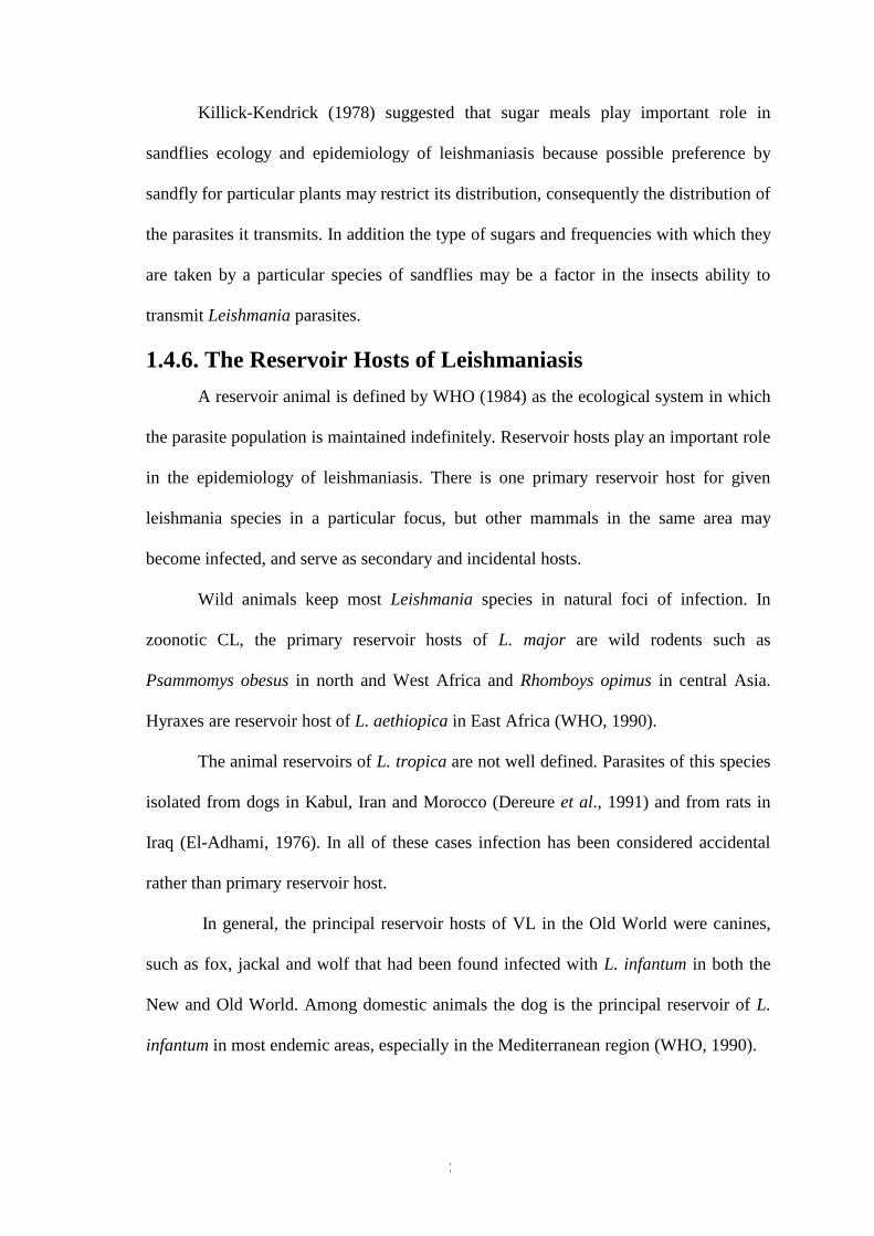
1.4.6. The Reservoir Hosts of Leishmaniasis
A reservoir animal is defined by WHO (1984) as the ecological system in which
the parasite population is maintained indefinitely. Reservoir hosts play an important role
in the epidemiology of leishmaniasis. There is one primary reservoir host for given
leishmania species in a particular focus, but other mammals in the same area may
become infected, and serve as secondary and incidental hosts.
Wild animals keep most Leishmania species in natural foci of infection. In
zoonotic CL, the primary reservoir hosts of L. major are wild rodents such as
Psammomys obesus in north and West Africa and Rhomboys opimus in central Asia.
Hyraxes are reservoir host of L. aethiopica in East Africa (WHO, 1990).
The animal reservoirs of L. tropica are not well defined. Parasites of this species
isolated from dogs in Kabul, Iran and Morocco (Dereure et al., 1991) and from rats in
Iraq (El-Adhami, 1976). In all of these cases infection has been considered accidental
rather than primary reservoir host.
In general, the principal reservoir hosts of VL in the Old World were canines,
such as fox, jackal and wolf that had been found infected with L. infantum in both the
New and Old World. Among domestic animals the dog is the principal reservoir of L.
infantum in most endemic areas, especially in the Mediterranean region (WHO, 1990).

33
1.4.7. Control of Leishmaniasis and Sandflies
The most successful integrated control programs of leishmaniasis combine the
control of the vector and the reservoir animal when the later is known. This method was
applied successfully in USSR, rodenticides and mechanical methods were used to
destroy the net work burrows of the great gerbil (Rhombomys opimus) which is the main
reservoir host of zoonotic CL (Ashford and Bettini, 1987).
In general, reservoir control must be considered as an important part of the
integrated control of leishmaniasis, however, different animals can act as reservoirs of
the same parasite and even if one species is completely eliminated another can take its
place. Also sandflies are generally opportunistic in their feeding habits and for this
reason they can turn to another host if any particular species of animal should be
eradicated. For these reasons reservoir control should be considered as only one method
in complex programs to control the leishmaniasis (WHO, 1990).
Vector control is considered to be the main method for prevention of leishmaniasis. For the proper organization of sandfly control, precise knowledge of their biology, ecology is important. The principle methods used in control of sandflies and
leishmaniasis are application of chemical insecticides sometimes in conjunction with environmental management. DDT is still
one of the recommended insecticides (Rozendaal, 1997). Other control methods, such as biological predators and genetical and ecological measures, are difficult to apply and sometimes not effective. An integrated program of control, including all other
measurements was recommended, but applied in very rare cases (Viokov, 1987).
Alternative control methods for sandflies are currently tested in different parts of world include the use of the insecticide-impregnated bed nets and curtains (Rozendaal, 1997). Organic remnants and piles of bricks and stones in construction sites
constitute potential breeding and resting sites for sandflies; elimination of these factors from human habitats decrease the risk of
leishmaniasis (WHO, 1990). The habitat of rural population to keep their animals in residence areas attracts both anthropophilic and zoophilic vectors and decrease the possibility of acquiring the disease (Rozendaal, 1997).
1.5. EPIDEMIOLOGY OF LEISHMANIASIS IN
PALESTINE AND VICINITY
In the West Bank, Palestine, both cutaneous and visceral leishmaniasis occur and
are a growing public health problem (Arda, 1983; Greenblatt et al., 1985; Klaus et al.,
1994; Qubain et al., 1997; Benth et al., 1998).
1.5.1. Cutaneous Leishmaniasis

34
Cutaneous leishmaniasis has been reported in Palestine since the
beginning of the last century. The Jordan Valley, and specially Jericho
town, is recognized as a hyperendemic area of CL in the country; where it
is called “Jericho Boil”. Huntimular was the first to detect Leishmania
parasites from cases of CL in the Jordan Valley in 1914; and since that
time many cases of CL still to occure every year as well as several
outbreaks were discribed from different parts of Palestine, and so that much
attention had been given to the diseas (Greenblatt, et al., 1985). The rate of
human cases depend on the number of people enter the zoonotic focus in
the nature (Naggan et al., 1970).
In Palestine, two forms of CL have been established i.e. L. major and
L. tropica. The epidemiology of the first form has been studied in great
detail. It is a zoonotic disease described from different parts of Palestine
and especially from the Jordan Valley; the parasite is harbored by P. obesus
and Meriones crassus and the vector is P. papatasi (Schlein et al., 1984).
Sporadic cases of CL caused by L. tropica were reported from mountainous
area in the northern parts of the West Bank, reservoir host and sandfly
vector were not identified (Klaus et al., 1994).
Blum (1978) found that the incidence of CL in the peripheral areas of
Salfit town was twice that found in the centre of town because of the
natural increasing of the researvoir animals (Rattus rattus) in peripheral
areas. 14 % of the sampled population were leishmanin positive and its

35
incidence was found to increase with age; and no active lesion was
observed above the age of 25 years.
Most CL cases were reported between June and November, but cases
have been reported in every month of the year (Y. Schlein, personal
communication). The main infectious period is during the summer months.
Based on the time of acquiring the infection of CL from different
Leishmaniasis foci the highest exposure months were June and July (Klaus
et al., 1994).
Dispite leishmaniasis is a reportable disease by law in West Bank and Israel,
many cases were not reported and the estimated cases are several handreds per year
(Greenblatt et al. 1985; Y. Schlein, unpublished work). Table 1 summarizes reported
cases and outbreaks of CL in the West Bank and Israel since early of the last century.
1.5.2. Visceral Leishmaniasis
Reports of VL cases were scattered; cases were reported from different parts
including Petah Tiqwa, Jerusalem and the Jezreel Valley (Greenblatt et al., 1985). In
addition, imported cases between Jewish immigrants from neighboring countries to
Palestine were also reported.
During 1960s, 45 sporadic cases of VL were reported from different Arab
villages in Gallilee; about 42 % of cases reported from three closed villages. All the
affected were children under 8 years old. The intensive uses of DDT in Israeli’s
settlements, during 1950s and thereafter, prevents the occurrence of VL in these
communities, also the decrease of cases in Arab villages was contributed to the same

36
reason. During 1971 to 1979 only one case of VL was reported in Israel (Greenblatt et
al., 1985).
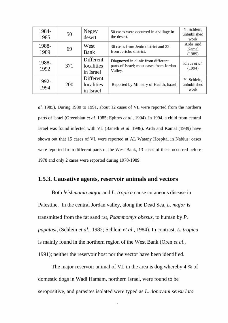
Table 1. Reported cases and outbreaks of CL in West Bank and neighboring Israel documented
from 1914 to 1994.
Year Number
of cases
Infected
locality(s) Comments Reference
1914 Few
The
Jordan
Valley
The first Parasitology proven cases Huntimular
(1914)
1914-
1948 Unknown Palestine Scattered reports of the presence of CL
Greenblatt et
al. (1985)
1967 125
The
Jordan
Valley
An epidemic resulted from entering
non-immune soldiers into hyper-
endemic area
Naggan et al.
(1970)
1967-
1974 377
Different
localities
in Israel
Reported by Ministry of Health, Israel Greenblatt et
al. (1985)
1972-
1980
131 Salfit
District
CL was unknown (or very rare) before
1972; its prevalence peaked in 1973
and then dropping. In 1978, 14.5 % of
residents were leishmanin positive.
Arda (1983);
Blum (1978)
57
Northern
districts of
West
Bank
33, 12 and 12 cases were reported
from Jenin, Tulkarm and Nablus
Districts respectively.
Arda (1983)
24 Jordan
Valley From different parts of the Jordan
Valley
11
Southern
districts of
West
Bank
8 cases from Rallah and 3 cases from
Hebron.
Total =
223
West
Bank In addition to 14 imported cases
1980-
1982 780
Different
localities
in Israel
Israeli Ministry of Health reported
cases. 60 were soldiers whom based in
an endemic area in Negev desert
during 1982.
Greenblatt et
al. (1985)
1983-
1987 173
Northern
districts of
West
Bank
Most of cases were diagnosed during
1986-87; 62 of these cases were found
in Qabatyah where the disease wasn’t
reported before.
Arda and
Kamal
(1989)
37
1984-
1985 50
Negev
desert 50 cases were occurred in a village in
the desert.
Y. Schlein,
unbublished
work
1988-
1989 69
West
Bank 36 cases from Jenin district and 22
from Jericho district.
Arda and
Kamal
(1989)
1988-
1992 371
Different
localities
in Israel
Diagnozed in clinic from different
parts of Israel; most cases from Jordan
Valley.
Klaus et al.
(1994)
1992-
1994 200
Different
localities
in Israel
Reported by Ministry of Health, Israel
Y. Schlein,
unbublished
work
al. 1985). During 1980 to 1991, about 12 cases of VL were reported from the northern
parts of Israel (Greenblatt et al. 1985; Ephros et al., 1994). In 1994, a child from central
Israel was found infected with VL (Baneth et al. 1998). Arda and Kamal (1989) have
shown out that 15 cases of VL were reported at AL Watany Hospital in Nablus; cases
were reported from different parts of the West Bank, 13 cases of these occurred before
1978 and only 2 cases were reported during 1978-1989.
1.5.3. Causative agents, reservoir animals and vectors
Both leishmania major and L. tropica cause cutaneous disease in
Palestine. In the central Jordan valley, along the Dead Sea, L. major is
transmitted from the fat sand rat, Psammomys obesus, to human by P.
papatasi, (Schlein et al., 1982; Schlein et al., 1984). In contrast, L. tropica
is mainly found in the northern region of the West Bank (Oren et al.,
1991); neither the reservoir host nor the vector have been identified.
The major reservoir animal of VL in the area is dog whereby 4 % of
domestic dogs in Wadi Hamam, northern Israel, were found to be
seropositive, and parasites isolated were typed as L. donovani sensu lato

38
(Jaffe, et al., 1988). Parasites were also isolated from seropositive dogs in
central Israel were typed as L. infantum; the rate of seropositive examined
animals from the area were 1-11.5 % in domestic dogs, 7.6 % in jackals
and 5 % in foxes (Baneth, et al., 1998).
Three species of rodents were found infected with L. major in the
southern parts of Israel, namely Psammomys obesus, Meriones crassus and
Nasokia indica. Other animals examined but found not to be infected
included two foxes, three porcupines and three hyraxes (Schlein, et al.
1984). It had been suggested that rocky hyrax may have a role in the
transmission of CL, because many cases of CL have been reported near
their colonies in Kfar Adumim (Klaus et al., 1994).
P.papatasi sandflies collected from three localities in the southern
parts were found infected with Leishmania parasites (7-23 %). One female
of P. sergenti caught from Arava area was found infected (Schlein, et al.,
1984).
Sawalha (2000) presumed on epidemiological and entomological surveys, that P.
papatasi might be the principal vector of CL in the northern parts of West Bank. Since
P. papatasi was the predominantly man biting species. Table 2 summarizes causative
agents, sandfly vectors and reservoir animals reported from Palestine and neighboring
Israel.

39
Table 2. Causative agents of leishmaniasis and their distribution, vectors
and reservoir hosts, documented from Palestine and neighboring Israel.
Causative agent
(clinical condition)
Geographical
Distribution
Sandfly
Vector(s)
Reservoir
Animal(s) References
L. major
(Cutaneous
leishmaniasis)
Central Jordan Valley P. papatasi P. obesus Schlein et al. (1984)
Dead Sea Region - P. obesus Schlein et al. (1984)
Kfar Adumim - - Klaus et al., (1994)
Rift Valley (Arava) P. sergenti P. obesus
M. crassus Schlein et al. (1984)
Keziot in the Negev
desert (South western
Israel)
P. papatasi P. obesus
M. crassus Giladi, et al. (1984)
L. tropica
(CL, VL* and
leishmania
recidivans)
Salfit P. papatasi
(suspected)
Rattus rattus
(not confirmed) Blum (1978)
Northern Jerusalem - - Y. Schlein (personal
communication)
Kfar Adumim - - Klaus et al., (1994)
Mountains between
Nablus and Jenin - Unknown Klaus et al. (1994)
L. infantum
(Visceral
Leishmaniasis)
Northern Israel - Domestic dogs Jaffe, et al., (1988)
Ephros et al., (1994).
Central Israel and West
Bank -
Dogs
Jackals
Foxes
Baneth, et al., (1998)
1.5.4. Ecology of leishmaniasis
The zoonotic nature of CL in the Jordan Valley had been well established, so
that infection is more likely to occur at the neighborhood of Jericho City, close to the
sand rat Psammomys obesus (Rodentia: Gerbillidae) colonies (Naggan et al., 1970;
Schlein et al., 1982). In this arid climate the sand rat burrows seem to provide sandflies
with moist and relatively cool microhabitat essential for larva and adults. This closeness
between the vector and reservoir animal lead to high rate of infection in both, which
may reached to 56 % in P. papatasi (Schlein et al., 1982). On the other hand the rate of
infection found in the same species of sandfly collected from Jericho city was less than
40
0.1 % (Adler and Theodor, 1926). Saliba et al. (1985) described an outbreak of CL,
which occurred in the outskirts of Amman in the Jordanian desert; peak transmission of
the outbreak took its place in the late summer of 1982. About two third of the patients
were less than 15 years old.
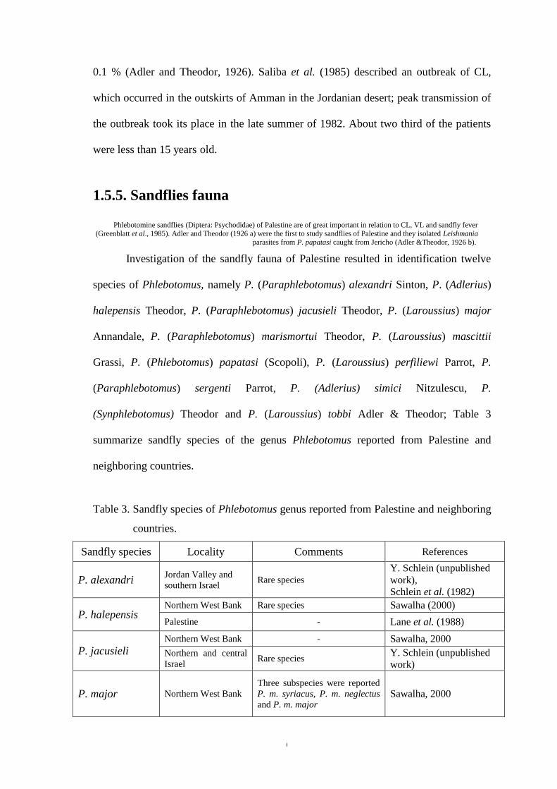
1.5.5. Sandflies fauna
Phlebotomine sandflies (Diptera: Psychodidae) of Palestine are of great important in relation to CL, VL and sandfly fever
(Greenblatt et al., 1985). Adler and Theodor (1926 a) were the first to study sandflies of Palestine and they isolated Leishmania
parasites from P. papatasi caught from Jericho (Adler &Theodor, 1926 b).
Investigation of the sandfly fauna of Palestine resulted in identification twelve
species of Phlebotomus, namely P. (Paraphlebotomus) alexandri Sinton, P. (Adlerius)
halepensis Theodor, P. (Paraphlebotomus) jacusieli Theodor, P. (Laroussius) major
Annandale, P. (Paraphlebotomus) marismortui Theodor, P. (Laroussius) mascittii
Grassi, P. (Phlebotomus) papatasi (Scopoli), P. (Laroussius) perfiliewi Parrot, P.
(Paraphlebotomus) sergenti Parrot, P. (Adlerius) simici Nitzulescu, P.
(Synphlebotomus) Theodor and P. (Laroussius) tobbi Adler & Theodor; Table 3
summarize sandfly species of the genus Phlebotomus reported from Palestine and
neighboring countries.
Table 3. Sandfly species of Phlebotomus genus reported from Palestine and neighboring
countries.
Sandfly species Locality Comments References
P. alexandri Jordan Valley and
southern Israel Rare species
Y. Schlein (unpublished
work),
Schlein et al. (1982)
P. halepensis Northern West Bank Rare species Sawalha (2000)
Palestine - Lane et al. (1988)
P. jacusieli Northern West Bank - Sawalha, 2000
Northern and central
Israel Rare species
Y. Schlein (unpublished
work)
P. major Northern West Bank
Three subspecies were reported
P. m. syriacus, P. m. neglectus
and P. m. major Sawalha, 2000

41
Northern and central
Israel
P. m. syriacus. Prevalent
subspecies
Y. Schlein (unpublished
work)
P. marismortui Israel Undefined location Lewis (1982)
P. mascittii
Northern West Bank P. mascitti canaaniticus and
P. mascitti mascitti Sawalha, 2000
Northern and central
Israel Rare species
Y. Schlein (unpublished
work)
Palestine P. mascitti canaaniticus Lane et al. (1988)
P. papatasi Northern West Bank - Sawalha, 2000
Jordan Valley and
southern parts of
Israel
The dominant species and a
proven vector of CL
Adler &Theodor (1926 a)
Schlein et al. (1982)
Schlein et al. (1984)
P. perfiliewi Northern West Bank - Sawalha, 2000
Palestine P. perfiliewi galilaeus Lane et al. (1988)
P. sergenti Northern West Bank - Sawalha, 2000
Different regions of
Israel
More prevalent in northern and
central regions
Y. Schlein (unpublished
work)
P. simici Palestine - Lane et al. (1988)
P. (Syn.) sp. Northern West Bank Reported from the area for the
first time Sawalha, 2000
P. tobbi
Northern West Bank - Sawalha, 2000
Different regions of
Israel
More prevalent in northern and
central parts
Y. Schlein (unpublished
work)
Jordan Valley Rare species Schlein et al. (1982)
Also eleven species of Sergentomyia were identified from the area, including S.
(Parrotomyia) africana asiatica Adler & Theodor, S. (Sergentomyia) antennata
(Newstead), S. christophersi (Sinton), S. clydei (Sinton), S. (Sergentomyia) fallax
Parrot, S. (Parrotomyia) palestinensis Adler & Theodor, and S. squamipleuris
(Newstead), S. (Sergentomyia) sinotoni Pringle, S. sp. a, S. theodori (Parrot), and S.
tiberiadis (Adler, Theodor & Lourie). Table 4 summarizes sandfly species of the genus
Sergentomya reported from Palestine and neighboring countries.
Table 4. Sandfly species of the genus Sergentomya reported from Palestine and
neighboring countries.
Sandfly species locality Comments References

42
S. africana
asiatica
Jordan Valley - Schlein et al. (1982)
Southern Israel - Schlein, et al., (1984)
S. antennata Jordan Valley Dominant species Schlein et al. (1982)
Southern Israel Dominant species Schlein, et al., (1984)
S. christophersi Northern West Bank - Sawalha, 2000
S. clydei Southern Israel Rare species Schlein, et al., (1984)
S. fallax
Northern West Bank - Sawalha, 2000
Jordan Valley Rare species Schlein et al. (1982)
Southern Israel Rare species Schlein, et al., (1984)
S. palestinensis Jordan Valley Rare species Schlein et al. (1982)
Palestine - Lane et al. (1988)
S. squamipleuris Southern Israel Rare species Schlein, et al., (1984)
S. sinotoni Jordan Valley Rare species Schlein et al. (1982)
S. sp. a Palestine - Lane et al. (1988)
S. theodori Northern West Bank Dominant species Sawalha, 2000
S. tiberiadis
Northern West Bank - Sawalha, 2000
Palestine - Lane et al. (1988)
Southern Israel - Schlein, et al. (1984)
1.5.6. Incrimination of Vectors
Janini et al. (1995) collected 1446 females of P. papatasi, and about 50 of P.
alexandri and P. alexandri. None of these flies were infected, but 14 of 686 of P.
papatasi collected from Ps. obesus burrows were infected.
Four females of 3624 P. papatasi, caught from Jericho during 1925, were found
infected with Leishmania parasites (Adler & Theodor, 1926 b). P. papatasi was the only
species of sandflies found in a focus of CL in Salfit area (Blum, 1978). Schlein et al.,
(1982) recorded 29 infected out of 70 P. papatasi females collected from Psammomys
burrows or near them in the Jordan Valley. Yuval (1991) found that 7.4% of P. papatasi

43
trapped from P. obesus burrows in southern Jordan Valley were infected with L. major,
with highest peak during July and August, and along the sandfly season.
Adler and Theodor (1957) have suggested that three species of sandflies P.
major, P. perniciosus and P. longicuspis are probable vectors of VL in the
Mediterranean region, and the first one is the only proven vector so far, in the region.
In Israel, it has been found that P. perfiliewi is a zoophilic species and rarely
feeds on man, and this explain the very low incidence of VL in the area although 20%
of dogs were infected during 1930s-1950s (Adler & Theodor, 1957).
1.5.7. Control of Leishmaniasis
The widespread spraying of insecticides such as residue Pirethroids
and Organo Phosphorous compounds, once a year in all infected areas,
were used during sandflies season. In addition, anticoagulant
compounds and Lenet were used for control of rodents and stray dogs
respectively. These procedures were employed since 1996, which
before the only control measure used was spraying patients’ houses,
who refer to governmental health clinics, with insecticides one to two
weeks after their referring.
44
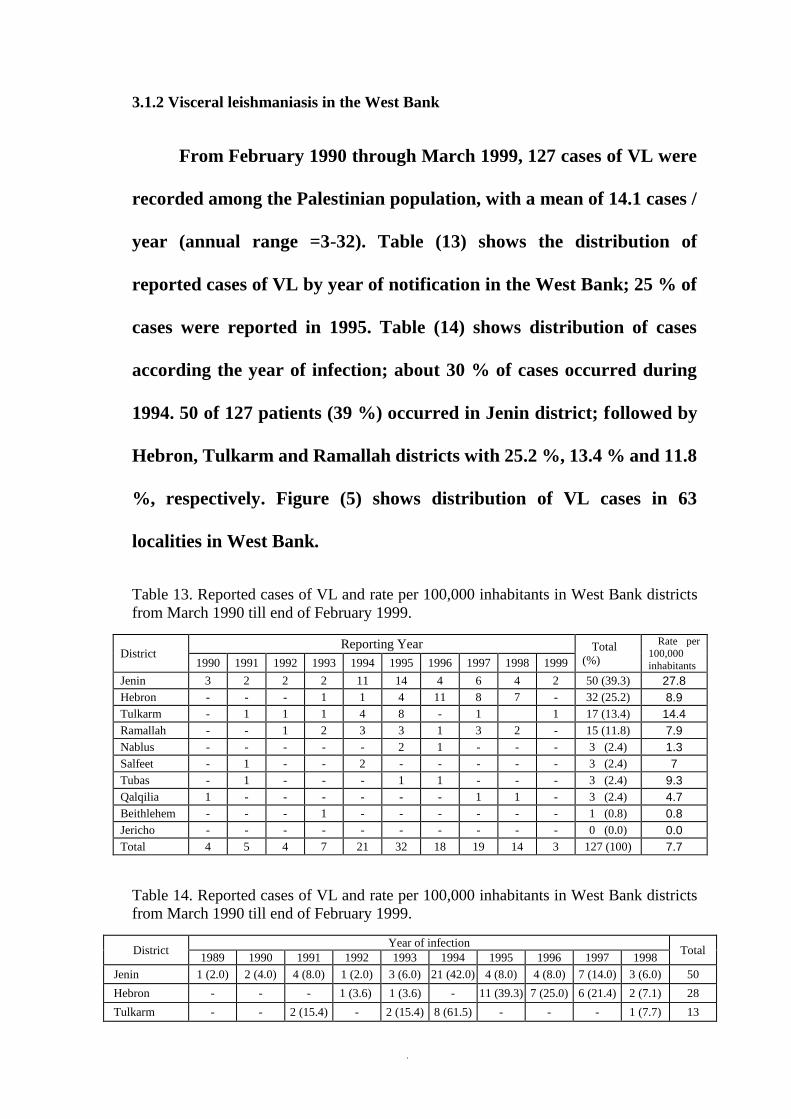
3.1.2 Visceral leishmaniasis in the West Bank
From February 1990 through March 1999, 127 cases of VL were
recorded among the Palestinian population, with a mean of 14.1 cases /
year (annual range =3-32). Table (13) shows the distribution of
reported cases of VL by year of notification in the West Bank; 25 % of
cases were reported in 1995. Table (14) shows distribution of cases
according the year of infection; about 30 % of cases occurred during
1994. 50 of 127 patients (39 %) occurred in Jenin district; followed by
Hebron, Tulkarm and Ramallah districts with 25.2 %, 13.4 % and 11.8
%, respectively. Figure (5) shows distribution of VL cases in 63
localities in West Bank.
Table 13. Reported cases of VL and rate per 100,000 inhabitants in West Bank districts
from March 1990 till end of February 1999.
District Reporting Year Total
(%)
Rate per
100,000
inhabitants 1990 1991 1992 1993 1994 1995 1996 1997 1998 1999
Jenin 3 2 2 2 11 14 4 6 4 2 50 (39.3) 27.8
Hebron - - - 1 1 4 11 8 7 - 32 (25.2) 8.9
Tulkarm - 1 1 1 4 8 - 1 1 17 (13.4) 14.4
Ramallah - - 1 2 3 3 1 3 2 - 15 (11.8) 7.9
Nablus - - - - - 2 1 - - - 3 (2.4) 1.3
Salfeet - 1 - - 2 - - - - - 3 (2.4) 7
Tubas - 1 - - - 1 1 - - - 3 (2.4) 9.3
Qalqilia 1 - - - - - - 1 1 - 3 (2.4) 4.7
Beithlehem - - - 1 - - - - - - 1 (0.8) 0.8
Jericho - - - - - - - - - - 0 (0.0) 0.0
Total 4 5 4 7 21 32 18 19 14 3 127 (100) 7.7
Table 14. Reported cases of VL and rate per 100,000 inhabitants in West Bank districts
from March 1990 till end of February 1999.
District Year of infection
Total 1989 1990 1991 1992 1993 1994 1995 1996 1997 1998
Jenin 1 (2.0) 2 (4.0) 4 (8.0) 1 (2.0) 3 (6.0) 21 (42.0) 4 (8.0) 4 (8.0) 7 (14.0) 3 (6.0) 50
Hebron - - - 1 (3.6) 1 (3.6) - 11 (39.3) 7 (25.0) 6 (21.4) 2 (7.1) 28
Tulkarm - - 2 (15.4) - 2 (15.4) 8 (61.5) - - - 1 (7.7) 13

45
Ramallah - - - 1 (8.3) 2 (16.7) 2 (16.7) 2 (16.7) 1 (8.3) 2 (16.7) 2 (16.7) 12
Tubas - - - - - 1 (33.3) 2 (66.7) - - - 3
Salfit - - 1 (33.3) - - - - - - - 3
Qalqilia - 1 (33.3) - - - - - 1 (33.3) 1 (33.3) - 3
Nablus - - - - - 1 (50.0) 1 (50.0) - - - 2
Beithlehem - - - - - - 1 (100) - - - 1
Total 1 (0.9) 3 (2.6) 7 (6.1) 3 (2.6) 9 (7.8) 34 (29.6) 21 (18.3) 13 (11.3) 16 (13.9) 8 (7.0) 115
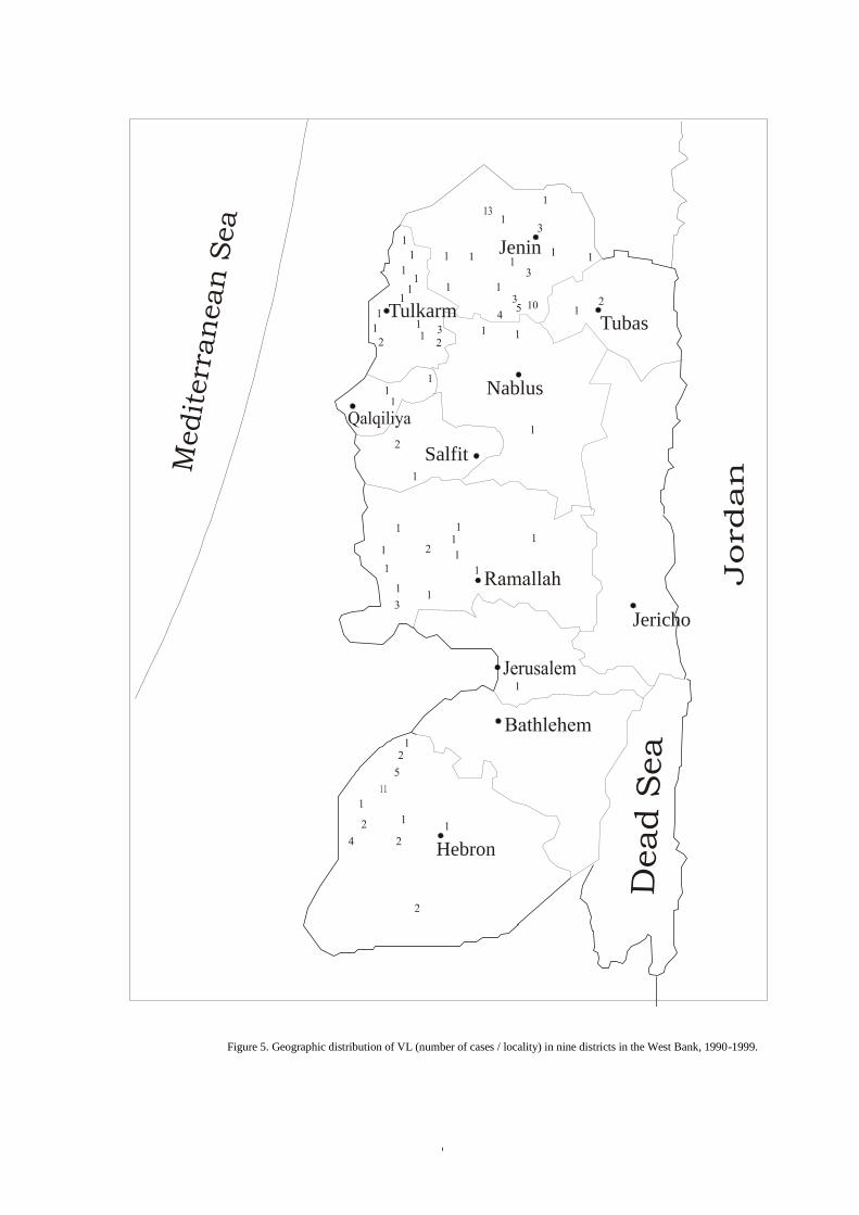
46
Figure 5. Geographic distribution of VL (number of cases / locality) in nine districts in the West Bank, 1990-1999.
Medit
err
an
ean
Sea
Jenin
Nablus
Tulkarm
Salfit
Tubas
Jericho
Hebron

47
Monthly distribution of VL cases showed that most cases were notified during
June. Disease symptoms were noticed during the same period Figure (6).
Table (15) shows distribution of VL patients by age group and sex. The age of
patients’ ranged from 5 months to 46 years; 114 (90 %) were younger than 5 years old,
and distribution by sex showed that there were no differences between both sexes.
Figure 6. Percentage of monthly distribution according to the date of infection (thick line) and
date of appearance of signs and symptoms (thin line) of VL cases reported from West Bank.
1990-1999.
Visceral disease mainly affects infants and young children throughout the northern region
of the West Bank. From 1990 through 1999, many cases (127) were recorded throughout the
0
5
10
15
20
Jan Feb Mar Apr May Jun Jul Aug Sep Oct Nov Dec
%
Month of notification
Month of signs appearance

48
West Bank, giving a mean of 12.7 cases / year; Jenin district constituted 50 (39%) of the total
cases 127 (61%).
Table 15. Distribution the numbers of VL patients by group and sex reported from the
West Bank, 1990-1999.
District Age (Years)
Sub-total Total 0-4 5-9 15-19 40+
Male Female Male Female Male Female Male Female Male Female
Jenin 19 26 2 1 - 1 - 1 21 29 50
Hebron 14 13 4 1 - - - - 18 14 32
Tulkarm 6 11 - - - - - - 6 11 17
Ramallah 9 5 1 - - - - - 10 5 15
Tubas 2 1 - - - - - - 2 1 3
Nablus 2 - - 1 - - - - 2 1 3
Salfit 2 - 1 - - - - - 3 - 3
Qalqilia 3 - - - - - - - 3 - 3
Beithlehem 1 - - - - - - - 1 - 1
Total 58 56 8 3 - 1 - 1 66 61 127

49
CHAPTER TWO
2. MATERIALS AND METHODS
Leishmaniases are zoonotic diseases that transmit from reservoir animals by
sandflies vectors to human. The ecological factors have large effects on disease
transmission. Thus, in a study about leishmaniasis epidemiology, it is important to study
the ecological factors which affect the prevalence and geographical distribution of the
disease in active foci. Also the sandfly species and suspected reservoir animals found in
a certain endemic area may be used as an indicator to the epidemiology of the disease.
Therefore, such a study can provide base line data about different leishmaniasis
foci in West Bank necessary to control the disease through its vector(s) and reservoir
host(s) as well as provide information which help in explaining and predicting
outbreaks of the disease.
All districts of West Bank were included in this study, except Jericho district,
which was excluded because the epidemiology of leishmaniasis had been well studied
in details (Schlein et al., 1984). Epidemiological data on CL and VL were collected
from the reports of other nine districts in West Bank, while ecological factors were
studied in Jenin district that lies in the northern parts of the West Bank. Sandflies were
collected from different localities in West Bank, except districts studied before, namely
Jenin (Sawalha, 2000) and Jericho (Schlein et al., 1982).
2.1. Design
The research design of this study was descriptive retrospective design. This
design was chosen to investigate the factors that lead to the increase of leishmaniasis
cases.

50
Although the study is descriptive, it attempts to put the problem in prospective
by showing the relative degree to which different districts are affected, how the patterns
of the disease occurrence vary between different areas and how the reservoir animal and
vectors of the disease are associated with the different patterns of occurrence.
2.2. The Study Area
2.2.1. West Bank
The West Bank is bordered by Israel on the north, west and south. On the east
the Jordan River and the Dead Sea from a natural border with Jordan. It is a hilly region
located between the coastal plain in the west and the Jordan valley in the east with an
average altitude of 2400 feet.
The total population size of the West Bank according to 1997 census was about
1,600,100 persons. Population density in the West Bank is high compared with
neighboring countries. It is around 320 / km 2
because the considerable part of the land
is still under Israel control.
The West Bank is divided into 10 districts: Jenin, Tubas, Tulkarm, Qalqiliya,
Nablus, Ramallah, Salfit, Bethlehem, Jericho, and Hebron. It has 10 large towns, 430
villages and 18 refugees’ camps. In general, 27 % of the population lived in urban
communities.
The West Bank has a Mediterranean type climate. The mean monthly
temperature over the last ten years ranged from 17.4° C in August. The mean annual
relative humidity ranged from 84 % to 39 % while the mean annual rainfall was 528
mm (source: Palestine Meteorological Department). The year may be divided into a hot
dry Summer, a warm Autumn, cold wet Winter and Spring.

51
Rainfall is limited to the winter and spring months from October to May. During
the period of April to June, hot Khamsin winds from the south may occur and
subsequently the atmosphere becomes hazy with dust from the desert. Rainfall in the
West Bank varies greatly from east to west and from south to north according to
topography. The annual rainfall ranges from 150 mm in the east to about 800 mm in the
northwest. In the summer months there is no precipitation at all.
The inhabitants mostly work in agriculture, especially in the rural areas that have
60% of the total population. Although raising animals has decreased in the last years,
domestic animals like sheep, goats, chickens, and others are still found, nearly, in all
rural localities with different numbers, but the number of poultry houses has increased
considerably and especially in certain localities. Most residents live in houses built of
concrete and / or stone. Many of these houses were constructed in the peripheral areas.
In general human behavior in the rural areas of the West Bank is not different
from district to another. During summer months (sandfly season) people spend the early
evening in different activities at out doors sites of their houses. While the majority of
people sleep indoors, young people may sleep at outdoors sites during the hot weather.
People do not have the habit of sleeping under bed nets.
A preliminary survey for reported cases of leishmaniasis in different districts of
West Bank was carried out at the beginning of the study. Based on the results of this
preliminary survey, Jenin district was chosen to study the ecological factors and
epidemiology of leishmaniasis with more emphasis for the following reasons: (1) It was
one of the most infected district during the study period as shown by the preliminary
survey. (2) Both forms of leishmaniasis were reported from many localities in the
district and some of these localities had both forms of the disease. (3) The large number
of leishmaniasis cases reported in the West Bank and the difficulties to reach to

52
different areas. And (4) Jenin district was pointed out to be a focus of leishmaniasis
since 1970s because of the many cases were reported from different district. The area
was thus considered as an endemic and active focus of the disease up to now.
2.2.2. Jenin District
Jenin district is located in the northern part of the West Bank. It has 96 localities
and covers an area of 592 km ² with altitudes ranging between 90 and 750 meters above
sea level (Applied Research Institute (ARIJ), 1996) and had a population of 195,299 in
1997 (Palestinian Central Bureau of Statistics, 1997).
Rainfall varies significantly in Jenin district from 778 mm in the west to 286 mm
in the east and with mean annual rainfall of 528 mm. The district receives rainfall
between middle October and end of the April, with peaks in January and February.
Precipitation decreases to 12 % of the annual rainfall in March (ARIJ, 1996).
Wind blows from southwest and northwest, and being more northerly during the
summer. The average wind speed ranged between 9.2 and 7.7 km / hour. There is also a
little fluctuation in relative humidity in the district especially during summer months. It
ranges from 63 to 66% during June to November, from 67 to 84 % during December to
April, and it falls to 39% during May as a result of the dry winds that blow from the
Arabian Desert (ARIJ, 1996).
Table 5 shows the dominant sandfly species, trees, topography, altitude and soil
type in the most affected localities of Jenin district where 92 % of CL and 86 % of VL
has been reported.

53
Table 5. Description of habitats of leishmaniasis foci in Jenin district where more than 90 %
of cases were reported.
Locality Topography Soil type* Altitude*
Plants of the area
(trees) Sandfly species**
Olive Fruit Stone
Fruit
Aba Hilly Grumusols 150-200 +++ + + P. papatasi
Al
Shuhada Hilly
Grumusols, and
Terra Rossas, Brown
Rendzinas.
270-366 +++ + + P. papatasi P. major syriacus
Beit Qad Hilly Grumusols 190-290 +++ + +
P. papatasi P. perfiliewi
P. tobbi
Bir El
Basha Hilly
Brown Rendzinas
and Pale Rendzinas 260-336 +++ + +
P. papatasi
P. tobbi
Deir Abu
Da’if Mountainous
Grumusols,and
Terra Rossas, Brown
Rendzinas.
180-302 +++ + +
P. papatasi P. perfiliewi
P. tobbi
El Yamun Mountainous
Grumusols, and
Terra Rossas, Brown
Rendzinas.
130-251 +++ + ++
P. papatasi
P. perfiliewi P. tobbi
P. major neglectus
Jaba Mountainous
Grumusols, And
Terra Rossas, Brown
Rendzinas.
350-610 +++ + ++
P. papatasii P.perfiliewi
P.tobbi
Jadeida Mountainous
Grumusols,And
Terra Rossas, Brown
Rendzinas.
360-539 +++ + ++
P. papatasii
P.perfiliewi
P.tobbi
P.major syriacus P.major neglectus
Jenein Mountainous Grumusols 134-248 ++ + ++
P. papatasi
P. perfiliewi P. tobbi
P. major syriacus
S. theodori
Maythalun Hilly Grumusols 350-423 ++ + ++ P. papatasii
P.perfiliewi
Qabatiya Mountainous
Grumusols,and
Terra Rossas, Brown
Rendzinas.
284-380 +++ + ++
P. papatasi P. perfiliewi
P. tobbi
P. major syriacus
Sanur Mountainous Grumusols 375-548 +++ + + P. papatasi
P. tobbi
Silat El
Harithiya Hilly Grumusols 150-195 +++ + +
P. papatasi P. perfiliewi
P. sergenti
Siris Mountainous
Grumusols,And
Terra Rossas, Brown
Rendzinas.
375-500 +++ + ++
P. papatasii P.perfiliewi
P.tobbi
P.major syriacus P.major neglectus
* Applied Research Institute, (1996)
** Sawalha (2000).
54
2.3. Collection of the Epidemiological Data
2.3.1. Medical records
Data on leishmaniasis cases, reported in the West Bank, over nine years (March
1990 to the end of February 1999) were collected from the Ministry of Health, records
of notifiable diseases.
The collected data included demographic (name, age, sex, and residence), type
of leishmaniasis disease, date of onset of illness, date of diagnosis, occupation, origin of
infection or travel history to other endemic foci, and the number and site of lesion or
scars.
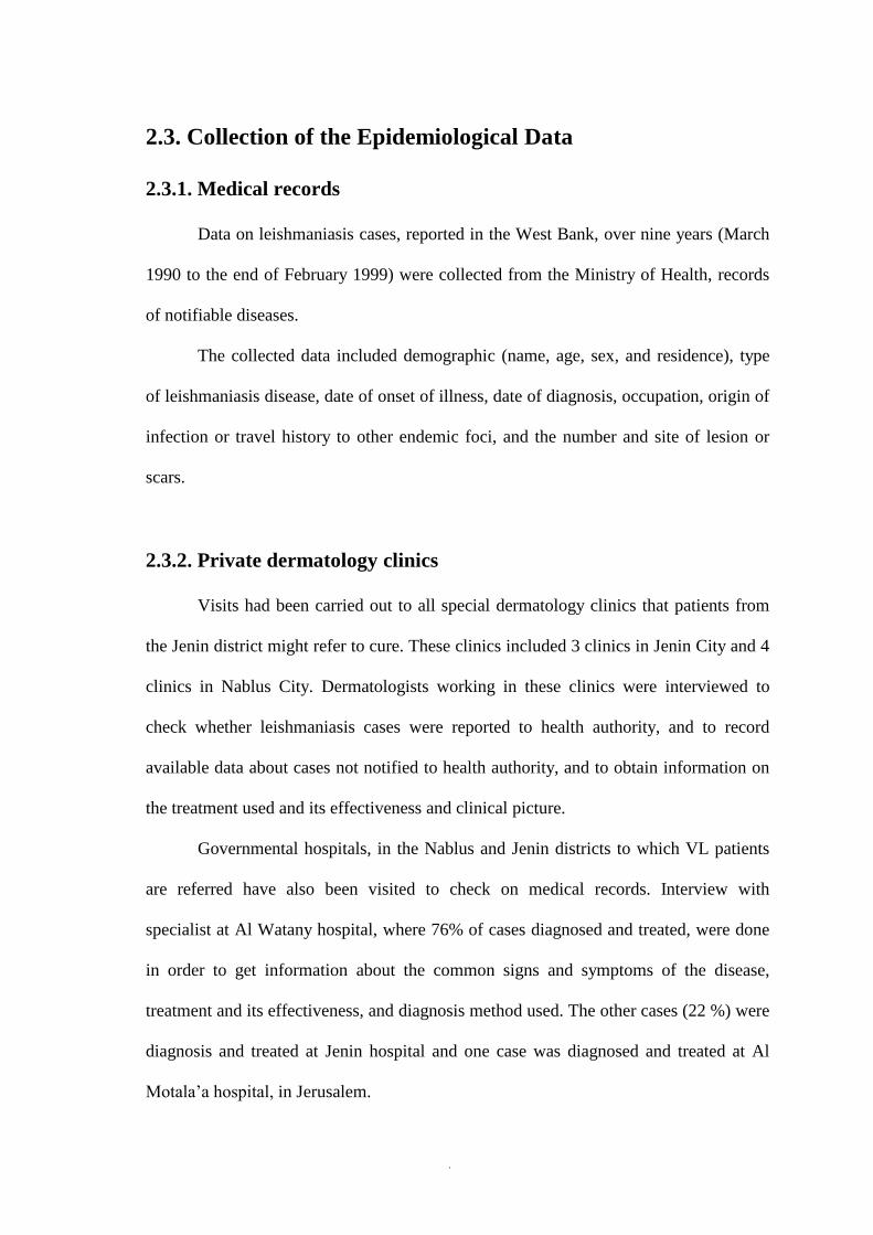
2.3.2. Private dermatology clinics
Visits had been carried out to all special dermatology clinics that patients from
the Jenin district might refer to cure. These clinics included 3 clinics in Jenin City and 4
clinics in Nablus City. Dermatologists working in these clinics were interviewed to
check whether leishmaniasis cases were reported to health authority, and to record
available data about cases not notified to health authority, and to obtain information on
the treatment used and its effectiveness and clinical picture.
Governmental hospitals, in the Nablus and Jenin districts to which VL patients
are referred have also been visited to check on medical records. Interview with
specialist at Al Watany hospital, where 76% of cases diagnosed and treated, were done
in order to get information about the common signs and symptoms of the disease,
treatment and its effectiveness, and diagnosis method used. The other cases (22 %) were
diagnosis and treated at Jenin hospital and one case was diagnosed and treated at Al
Motala’a hospital, in Jerusalem.

55
2.3.3. Active case finding
The last source of data collection was active investigation about leishmaniasis
cases in the infected areas. During visiting to houses of leishmaniasis cases in Jenin
district interviewees were asked whether they knew of other persons that have been
infected with leishmaniasis. These names were compared those in medical records, and
unreported cases were visited and epidemiological data were collected.
2.3.4. Diagnosis methods used
Collected data on diagnosis CL from dermatologist clinic and health centers in
Jenin and Nablus districts included clinical picture and microscopic examination (Table
6). Demonstration of Leishmania parasite by microscopic method was used to confirm
diagnosis especially for cases from a new foci and in case of infected wounds with long
duration, recent scars not typical to CL but with no history of traumatic injury, etc.
Table 6. Number and percentage of CL cases in Jenin district diagnosed by different
methods, 1990-1999.
Diagnosis Method used Number Percent
Clinical Picture 299 64.9
Microscopic Examination and Clinical Picture 72 15.6
Unknown 90 19.5
Total 461 100.0
A visceral leishmaniasis case was only considered if diagnosis was proven
parasitologically, either by microscopic examination or culture of material for bone

56
marrow accompanied by the following clinical manifestations: (1) high fever; (2)
hepatosplenomegaly; (3) anemia and (4) leukopenia.
2.3.5. Determination of the year of infection
Determination of the year in which infection occurred is important to study
factors affecting disease transmission. Year of infection was determined based on: (1)
Incubation period of CL 7-60 days (Sacks et al., 1993; Herwaldt, 1999) and incubation
period of VL 1-3 months mean (Sacks et al., 1993, Herwaldt, 1999). (2) The period
consumed before seeking medical care for CL 1-4 months (Arda and Kamal, 1989) and
nearly the same period is needed for seeking medical care and diagnosis of VL (Dr Loai
Shahen personal communication; Qubain, 1997). (3) Multiplication of Leishmania
parasites in the sandfly during 3-6 days (Sacks et al., 1984). (4) The beginning of the
sandfly season in May (Sawalha, 2000). (5) Both the date of referring to medical care
and the date of the onset of signs and symptoms appearance. So that all cases referred to
medical care or onsets of the disease appeared before July and August for CL and VL,
respectively, were considered to have acquired infection of the previous year.
2.3.6. Mapping and interviews of leishmaniasis cases in Jenin district
Patients from the most affected localities in Jenin district (14 localities described
in Table 5) were traced by their last known addresses. The location of patients’ house
where infection was suspected to have taken place were defined on a topographic maps
from the Survey of Israel, 1996.

57
Location of patients’ houses were plotted on a separate topographic map (Scale
1: 10,000) for each locality (Appendix B). Then altitude of the house and location in the
central (built-up area on the map) or peripheral (far from built-up area on the map) of
the locality were detected from the map. Every cluster of houses less than 300 m a part
was given a symbol and plotted on the district map (figures 7 and 13).
Interviews were conducted with patients or their parents whose addresses were
known. Information was collected about (1) the history of the disease in the area, (2)
type of domestic and wild animals around the patients’ houses, and (3) distribution of
caves and crevices around the houses.
2.4. Sandflies Vectors
2.4.1. Collection methods used
Sandflies collections were carried throughout the sandfly season. Sampling
techniques used were CDC light trap and knock down collection as described by Lewis
(1973), Killick-Kendrick (1987) and WHO (1984).
Light traps: Centers for Disease Control (CDC) miniature light traps (model
512; John W. Hock Co., Gainesville, Florida, USA) were hanged at a height of 0.5 m
above the ground level in collection sites. Traps were placed overnight, one to half an
hour before sunset and collected within two hours after sunrise.
Sticky-paper traps: made of ordinary white paper sheets (21X 29 cm), coated
on both sides with caster oil and fixed on sticks held vertically at a height of 0.25-0.3 m
above the ground level. These traps were fixed overnight. Sandflies and other insects
were trapped on the sticky oiled surface when they land. Sandflies were removed from
oiled paper using a small fine brush, washed in 10 % domestic detergent and processed
for identification.

58
2.4.2. Mounting and identification of sandflies
Sandfly specimens preserved in alcohol were carefully mounted in Berlese’s
medium on glass slide as described by Lewis (1967) and Killick-Kendrick (1987).
Using a pair of fine dissecting needles, under binocular dissecting microscope, the head
of each fly was severed and placed near the rest of the body. In case of the female
samples, the last two abdominal segments were dissected out and mounted separately to
examine the spermatheca. Care was taken to place the cover slip gently on each
specimen, which was then left for at least 12 hours to clear. Sandfly identification was
done under binocular microscope at 40X. The main features used for sandfly
identification were the spermatheca of the female, the terminalia of the male and the
pharyngeal and the cibarial toothed structures of both sexes. The taxonomic keys of
Theodor (1958), Lewis (1967; 1978; 1982), Lane (1986), and Lane et al. (1988), were
used for the identification of sandflies species.
2.5. Reservoir Hosts
In this part of the study of reservoir animals more attention was paid to rocky
hyraxes, rodents, wild canines and dogs as possible reservoir hosts for leishmania
parasites, because most of these animals were proved or suspected to act as such in
many countries of Africa and Mediterranean region. Data about abundance and samples
of blood and tissues for parasite infection of these animals were collected.
2.5.1. Abundance of suspected reservoir animals
In order to provide base line data about suspected reservoir animals and their
role in disease transmission in different leishmaniasis foci abundance of different
suspected reservoir animals, was approximately assessed by different ways. Data were
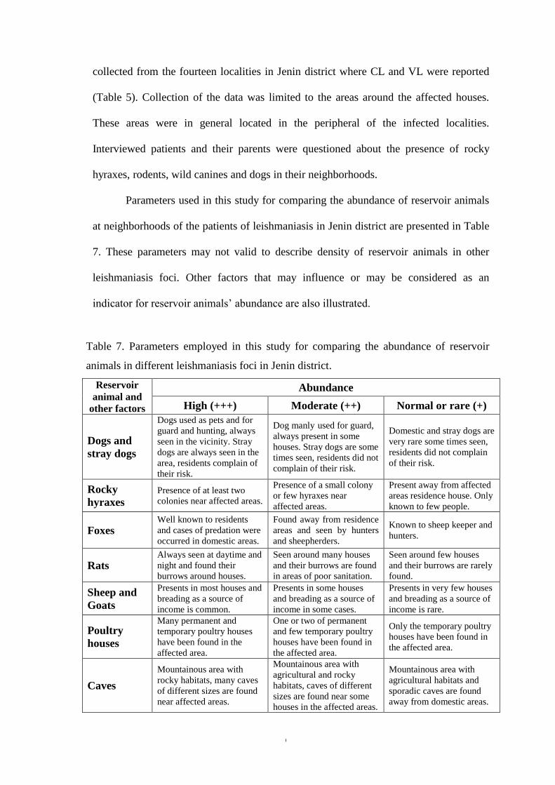
59
collected from the fourteen localities in Jenin district where CL and VL were reported
(Table 5). Collection of the data was limited to the areas around the affected houses.
These areas were in general located in the peripheral of the infected localities.
Interviewed patients and their parents were questioned about the presence of rocky
hyraxes, rodents, wild canines and dogs in their neighborhoods.
Parameters used in this study for comparing the abundance of reservoir animals
at neighborhoods of the patients of leishmaniasis in Jenin district are presented in Table
7. These parameters may not valid to describe density of reservoir animals in other
leishmaniasis foci. Other factors that may influence or may be considered as an
indicator for reservoir animals’ abundance are also illustrated.
Table 7. Parameters employed in this study for comparing the abundance of reservoir
animals in different leishmaniasis foci in Jenin district.
Reservoir
animal and
other factors
Abundance
High (+++) Moderate (++) Normal or rare (+)
Dogs and
stray dogs
Dogs used as pets and for
guard and hunting, always
seen in the vicinity. Stray
dogs are always seen in the
area, residents complain of
their risk.
Dog manly used for guard,
always present in some
houses. Stray dogs are some
times seen, residents did not
complain of their risk.
Domestic and stray dogs are
very rare some times seen,
residents did not complain
of their risk.
Rocky
hyraxes
Presence of at least two
colonies near affected areas.
Presence of a small colony
or few hyraxes near
affected areas.
Present away from affected
areas residence house. Only
known to few people.
Foxes Well known to residents
and cases of predation were
occurred in domestic areas.
Found away from residence
areas and seen by hunters
and sheepherders.
Known to sheep keeper and
hunters.
Rats Always seen at daytime and
night and found their
burrows around houses.
Seen around many houses
and their burrows are found
in areas of poor sanitation.
Seen around few houses
and their burrows are rarely
found.
Sheep and
Goats
Presents in most houses and
breading as a source of
income is common.
Presents in some houses
and breading as a source of
income in some cases.
Presents in very few houses
and breading as a source of
income is rare.
Poultry
houses
Many permanent and
temporary poultry houses
have been found in the
affected area.
One or two of permanent
and few temporary poultry
houses have been found in
the affected area.
Only the temporary poultry
houses have been found in
the affected area.
Caves
Mountainous area with
rocky habitats, many caves
of different sizes are found
near affected areas.
Mountainous area with
agricultural and rocky
habitats, caves of different
sizes are found near some
houses in the affected areas.
Mountainous area with
agricultural habitats and
sporadic caves are found
away from domestic areas.

60
To ensure validity and reliability of the presence of suspected reservoir animals
and their abundance, visits to infected locality and observations were recorded. Other
appropriate persons including sheepherders, hunters and health workers were
interviewed. In addition colored photographs for wild animals were used during
interviews.
2.5.2. Trapping and examining of the suspected reservoir animals
Different kinds of traps were used to capture wild animals (rats, rocky hyraxes
and wild canine). Traps were set in four leishmaniasis foci in Jenin district namely Al
Shuhada, Deir Abu daif and Qabatyah.
2.5.2.1. Rats
Sticky traps made from plastic sheets, covered with glue were used to catch rats.
The trap consisted of a rectangle sheet 30 cm. in length and 17 cm. in width. About 400
traps were set for two nights, at different places outside houses of leishmaniasis cases
on the outskirts of Qabatia. They were set early in the afternoon and collected early in
the next morning. Traps baited with groundnuts, raw meat and cheese were set under
small bushes, in the daily routes of rats and near rodents’ barrows.
2.5.2.2. Rocky hyraxes
Deir Abu daif village was chosen for trapping of the hyraxes after the data about
the distribution of hyraxes in leishmaniasis foci were known. Many attempts to collect
the rocky hyraxes were done by using different methods cage traps, hunting dogs, and
smoking by burning wood in their caves.
Cage traps with spring operated door were used to capture rocky hyraxes. These
traps consisted of metallic frame 50 cm in length 30 cm in width and 40 cm high
surrounded by wire gauze having a mesh of 1 cm, the cage provided with a door on its

61
side which tied with spring. The door shuts immediately when the animal enters the
cage for eating. Cages were baited with green leaves of almond, figs and different types
of grasses and they were placed near burrows and along the routes, which the hyraxes
used. Four traps were placed for ten hours during daytime for five days.
Two well-trained hunting dogs were used to catch hyraxes away from their
colonies upon leaving burrows for grazing during daytime. Blocked hyraxes entered the
metallic cage. Hunting was carried out during daytime for three times.
Wood were flamed inside burrows of hyraxes, in order to generate smoking which
might obligate hyraxes to leave their burrows. Cages traps were placed on the openings
burrows to catch escaped hyraxes. Three different colonies were flamed during daytime.
2.5.2.3. Wild Canine
Areas surrounding Al Shuhada village were selected to catch wild canine. Cage
traps (similar to traps mentioned above) were used to capture wild canine. These traps
were 80 cm in length, 40 cm in width and 60 cm. Traps were baited with meat and
placed away from domestic areas overnight. Two traps were used for five night traps.
2.5.2.4. Domestic dogs
As part of interviews and visiting the affected houses in the Jenin district
domestic dogs were examined for clinical symptoms of VL including skin lesion,
lymphadenomegaly, weight loss and general weakness. A total of 23 dogs from Al
Shuhda, Jaba’a, Kafr Dan and Siris were inspected visually. In other occasions, visits
were made to houses of VL patients in Bet Anan Ramallah district and Kafr Thulth in
Qalqiliya districts.
62
2.5.3. Detection of parasite in captured animals
Eight hyraxes and one wild canine (Marbled polecat, Vormela peregusna) were
sent to the laboratory of College of Science and Technology, Al-Quds University for
dissection. From each animal eight smears were made. Two blood films, two impression
smears from the liver, two from bone marrow and two from the spleen. In addition
biopsies of spleen, bone marrow and liver were cultured on NNN culture medium
(WHO, 1996).
One hyrax with lesions suspected to be CL and two blood samples from dogs with
signs of VL were sent to the WHO Jerusalem Reference Center for the Leishmaniasis-
Kuvin Center for Enzyme-Linked Immunosorbent Assay (ELISA).
2.6. Meteorological Data
Meteorological data were obtained from the Meteorological Office, Ministry of
Transport, Palestinian National Authority. The meteorological data included the annual
and monthly rainfall in Jenin District from 1989 to 1998.