Embed Size (px)
Citation preview

MicrobiologyOpen. 2017;e469. | 1 of 13https://doi.org/10.1002/mbo3.469
www.MicrobiologyOpen.com
Received:4October2016 | Revised:6February2017 | Accepted:17February2017DOI: 10.1002/mbo3.469
O R I G I N A L R E S E A R C H
Changes to cholesterol trafficking in macrophages by Leishmania parasites infection
Geo Semini1 | Daniel Paape2 | Athina Paterou2 | Juliane Schroeder2 | Martin Barrios-Llerena2 | Toni Aebischer1,2
ThisisanopenaccessarticleunderthetermsoftheCreativeCommonsAttributionLicense,whichpermitsuse,distributionandreproductioninanymedium,providedtheoriginalworkisproperlycited.©2017TheAuthors.MicrobiologyOpenpublishedbyJohnWiley&SonsLtd.
1MycoticandParasiticAgentsandMycobacteria,DepartmentofInfectiousDiseases,RobertKoch-Institute,Berlin,Germany2InstituteofImmunologyandInfectionResearch,TheUniversityofEdinburgh,Edinburgh,UK
CorrespondenceToniAebischer,MycoticandParasiticAgentsandMycobacteria,DepartmentofInfectiousDiseases,RobertKoch-Institute,Berlin,Germany.Email:[email protected]
Present addressesDanielPaapeandJulianeSchroeder,WelcomeTrustCentreforMolecularParasitologyandInstituteofInfection,ImmunityandInflammation,CollegeofMedical,VeterinaryandLifeSciences,UniversityofGlasgow,Glasgow,UK
MartinBarrios-Llerena,CentreforCardiovascularSciences,Queen’sMedicalResearchInstitute,UniversityofEdinburgh,Edinburgh,UK
Funding informationThisworkwassupportedbytheEuropeanCommissionMarieCurieExcellenceGrantExt-25435andDeutscheForschungsgemeinschaftgrantAE16/5-1,2bothtoTA.
AbstractLeishmaniaspp.areprotozoanparasitesthataretransmittedbysandflyvectorsduringbloodsuckingtovertebratehostsandcauseaspectrumofdiseasescalledleishmani-ases. IthasbeendemonstratedthathostcholesterolplaysanimportantroleduringLeishmaniainfection.Nevertheless,littleisknownabouttheintracellulardistributionofthislipidearlyafterinternalizationoftheparasite.Here,pulse-chaseexperimentswithradiolabeledcholesterylesterifiedtofattyacidsboundto low-density lipopro-teins indicated that retentionof this sourceof cholesterol is increased inparasite-containingsubcellularfractions,whileuptakeisunaffected.ThisiscorrelatedwithareductionorabsenceofdetectableNPC1(Niemann–Pickdisease,typeC1),aproteinresponsible for cholesterol efflux from endocytic compartments, in the Leishmania mexicanahabitatandinfectedcells.Filipinstainingrevealedahaloaroundparasiteswithinparasitophorousvacuoles (PV) likely representing freecholesterolaccumula-tion.LabelingofhostcellmembranouscholesterolbyfluorescentcholesterolspeciesbeforeinfectionrevealedthatthispoolisalsotraffickedtothePVbutbecomesincor-poratedintotheparasites’membranesandseemsnottocontributetothehalode-tectedbyfilipin.ThischolesterolsequestrationhappenedearlyafterinfectionandwasfunctionallysignificantasitcorrelatedwiththeupregulationofmRNA-encodingpro-teins required for cholesterol biosynthesis. Thus, sequestration of cholesterol byLeishmaniaamastigotesearlyafterinfectionprovidesabasistounderstandperturba-tionofcholesterol-dependentprocessesinmacrophagesthatwereshownpreviouslybyothers tobenecessary for theirproper function in innateandadaptive immuneresponses.
K E Y W O R D S
cholesterol,filipinstaining,Leishmania,macrophages,Niemann–PickC1(NPC1),parasitophorousvacuole

2 of 13 | SEMINI Et al.
1 | INTRODUCTION
Leishmania spp. are trypanosomatid parasites that are transmittedasflagellatedextracellularmetacyclicpromastigotesintotheskinofthemammalianhostbythebiteofafemalesandfly.Afteruptakebyhostphagocytes,promastigotesdifferentiatetononflagellatedintra-cellularamastigotes.Amastigotesrepresenttheparasitesformsthatpersist intheinfectedhostandareresponsiblefortheinfectionofother cells recruited to the lesions, thereby inducing the propaga-tionofthedisease(Kima,2007). Inthehostcell,similartoCoxiella burnetii but in contrast to mycobacteria or Salmonella, Leishmania amastigotesthriveinahabitatdesignatedasparasitophorousvacu-ole(PV)thatisthoughttoresemblealateendosome/earlylysosomebased on the presence of marker proteins characteristic of thesecompartmentssuchasRab7andLAMP-1/-2,respectively(Antoine,Prina, Jouanne, & Bongrand, 1990; Kima etal., 2010; Russell, Xu,&Chakraborty,1992).Weandothershaveshownthatmaturationkineticsofparasite-infestedphagosomes followauniformmatura-tionsequencecompatiblewith thatof thegeneralendocyticpath-way (Collins, Schaible, Ernst, & Russell, 1997; Duclos etal., 2000;Lippuneretal.,2009).
Previous proteome analysis of intracellular Leishmania para-sites performed in our laboratory (Paape, Barrios-Llerena, LeBihan,Mackay,&Aebischer, 2010;Paapeetal., 2008) confirmed adrasticdifference in metabolism between amastigotes and promastigotes(Hart&Coombs,1982).Aswitchtowardfattyacidcatabolisminamas-tigoteshasalsobeenreportedforLeishmania donovani (Rosenzweig,Smith,Myler,Olafson,&Zilberstein,2008).Inaddition,thisoutcomehasbeencorroboratedbystudiesusingisotope-resolvedmetabolom-ics,whereamastigotesexhibitedaglucose-sparingmetabolismasso-ciatedwithincreasedratesofbeta-oxidation(Saundersetal.,2014).Thus,thesedatatogetherwiththerisingsignificanceofendocyticallylow-density lipoprotein(LDL)-derivedcholesterolduringparasitic in-fections(Bansal,Bhatti,&Sehgal,2005;Hamidetal.,2015;Johndrow,Nelson,Tanowitz,Weiss,&Nagajyothi,2014)promptedourinterestinlipidtraffickingandmetabolisminthehostcell,andintheLeishmania-containingPVinparticular.
SeveralrecentinvestigationsalreadyrecognizedtheimportanceofcholesterolduringLeishmaniainfection.Ithasbeendemonstratedthat parasite phagocytosis is dependent on normal plasma mem-branecholesterollevels(Chattopadhyay&Jafurulla,2011;Pucadyil,Tewary, Madhubala, & Chattopadhyay, 2004; Tewary, Veena,Pucadyil, Chattopadhyay, &Madhubala, 2006).Moreover, throughayettobedefinedmechanism,infectionleadstoreducedlevelsofthis lipid in theplasmamembrane therebyaffectingwhatarecon-sidered lipid raft-dependent processes (Chakraborty etal., 2005;Sen,Roy,Mukherjee,Mukhopadhyay,&Roy,2011).TranscriptomicstudiesofthehostcellsshowedthatLeishmaniainfectionmodulatedthe expression of genes associated with cholesterol biosynthesis,uptake,andefflux(OsorioyForteaetal.,2009;Rabhietal.,2012).Finally,mousemodels(Fernandesetal.,2013;Ghosh,Bose,Roy,&Bhattacharyya, 2013; Ghosh etal., 2012; Shamshiev etal., 2007)andepidemiologicaldata(Ghoshetal.,2011;Laletal.,2007;Soares
etal.,2010)suggestacorrelationbetweenhypocholesterolemiaandhypolipidemiaandprogressionofthedisease.However,thekineticsandmechanism (s) leading toalterations incholesteroldistributioninhostcellmembranes,itsextent,andeffectonitsbiosynthesisarestillnotunderstood.
This study reports new insight into the potential cause ofthe abovementioned findings by establishing that infection withLeishmania is associated with an increased retention of exogenouscholesterol and fatty acids bound to LDL in thePV.This correlateswithaconcentrationofsequesteredfreecellularcholesterolaroundtheparasite,detectableasahalo.Theextentofsequestrationisde-pendenton thenumberof intracellularparasites.Furthermore,hostcellmembrane-boundcholesterol,whilenotcontributingtothecho-lesterolhalo,becomesquicklyincorporatedintotheparasites.Finally,thistwo-waycholesterolsequestrationandincorporationbythepar-asitesamountstoonethirdofthehostcellwholecholesterolandisassociatedwithtranscriptionalupregulationofhostcellgenesthatareinvolved in cholesterolbiosynthesis andare inducedby theER res-ident, cholesterol-level-sensitive transcription factor SREBP (sterolregulatoryelement-bindingprotein).
2 | EXPERIMENTAL PROCEDURES
2.1 | Growth and differentiation of L. mexicana
A clone of L. mexicana mexicana (MNYC/BZ/62/M379) express-ing DsRed(Sorensenetal.,2003)andL. majorLRC-L137V121wildtype(Misslitz,Mottram,Overath,&Aebischer,2000)wereculturedin semidefined culture medium (SDM) supplemented with 10%heat-inactivatedfetalbovineserum,0.1mmol/Ladenine,1μg ml−1 biotin, 5μg ml−1 hemin, and 2μg ml−1 biopterin (all from Sigma-Aldrich). Differentiation of L. mexicana promastigotes to amas-tigotes in axenic culturewas carried out as described previously(Paapeetal.,2008).
2.2 | Ethics statement
All animal experiments adhered to the UK Animals (ScientificProcedures) Act 1986 and were approved by Project License No.6003581grantedbytheHomeOffice(U.K.)toT.A.andconductedinaccordancewithlocalguidelines.
2.3 | Infection of bone marrow- derived macrophages
Bone marrow was flushed from femura of CBA/J mice purchasedfromHarlanUK(Loughborough,UK)andmaintainedinaconventionalanimal facility.Macrophagesweredifferentiatedfrombonemarrowof 6–8weeks old femalemice as described previously (Weinheber,Wolfram, Harbecke, & Aebischer, 1998), and infected with axenicL. Mexicana::DsRedamastigotesorL. majorpromastigotesatamulti-plicityofinfectionof5or10,respectively.Twohoursafteraddition,excessparasiteswerewashedoutandmacrophageswerefurtherin-cubatedat33°Cand5%CO2beforeharvesting.

| 3 of 13SEMINI Et al.
2.4 | Low- density lipoprotein labeling
ForlabelingofLDL,thelipiddispersiontechniquewasdoneaccordingtoGroener,Pelton,&Kostner(1986).Briefly,5μCiofcholesteryllinoleate[cholesteryl-1267-3H(N)](AmericanRadiolabeledChemicals,St.Louis,USA) was mixed with 1μmol phosphatidylcholine (Sigma-Aldrich, St.Louis,USA) and20nmolofbutylatedhydroxytoluene (Sigma-Aldrich,St.Louis,USA)inchloroform.Chloroformwasevaporatedinastreamofnitrogen.Esterandphosphatidylcholineweredissolvedin50mmol/LTrisHC1,pH7.4,containing0.1gL−1EDTAandthetubewasflushedwithnitrogen.Thesuspensionwassonicatedtwice(RK100HSonorex,Bandelin, Berlin, Germany) for 5min at RT. Sonicated mixture wasaddedto5mgLDL(Sigma-Aldrich,St.Louis,USA)and5mllipoprotein-deficient serum (LPDS; Sigma-Aldrich, St. Louis, USA), 0.6ml of a10mmolL−15,5-dithiobis(2-nitrobenzoicacid)(Sigma-Aldrich,St.Louis,USA)solution,and80μlofa100gL−1EDTAsolutionwereadded.Thetubewasflushedwithnitrogenandincubatedfor24hrat37°C.
Afterlabeling,theLDLwasobtainedbydensity-gradientultracen-trifugationasdescribedbyRedgraveetal.(1975).Briefly,thedensityofmixturewasadjustedwithKBr (Sigma-Aldrich,St. Louis,USA) to1.063 g ml−1andcentrifugedfor24hrat~286,000gat4°C.LDLwasfloatingontopofgradientandtransferredinto1mlofLPDS.Tosta-bilizetheLDL,porcinealbuminwasaddedtoobtainafinalconcentra-tionof80gL−1.
MacrophageswereinfectedwithL. mexicanaamastigotes(MOIof5)orbeads(MOIof5).MacrophagesweremaintainedinDMEM/10%FCS/4% L929 conditioned medium. Medium was changed every24hr.Ninety-sixhourspostinfection,infectedaswellasuninfectedcellswereincubatedfor1hrinmediumcontaining10%LDL-depletedplasmaand3H-labeledcholesteryllinoleate.Aliquotsweretakenbe-foreandafterincubation.Labeledcholesterolcontainingmediumwasremovedandcellswerewashedwithnormalmediumandincubatedfor2.5hrat34°C.Analiquotwaswithdrawnafterthe2.5hrincuba-tionandcellswerewashedin4°CDMEMmedium.Next,cellswerelysed in DMEM/0.008% SDS, transferred into a 1.5-ml tube, andcentrifugedat1100g.The supernatantwas retainedand thepelletwaswashedthreetimeswithcoldPBS.Subsequentlyaliquotsofallsamplesexceptforthepelletfractionwereappliedontoafiltermatinduplicate.Thematwasdriedandawaxscintillantwasmeltedontothe filtermat and subsequently evaluated in a scintillation counter(Wallac MicroBeta TriLux, PerkinElmer, Waltham, MA, USA). Datawere analyzed using a general linear model whichwas conductedin Minitab version 15. Residual were log10 transformed and theyshowedhomogeneityofvariance.ps<.05wereconsideredstatisti-callysignificant.
2.5 | Fluorescence microscopy
Culturedmacrophageswereattachedon13-mmdiameterglasscov-erslips with 2 × 10⁵cells.MacrophagesweretheninfectedeitherwithaxenicL. mexicanaamastigotes,L. majorpromastigotes,orIgG-coated3-μmlatexbeads(Polysciences,Eppelheim,Germany)atamultiplicityofinfectionof5and10(forpromastigotes).Inthecaseofcoinfection
ofmacrophageswithparasites and latexbeads ratioof oneor fivebead(s)permacrophagewasusedasindicatedinthefigurelegends.Infected or bead exposedmacrophageswere incubated for 2hr at33°Cand5%CO2.Forkinetic studies,cellswerewashedwithPBSandfixedattheindicatedtimepointspostinfection.
ImmunostainingofNPC-1 and LAMP-1was carriedout as fol-lows: cellswere fixedwith4% (v/v)PFA for30min,permeabilizedwith0.5%saponininbufferA(2%FCS,0.1%BSA,0.1%NaN3inPBS)for30min,blockedin3%BSA/PBS,andincubatedfor1hrwiththeappropriateprimaryantibody.NPC-1wasdetectedusingaprimarypolyclonal rabbit anti-mouseNPC-1-antibody (AbCam, Cambridge,UK) and a secondary Cy5® goat anti-rabbit IgG. LAMP-1was de-tectedusinganAlexafluor488-conjugatedratanti-mouseLAMP-1antibody,clone1D4B(BDBiosciences,SanJose,USA).Primaryandsecondaryantibodiesweredilutedin0.5%saponin inbufferA.Forcholesterolstainingweusedthepolyenefilipin.CellswerewashedthreetimeswithPBSandfixedfor30minatRTwith4%paraformal-dehyde.After fixation, cellswerewashedwith PBS and incubatedwith1.5gglycineinPBS10minatRT.StainingwasperformedwithPBS/10%FCScontaining0.05mgml−1filipin(25mgml−1inDMSO,Sigma-Aldrich,St.Louis,USA)for1hratRT.Afterstaining,cellswerewashed three times with PBS and the cover slips were mountedin Vectashield HardSet Mounting Medium (Vector Laboratories,Burlingame,USA).
For labeling of host cell cholesterol pool by fluorescent cho-lesterol species, macrophages were cultured as indicated above.Sixteen hours prior to infection,macrophageswere incubatedwith22-NBD-cholesterol (2.5μmol/L, Life Technologies, Darmstadt,Germany)orTopFluor(BODIPY)-cholesterol(0.5μmol/L,AvantiPolarLipids, Inc., Alabaster, USA), washed with PBS, and infected withL. mexicanaamastigotesfortheindicatedtimepoints.Then,cellsweretreatedasdescribedforfilipinstaining.
CellswereexaminedwithaLeicaTCSSP5orZeissLSM780con-focal laser scanningmicroscopewithAxioObserver Z1 (Carl Zeiss,Jena,Germany).ImageswereacquiredandanalyzedwithZEN(BlackEdition; Carl Zeiss, Jena, Germany) or Volocity 3D (Improvision/PerkinElmer)software.FilipinimageswererecordedwithanAxioCamMRmcamera (CarlZeiss,Jena,Germany),63×magnification,oil im-mersion.Filipinwasexcitedwitha405-nmlaser,andfluorescencewasdetected in anemissionwindowat400–480nm.The singleopticalsectionwasestimatedtobeabout0.5-μmthick.Processing,linescans,andcross-sectionvisualizationof2Dand3DimageswasperformedwithZENLite2012BlueandBlackEdition(CarlZeiss,Jena,Germany).Quantification of the filipin intensity and NBD cholesterol fluores-cencewasperformedbyImageJ(version1.47m).
2.6 | Purification of RNA and generation of complementary DNA
RNA was purified from 1.1×10⁶ L. mexicana amastigotes infected(MOI 5) or uninfected macrophages at 0, 24, 48, 72, and 96hrpostinfection. Infectionswere routinely done in six-well tissue cul-tureplates.CellswerewashedthreetimeswithPBS,and1mlTRIzol
4 of 13 | SEMINI Et al.
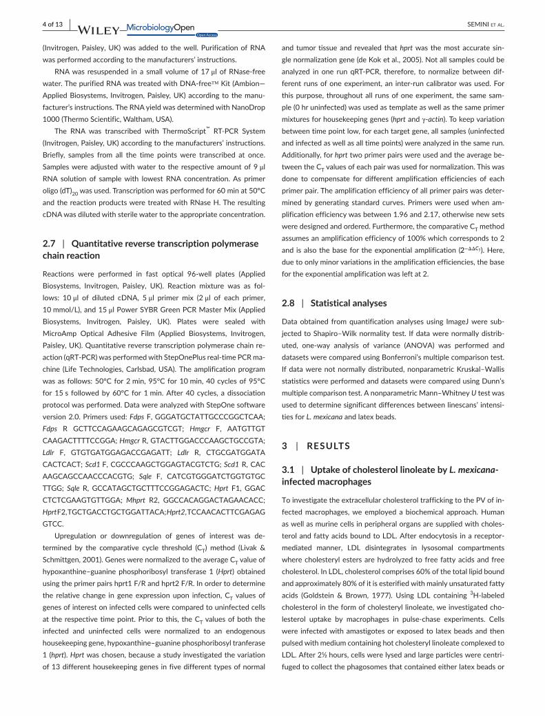
(Invitrogen,Paisley,UK)wasaddedtothewell.PurificationofRNAwasperformedaccordingtothemanufacturers’instructions.
RNAwas resuspended ina smallvolumeof17μl ofRNase-freewater.ThepurifiedRNAwastreatedwithDNA-free™Kit(Ambion—AppliedBiosystems, Invitrogen,Paisley,UK)accordingtothemanu-facturer’sinstructions.TheRNAyieldwasdeterminedwithNanoDrop1000(ThermoScientific,Waltham,USA).
The RNA was transcribed with ThermoScript™ RT-PCR System(Invitrogen,Paisley,UK)accordingtothemanufacturers’instructions.Briefly, samples from all the time pointswere transcribed at once.Sampleswereadjustedwithwater to the respectiveamountof9μl RNA solution of samplewith lowest RNA concentration.As primeroligo(dT)20wasused.Transcriptionwasperformedfor60minat50°CandthereactionproductsweretreatedwithRNaseH.TheresultingcDNAwasdilutedwithsterilewatertotheappropriateconcentration.
2.7 | Quantitative reverse transcription polymerase chain reaction
Reactions were performed in fast optical 96-well plates (AppliedBiosystems, Invitrogen, Paisley, UK). Reaction mixture was as fol-lows: 10 μl of diluted cDNA, 5μl primer mix (2μl of each primer,10mmol/L),and15μlPowerSYBRGreenPCRMasterMix(AppliedBiosystems, Invitrogen, Paisley, UK). Plates were sealed withMicroAmp Optical Adhesive Film (Applied Biosystems, Invitrogen,Paisley,UK).Quantitativereversetranscriptionpolymerasechainre-action(qRT-PCR)wasperformedwithStepOnePlusreal-timePCRma-chine (LifeTechnologies,Carlsbad,USA).Theamplificationprogramwasasfollows:50°Cfor2min,95°Cfor10min,40cyclesof95°Cfor15s followedby60°C for1min.After40cycles, adissociationprotocolwasperformed.DatawereanalyzedwithStepOnesoftwareversion2.0.Primersused:FdpsF,GGGATGCTATTGCCCGGCTCAA;Fdps R GCTTCCAGAAGCAGAGCGTCGT; Hmgcr F, AATGTTGT CAAGACTTTTCCGGA;HmgcrR,GTACTTGGACCCAAGCTGCCGTA;Ldlr F, GTGTGATGGAGACCGAGATT; Ldlr R, CTGCGATGGATA CACTCACT;Scd1F,CGCCCAAGCTGGAGTACGTCTG;Scd1R,CAC AAGCAGCCAACCCACGTG; Sqle F, CATCGTGGGATCTGGTGTGC TTGG; Sqle R, GCCATAGCTGCTTTCCGGAGACTC;Hprt F1, GGAC CTCTCGAAGTGTTGGA;Mhprt R2, GGCCACAGGACTAGAACACC;HprtF2,TGCTGACCTGCTGGATTACA;Hprt2,TCCAACACTTCGAGAG GTCC.
Upregulation or downregulation of genes of interest was de-termined by the comparative cycle threshold (CT) method (Livak &Schmittgen,2001).GeneswerenormalizedtotheaverageCTvalueofhypoxanthine–guaninephosphoribosyl transferase1 (Hprt)obtainedusingtheprimerpairshprt1F/Randhprt2F/R.Inordertodeterminethe relative change in geneexpressionupon infection,CTvaluesofgenesofinterestoninfectedcellswerecomparedtouninfectedcellsat therespectivetimepoint.Priortothis, theCTvaluesofboththeinfected and uninfected cells were normalized to an endogenoushousekeepinggene,hypoxanthine–guaninephosphoribosyltranferase1(hprt).Hprtwaschosen,becauseastudyinvestigatedthevariationof13differenthousekeepinggenesinfivedifferenttypesofnormal
and tumor tissueandrevealed thathprtwas themostaccuratesin-glenormalizationgene(deKoketal.,2005).Notallsamplescouldbeanalyzed in one run qRT-PCR, therefore, to normalize betweendif-ferentrunsofoneexperiment,an inter-runcalibratorwasused.Forthispurpose, throughoutall runsofoneexperiment, thesamesam-ple(0hruninfected)wasusedastemplateaswellasthesameprimermixturesforhousekeepinggenes(hprtandγ-actin).Tokeepvariationbetweentimepointlow,foreachtargetgene,allsamples(uninfectedandinfectedaswellasalltimepoints)wereanalyzedinthesamerun.Additionally,forhprttwoprimerpairswereusedandtheaveragebe-tweentheCTvaluesofeachpairwasusedfornormalization.Thiswasdone to compensate for different amplification efficiencies of eachprimerpair.Theamplificationefficiencyofallprimerpairswasdeter-minedbygenerating standardcurves.Primerswereusedwhenam-plificationefficiencywasbetween1.96and2.17,otherwisenewsetsweredesignedandordered.Furthermore,thecomparativeCT method assumesanamplificationefficiencyof100%whichcorrespondsto2and isalsothebasefortheexponentialamplification (2−ΔΔCT).Here,duetoonlyminorvariationsintheamplificationefficiencies,thebasefortheexponentialamplificationwasleftat2.
2.8 | Statistical analyses
Dataobtained fromquantificationanalysesusing ImageJwere sub-jectedtoShapiro–Wilknormalitytest. Ifdatawerenormallydistrib-uted, one-way analysis of variance (ANOVA) was performed anddatasetswerecomparedusingBonferroni’smultiplecomparisontest.Ifdatawerenotnormallydistributed,nonparametricKruskal–WallisstatisticswereperformedanddatasetswerecomparedusingDunn’smultiplecomparisontest.AnonparametricMann–WhitneyUtestwasusedtodeterminesignificantdifferencesbetweenlinescans’intensi-tiesforL. mexicanaandlatexbeads.
3 | RESULTS
3.1 | Uptake of cholesterol linoleate by L. mexicana- infected macrophages
ToinvestigatetheextracellularcholesteroltraffickingtothePVofin-fectedmacrophages,weemployedabiochemical approach.Humanaswellasmurinecellsinperipheralorgansaresuppliedwithcholes-terolandfattyacidsboundtoLDL.Afterendocytosis inareceptor-mediated manner, LDL disintegrates in lysosomal compartmentswherecholesterylestersarehydrolyzedtofreefattyacidsandfreecholesterol.InLDL,cholesterolcomprises60%ofthetotallipidboundandapproximately80%ofitisesterifiedwithmainlyunsaturatedfattyacids (Goldstein & Brown, 1977). Using LDL containing 3H-labeledcholesterolintheformofcholesteryllinoleate,weinvestigatedcho-lesterol uptake by macrophages in pulse-chase experiments. Cellswere infectedwithamastigotesorexposedto latexbeadsandthenpulsedwithmediumcontaininghotcholesteryllinoleatecomplexedtoLDL.After2½hours,cellswerelysedandlargeparticleswerecentri-fugedtocollectthephagosomesthatcontainedeitherlatexbeadsor

| 5 of 13SEMINI Et al.
parasites.Theradioactivityofthesupernatantandthepelletwasde-terminedseparatelyandtotalcell-associatedradioactivitywascalcu-latedandcorrelatedwithparticlepelletretainedactivity.Inthepelletfractionofuninfectedmacrophages,wemeasuredonlybackground-levelradioactivity,hencenocholesterolwassequesteredinpelletedmaterialofuninfectedcells.Macrophagesinfectedwithamastigotesorexposedto latexbeadsexhibitedproportionsofretainedcholes-terol in thepellet fractionof32%and13%, respectively.Thus, theamastigote-containing fraction of infected macrophages retainedmore than twice the amount of cholesterol than bead-containingphagosomes,andthisdifferencewasstatisticallysignificant(Table1,p=.011).
3.2 | Localization of Niemann–Pick C1 (NPC1) protein in L. mexicana- infected macrophages
Cholesterollevelsinmacrophagesarenormallyregulatedbycholes-terol uptake, biosynthesis and efflux. Tubulovesicular trafficking ofcholesteroloutof the lateendosome/early lysosome to theplasmamembraneand/ortotheendoplasmicreticulumismediatedatleastby two proteins: Niemann–Pick C1 (NPC1) andNPC2 (Blanchette-Mackie,2000;Neufeldetal.,1999).Ithasbeenproposedthatsolu-bleNPC2acceptscholesterolderivingfromLDLandtransportsittomembrane-boundNPC1forexport(Infanteetal.,2008;Kwonetal.,2009).Consequently,mutationsineitherNPC1orNPC2areresponsi-bleforanaccumulationofunesterifiedfreecholesterolinlateendoso-mal/lysosomalcompartments(Carsteaetal.,1997;Naureckieneetal.,2000).WeinvestigatedwhethercholesterolretentioninPVsmaytoa certain extent related to presence or absence of NPC1 protein.Analysesusing immunofluorescentstainingandconfocal laserscan-ningmicroscopy (CLSM) demonstrated thatNPC1 protein levels inbonemarrow-derivedmacrophagesinfectedwithL. mexicana were re-duced,whiletheproteinwaseasilydetectedatlatexbead-containingphagosomes (Figure1a). Representative linescans of macrophagesthatenclosedbothparasitesand latexbeadsdisplayedadecreasedNPC1 signal associatedwith the PVs compared to theNPC1 posi-tivebeads-containingphagosomes (Figure1b).QuantitativeanalysisshowedthatL. mexicana, incontrasttothelatexbeads,significantlydiminishedthecellularcontentofNPC1toabout35%,independentlyof thepresenceof latexbeads in thesamecell (Figure1c). Inaddi-tion,similarresultswerealsoobservedwhenonlytheregionthatim-mediatelysurroundedparasitesand/or latexbeadswasanalyzed. Incellscontainingparasitesorbeads,intensityoftheNPC1signalinthe
areaaroundtheparasiteswas37%lowercomparedtothesameareaaround the latexbeads (Figure1d). Incellscontainingparasitesandbeads, L. mexicana-dependent NPC1 depletion was notable mostlyaroundbothphagosometypes (Figure1d); incertaincells, affectingmorenotablyparasite-containingphagosomes(Figure1b).
3.3 | Fate of cellular free cholesterol in the presence of Leishmania parasites
As mentioned in the Introduction, previous studies indicate thatLeishmaniauptakebymacrophagesaswellasinfectionmaintenanceis dependent on serum and cellular cholesterol. Moreover, it hasbeendescribedthat infectionleadstodepletionofcholesterolfromthehostcellplasmamembrane,disturbing lipidraft-dependentpro-cesses(Chakrabortyetal.,2005;Rabhietal.,2012).Nevertheless,lit-tle isknownabout thesequenceofevents,distribution,andextentand kinetics of changes in free cholesterol thatmight occurwithinthe host cell in the course of Leishmania infection. For cholesterolstaining,weusedthepolyenefilipin.Althoughthisantibioticcanalsobind other β-hydroxysterols and lipids (e.g., GM1, sphingomyelin,phosphatidylcholine),itis—becauseofthescarcityofreagentsavail-able—commonlyusedtovisualizefreecholesteroldistributionincells.Usingthisreagent,wefirstinvestigatedthedistributionoffreecho-lesterolusingCLSM.Imageanalysisrevealedthatfilipinstainingac-cumulatedaroundL. mexicanaamastigoteswithinthePVofinfectedmacrophages.Thisaccumulationoflikelyfreecholesterolwasdetect-ableasahalothroughoutatimecourseof96hr(Figure2).Moreover,filipinsignalaccumulatedalready1hrafterinfectionsuggestingthatthisprocessbeginsveryearlyduringtheinfection(seefurtherdownand Figure 5b). The filipin halo was also detected in PVs of mac-rophagesinfectedwithL. majorpromastigotesunderthesameexperi-mentalconditions (FigureS1), indicatingthat thiseventoccurswithspeciesthat live incommunal (L. mexicana)aswellas individualPVs(L. major). In contrast, macrophages incubatedwith latex beads didnotshowanaugmentedfilipinaccumulationaroundthelatexbeadsintheirphagosomes(FigureS2).Thisresultisfurthersupportedinex-perimentswithmacrophagesthatweresimultaneouslyinfectedwithL. mexicana amastigotes and incubatedwith latexbeads (Figure3a).Filipin staining of axenically grown amastigotes and promastigotesalone did not exhibit the characteristic cholesterol halo around theparasite,indicatingthatthisprocessoccurredspecificallyinPVs(datanotshown).Wealsodeterminedtheextentofthefilipinsignalaroundboth,internalizedparasitesandlatexbeads.Representativelinescans
TABLE 1 Uptakeandretentionofcholesterolinuninfectedamastigotesorbeadsexposedmacrophages
Uninfected macrophagesMacrophages infected with amastigotes (MOI 5)
Macrophages exposed to beads (5 beads per cell)
Activityassociatedwithtotalcells(cpm) 1,135±195 1,199±32 1,570±325
Activityretainedbyparticlepellet(cpm) 33±18 386±81 209±55
Proportionofretainedcholesterolinpellet 0.03±0.01 0.32±0.07* 0.13±0.03
*Significantdifference,p=.011,incholesterolretentionincompartmentsharboringamastigotescomparedtobeads,96hrpostinfection(analyzedwithgenerallinearmodel,F1,4 = 20.13; n=3).

6 of 13 | SEMINI Et al.
F IGURE 1 Niemann–PickC1(NPC1)accumulationisreducedin Leishmania mexicana-infectedmacrophages.MacrophagesweresimultaneouslyinfectedwithaxenicL. mexicanaamastigotesexpressingDsRed(MOIof3)andexposedtoIgG-coatedlatexbeads(MOIof3).Threehoursafterinfection,macrophageswerefixed,blocked,andincubatedwithantibodiesrecognizingNPC1andLAMP-1(lysosomalmembraneproteinusedasamarkerofPVs).NucleiwerestainedwithDAPI.(a)Confocalimageoftworepresentativemacrophages,whicheitherexclusivelycontainL. mexicanaamastigotesorlatexbeads.(b)ConfocalimageofonerepresentativecellcontainingbothL. mexicanaamastigotesandlatexbeads.Arrowsandchartsoutlinedwiththerespectivecolorrefertolinescans(measuredwithVolocity3Dsoftware)ofadjacentparasiteandlatexbeads,inordertohighlightcolocalizationofNPC1andLAMP-1.(c)QuantificationofthecytosolicNPC1signalinmacrophagesusingImageJsoftware.ThemeanNPC1signalintensityofthewholemacrophagewasnormalizedremovingtheunspecificNPC1signalintensityandareaassociatedwithnucleus,parasitebody,andlatexbead.Fourgroupsweredefined:uninfectedmacrophages(U;n=32),macrophagescontaininglatexbeadsonly(B;n=28),macrophagescontainingparasitesonly(L;n=44),andmacrophagescontainingbothparasitesandbeads(B+L;n=16).Uninfectedmacrophagesweresetascontrolgroupforstatisticalanalysis(***p<.001,**p<.01).(d)QuantificationoftheNPC1signalintheouterringofparasitesandlatexbeads.ThemeanNPC1signalintensityoftheareathatimmediatelysurroundslatexbeadsandparasiteswithathicknessof0.25μmwascalculatedusingImageJsoftware.Threegroupsweredefined:intensityringoflatexbeadsfrommacrophageswithoutLeishmaniaparasites(B;n=32),intensityringoflatexbeadsfrommacrophagescontainingparasites(B+L;n=20),andintensityringofparasites(L;n=46).DataforB+LandLwerecomparedwithB(***p<.001).AllimageswereacquiredusingLeicaTCSSP5microscopewitha100×oil-immersionobjective
L. mexicanaNPC1Nucleus
10 µm
L. mexicana LAMP-1NPC1 Nucleus
(a)
(b)
(c) (d)
Ave
rage
Inte
nsity
0
10
20
30
40
50
60
70
Ave
rage
Inte
nsity
0
10
20
30
40
50
60
70
*** **
U B L B+L
Cellular NPC1
*** ***
B LB+L
NPC1 aroundBeads/Leishmania
F IGURE 2 FilipinaccumulationinmacrophagesuponLeishmania mexicana amastigotesinfection.MacrophageswereinfectedwithL. mexicanaamastigotesexpressingDsRed(MOIof5)for2hr.Distributionoffilipinwasanalyzedoveratimecourseof96hr,withtimepointsevery24hr.Cellswerefixedandsubsequentlystainedwithfilipin(white,excitedat405nm)andSYTO®13(nuclei,green,excitedat488nm).ArrowsindicatethepresenceofLeishmaniaparasites(red)and/orfilipinaroundtheparasites.Onerepresentativeofthreeindependentexperimentsisshown.AllimagesareconfocalandwereacquiredusingaZeissLSM780microscope,63×oil-immersionobjective,andprocessedusingidenticalconditions
Filipin
FilipinNucleiL. mexicana
20 µm
24 hr 48 hr 72 hr 96 hr
5 µm 5 µm

| 7 of 13SEMINI Et al.
displayedanincreasedfilipinsignalaroundtheL. mexicanaparasites,butnotaroundlatexbeads(Figure3b).Relativesignalintensitieswerethenquantifiedrevealingathreefoldenhancedaccumulationoffilipin-stainablecholesterolaroundLeishmaniaparasitescomparedto latexbeads(Figure3c).SignalaccumulationaroundinternalizedL. mexicana 24hrafterinfectionwasfurtheranalyzedbya3Dvisualizationofaseriesofimagesusingasurfacereconstructionmode(FigureS3A)andbyacross-sectionalprofileofaz-stack(FigureS3B),whichindicatedacoat-likedistributiondetectedasahalo in2Dprojections.Similarresultswere alsoobtained inL. mexicana-infectedmacrophages48,72,and96hrpostinfection(FigureS4–S6,respectively). Inaddition,2Dsectionswerequantifiedmeasuring the intensity (referred toasintegrateddensity)offilipin-stainablecholesterolaroundtheparasitesaswellasincorporatedbyhostcells.Thisanalysisshowedthatabout15%and44%ofthehostcellfilipinsignalbecomesassociatedwiththe parasites in infectedmacrophageswith low and high pathogen
load,respectively (Figure4a).Regressionanalysisofthesedatasug-gestedthatthemaximalamountofsignalsequesteredhadalimitandconsequentlytheamountperparasitewasafunctionofthenumberofparasitespercell,andwasnottimedependent(datanotshown).3Dconfocalmicroscopeimagestacks,whichwerethenprocessedfor2Drepresentation bymaximum intensity projection, further supportedthisinterpretation(Figure4b).Themostparsimoniousinterpretationofthesedataisthatfilipinsignalsrevealaccumulationoffreecholes-terolaroundintracellularparasitesthathappensearlyininfectionanddependingonparasiteloadsequesterssignificantamounts.
3.4 | Analysis of cholesterol distribution with fluorophore- labeled probes
InordertogainmoreinsightintocholesteroltraffickingtothePV,weconductedinvitroinfectionsofcellsinwhichthecholesterolpoolwas
F IGURE 3 DistributionandquantificationofthefilipinsignalaroundLeishmania mexicanaamastigotesandlatexbeads.MacrophagesweresimultaneouslyinfectedwithL. mexicanaamastigotes(MOIof5)andincubatedwithlatexbeads(fivepercell)for2hr.Distributionoffilipinwasanalyzed72hrafterinfection.Cellswerefixedandsubsequentlystainedwithfilipin(white,excitedat405nm)andSYTO®13(nuclei,green,excitedat488nm).Latexbeadsarevisualizedusingthetransmissionchannel.TheimagerepresentsaconfocalsectionacquiredusingaZeissLSM780microscope,63×oil-immersionobjective.(leftpanel)ArrowsindicatethepresenceLeishmaniaparasites(red,excitedat543nm)and/orfilipin-stainablefreecholesterolaroundtheparasites.Arrowheadsindicateselectedlatexbeadsincorporatedbymacrophages.Onerepresentativeofthreeindependentexperimentsisshown.(rightpaneltop)Redarrowsandchartsoutlinedinredrefertorepresentativelinescans(measuredwithZENsoftware)ofL. mexicanaparasitesphagocytosedbymacrophage.Arrowsandchartsoutlinedinbluerefertorepresentativelinescansoflatexbeadsincorporatedbymacrophages.Whitearrowsindicatesignalintensitiesforfilipin(whiteline)aroundlatexbeads(yellowline)andparasites(redline)thatwereusedtoquantifyintensitydifferencesinfilipinstaining.(rightpanelbottom)ForquantificationoffilipinsignalaroundinternalizedL. mexicanaamastigotesandlatexbeads,maximalsignalintensity(indicatedasmeanpeakvalueinarbitraryunits(AU)±SD,n=36alongsectionpaths)wasdeterminedusingZEN(blueedition)software.Backgroundvaluesweresubtractedfromtheabsolutevalues.Differencesbetweenparasitesandlatexbeadswerehighlysignificantp < .0001
L. mexicana amastigotes+ latex beads
72 h p.i.
Filipin signal intensity aroundL. mexicana parasites/ latex beads
L. mexicana Latex beads
AU
(Int
ensi
ty o
f fili
pin)
0
20
40
60
80
10 µm
Filipin
FilipinNucleiL. mexicana

8 of 13 | SEMINI Et al.
spikedwithfluorescentcholesterolprobes.Forthispurpose,weusedtwofluorophore-labeledcholesterols,NBD-andTopFluor(BODIPY)cholesterol,whicharewidelyusedforthestudyofcholesteroltraf-fickinganddistributioninlivingcells(Maxfield&Wustner,2012).
Macrophages were preincubated with NBD- or TopFluor-cholesterolandtheninfectedwithL. mexicanaamastigotesandfixedat different times postinfection. Figure5a shows that 72hr postin-fection, both fluorescent cholesterol formswere present in the PVbut,surprisingly,didnotformahaloaroundtheparasitesratherbe-came incorporated into parasite membrane compartments. Doublecholesterol stainingusing filipinandNBD-cholesterol indicated thatthecholesterolhalowasalreadypresent1hrpostinfectionandthatfluorophore-labeled cholesterol did indeed not contribute at a de-tectableleveltoitsformation(Figure5b).Moreover,thepresenceofNBD-andTopFluor-cholesteroldidnot interferewith thecourseofthe infection (FigureS7AandS7B, respectively).Thus, these resultsdemonstrate that cholesterol is delivered by two distinct pathwaysand isdifferentiallyaccumulatedbytheparasitessuggestingcholes-terolsourcespecificityforthegenerationofthehalo.
As carried out for filipin-stainable cholesterol,we quantified theamountofNBD-cholesterol takenupby theparasitesduring the in-fection.Weobserved that about 10%of totalNBD-cholesterolwasretained by a single parasite, whereas parasites in highly infectedmacrophages sequestered 37% of this fluorescent cholesterol form(Figure5c).Asdemonstratedforfilipinstaining,weidentifiedthattheamountofNBD-cholesterolassociatedwiththeparasitesisafunction
ofthenumberofparasitesinternalizedbythehostcell(Figure5c),show-ingasimilartrendoveralltimepoints(datanotshown).QuantificationofTopFluor-cholesterolwasnotpossible,becauselipiddropletslabeledbytheTopFluor-dyearelocalizedtooclosetotheparasites(observa-tionreminiscentofthestudyofLecoeur,Giraud,Prevost,Milon,&Lang,2013,where dendritic cells have been infectedwith L. amazonensis)andwould falsify thecalculationofcholesterol sequestration (FigureS7B). Incorporation of NBD-cholesterol by L. mexicana expressing aDsRedtransgene16hrafterinfectionhasalsobeenconfirmedbya3Dvisualizationofaseriesofimagesusingsurfacereconstruction(FigureS8A).Thecross-sectionalprofileofaz-stackevidencedanenhancedDsRed-NBDcolocalizationwithintheparasiteandtheabsenceoftheNBD-cholesterolaroundtheparasites(FigureS8B).
3.5 | Effect of infection on transcription of genes regulating lipid biosynthesis and metabolism
Because internalized parasites are assumed to reside in the “foodchain” of thehost cell itself and are therefore able to sequester atleastapartofthefattyacidsthathavebeenproposedtofueltheirenergymetabolism(Coombs,Craft,&Hart,1982;Ginger,2006;Hart&Coombs,1982),weaimedtofurtherinvestigatetheconsequencesof this adaptive strategy on the lipid metabolism of infected mac-rophages.Itiscommonlyknownthatseveraltranscriptionfactorsareactivatedeitherbythepresence(PPAR-γ,LXR-α;Desvergne&Wahli,1999; Edwards, Kennedy, & Mak, 2002; Tontonoz & Spiegelman,
F IGURE 4 Amountofsequesteredfilipinperparasiteisafunctionofthenumberofparasitespercell.Relativefilipinsignalof(a)confocal2Dsectionsand(b)3Dconfocalmicroscopeimagestackprocessedina2Dprojectionbymaximumintensityprojectionisrepresentedasafunctionofthenumberofparasitesinternalizedbythemacrophagesoveraperiodof96hrpostinfection.TheexponentialcurveregressionswerecalculatedusingGraphPadPrism
Filipin-stainable cholesterolassociated with all L. mexicana
parasites
Filipin-stainable cholesterolassociated with all L. mexicana
parasites
Filipin-stainable cholesterolper L. mexicana parasite
Filipin-stainable cholesterolper L. mexicana parasite
Number of Parasites per Macrophage
% F
ilipi
n-si
gnal
0 5 10 15 20 25 300
2
4
6
8
10
Number of Parasites per Macrophage
% F
ilipi
n-si
gnal
0 5 10 15 20 25 300
10
20
30
40
50
60
Number of Parasites per Macrophage
% F
ilipi
n-si
gnal
0 2 4 6 8 10 12 140
2
4
6
8
10
Number of Parasites per Macrophage
% F
ilipi
n-si
gnal
0 2 4 6 8 10 12 140
10
20
30
40
50
(a)
(b)

| 9 of 13SEMINI Et al.
2008)orabsence(sterolregulatoryelement-bindingproteinSREBP;Ikonen, 2008) of particular lipids. Thus, we looked at the possibleeffectsofLeishmania infectiononhostcell lipidhomeostasisbyan-alyzingtherelativechangeinthemRNAoftargetgenesofthesetran-scriptionfactors.
WefoundthatgenesregulatedbyPPAR-γandLXR-αareessen-tiallynotaffectedbyinfection(datanotshown).Incontrast,genesin-volvedincholesterolsynthesis(Fdps,Hmgcr,Scd1,andSqle)underthetranscriptionalcontrolofSREBPwereupregulated24–48hrpostinfec-tion(Figure6).TheLDLreceptor(Ldlr),alsoregulatedbySREBP,and
involvedinLDLuptake,wasnotconsistentlyupregulatedthroughoutthecourseofaninfection.However,wedidobserveincreasedlevelsofLdlrmRNAat24and96hrpostinfectionandatthe72hrtimepoint,theexpressionofthisgenewasincreasedmorethantwofold(Figure6).
4 | DISCUSSION
In the present work, we investigated the fate of cholesterol inmacrophages infected with Leishmania parasites and observed a
F IGURE 5 Fluorophore-labeledcholesteroldoesnotaccumulatearoundtheparasite.(a)MacrophageswerepreincubatedwithNBD-orTopFluor-cholesterolfor16hr.Afterward,macrophageswereinfectedwithLeishmania mexicanaamastigotes(MOIof5)for2hr.Seventy-twohoursafterinfection,cellswerefixedandsubsequentlystainedwithDAPI(nuclei,excitedat405nm).BothNBDandTopFluorgroupswereexcitedat488nm.(b)Distributionoffilipin-stainableandfluorophore-labeledcholesterolintheearlyphasesofinfectionwithL. mexicana. Mousebonemarrow-derivedmacrophageswerepreincubatedwithNBD-cholesterolfor16hrandtheninfectedwithL. mexicanaamastigotes(MOIof5)for2hr.Anhourafterinfection,cellswerefixedandsubsequentlystainedwithfilipin(excitedat405nm).Forabetteridentification,filipin-stainedcholesterolinthepresenceofNBD-cholesterolisvisualizedinpurple.ArrowsindicateLeishmaniaparasitesand/orfilipinaroundtheparasites.AllimagesareconfocalandwereacquiredusingaZeissLSM780microscope,63×oil-immersionobjective,andprocessedusingidenticalconditions.(c)RelativefluorescencecorrespondingtoNBD-cholesterolincorporatedbyalltheparasites(leftpanel)orasingleparasite(rightpanel)inaninfectedmacrophageisrepresentedasafunctionofthenumberofparasitesinternalizedbythemacrophagesoveraperiodof48hrpostinfection.TheexponentialcurveregressionswerecalculatedusingGraphPadPrism
NBD-cholesterol TopFluor-cholesterol
20 µm 10 µm
NBD-cholesterol
NBD-cholesterolNucleiL. mexicana
TopFluor-cholesterol
TopFluor-cholesterolNucleiL. mexicana
FilipinL. mexicana
5 µm10 µm
NBD-cholesterolL. mexicana
5 µm
5 µm
NBD-cholesterolFilipin
1 h p.i.
NBD-cholesterol associated withall L. mexicana parasites
NBD-cholesterolper L. mexicana parasite
Number of parasites per macrophage
% N
BD
-cho
lest
erol
0 2 4 6 8 10 12 140
10
20
30
40
50
60
Number of parasites per macrophage
% N
BD
-cho
lest
erol
0 2 4 6 8 10 12 1402468
101214161820
(a)
(c)
(b)

10 of 13 | SEMINI Et al.
cholesterol redistributionof cellular cholesterol.Basedon the find-ings,weproposeincreasedretentionofserum-derivedcholesterolinthe PV during the leishmanial infection as a consequence or causeofreducedlevelsofNPC1,aproteinimplicatedincholesteroleffluxfrom late endosomes and lysosomes. Retained cholesterol concen-tratesaroundparasitesformingakindofcoatwhosefunctionhasyettobeelucidated.Moreover,parasitesincorporatehostcellmembra-neouscholesterol.Thistwo-waycholesterolsequestrationcanaffectsignificantproportionsthatbecomeregisteredbythehostcell’scho-lesterolhomeostasismechanismswhichincludetriggeringofSREBP-mediatedtranscriptionofkeycholesterolbiosynthesisgenes.
Cholesterolisamajorconstituentofeukaryoticmembranesandisimplicatedinseveralcellularprocessessuchasmembraneorganizationanddynamics,signaltransduction,andmolecularsorting.Itisknownthat serum levels correlatewith distinct occurrence of disease. Forinstance, hypercholesterolemia protects from,whereas hypocholes-terolemiapromotes, infectionofmacrophagesbyL. donovani (Ghoshetal.,2012).Recently,ithasbeendocumentedthatlowserumcholes-terolinmicewithvisceralleishmaniasisisassociatedwithdownregu-lationofmiR-122throughthedegradationofitsprocessor,Dicer1,bythemetalloproteasegp63,which inhibitscholesterolbiosynthesis ininfectedmouseliver(Ghoshetal.,2013).Inaddition,lipoproteinlipasegenepolymorphismisassociatedwithhighlevelsoftriglyceridesandvery low-density lipoproteins (VLDL) and low levels of high-densitylipoprotein(HDL),whichfavorthedevelopmentofvisceralleishmani-asis(Carvalhoetal.,2014).
At the cellular level, cholesterol-enriched membrane micro-domains termed lipid rafts seemessential inLeishmania uptake andinfection (Majumder etal., 2012; Pucadyil etal., 2004). Cholesteroldepletion from the plasmamembrane noted after Leishmania infec-tion has been proposed to represent an immune evasion strategyused by the intracellular parasites. Lipid rafts-dependent processes
suchasMHC–antigencomplexformationonthesurfaceofantigen-presenting cells which is required for a productive T cell response(Anderson,Hiltbold,&Roche,2000;Burack,Lee,Holdorf,Dustin,&Shaw, 2002) and the function of raft-associatedmembrane-locatedreceptors,suchas IFNreceptors (Rubetal.,2009;Senetal.,2011),would thereby be impaired and intracellular parasite survivalwouldbemorelikely.
TrypanasomatidsincludingLeishmaniaamastigotescannotsynthe-sizecholesteroldenovo(Robertsetal.,2003).Cholesteroldetectedintheparasitesmustbeobtainedfromtheirhostcellorenvironment.Itisofnotethatinlesionderivedamastigotes,thecontentofcholesterolrelatedtothetotalamountoffreesterolsincreases(Tetley,Coombs,&Vickerman,1986)raisingfrom7%to39%whencomparedtopromas-tigotes(Berman,Goad,Beach,&Holz,1986;Ginger,Chance,Sadler,&Goad,2001).Recently, it hasbeendemonstrated thatLeishmania parasitesareabletoacquiresignificantamountsofcholesterolfromLDLparticleendocytosis(Andrade-Netoetal.,2011;DeCiccoetal.,2012).Thisstrategyhasalsobeenillustratedforotherprotozoanpara-sitessuchasToxoplasma gondii,Trypanosoma cruzi,andCryptosporidium parvum,which indicate that cholesterol iswidely important for theintracellular survival and replication of pathogens (Coppens, Sinai,& Joiner, 2000; Ehrenman,Wanyiri, Bhat,Ward, & Coppens, 2013;Johndrowetal.,2014;Pereiraetal.,2011;Portugaletal.,2008).Ourexperiments using LDL containing 3H-labeled cholesteryl linoleatesuggestthatLeishmaniaamastigote-infectedcellsdonotprimarilyin-creasetheoverallcellularuptakeofcholesterol,butratherincreasedretentionofcholesterolintheparasite-containingsubcellularfractionisobservedwhencomparedtoasimilarlypreparedfractioncontaininglatexbeads.Theabsence,exclusion,orinhibitionofproteinsthatnor-mally regulatecholesterolefflux fromsuchcompartmentsasshownhereforNPC-1offersaparsimoniousexplanationforthisobservation.Thisdifferssubstantiallyfromthehypothesis(Rabhietal.,2012)thatLeishmaniamightinduceaccumulationofcellularcholesterolbydown-regulatingexpressionofmembersofthelipideffluxmachinery,suchas the ATP-binding cassette transporter A1 (ABCA1), as illustratedforL. major-infectedcells,whichwecouldnotconfirm(D.Paape&T.Aebischer,unpubl.data).
AlthoughtheaforementionedstudiesprovideteleologicalsupportforabeneficialroleofsequestrationofserumandplasmamembranecholesterolduringLeishmania infection inorder toprotect thepara-sites from the host cell’s antileishmanial activity, the chronology ofredistributionandaccumulationofcholesterolafterinfectionhasnotbeen investigated. InL. major-infectedmacrophages, ithasbeen re-portedthatcholesterol is localizedinthecytosolasfreecholesteroland/oresterifiedwithfattyacidsandstoredinlipiddropletsincloseproximity to PV, suggesting that lipid droplets act as energy repos-itories for internalized amastigotes (Rabhi etal., 2012). The lattermechanismhasalsobeenproposed fordendritic cells infectedwithL. amazonensis(Lecoeuretal.,2013).
Withafocusontheparasite,ourworksuggeststhatLeishmania in-fectionleadstothesequestrationandaccumulationoffilipin-stainablefreecholesterolaroundtheparasitesformingacoatwhichisalreadydetectableintheearlyphasesoftheinfection.Thisphenotypeappears
F IGURE 6 GenesinvolvedincholesterolsynthesisareupregulateduponLeishmania mexicanaamastigotesinfection.RelativemRNAlevelchangesbetweeninfectedanduninfectedbonemarrow-derivedmacrophagesofgenesregulatedbySREBP.Avalueof1correspondstonochangeingeneexpression.Dottedlinesshoweithertwofoldup-ordownregulation.Fdps,farnesyldiphosphatesynthase;Hmgcr,3-hydroxy-3-methylglutaryl-CoAreductase;Ldlr,low-densitylipoproteinreceptor;Scd1,stearoyl-CoenzymeAdesaturase1,Sqle:squaleneepoxidase
SREBP target genes
0 hr
24 h
r48
hr
72 h
r96
hr
0 hr
24 h
r48
hr
72 h
r96
hr
0 hr
24 h
r48
hr
72 h
r96
hr
0 hr
24 h
r48
hr
72 h
r96
hr
0 hr
24 h
r48
hr
72 h
r96
hr
0.1
1
102–
∆∆C
T
Fdps Hmgcr Ldlr Scd1 Sqle

| 11 of 13SEMINI Et al.
tobecommonsinceitwasobservedwithboth,NewWorld(L. mexi-cana)andOldWorld(L. major)species,despitetheirsignificantlydif-ferentPVs. In infectionswithToxoplasma gondii andCryptosporidium parvum,LDL-derivedcholesterolhasbeenproposedtobecomelocal-ized in parasitemembranes, but filipin staining suggested a surfaceaccumulationof freecholesterolalso in thesecases (Coppensetal.,2000; Ehrenman etal., 2013). Because analyses with NBD- andTopFluor-cholesterolprobes,whichprincipallybecomeincludedinthecytoplasmaandplasmamembranelipidrafts,respectively(McIntoshetal.,2008),aretraffickedtothePVandincorporatedintoparasites,butareapparentlynotaccumulatingaroundtheparasite,wesuggestthatthehalo ispredominantlyconstitutedbythepartofexogenouslikelyserum-derivedcholesterolthatistraffickeddirectlyintothePVs.Nevertheless,furtheranalysesarerequiredtoconfirmthishypothesis.
The semiquantitative analysis of cholesterol sequestration re-vealed a general correlation of parasite number per cell and extentofsequestration.Afindingthatisanimportantadditiontopreviouslypublishedworks.Onlyathigherparasiteloadsdo~25%ofbothhostcell total filipin-stainable freecholesteroland totalNBD-cholesterolbecome associatedwith parasiteswithmaximal values of 50% ob-servable.ThesearevaluesintherangesreportedforL. majorthatinexperimentswith anMOIof 10diminished the cholesterol contentinmacrophagesderivedfromBALB/cmicebyaboutone-third72hrafter infection and this correlated with altered CD40 signalosomecompositionandfunction(Rubetal.,2009).Similarly,inmacrophagesderivedfromC57BL/6miceandinfectedwithL. donovaniatanMOIof10,thetotalhostcellmembranecholesterolbecamedecreasedbyalmost50%48hrpostinfection(Ghoshetal.,2012).Thesamepara-sitespeciesusedagainatanMOIof10reducedplasmacellmembranecholesterolreportedlybytwothirdsinRAW264.7macrophages,8hrpostinfection.ThiswasaccompaniedbydownregulationoftheIFN-γ receptor signaling in these host cells (Sen etal., 2011).Thus,whilecholesterolhalo formation thatcovers theparasite is fastand in itsextentperparasitemostpronouncedatlowparasiteload,cholesterolsequestrationfromthehostcellspointofviewbecomessizableandfunctionally relevantonlyathigherparasitenumbers.Therefore,weliketospeculatethathaloformationservesadifferentpurposethatisnotyetknown.
The obvious consequence of cholesterol retention and depriva-tionreachingfunctionallyrelevantlevelswouldbetheattemptofthehostcelltocounteractthistrendandtorestoretheinitialcholesterollevels.AsreportedinourworkwithL. mexicana,upregulationoftheexpressionof genes related to the lipidmetabolismand cholesterolbiosynthesis has been also shown in other studies conductedwithL. major andL. amazonensis inmacrophagesofBALB/cmice (OsorioyFortea etal., 2009;Rabhi etal., 2012).However,whether this bi-ologicalresponseisuniversalneedsfurtherstudies,since,forexam-ple,indendriticleukocytesL. amazonensisdidnotinfluence(Lecoeuretal.,2013),whereasinmacrophagesderivedfromhumanlunglym-phoblasts L. viannia braziliensis even lead to downregulation of theexpressionofgenesbelongingtosteroidandsterolbiosyntheticpro-cesses (Ovalle-Bracho, Franco-Munoz, Londono-Barbosa, Restrepo-Montoya,&Clavijo-Ramirez,2015).
Insummary,ourstudyshowsanoveldynamiccholesterolseques-trationpatternbyLeishmaniaspp. intheparasitophorousvacuoleofhostcellmacrophagesandidentifieschangestotraffickingofatleasttwopoolsofcellularcholesterolasitsmechanisticbasis.Quantitativeanalysesrevealedthatcholesterolsequestrationisdependentonthenumber of parasites per cell, but is time independent. This findingsuggeststhatLeishmanianeedtoreacharelativelyhigh intracellularnumbertoaffectthehostcell’scholesterolhomeostasistoadegreethatimpairsitsimmunesignalingfunctions.Incontrast,theamountofcholesterolsequesteredperparasiteismostpronouncedatlownum-berof intracellular amastigotes.Thismight indicate a response to ayettobeelucidatedselectionpressureencounteredduringevolution.
ACKNOWLEDGMENTS
WethankPaulaMacGregorforadviceonthestatisticalanalysisofthecholesterollinoleateretentionandSamuelDeanforhelpfulcommentsonthemanuscript.
CONFLICT OF INTEREST
Nonedeclared.
REFERENCES
Anderson,H.A.,Hiltbold,E.M.,&Roche,P.A. (2000).ConcentrationofMHCclass IImolecules in lipid rafts facilitates antigenpresentation.Nature Immunology,1,156–162.
Andrade-Neto,V.V.,Cicco,N.N.,Cunha-Junior, E. F.,Canto-Cavalheiro,M.M.,Atella,G.C.,&Torres-Santos,E.C.(2011).Thepharmacologicalinhibitionof sterolbiosynthesis inLeishmania is counteractedbyen-hancementofLDLendocytosis.Acta Tropica,119,194–198.
Antoine,J.C.,Prina,E.,Jouanne,C.,&Bongrand,P.(1990).Parasitophorousvacuoles of Leishmania amazonensis-infected macrophages maintainanacidicpH.Infection and Immunity,58,779–787.
Bansal,D.,Bhatti,H.S.,&Sehgal,R.(2005).Roleofcholesterolinparasiticinfections.Lipids in Health and Disease,4,10.
Berman,J.D.,Goad,L.J.,Beach,D.H.,&Holz,G.G.Jr. (1986).Effectsof ketoconazoleon sterol biosynthesis byLeishmania mexicana mexi-cana amastigotes in murine macrophage tumor cells.Molecular and Biochemical Parasitology,20,85–92.
Blanchette-Mackie,E.J.(2000).Intracellularcholesteroltrafficking:RoleoftheNPC1protein.Biochimica et Biophysica Acta,1486,171–183.
Burack,W.R.,Lee,K.H.,Holdorf,A.D.,Dustin,M.L.,&Shaw,A.S.(2002).Cuttingedge:Quantitativeimagingofraftaccumulationintheimmu-nologicalsynapse.The Journal of Immunology,169,2837–2841.
Carstea, E. D., Morris, J. A., Coleman, K. G., Loftus, S. K., Zhang, D.,Cummings,C.,…Tagle,D.A. (1997).Niemann-PickC1diseasegene:Homology to mediators of cholesterol homeostasis. Science, 277, 228–231.
Carvalho,M. D.,Alonso, D. P.,Vendrame, C.M., Costa, D. L., Costa, C.H.,Werneck,G. L.,…Goto,H. (2014). Lipoprotein lipase and PPARalpha gene polymorphisms, increased very-low-density lipoproteinlevels, and decreased high-density lipoprotein levels as riskmarkersforthedevelopmentofvisceral leishmaniasisbyLeishmania infantum. Mediators of Inflammation,2014,230129.
Chakraborty,D., Banerjee, S., Sen,A., Banerjee,K.K.,Das, P.,&Roy, S.(2005). Leishmania donovani affects antigen presentation of mac-rophage by disrupting lipid rafts. The Journal of Immunology, 175,3214–3224.

12 of 13 | SEMINI Et al.
Chattopadhyay, A., & Jafurulla, M. (2011). A novel mechanism for anold drug:Amphotericin B in the treatment of visceral leishmaniasis.Biochemical and Biophysical Research Communications,416,7–12.
Collins,H.L.,Schaible,U.E.,Ernst,J.D.,&Russell,D.G.(1997).TransferofphagocytosedparticlestotheparasitophorousvacuoleofLeishmania mexicana isatransientphenomenonprecedingtheacquisitionofan-nexinIbythephagosome.Journal of Cell Science,110,191–200.
Coombs,G.H.,Craft,J.A.,&Hart,D.T. (1982).AcomparativestudyofLeishmania mexicana amastigotes and promastigotes. Enzyme activi-ties and subcellular locations.Molecular and Biochemical Parasitology,5,199–211.
Coppens,I.,Sinai,A.P.,&Joiner,K.A.(2000).Toxoplasmagondiiexploitshost low-density lipoprotein receptor-mediatedendocytosis for cho-lesterolacquisition.Journal of Cell Biology,149,167–180.
DeCicco,N.N.,Pereira,M.G.,Correa,J.R.,Andrade-Neto,V.V.,Saraiva,F.B.,Chagas-Lima,A.C.,…Atella,G.C.(2012).LDLuptakebyLeishmania amazonensis: Involvement of membrane lipid microdomains.Experimental Parasitology,130,330–340.
Desvergne,B.,&Wahli,W.(1999).Peroxisomeproliferator-activatedrecep-tors:Nuclearcontrolofmetabolism.Endocrine Reviews,20,649–688.
Duclos,S.,Diez,R.,Garin,J.,Papadopoulou,B.,Descoteaux,A.,Stenmark,H.,&Desjardins,M.(2000).Rab5regulatesthekissandrunfusionbe-tweenphagosomesandendosomesandtheacquisitionofphagosomeleishmanicidalproperties inRAW264.7macrophages. Journal of Cell Science,113,3531–3541.
Edwards, P. A., Kennedy, M. A., & Mak, P. A. (2002). LXRs; oxysterol-activated nuclear receptors that regulate genes controlling lipid ho-meostasis.Vascular Pharmacology,38,249–256.
Ehrenman,K.,Wanyiri,J.W.,Bhat,N.,Ward,H.D.,&Coppens,I.(2013).CryptosporidiumparvumscavengesLDL-derivedcholesterol andmi-cellar cholesterol internalized into enterocytes.Cellular Microbiology,15,1182–1197.
Fernandes,L.R.,Ribeiro,A.C.,Segatto,M.,Santos,L.F.,Amaral,J.,Portugal,L. R., & Leite, J. I. (2013). Leishmaniamajor self-limited infection in-creasesbloodcholesterolandpromotesatherosclerosisdevelopment.Cholesterol,2013,754580.
Ghosh, J., Bose, M., Roy, S., & Bhattacharyya, S. N. (2013). Leishmania donovani targetsDicer1todownregulatemiR-122, lowerserumcho-lesterol, and facilitatemurine liver infection.Cell Host & Microbe,13, 277–288.
Ghosh,J.,Das,S.,Guha,R.,Ghosh,D.,Naskar,K.,Das,A.,&Roy,S.(2012).Hyperlipidemia offers protection against Leishmania donovani in-fection: Role ofmembrane cholesterol. Journal of Lipid Research,53,2560–2572.
Ghosh,J.,Lal,C.S.,Pandey,K.,Das,V.N.,Das,P.,Roychoudhury,K.,&Roy,S.(2011).Humanvisceralleishmaniasis:Decreaseinserumcholesterolasa functionofsplenicparasite load.Annals of Tropical Medicine and Parasitology,105,267–271.
Ginger,M.L.(2006).Nichemetabolisminparasiticprotozoa.Philosophical Transactions of the Royal Society of London. Series B, Biological sciences,361,101–118.
Ginger,M.L.,Chance,M.L.,Sadler,I.H.,&Goad,L.J.(2001).Thebiosyn-thetic incorporationof the intact leucine skeleton into sterol by thetrypanosomatid Leishmania mexicana. Journal of Biological Chemistry,276,11674–11682.
Goldstein,J.L.,&Brown,M.S.(1977).Thelow-densitylipoproteinpathwayand its relation toatherosclerosis.Annual Review of Biochemistry,46,897–930.
Groener,J. E., Pelton,R.W.,&Kostner,G.M. (1986). Improvedestima-tionofcholesterylestertransfer/exchangeactivityinserumorplasma.Clinical Chemistry,32,283–286.
Hamid,P.H.,Hirzmann,J.,Kerner,K.,Gimpl,G.,Lochnit,G.,Hermosilla,C.R.,&Taubert,A.(2015).Eimeriabovisinfectionmodulatesendothelialhostcellcholesterolmetabolismforsuccessful replication.Veterinary Research,46,100.
Hart,D.T.,&Coombs,G.H.(1982).Leishmania mexicana:Energymetab-olismofamastigotesandpromastigotes.Experimental Parasitology,54,397–409.
Ikonen, E. (2008). Cellular cholesterol trafficking and compartmentaliza-tion. Nature Reviews Molecular Cell Biology,9,125–138.
Infante,R.E.,Wang,M.L.,Radhakrishnan,A.,Kwon,H.J.,Brown,M.S.,&Goldstein,J.L.(2008).NPC2facilitatesbidirectionaltransferofcholes-terolbetweenNPC1andlipidbilayers,astepincholesterolegressfromlysosomes. Proceedings of the National Academy of Sciences of the United States of America,105,15287–15292.
Johndrow,C.,Nelson,R.,Tanowitz,H.,Weiss,L.M.,&Nagajyothi,F.(2014).Trypanosomacruziinfectionresultsinanincreaseinintracellularcho-lesterol. Microbes and Infection,16,337–344.
Kima, P. E. (2007). The amastigote forms of Leishmania are experts atexploiting host cell processes to establish infection and persist.International Journal for Parasitology,37,1087–1096.
Kima, P.E., Bonilla, J.A., Cho, E., Ndjamen, B., Canton, J., Leal, N., &Handfield,M., (2010). IdentificationofLeishmania proteinspreferen-tiallyreleasedininfectedcellsusingchangemediatedantigentechnol-ogy(CMAT).PLoS Neglected Tropical Diseases,4:e842.
deKok, J. B., Roelofs, R.W.,Giesendorf, B.A., Pennings, J. L.,Waas, E.T.,Feuth,T.,…Span,P.N. (2005).Normalizationofgeneexpressionmeasurementsintumortissues:Comparisonof13endogenouscontrolgenes. Laboratory Investigation,85,154–159.
Kwon,H.J.,Abi-Mosleh,L.,Wang,M.L.,Deisenhofer,J.,Goldstein,J.L.,Brown,M.S.,&Infante,R.E.(2009).StructureofN-terminaldomainofNPC1revealsdistinctsubdomainsforbindingandtransferofcholes-terol. Cell,137,1213–1224.
Lal, C. S., Kumar,A., Kumar, S., Pandey,K., Kumar,N., Bimal, S.,…Das,P.(2007).Hypocholesterolemiaandincreasedtriglycerideinpediatricvisceralleishmaniasis.Clinica Chimica Acta,382,151–153.
Lecoeur, H., Giraud, E., Prevost, M. C., Milon, G., & Lang, T. (2013).Reprogramming neutral lipid metabolism in mouse dendritic leuco-cytes hosting live Leishmaniaamazonensisamastigotes.PLoS Neglected Tropical Diseases,7,e2276.
Lippuner,C.,Paape,D.,Paterou,A.,Brand,J.,Richardson,M.,Smith,A.J.,etal.(2009).Real-timeimagingofLeishmania mexicana-infectedearlyphagosomes: A study using primary macrophages generated fromgreen fluorescent protein-Rab5 transgenic mice. FASEB Journal, 23,483–491.
Livak,K.J.,&Schmittgen,T.D. (2001).Analysisof relativegeneexpres-siondatausingreal-timequantitativePCRandthe2(-DeltaDeltaC(T))Method. Methods,25,402–408.
Majumder,S.,Dey,R.,Bhattacharjee,S.,Rub,A.,Gupta,G.,BhattacharyyaMajumdar,S.,…Majumdar,S.(2012).Leishmania-inducedbiphasicce-ramidegenerationinmacrophagesiscrucialforuptakeandsurvivaloftheparasite.Journal of Infectious Diseases,205,1607–1616.
Maxfield, F. R., &Wustner,D. (2012).Analysis of cholesterol traffickingwithfluorescentprobes.Methods in Cell Biology,108,367–393.
McIntosh, A.L., Huang, H., Atshaves, B.P., Storey, S.M., Gallegos, A.M., &Spencer,T.A.,…Schroeder,F.(2008).Fluorescentsterolsforthestudyofcholesteroltraffickinginlivingcells.In:L.W.Miller(Ed.),Probes and tags to study biomolecular function: For proteins, RNA, and membranes(pp.1–25).Wiley.
Misslitz,A.,Mottram,J.C.,Overath, P.,&Aebischer,T. (2000).TargetedintegrationintoarRNAlocusresultsinuniformandhighlevelexpres-sionoftransgenesinLeishmaniaamastigotes.Molecular and Biochemical Parasitology,107,251–261.
Naureckiene, S., Sleat, D. E., Lackland, H., Fensom, A., Vanier, M. T.,Wattiaux,R.,…Lobel,P. (2000). IdentificationofHE1as thesecondgeneofNiemann-PickCdisease.Science,290,2298–2301.
Neufeld,E.B.,Wastney,M.,Patel,S.,Suresh,S.,Cooney,A.M.,Dwyer,N.K.,…Blanchette-Mackie,E.J.(1999).TheNiemann-PickC1proteinre-sidesinavesicularcompartmentlinkedtoretrogradetransportofmul-tiplelysosomalcargo.Journal of Biological Chemistry,274,9627–9635.

| 13 of 13SEMINI Et al.
Osorioy Fortea, J., de La Llave, E., Regnault, B.,Coppee,J.Y.,Milon,G.,Lang,T.,&Prina,E.(2009).TranscriptionalsignaturesofBALB/cmousemacrophages housing multiplying Leishmania amazonensis amastig-otes. BMC Genomics,10,119.
Ovalle-Bracho, C., Franco-Munoz, C., Londono-Barbosa, D., Restrepo-Montoya, D., & Clavijo-Ramirez, C. (2015). Changes in macrophagegeneexpressionassociatedwithLeishmania(Viannia)braziliensisinfec-tion. PLoS ONE,10,e0128934.
Paape,D.,Barrios-Llerena,M.E.,LeBihan,T.,Mackay,L.,&Aebischer,T.(2010). Gel free analysis of the proteome of intracellular Leishmania mexicana. Molecular and Biochemical Parasitology,169,108–114.
Paape, D., Lippuner, C., Schmid, M., Ackermann, R., Barrios-Llerena, M.E., Zimny-Arndt, U., … Aebischer, T. (2008). Transgenic, fluorescentLeishmania mexicanaallowdirectanalysisoftheproteomeofintracel-lularamastigotes.Molecular & Cellular Proteomics: MCP,7,1688–1701.
Pereira,M.G.,Nakayasu,E.S.,Sant’Anna,C.,DeCicco,N.N.,Atella,G.C.,&deSouza,W.,…Cunha-e-Silva,N.(2011).Trypanosomacruziepimas-tigotesareabletostoreandmobilizehighamountsofcholesterol inreservosomelipidinclusions.PLoS ONE,6,e22359.
Portugal, L. R., Fernandes, L. R., Pietra Pedroso, V. S., Santiago, H. C.,Gazzinelli,R.T.,&Alvarez-Leite,J. I. (2008). Influenceof low-densitylipoprotein(LDL)receptoronlipidcomposition,inflammationandpar-asitismduringToxoplasmagondiiinfection.Microbes and Infection,10,276–284.
Pucadyil, T. J., Tewary, P., Madhubala, R., & Chattopadhyay, A. (2004).CholesterolisrequiredforLeishmania donovaniinfection:Implicationsin leishmaniasis. Molecular and Biochemical Parasitology, 133, 145– 152.
Rabhi, I.,Rabhi,S.,Ben-Othman,R.,Rasche,A.,Daskalaki,A.,Trentin,B.,…Guizani-Tabbane,L. (2012).Transcriptomic signatureofLeishmania infectedmicemacrophages:Ametabolicpointofview.PLoS Neglected Tropical Diseases,6,e1763.
Redgrave,T.G.,Roberts,D.C.,&West,C.E.(1975).Separationofplasmalipoproteinsbydensity-gradientultracentrifugation.Anal Biochem,65,42–49.
Roberts,C.W.,McLeod,R.,Rice,D.W.,Ginger,M.,Chance,M.L.,&Goad,L. J. (2003). Fatty acid and sterol metabolism: Potential antimicro-bial targets in apicomplexan and trypanosomatid parasitic protozoa.Molecular and Biochemical Parasitology,126,129–142.
Rosenzweig, D., Smith, D.,Myler, P. J., Olafson, R.W., & Zilberstein, D.(2008). Post-translational modification of cellular proteins duringLeishmania donovanidifferentiation.Proteomics,8,1843–1850.
Rub,A.,Dey,R.,Jadhav,M.,Kamat,R.,Chakkaramakkil,S.,Majumdar,S.,… Saha, B. (2009). Cholesterol depletion associatedwith Leishmania majorinfectionaltersmacrophageCD40signalosomecompositionandeffectorfunction.Nature Immunology,10,273–280.
Russell,D.G.,Xu,S.,&Chakraborty,P.(1992).IntracellulartraffickingandtheparasitophorousvacuoleofLeishmania mexicana-infectedmacro-phages.Journal of Cell Science,103,1193–1210.
Saunders,E.C.,Ng,W.W.,Kloehn,J.,Chambers,J.M.,Ng,M.,&McConville,M.J.(2014).InductionofastringentmetabolicresponseinintracellularstagesofLeishmaniamexicanaleadstoincreaseddependenceonmi-tochondrialmetabolism.PLoS Pathog,10,e1003888.
Sen, S., Roy, K., Mukherjee, S., Mukhopadhyay, R., & Roy, S. (2011).RestorationofIFNgammaRsubunitassembly,IFNgammasignalingandparasiteclearanceinLeishmania donovaniinfectedmacrophages:Roleofmembranecholesterol.PLoS Pathogens,7,e1002229.
Shamshiev,A.T.,Ampenberger,F.,Ernst,B.,Rohrer,L.,Marsland,B.J.,&Kopf,M. (2007).Dyslipidemia inhibitsToll-likereceptor-inducedacti-vationofCD8alpha-negativedendritic cells andprotectiveTh1 typeimmunity.Journal of Experimental Medicine,204,441–452.
Soares,N.M.,Leal,T.F.,Fiuza,M.C.,Reis,E.A.,Souza,M.A.,Dos-Santos,W.L.,&Pontes-de-Carvalho,L.(2010).Plasmalipoproteinsinvisceralleishmaniasis and their effect on Leishmania-infected macrophages.Parasite Immunology,32,259–266.
Sorensen,M.,Lippuner,C.,Kaiser,T.,Misslitz,A.,Aebischer,T.,&Bumann,D. (2003). Rapidly maturing red fluorescent protein variants withstronglyenhancedbrightnessinbacteria.FEBS Letters,552,110–114.
Tetley, L., Coombs, G. H., & Vickerman, K. (1986). The surface mem-brane of Leishmania mexicana mexicana: Comparison of amastigoteand promastigote using freeze-fracture cytochemistry. Zeitschrift für Parasitenkunde,72,281–292.
Tewary,P.,Veena,K.,Pucadyil,T.J.,Chattopadhyay,A.,&Madhubala,R.(2006). The sterol-binding antibiotic nystatin inhibits entry of non-opsonized Leishmania donovani into macrophages. Biochemical and Biophysical Research Communications,339,661–666.
Tontonoz,P.,&Spiegelman,B.M.(2008).Fatandbeyond:Thediversebiol-ogyofPPARgamma.Annual Review of Biochemistry,77,289–312.
Weinheber, N., Wolfram, M., Harbecke, D., & Aebischer, T. (1998).Phagocytosis of Leishmania mexicana amastigotes by macrophagesleadstoasustainedsuppressionofIL-12production.European Journal of Immunology,28,2467–2477.
SUPPORTING INFORMATION
Additional Supporting Information may be found online in thesupportinginformationtabforthisarticle.
How to cite this article:SeminiG,PaapeD,PaterouA,SchroederJ,Barrios-LlerenaM,AebischerT.ChangestocholesteroltraffickinginmacrophagesbyLeishmania parasitesinfection.MicrobiologyOpen. 2017;00:e469. http://doi.org/10.1002/mbo3.469