Embed Size (px)
Citation preview
CASE REPORT
Leiomyoma of the third eyelid in a dog
Rachel L. Mathes,* Sarah J. Noble† and Angela E. Ellis‡*MedVet Medical and Cancer Centers for Pets, Cincinnati, OH, USA; †Portland Veterinary Specialists, Portland, ME, USA; and ‡College of VeterinaryMedicine, Athens Veterinary Diagnostic Laboratory, University of Georgia, Athens, GA, USA
Address communications to:
R.L. Mathes
Tel.: (513) 561 0069Fax: (614) 846 5803e-mail: [email protected]
AbstractA 14-year-old neutered male Dachshund presented for the evaluation of oculus dexter(OD) third eyelid elevation ongoing for approximately 2 months. Complete oph-thalmic examination revealed a large, nonpainful, well-demarcated, soft mass at thebase of the right third eyelid causing elevation and mild hyperemia. The mass wasfreely moveable with the third eyelid, and no right globe deviation was noted. Noother abnormalities were noted on physical examination, routine blood chemistry,complete blood count, serum T4, urinalysis, or urine cortisol/creatinine ratio. OcularB-mode ultrasonography showed an anechoic, well-demarcated, homogenous, soft tis-sue mass at the base of the third eyelid with no orbital extension. A leiomyoma wasdiagnosed after multiple punch biopsies were obtained from the palpebral surface ofthe mass. The right third eyelid was excised surgically. Histopathology confirmed acompletely excised, nodular, unencapsulated, expansile mass within the third eyelid.Positive smooth muscle actin and negative S-100 immunohistochemistry confirmed aleiomyoma. Bundles of normal smooth muscle were also present adjacent to the mass.The mass was compressing the adjacent lacrimal gland and associated with moderatedacryoadenitis. Twelve months postoperatively, the right globe position and motilityremain normal with no evidence of mass regrowth. To the author’s knowledge, this isthe first reported case of a leiomyoma of the third eyelid in any species. In this case,the mass was completely excised and no regrowth has occurred twelve months aftersurgery. This case along with independently reviewed canine third eyelids clearlydemonstrates the presence of smooth muscle within the canine third eyelid.
Key Words: canine, eyelid leiomyoma, leiomyoma, smooth muscle, third eyelid
INTRODUCTION
Canine leiomyomas are benign tumors arising fromsmooth muscle and are most commonly found in the gas-trointestinal tract.1–7 These tumors may also occur in avariety of other locations including the heart,8 urinary sys-tem,9–12 uterus,13 mesosalpinx,14 vagina,15,16 trachea,17 andnasopharynx.18 Angioleiomyomas, uncommonly reportedin dogs, arise from vascular smooth muscle and may occurin a variety of locations. Although leiomyomas are benign,they may cause serious disease secondary to compressionor displacement of adjacent abdominal or alimentarystructures.1,2,5,6 Leiomyomas may also produce insulin-likegrowth factors that cause hypoglycemia in affected caninepatients.19–21 In spite of their relative commonness in
other locations in dogs, there is only one report of acanine intraocular leiomyoma, arising from the iris in adog.22
In the human literature, there are multiple reports ofocular or periocular leiomyomas arising from the orbit,23
eyelid,24,25 conjunctiva,26 iris,27–29 ciliary body,30–33 andchoroid.34,35 Ocular leiomyomas are considered rare36,37
as these case reports are very sporadic throughout the lit-erature. Additionally, a 1994 study revealed that many ear-lier intraocular ‘leiomyomas’ were misdiagnosed;immunohistochemistry on previous cases confirmed manyof these tumors to be melanocytic lesions.38 Thus, ocularleiomyomas may be even rarer than the literature wouldsuggest. Mesectodermal leiomyomas are benign intraocu-lar tumors arising from the ciliary body epithelium, a cell
© 2015 American College of Veterinary Ophthalmologists
Veterinary Ophthalmology (2016) 19, 4, 347–354 DOI:10.1111/vop.12305
of neural crest origin.39 Although routine histopathologymay be highly suggestive of a leiomyoma, a definitivediagnosis of leiomyoma can only be made with immuno-histochemistry, as the clinical and microscopic appearanceof these tumors may be similar to other tumors such asmelanocytic neoplasia, gastrointestinal stromal tumors(GISTs), and other spindle cell tumors.2,3,38 This was alsohighlighted in a 2007 study of canine small intestinal andcecal tumors that demonstrated distinct immunohisto-chemical differences between gastrointestinal smooth mus-cle and stromal tumors, resulting in many tumors beingreclassified.40
The herein present case report describes a primarycanine third eyelid leiomyoma diagnosed with histopathol-ogy and immunohistochemistry. To the authors’ knowl-edge, this is the first report of a primary third eyelidleiomyoma in any species. The mass was surgically excisedwith no regrowth reported twelve months postoperatively.This case report also definitively demonstrates the presenceof native smooth muscle within the canine third eyelid, thepresence of which has previously been indeterminate.
CLINICAL REPORT
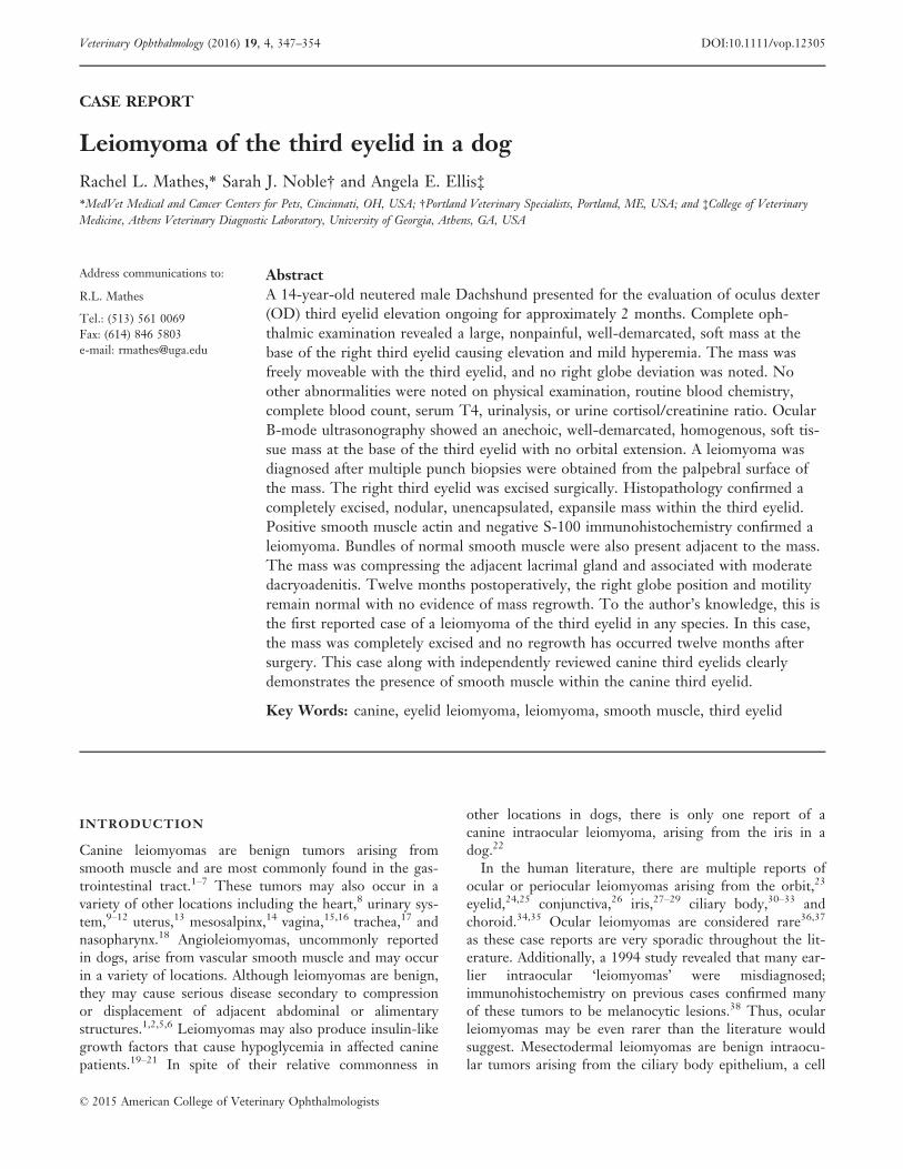
Ophthalmic examinationA fourteen-year-old neutered male short-haired Dachs-hund presented to a private referral hospital’s Ophthal-mology Service for the evaluation of recurrent cornealulceration oculus uterque (OU), blepharospasm OU, andwhite corneal opacities OU. Full ophthalmic examinationwith slit-lamp biomicroscopy (Kowa SL-15; Kowa Opti-med, Torrance, CA, USA) and indirect ophthalmoscopy(Keeler Vantage Plus; Dan Scott and Associates, Wester-ville, OH, USA) performed by a board-certified veterinaryophthalmologist (RM) revealed mild enophthalmos OU,mild intermittent blepharospasm OU, mild-to-moderatehyperemia OU, paraxial ventrotemporal dense mid toanterior corneal stromal lipid opacifications OU and mul-tifocal wispy arborizing anterior stromal limbal-based cor-neal vascularization temporally and ventrotemporallyextending variable distances into the corneal stroma OU.A large, nonpainful, well-demarcated, soft mass at the baseof the right third eyelid causing third eyelid elevation wasalso identified (Fig. 1). Save for the previously mentionedmild OU enophthalmos, there was no OD globe deviationor strabismus. There was normal ocular motility and anotherwise normal neuro-ophthalmic examination. Themass was freely movable within the right third eyelid.Schirmer tear test values were 18 mm/min OD and15 mm/min OS (Schirmer Tear Test; Merck and Com-pany, Inc., Whitehouse Station, NJ, USA), and intraocularpressures were 8 mmHg OD and 8 mmHg OS measuredby applanation tonometry (Tonopen XL; Reichert Tech-nologies, Depew, NY, USA). The tear film breakup time(TBUT), measured as previously described,41 wasdecreased in both eyes (3 s OD, 5 s OS), consistent with a
qualitative tear deficiency. There was no corneal or con-junctival fluorescein stain retention OU. The patient wasvisual with a normal intraocular examination OU.
Based on the ophthalmic examination, diagnoses of OUqualitative tear deficiency, OU corneal lipidosis, OU mildkeratitis, and OD third eyelid mass were made. Recom-mendations were made for systemic workup to identifyany underlying systemic factors contributing to the cor-neal lipidosis along with an ocular ultrasound and biopsiesof the third eyelid mass. Topical therapy was institutedwith 0.2% cyclosporine (small strip OU BID; Optim-mune, Merck, White House Station, NJ, USA) and anartificial lacrimomimetic (1 drop OU BID; Refresh Cellu-visc, Allergan, Irvine, CA, USA) for the qualitative teardeficiency.
DiagnosticsSystemic diagnostics including routine blood chemistry,complete blood count, serum triglycerides, serum T4, andurinalysis were within normal limits. A urine cortisol/crea-tinine ratio was 6, which is consistent with normal adrenalfunction. Based on these diagnostics, no further investiga-tion of adrenal or thyroid function was performed.
The patient presented again for ocular ultrasound andbiopsies of the third eyelid mass. Ophthalmic examinationrevealed marked improvement in the patient’s overall
(a)
(b)
Figure 1. Front (a) and side (b) photographs of the patient at initialpresentation are depicted. There is elevation of the third eyelid ODand a ventral periocular mass effect. The position and motility of theright globe were normal.
© 2015 American College of Veterinary Ophthalmologists, Veterinary Ophthalmology, 19, 347–354
348 MATH E S E T A L .
comfort. The hyperemia and enophthalmos had resolvedOU. The limbal-based corneal vascularization hadresolved, and the tear film breakup time (TBUT) was nor-mal in both eyes (>15 s OU). No other changes in theophthalmic examination were noted. The right third eye-lid mass appeared unchanged.
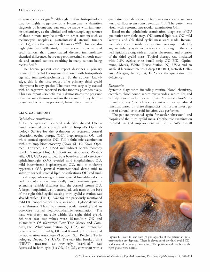
The patient was sedated with dexmedetomidine(375 mcg/m2 IV; Dexdomitor, Zoetis, Florham Park, NJ,USA) and butorphanol (0.4 mg/kg IV; Torbugesic, FortDodge, Fort Dodge, IA, USA). After sedation, ocularultrasound imaging (10 mHz UBM probe; Aviso, QuantellMedical, Bozeman, MT, USA) was obtained and showedan anechoic to mildly hypo-echoic, well-demarcated,homogenous, soft tissue mass at the base of the third eye-lid with no orbital extension (Fig. 2). Based on the ultra-sound and readily accessible location for tissue sampling,biopsies of the mass were pursued. After routine surgicalpreparation, local anesthetic was injected subconjunctivallyon the palpebral surface of the third eyelid overlying themass with 2% injectable lidocaine (0.15 mL subconjuncti-val; MWI, Boise, ID, USA). Several 4-mm punch biopsies(disposable biopsy punch; Miltex, York, PA, USA) wereobtained from the palpebral surface of the third eyelid inthe body of the firm, intrapalpebral mass and submittedfor histopathologic examination. The conjunctival defectwas closed with simple interrupted 6-0 vicryl (Ethicon;Johnson and Johnson, New Brunswick, NJ, USA) sutures.The patient’s dexmedetomidine sedation was then reversedusing atipamezole (375 mcg/m2 IM; Antisedan, Zoetis,Florham Park, NJ, USA). The patient recovered routinelyand was discharged with topical diclofenac (1 drop Q12 hOD; Nexus Pharmaceuticals, Vernon Hills, IL, USA) andtopical Optimmune as previously prescribed.
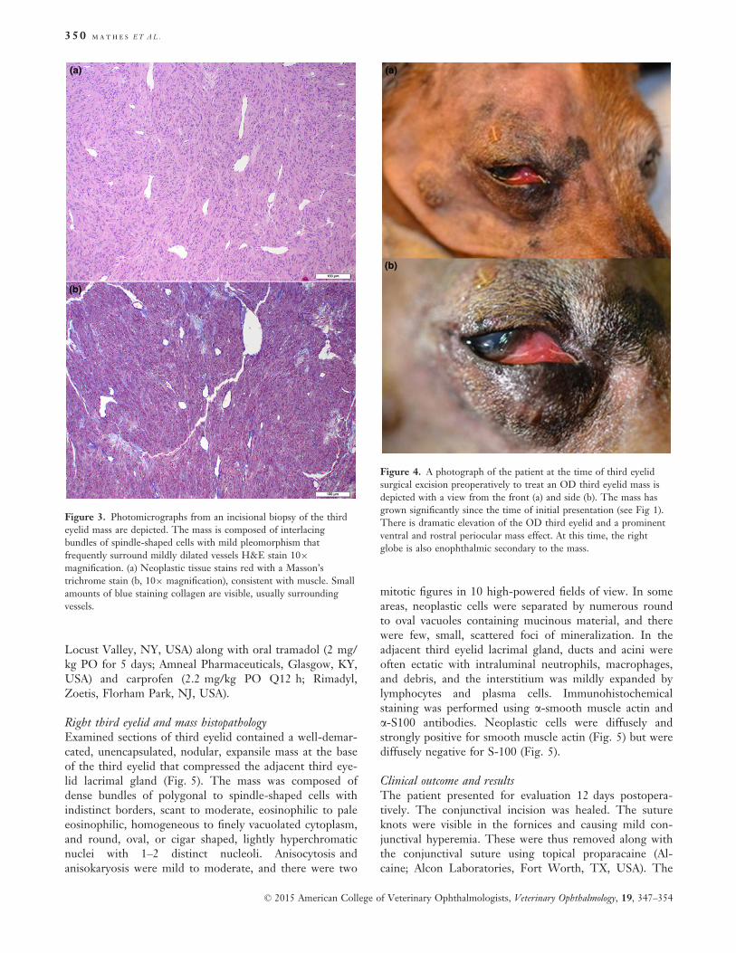
Third eyelid biopsy histopathologyExamined sections contained sections of a mass that werecomposed of interlacing bundles and fascicles of spindle-shaped to polygonal cells with indistinct borders andscant, clear to eosinophilic cytoplasm that extended awayfrom the nucleus in delicate streams (Fig. 3). Nuclei wereround to oval with finely stippled chromatin and 1-2 dis-tinct nucleoli. Anisocytosis and anisokaryosis were mild.The mass also had numerous, mildly dilated, occasionallybranching vessels lined by flattened to slightly plumpendothelial cells. A Masson’s trichrome stain was per-formed, and staining was consistent with muscle (Fig. 3).A diagnosis of leiomyoma was made; however, an angi-oleiomyoma could not be excluded.
Right third eyelid excisionBased on the histopathology results and location of themass, recommendations were made for surgical excision ofthe right third eyelid with histopathologic evaluation. Thepatient was premedicated with acepromazine (0.02 mg/kgIM; Fort Dodge, Fort Dodge, IA, USA) and hydromor-phone (0.1 mg/kg IM; West Ward, Eatontown, NJ, USA)prior to general anesthesia. Anesthetic induction wasachieved with propofol (4 mg/kg IV; Propoflo, AbbottAnimal Health, North Chicago, IL, USA) to effect. Gen-eral anesthesia was maintained after endotracheal intuba-tion with inhalant isoflurane 1–2% in oxygen (Isoflo;Abbott Animal Health, North Chicago, IL, USA). Afterendotracheal intubation and anesthesia maintenance, cefa-zolin (22 mg/kg IV; WG Critical Care LLC, Paramus,NJ, USA) and carprofen (2.2 mg/kg SQ; Rimadyl, Zoetis,Florham Park, NJ, USA) were then administered. Thepatient was positioned in sternal recumbency, and theright eye was prepped routinely for surgery (Fig. 4). Priorto excision, an encircling conjunctival incision was madeat the base of the third eyelid with a #15 blade. Metzen-baum scissors were used to undermine the bulbar andpalpebral conjunctiva on either side of the third eyelidwithin the fornices to provide a section of conjunctivaavailable for surgical closure after third eyelid excision.The right third eyelid was excised at the base as previouslydescribed42 and submitted for histopathology. The con-junctiva was closed with simple continuous 6-0 vicryl(Ethicon; Johnson and Johnson, New Brunswick, NJ,USA) sutures using with care to place the knots deepwithin the dorsonasal and ventrotemporal fornices oneither end of the incision to avoid contact with thecornea.
The patient recovered from general anesthesia rou-tinely. Postoperatively, the patient received hydromor-phone (0.05 mg/kg IV Q6 h). The patient was dischargedwith topical OD diclofenac (1 drop OD Q8 h), Optim-mune (small strip OU Q12 h) neomycin–polymyxin–gramicidin (1 drop OD Q8 h; Neopolygram, Bausch andLomb, Inc., Tampa, FL, USA), and artificial tear ointment(small strip OD Q6 h; Puralube, Fera Pharmaceuticals,
Figure 2. A representative ultrasonographic image of the patient’sthird eyelid is shown. The globe is positioned toward the top of thephotograph (asterisk), and the mass is depicted by the arrow. Thesoft tissue density mass is well-demarcated, homogenous, andanechoic to mildly hypo-echoic. The mass was freely moveable withthe third eyelid and showed no further orbital extension orinvolvement.
© 2015 American College of Veterinary Ophthalmologists, Veterinary Ophthalmology, 19, 347–354
C AN I N E TH I R D E Y E L I D L E I OMYOMA 349
Locust Valley, NY, USA) along with oral tramadol (2 mg/kg PO for 5 days; Amneal Pharmaceuticals, Glasgow, KY,USA) and carprofen (2.2 mg/kg PO Q12 h; Rimadyl,Zoetis, Florham Park, NJ, USA).
Right third eyelid and mass histopathologyExamined sections of third eyelid contained a well-demar-cated, unencapsulated, nodular, expansile mass at the baseof the third eyelid that compressed the adjacent third eye-lid lacrimal gland (Fig. 5). The mass was composed ofdense bundles of polygonal to spindle-shaped cells withindistinct borders, scant to moderate, eosinophilic to paleeosinophilic, homogeneous to finely vacuolated cytoplasm,and round, oval, or cigar shaped, lightly hyperchromaticnuclei with 1–2 distinct nucleoli. Anisocytosis andanisokaryosis were mild to moderate, and there were two
mitotic figures in 10 high-powered fields of view. In someareas, neoplastic cells were separated by numerous roundto oval vacuoles containing mucinous material, and therewere few, small, scattered foci of mineralization. In theadjacent third eyelid lacrimal gland, ducts and acini wereoften ectatic with intraluminal neutrophils, macrophages,and debris, and the interstitium was mildly expanded bylymphocytes and plasma cells. Immunohistochemicalstaining was performed using a-smooth muscle actin anda-S100 antibodies. Neoplastic cells were diffusely andstrongly positive for smooth muscle actin (Fig. 5) but werediffusely negative for S-100 (Fig. 5).
Clinical outcome and resultsThe patient presented for evaluation 12 days postopera-tively. The conjunctival incision was healed. The sutureknots were visible in the fornices and causing mild con-junctival hyperemia. These were thus removed along withthe conjunctival suture using topical proparacaine (Al-caine; Alcon Laboratories, Fort Worth, TX, USA). The
(a)
(b)
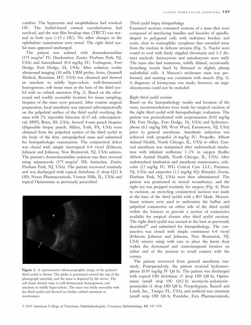
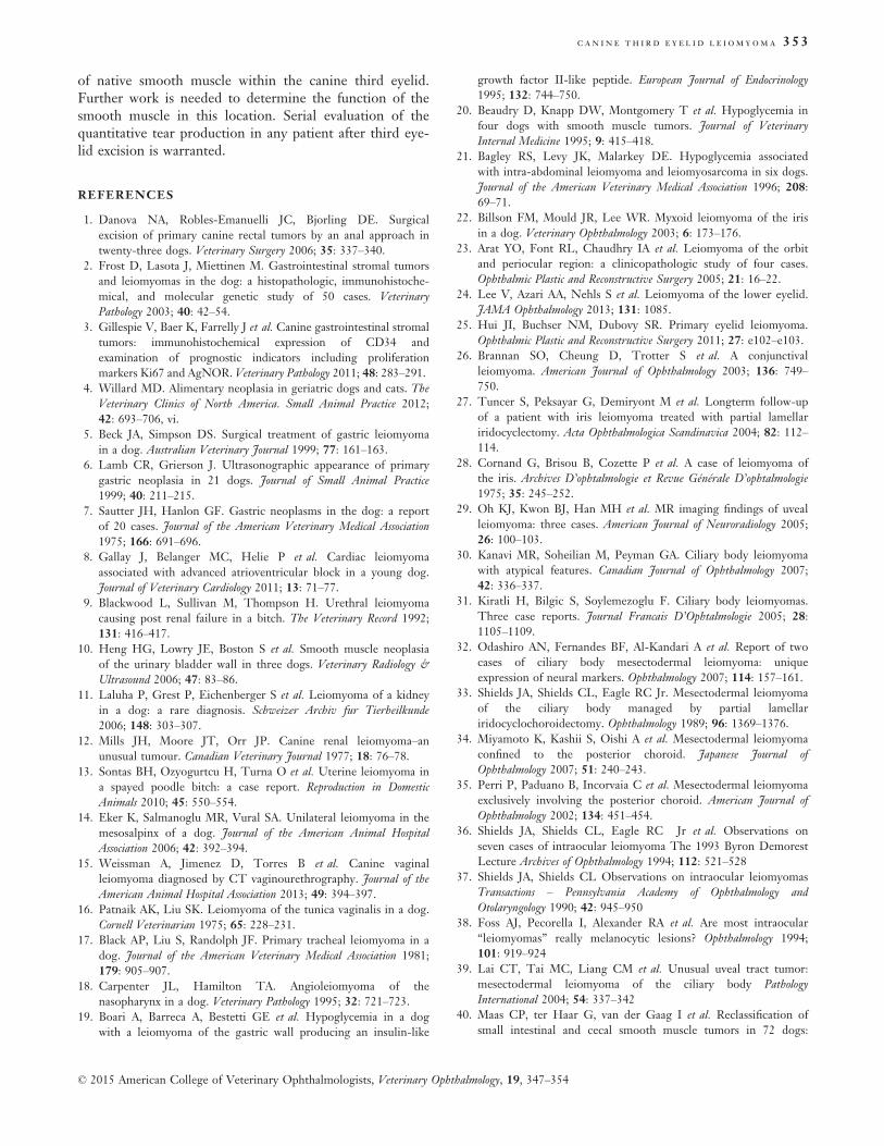
Figure 4. A photograph of the patient at the time of third eyelidsurgical excision preoperatively to treat an OD third eyelid mass isdepicted with a view from the front (a) and side (b). The mass hasgrown significantly since the time of initial presentation (see Fig 1).There is dramatic elevation of the OD third eyelid and a prominentventral and rostral periocular mass effect. At this time, the rightglobe is also enophthalmic secondary to the mass.
(a)
(b)
Figure 3. Photomicrographs from an incisional biopsy of the thirdeyelid mass are depicted. The mass is composed of interlacingbundles of spindle-shaped cells with mild pleomorphism thatfrequently surround mildly dilated vessels H&E stain 109magnification. (a) Neoplastic tissue stains red with a Masson’strichrome stain (b, 109 magnification), consistent with muscle. Smallamounts of blue staining collagen are visible, usually surroundingvessels.
© 2015 American College of Veterinary Ophthalmologists, Veterinary Ophthalmology, 19, 347–354
350 MATH E S E T A L .

ophthalmic examination was otherwise unremarkable,excepting the previously described small lipid cornealopacities. The Schirmer tear test values were 24 mm/minOD and 17 mm/min OS (Schirmer Tear Test; Merck andCompany, Inc., Whitehouse Station, NJ, USA), andintraocular pressures were 14 mmHg OD and 13 mmHgOS measured by applanation tonometry (Tonopen XL;Reichert Technologies, Depew, NY, USA). Recommenda-tions were made for long-term topical Optimmune (smallstrip OU Q24 h). Artificial tear ointment OD was recom-mended for 2 weeks with instructions to discontinue afterthat. All other medications were discontinued.
The patient presented for evaluation 3 months aftersurgery (Fig. 6). The right conjunctival surfaces were nor-mal with the absence of the right third eyelid. Except forthe previously described corneal lipidosis, the ophthalmicexamination was normal. Schirmer tear test values were17 mm/min OD and 22 mm/min OS (Schirmer TearTest; Merck and Company, Inc., Whitehouse Station, NJ,USA). No corneal ulceration or episodes of discomforthad occurred since surgery.
Telephone conversation with the owners 12 monthsafter surgery revealed that there had been no change inthe patient’s general or ocular condition or appearance.No clinical signs of keratoconjunctivitis sicca had beennoted as the owners described bright, clear, comfortableeyes in the patient. The owners had continued the Optim-mune therapy OU Q24 h and were still administering themedication.
Histology of selected canine third eyelidsSeveral sections of separate canine third eyelids wereexamined histologically to evaluate for the presence ofnative smooth muscle. Strips of normal smooth musclewere consistently noted within the canine third eyelid.
There were variations among patients and sections as tothe amount of smooth muscle present (Fig. 7), but it wasconsistently noted on canine third eyelid histology.
DISCUSSION
The described case herein is the first reported leiomyomaof the third eyelid in any species, to the authors’ knowl-edge. This tumor was diagnosed based on histopathologyand immunohistochemistry. As has been previously docu-mented,3,38,40 it is very important to perform immunohis-tochemistry on suspected leiomyomas as these tumors mayhistologically appear similar to other tumors. In our case,the tumor displayed clear histologic characteristics ofmesenchymal origin. The tumor exhibited polygonal to
(a) (b)
(c) (d)
Figure 5. Photomicrographs from the excisedthird eyelid mass are depicted. The mass is welldemarcated and is composed of densely packed,interlacing bundles of spindle-shaped cells. Asmall bundle of normal smooth muscle is evidentat the base of the tumor (arrow) H&E stain (a,29 magnification). Neoplastic cells are spindleshaped and are arranged in relatively well-organized bundles and fascicles. Anisocytosis andanisokaryosis are mild, and mitoses are rare.Nuclei are round, oval, or cigar shaped andlightly hyperchromatic. Occasional cells haveclear or lightly basophilic vacuoles H&E stain (b,109 magnification). Immunohistochemistry usinganti-smooth muscle actin antibody (c, 409magnification). Neoplastic cells exhibit strongpositive staining Inset: positive controlImmunohistochemistry using anti-S100 antibody(d, 109 magnification). Neoplastic cells arediffusely negative for S100 Inset: Positive control.
Figure 6. A photograph of the patient 3 months after third eyelidexcision OD is depicted. The right globe position and motility arenormal with normal facial symmetry. Both eyes are comfortable witha normal pre-ocular tear film.
© 2015 American College of Veterinary Ophthalmologists, Veterinary Ophthalmology, 19, 347–354
C AN I N E TH I R D E Y E L I D L E I OMYOMA 351

spindle cell shape, low mitotic index, scant intercellularconnective tissue matrix, and an intermittent ‘whorling’cell pattern, which may be histologic features of othertypes of tumors. Because this tumor was not locatedwithin the gastrointestinal tract, a gastrointestinal stromaltumor, which may appear similar to a leiomyoma, was notconsidered. Peripheral nerve sheath tumors and melanocy-tic neoplasia have been reported within the globe and inthe facial region in dogs;43–46 therefore, these tumors wereconsidered important differential diagnoses. In this case,smooth muscle actin (SMA) staining was positive andS-100 staining was negative, clearly differentiating thetumor as a leiomyoma. The initial biopsy samples showedfrequent, prominent, often dilated blood vessels within thetumor; thus, an angioleiomyoma was initially considered.However, once the tumor was available in its entirety forreview, an angioleiomyoma was ruled out based on a his-tologic lack of similarly prominent vasculature, and aleiomyoma was diagnosed.
Leiomyomas are benign tumors and, as such, do notmetastasize, but may be locally expansive and can becomevery large. In dogs, these tumors are most commonlyfound in the GI tract and are one of the most commonGI tumors.2–4,40,47 In the GI tract, these tumors causemorbidity related to adjacent structure involvement;effects are usually due to compression or obstruction. Inour case, the leiomyoma caused third eyelid elevation, butwas found primarily as an incidental finding. This patientpresented to the ophthalmologist for clinical signs of hisqualitative tear deficiency. By the time the mass wasremoved, it had grown to a large enough size that thefacial appearance of the patient was altered and the thirdeyelid was significantly elevated (Fig. 4). Likely, had themass continued to grow and this been the only clinicalocular abnormality, the patient would have been presentedto the ophthalmologist for these changes. Because themass was limited to the third eyelid, removal of the thirdeyelid in its entirety was warranted. In retrospect, excisionof the mass without removal of the third eyelid could hasbeen attempted. However, this mass was quite extensiveand it is unlikely that both complete excision of the massand preservation of the third eyelid anatomy would have
been achieved. A primary mass of the third eyelid is oneof the rare indications for complete removal of this struc-ture as removal of the associated lacrimal gland is consid-ered detrimental.48 In our case, close monitoring of thequantitative tear production was considered importantduring follow-up. In addition, the possibility of clinicalkeratoconjunctivitis sicca was discussed with the clientprior to surgical removal of the third eyelid. The patient’squantitative and qualitative tear production remains nor-mal 12 months after surgery on once-per-day lacrimostim-ulant therapy. Whether the quantitative tear productionwould have been affected had this medication not beenadministered is unknown. In this patient’s case, this medi-cation was administered to treat a qualitative tear defi-ciency, for which the patient was already clinical prior toremoval of the lacrimal gland.
This case is interesting in that it is the first reportedcase of leiomyoma in the periocular region of a dog, butalso because it clearly demonstrates the presence of nativesmooth muscle within the canine third eyelid. Not onlydoes this tumor originate from smooth muscle and bydefault shows the presence of smooth muscle in this loca-tion, and there were also bundles of normal smooth mus-cle at the base of the mass within our patient’s thirdeyelid (Fig. 5). This prompted the authors to criticallyexamine other canine third eyelids. A random selection ofnormal canine third eyelids or third eyelids submitted forother conditions clearly demonstrated the presence ofsmooth muscle (Fig. 7). There were variations amongpatients and sections as to the amount of smooth musclepresent (Fig. 7), but it was consistently noted on caninethird eyelid histology and was exclusively at or near thebase of the third eyelid along the margins of, and usuallyparallel to, the gland. Previously, the presence of nativesmooth muscle within the canine eyelid was undecided,being more commonly reported as a consistent feature ofthe feline third eyelid. Further research is needed to eluci-date the full role of this muscle in the dog.
Leiomyoma may occur within the canine third eyelid.In our case, surgical excision carried an excellent progno-sis with no tumor recurrence to date 12 months after sur-gery. This report also definitively illustrates the presence
(a) (b)
Figure 7. Photomicrographs of two canine thirdeyelids are depicted (109 magnification a, 29magnification b) highlighting the variability inamount of smooth muscle present in canine thirdeyelid sections. Smooth muscle is present aslarger bundles near the base of the third eyelid(arrowhead, a) and as thin, barely perceptiblestrips extending along most of the length of thethird eyelid (arrows, b) in two representativesamples H&E stain.
© 2015 American College of Veterinary Ophthalmologists, Veterinary Ophthalmology, 19, 347–354
352 MATH E S E T A L .
of native smooth muscle within the canine third eyelid.Further work is needed to determine the function of thesmooth muscle in this location. Serial evaluation of thequantitative tear production in any patient after third eye-lid excision is warranted.
REFERENCES
1. Danova NA, Robles-Emanuelli JC, Bjorling DE. Surgicalexcision of primary canine rectal tumors by an anal approach intwenty-three dogs. Veterinary Surgery 2006; 35: 337–340.
2. Frost D, Lasota J, Miettinen M. Gastrointestinal stromal tumorsand leiomyomas in the dog: a histopathologic, immunohistoche-mical, and molecular genetic study of 50 cases. VeterinaryPathology 2003; 40: 42–54.
3. Gillespie V, Baer K, Farrelly J et al. Canine gastrointestinal stromaltumors: immunohistochemical expression of CD34 andexamination of prognostic indicators including proliferationmarkers Ki67 and AgNOR. Veterinary Pathology 2011; 48: 283–291.
4. Willard MD. Alimentary neoplasia in geriatric dogs and cats. TheVeterinary Clinics of North America. Small Animal Practice 2012;42: 693–706, vi.
5. Beck JA, Simpson DS. Surgical treatment of gastric leiomyomain a dog. Australian Veterinary Journal 1999; 77: 161–163.
6. Lamb CR, Grierson J. Ultrasonographic appearance of primarygastric neoplasia in 21 dogs. Journal of Small Animal Practice1999; 40: 211–215.
7. Sautter JH, Hanlon GF. Gastric neoplasms in the dog: a reportof 20 cases. Journal of the American Veterinary Medical Association1975; 166: 691–696.
8. Gallay J, Belanger MC, Helie P et al. Cardiac leiomyomaassociated with advanced atrioventricular block in a young dog.Journal of Veterinary Cardiology 2011; 13: 71–77.
9. Blackwood L, Sullivan M, Thompson H. Urethral leiomyomacausing post renal failure in a bitch. The Veterinary Record 1992;131: 416–417.
10. Heng HG, Lowry JE, Boston S et al. Smooth muscle neoplasiaof the urinary bladder wall in three dogs. Veterinary Radiology &Ultrasound 2006; 47: 83–86.
11. Laluha P, Grest P, Eichenberger S et al. Leiomyoma of a kidneyin a dog: a rare diagnosis. Schweizer Archiv fur Tierheilkunde2006; 148: 303–307.
12. Mills JH, Moore JT, Orr JP. Canine renal leiomyoma–anunusual tumour. Canadian Veterinary Journal 1977; 18: 76–78.
13. Sontas BH, Ozyogurtcu H, Turna O et al. Uterine leiomyoma ina spayed poodle bitch: a case report. Reproduction in DomesticAnimals 2010; 45: 550–554.
14. Eker K, Salmanoglu MR, Vural SA. Unilateral leiomyoma in themesosalpinx of a dog. Journal of the American Animal HospitalAssociation 2006; 42: 392–394.
15. Weissman A, Jimenez D, Torres B et al. Canine vaginalleiomyoma diagnosed by CT vaginourethrography. Journal of theAmerican Animal Hospital Association 2013; 49: 394–397.
16. Patnaik AK, Liu SK. Leiomyoma of the tunica vaginalis in a dog.Cornell Veterinarian 1975; 65: 228–231.
17. Black AP, Liu S, Randolph JF. Primary tracheal leiomyoma in adog. Journal of the American Veterinary Medical Association 1981;179: 905–907.
18. Carpenter JL, Hamilton TA. Angioleiomyoma of thenasopharynx in a dog. Veterinary Pathology 1995; 32: 721–723.
19. Boari A, Barreca A, Bestetti GE et al. Hypoglycemia in a dogwith a leiomyoma of the gastric wall producing an insulin-like
growth factor II-like peptide. European Journal of Endocrinology1995; 132: 744–750.
20. Beaudry D, Knapp DW, Montgomery T et al. Hypoglycemia infour dogs with smooth muscle tumors. Journal of VeterinaryInternal Medicine 1995; 9: 415–418.
21. Bagley RS, Levy JK, Malarkey DE. Hypoglycemia associatedwith intra-abdominal leiomyoma and leiomyosarcoma in six dogs.Journal of the American Veterinary Medical Association 1996; 208:69–71.
22. Billson FM, Mould JR, Lee WR. Myxoid leiomyoma of the irisin a dog. Veterinary Ophthalmology 2003; 6: 173–176.
23. Arat YO, Font RL, Chaudhry IA et al. Leiomyoma of the orbitand periocular region: a clinicopathologic study of four cases.Ophthalmic Plastic and Reconstructive Surgery 2005; 21: 16–22.
24. Lee V, Azari AA, Nehls S et al. Leiomyoma of the lower eyelid.JAMA Ophthalmology 2013; 131: 1085.
25. Hui JI, Buchser NM, Dubovy SR. Primary eyelid leiomyoma.Ophthalmic Plastic and Reconstructive Surgery 2011; 27: e102–e103.
26. Brannan SO, Cheung D, Trotter S et al. A conjunctivalleiomyoma. American Journal of Ophthalmology 2003; 136: 749–750.
27. Tuncer S, Peksayar G, Demiryont M et al. Longterm follow-upof a patient with iris leiomyoma treated with partial lamellariridocyclectomy. Acta Ophthalmologica Scandinavica 2004; 82: 112–114.
28. Cornand G, Brisou B, Cozette P et al. A case of leiomyoma ofthe iris. Archives D’ophtalmologie et Revue G!en!erale D’ophtalmologie1975; 35: 245–252.
29. Oh KJ, Kwon BJ, Han MH et al. MR imaging findings of uvealleiomyoma: three cases. American Journal of Neuroradiology 2005;26: 100–103.
30. Kanavi MR, Soheilian M, Peyman GA. Ciliary body leiomyomawith atypical features. Canadian Journal of Ophthalmology 2007;42: 336–337.
31. Kiratli H, Bilgic S, Soylemezoglu F. Ciliary body leiomyomas.Three case reports. Journal Francais D’Ophtalmologie 2005; 28:1105–1109.
32. Odashiro AN, Fernandes BF, Al-Kandari A et al. Report of twocases of ciliary body mesectodermal leiomyoma: uniqueexpression of neural markers. Ophthalmology 2007; 114: 157–161.
33. Shields JA, Shields CL, Eagle RC Jr. Mesectodermal leiomyomaof the ciliary body managed by partial lamellariridocyclochoroidectomy. Ophthalmology 1989; 96: 1369–1376.
34. Miyamoto K, Kashii S, Oishi A et al. Mesectodermal leiomyomaconfined to the posterior choroid. Japanese Journal ofOphthalmology 2007; 51: 240–243.
35. Perri P, Paduano B, Incorvaia C et al. Mesectodermal leiomyomaexclusively involving the posterior choroid. American Journal ofOphthalmology 2002; 134: 451–454.
36. Shields JA, Shields CL, Eagle RC Jr et al. Observations onseven cases of intraocular leiomyoma The 1993 Byron DemorestLecture Archives of Ophthalmology 1994; 112: 521–528
37. Shields JA, Shields CL Observations on intraocular leiomyomasTransactions – Pennsylvania Academy of Ophthalmology andOtolaryngology 1990; 42: 945–950
38. Foss AJ, Pecorella I, Alexander RA et al. Are most intraocular“leiomyomas” really melanocytic lesions? Ophthalmology 1994;101: 919–924
39. Lai CT, Tai MC, Liang CM et al. Unusual uveal tract tumor:mesectodermal leiomyoma of the ciliary body PathologyInternational 2004; 54: 337–342
40. Maas CP, ter Haar G, van der Gaag I et al. Reclassification ofsmall intestinal and cecal smooth muscle tumors in 72 dogs:
© 2015 American College of Veterinary Ophthalmologists, Veterinary Ophthalmology, 19, 347–354
C AN I N E TH I R D E Y E L I D L E I OMYOMA 353
clinical, histologic, and immunohistochemical evaluationVeterinary Surgery 2007; 36: 302–313
41. Saito A, Kotani T Estimation of lacrimal level and testing methodson normal beagles Veterinary Ophthalmology 2001; 4: 7–11
42. Slatter DH Textbook of Small Animal Surgery, Vol 2, SaundersElsevier, Philadelphia, 2003; Chapter 90, 1366
43. Sato T, Yamamoto A, Shibuya H et al. Intraocular peripheralnerve sheath tumor in a dog Veterinary Ophthalmology 2005; 8:283–286
44. Pumarola M, Anor S, Borras D et al. Malignant epithelioidschwannoma affecting the trigeminal nerve of a dog VeterinaryPathology 1996; 33: 434–436
45. Sawamoto O, Yamate J, Kuwamura M et al. A canine peripheralnerve sheath tumor including peripheral nerve fibers Journal ofVeterinary Medical Science 1999; 61: 1335–1338
46. Kircher CH, Garner FM, Robinson FR Tumours of the eye andadnexa Bulletin of the World Health Organization 1974; 50: 135–142
47. Bettini G, Morini M, Marcato PS Gastrointestinal spindle celltumours of the dog: histological and immunohistochemical studyJournal of Comparative Pathology 2003; 129: 283–293
48. Saito A, Izumisawa Y, Yamashita K et al. The effect of thirdeyelid gland removal on the ocular surface of dogs VeterinaryOphthalmology 2001; 4: 13–18
© 2015 American College of Veterinary Ophthalmologists, Veterinary Ophthalmology, 19, 347–354
354 MATH E S E T A L .