Embed Size (px)
Citation preview
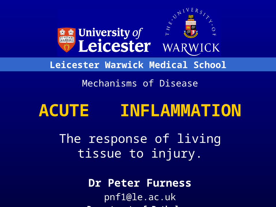
Leicester Warwick Medical School
Mechanisms of Disease
ACUTE INFLAMMATION
The response of living tissue to injury.
Dr Peter [email protected]
Department of Pathology
ACUTE INFLAMMATION
The response of living tissue to injury.
Dr P. N. Furness [email protected]
Mechanisms of Disease
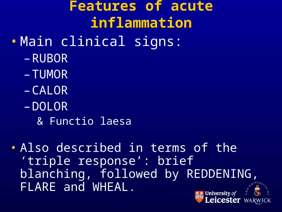
Features of acute inflammation• Main clinical signs:
– RUBOR– TUMOR– CALOR– DOLOR
& Functio laesa
• Also described in terms of the ‘triple response’: brief blanching, followed by REDDENING, FLARE and WHEAL.
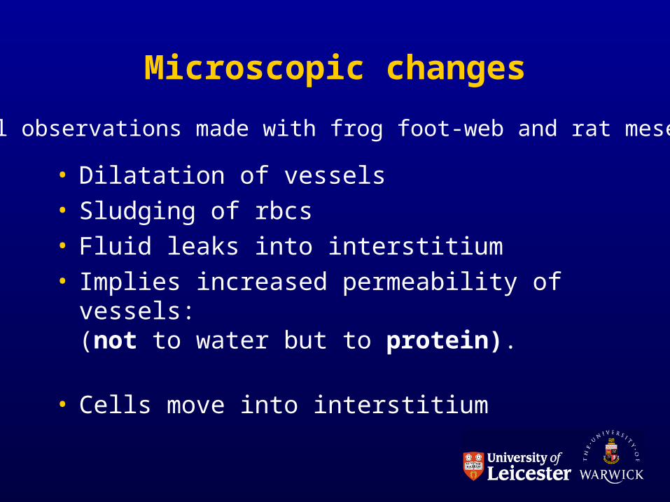
Microscopic changes
• Dilatation of vessels• Sludging of rbcs• Fluid leaks into interstitium• Implies increased permeability of vessels:
(not to water but to protein).
• Cells move into interstitium
Original observations made with frog foot-web and rat mesentery.
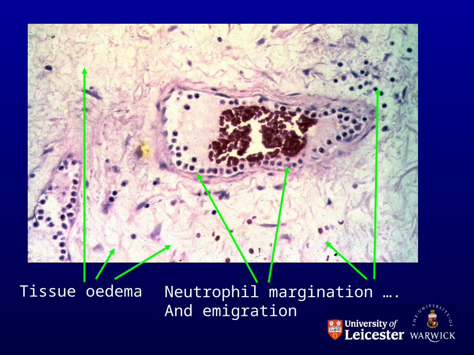
Tissue oedema Neutrophil margination …. And emigration
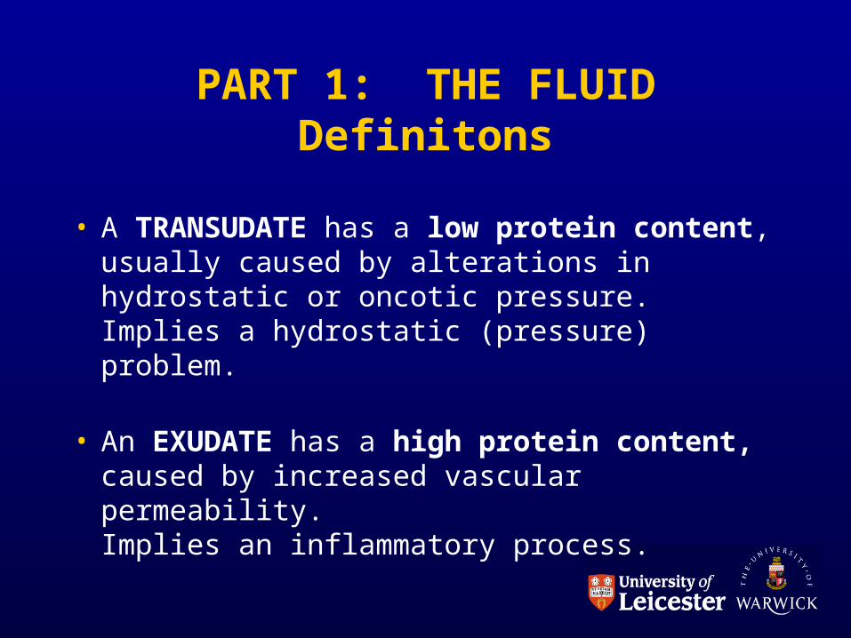
PART 1: THE FLUIDDefinitons
• A TRANSUDATE has a low protein content, usually caused by alterations in hydrostatic or oncotic pressure.Implies a hydrostatic (pressure) problem.
• An EXUDATE has a high protein content, caused by increased vascular permeability.Implies an inflammatory process.
a
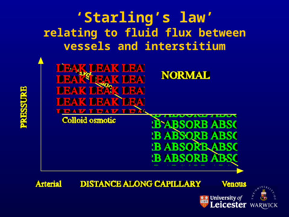
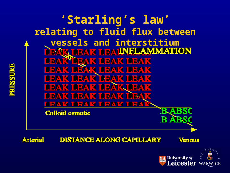
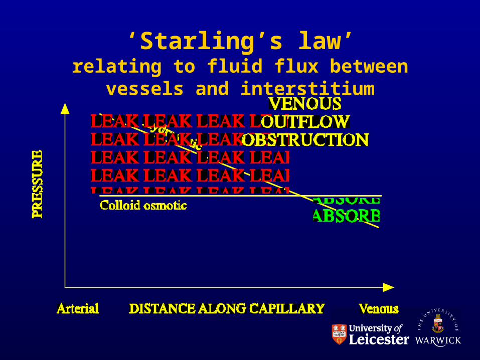
‘Starling’s law’relating to fluid flux between vessels and
interstitium
a
‘Starling’s law’relating to fluid flux between vessels and
interstitium
a
‘Starling’s law’relating to fluid flux between vessels and
interstitium
PART 2: THE CELLS
• White blood cells MARGINATION and EMIGRATION.
• Implies binding to endothelium then directional movement through vessel wall towards injured area.
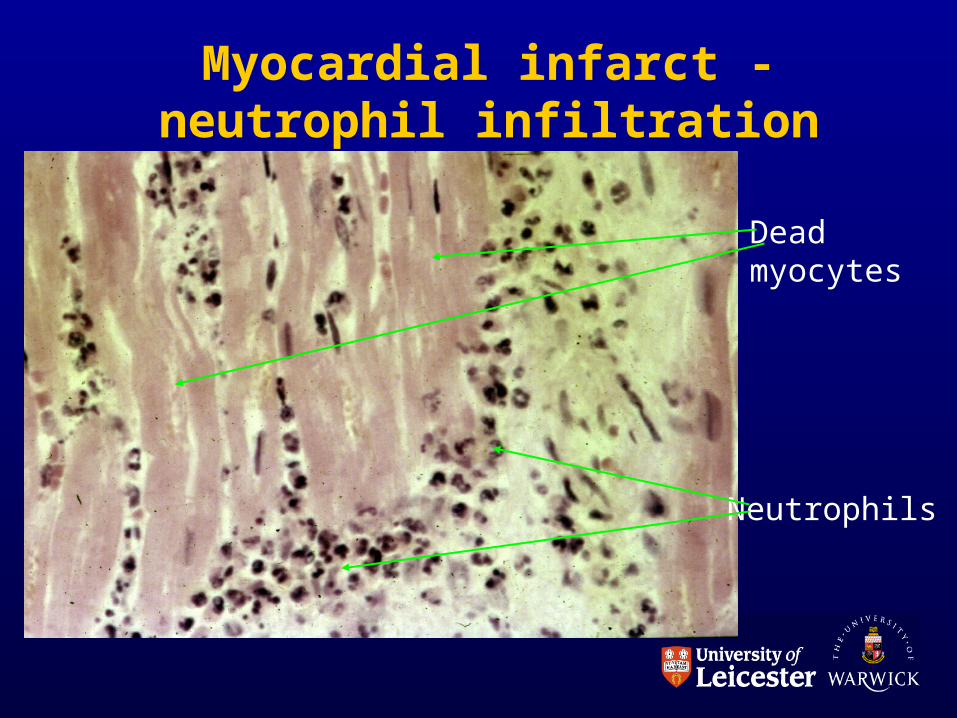
Myocardial infarct - neutrophil infiltration
Deadmyocytes
Neutrophils
How do these changes combat injury?• Vasodilatation:
– Increases delivery, increases temperature, removes toxins.
• Exudate:– Delivers immunoglobulins etc., dilutes toxins, delivers
fibrinogen, increases lymphatic drainage.
• Increased lymphatic drainage:– Delivers bugs to phagocytes and antigens to immune system.
• Cells:– Removes pathogenic organisms, necrotic debris etc.
• Pain and loss of function:– Enforces rest, reduces chance of further traumatic damage.
• How is all this brought about?
What are the mechanisms?CHEMICAL MEDIATORS.
Three phases:
1) Immediate early response (1/2 hr):• HISTAMINE
– Released from mast cells, basophils and platelets, in response to many stimuli: physical damage, immunologic reactions, C3a, C5a, IL1, factors from neutrophils and platelets
– Effects: Largely vascular. Pain. Not chemotactic.
2) Immediate sustained response:
(Not always seen. Due to direct damage to endothelial cells.)
3) Delayed response: (Peaks about 3hrs):
• Many and varied chemical mediators, interlinked and of varying importance
• Incompletely understood.
• IMPORTANT because of possibility of therapeutic intervention
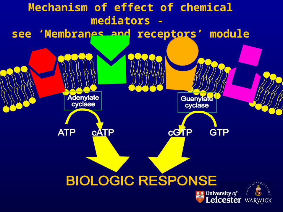
Mechanism of effect of chemical mediators - see ‘Membranes and receptors’ module
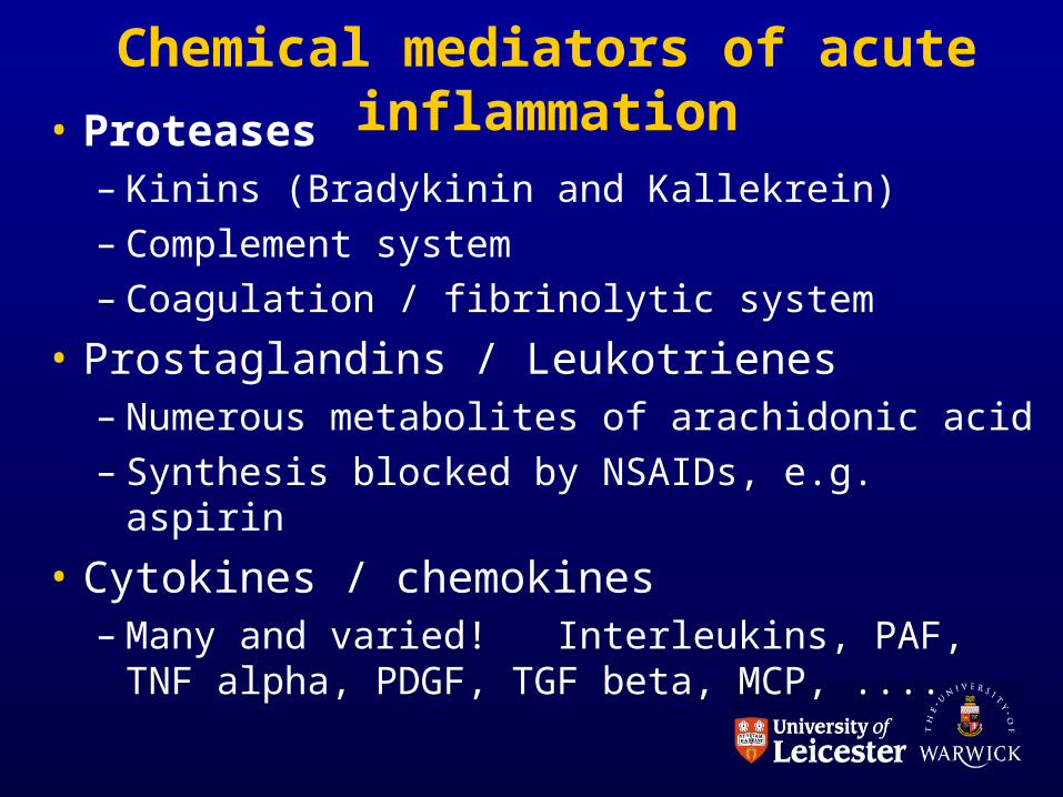
Chemical mediators of acute inflammation• Proteases
– Kinins (Bradykinin and Kallekrein)– Complement system– Coagulation / fibrinolytic system
• Prostaglandins / Leukotrienes– Numerous metabolites of arachidonic acid– Synthesis blocked by NSAIDs, e.g. aspirin
• Cytokines / chemokines– Many and varied! Interleukins, PAF, TNF
alpha, PDGF, TGF beta, MCP, ....
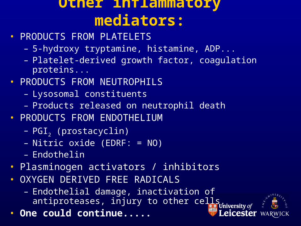
Other inflammatory mediators:• PRODUCTS FROM PLATELETS
– 5-hydroxy tryptamine, histamine, ADP...– Platelet-derived growth factor, coagulation proteins...
• PRODUCTS FROM NEUTROPHILS – Lysosomal constituents– Products released on neutrophil death
• PRODUCTS FROM ENDOTHELIUM– PGI2 (prostacyclin)– Nitric oxide (EDRF: = NO)– Endothelin
• Plasminogen activators / inhibitors• OXYGEN DERIVED FREE RADICALS
– Endothelial damage, inactivation of antiproteases, injury to other cells.
• One could continue.....
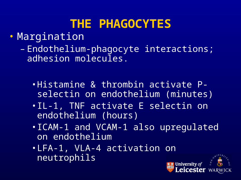
THE PHAGOCYTES• Margination
– Endothelium-phagocyte interactions; adhesion molecules.
• Histamine & thrombin activate P-selectin on endothelium (minutes)
• IL-1, TNF activate E selectin on endothelium (hours)
• ICAM-1 and VCAM-1 also upregulated on endothelium
• LFA-1, VLA-4 activation on neutrophils
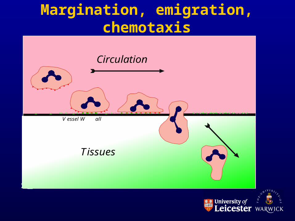
Margination, emigration, chemotaxis
a
V essel W all
Circulation
Tissues
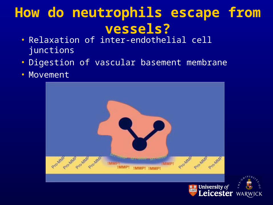
How do neutrophils escape from vessels?
• Relaxation of inter-endothelial cell junctions• Digestion of vascular basement membrane• Movement
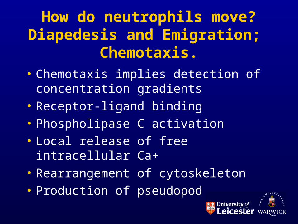
How do neutrophils move?Diapedesis and Emigration;
Chemotaxis.
• Chemotaxis implies detection of concentration gradients
• Receptor-ligand binding
• Phospholipase C activation
• Local release of free intracellular Ca+
• Rearrangement of cytoskeleton
• Production of pseudopod
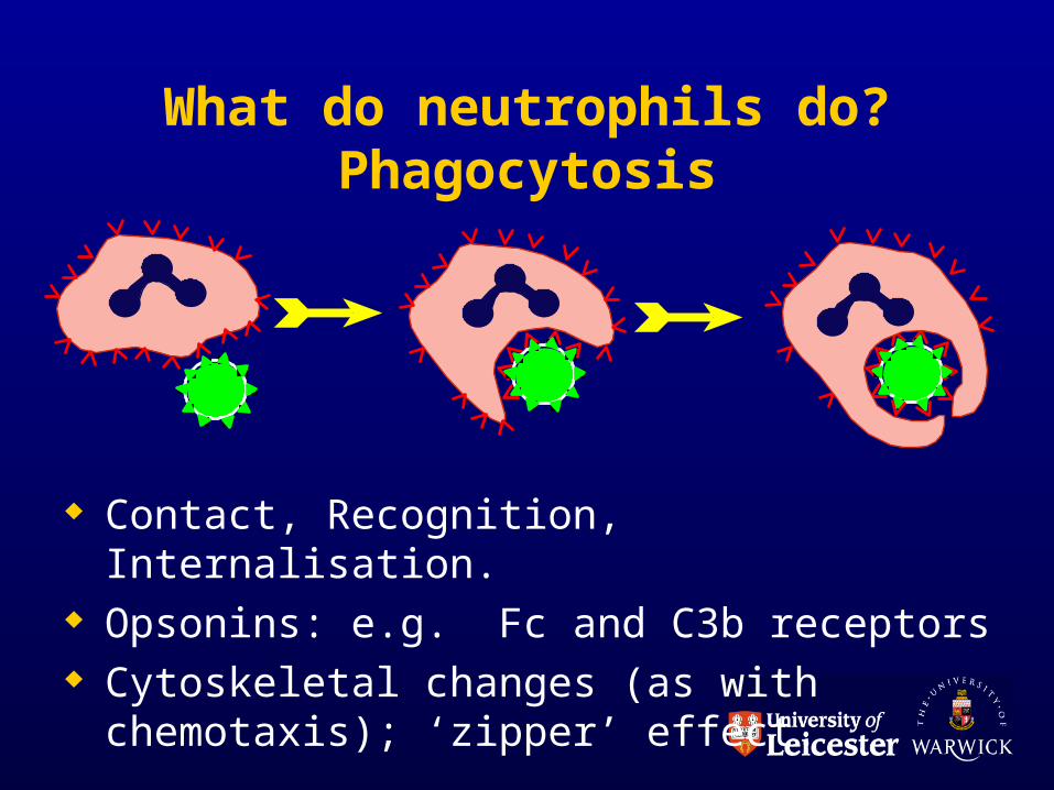
What do neutrophils do?Phagocytosis
Contact, Recognition, Internalisation. Opsonins: e.g. Fc and C3b receptors Cytoskeletal changes (as with chemotaxis); ‘zipper’
effect.
What do neutrophils do?Microbial killing
• Phagosomes fuse with lysosomes to produce secondary lysosomes.Mechanisms:
• O2 dependent– NADPH oxidase activated; produces superoxide ion.
This converts to hydrogen peroxide.– H2O2-Myeloperoxidase-halide system: produces
HOCl. (i.e. bleach!)
– Myeloperoxidase independent:– Uses superoxide and hydroxyl radicals. Less efficient.
O2 independent killing mechanisms
• Lysozyme & hydrolases
• Lactoferrin
• Bactericidal Permeability Increasing Protein (BPI)
• Cationic proteins (‘Defensins’)
• Major Basic Protein (MBP; Eosinophils)
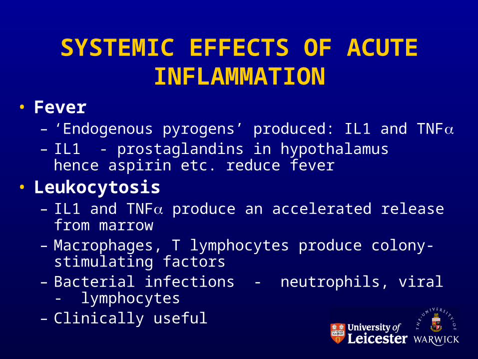
SYSTEMIC EFFECTS OF ACUTE INFLAMMATION
• Fever– ‘Endogenous pyrogens’ produced: IL1 and TNF– IL1 - prostaglandins in hypothalamus
hence aspirin etc. reduce fever
• Leukocytosis– IL1 and TNF produce an accelerated release from
marrow– Macrophages, T lymphocytes produce colony-
stimulating factors– Bacterial infections - neutrophils, viral - lymphocytes– Clinically useful

SYSTEMIC EFFECTS OF ACUTE INFLAMMATION
• Acute phase response– Decreased appetite, altered sleep patterns and
changes in plasma concentrations of:
• Acute phase proteins:– C-reactive protein (CRP) (Clinically useful) 1 antitrypsin
– Haptoglobin– Fibrinogen– Serum amyloid A protein
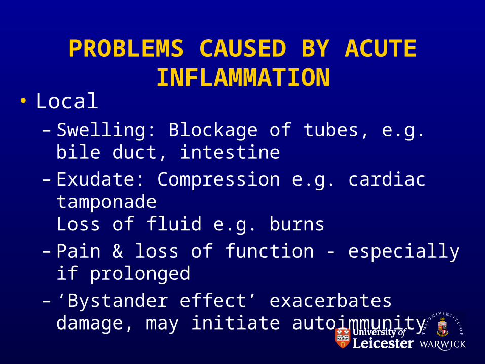
PROBLEMS CAUSED BY ACUTE INFLAMMATION
• Local– Swelling: Blockage of tubes, e.g. bile duct,
intestine– Exudate: Compression e.g. cardiac tamponade
Loss of fluid e.g. burns– Pain & loss of function - especially if prolonged– ‘Bystander effect’ exacerbates damage, may
initiate autoimmunity
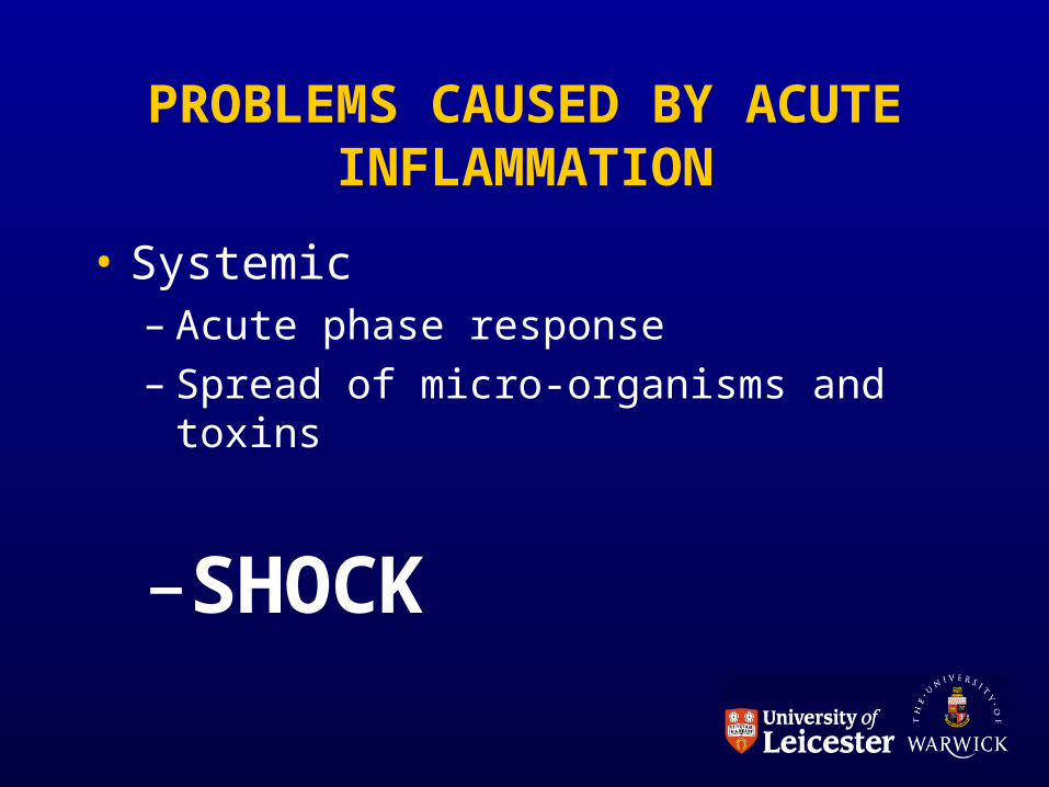
PROBLEMS CAUSED BY ACUTE INFLAMMATION
• Systemic– Acute phase response– Spread of micro-organisms and toxins
–SHOCK
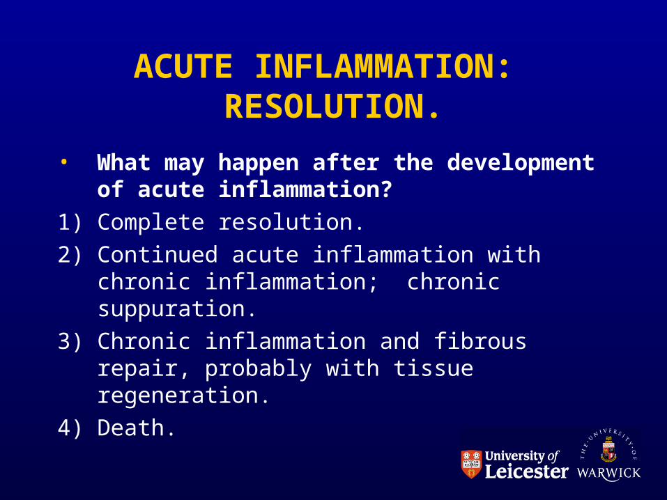
ACUTE INFLAMMATION: RESOLUTION.
• What may happen after the development of acute inflammation?
1) Complete resolution.
2) Continued acute inflammation with chronic inflammation; chronic suppuration.
3) Chronic inflammation and fibrous repair, probably with tissue regeneration.
4) Death.
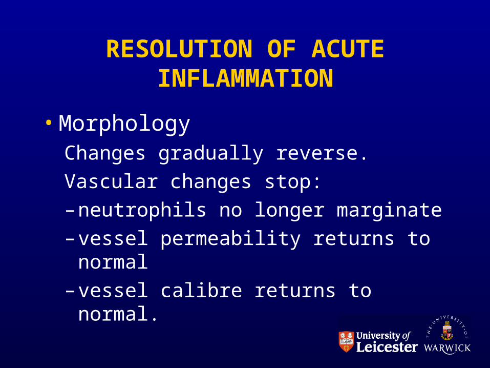
RESOLUTION OF ACUTE INFLAMMATION
• MorphologyChanges gradually reverse.
Vascular changes stop:
– neutrophils no longer marginate
– vessel permeability returns to normal
– vessel calibre returns to normal.
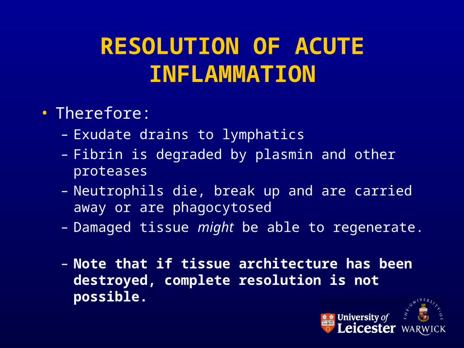
RESOLUTION OF ACUTE INFLAMMATION
• Therefore:– Exudate drains to lymphatics– Fibrin is degraded by plasmin and other proteases– Neutrophils die, break up and are carried away or
are phagocytosed– Damaged tissue might be able to regenerate.
– Note that if tissue architecture has been destroyed, complete resolution is not possible.
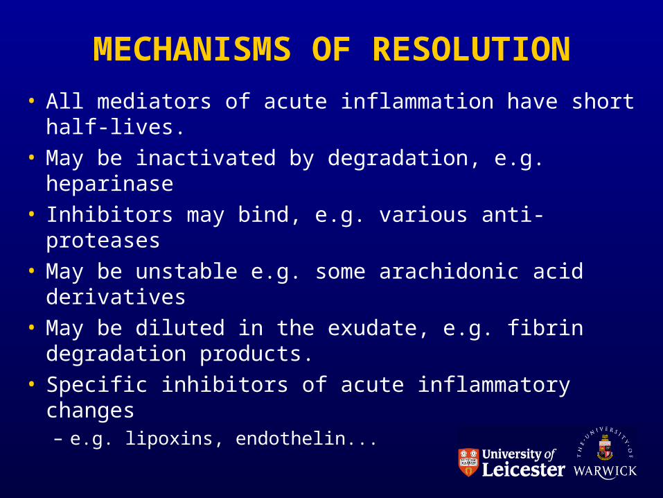
MECHANISMS OF RESOLUTION
• All mediators of acute inflammation have short half-lives.
• May be inactivated by degradation, e.g. heparinase• Inhibitors may bind, e.g. various anti-proteases• May be unstable e.g. some arachidonic acid
derivatives• May be diluted in the exudate, e.g. fibrin
degradation products.• Specific inhibitors of acute inflammatory changes
– e.g. lipoxins, endothelin...
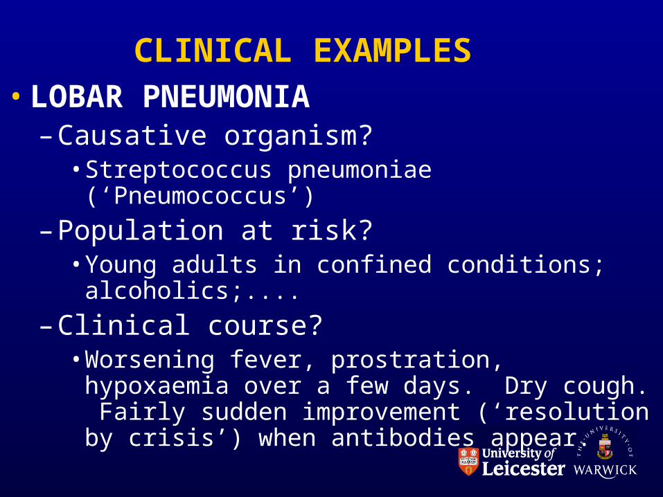
CLINICAL EXAMPLES• LOBAR PNEUMONIA
– Causative organism?• Streptococcus pneumoniae (‘Pneumococcus’)
– Population at risk?• Young adults in confined conditions;
alcoholics;....
– Clinical course?• Worsening fever, prostration, hypoxaemia over a
few days. Dry cough. Fairly sudden improvement (‘resolution by crisis’) when antibodies appear.
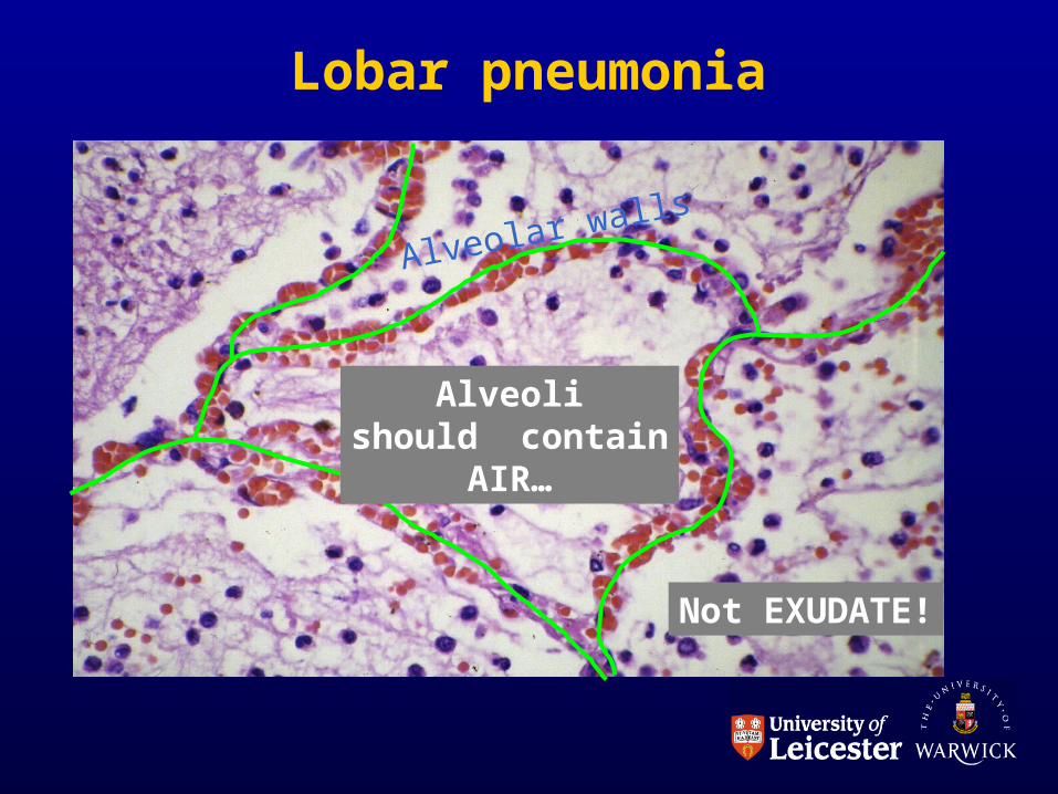
Lobar pneumonia
Alveolar walls
Alveolishould contain
AIR…
Not EXUDATE!
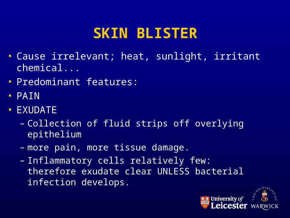
SKIN BLISTER
• Cause irrelevant; heat, sunlight, irritant chemical...• Predominant features:• PAIN• EXUDATE
– Collection of fluid strips off overlying epithelium
– more pain, more tissue damage.
– Inflammatory cells relatively few:therefore exudate clear UNLESS bacterial infection develops.
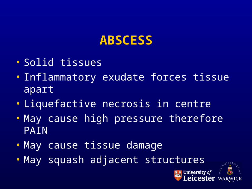
ABSCESS
• Solid tissues
• Inflammatory exudate forces tissue apart
• Liquefactive necrosis in centre
• May cause high pressure therefore PAIN
• May cause tissue damage
• May squash adjacent structures
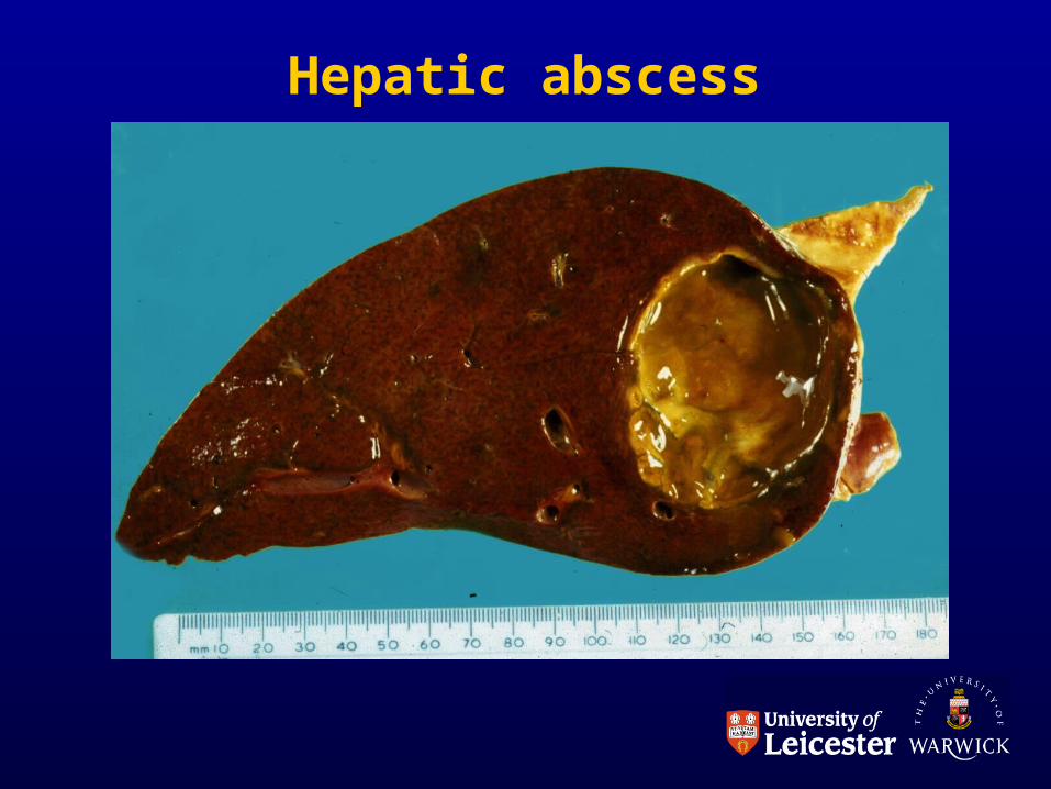
Hepatic abscess
ACUTE INFLAMMATION IN SEROUS CAVITIES
• Exudate pours into cavity
• ascites, pleural or pericardial effusion
• respiratory or cardiac impairment
• Localised fibrin deposition
• ‘bread and butter’ pericarditis
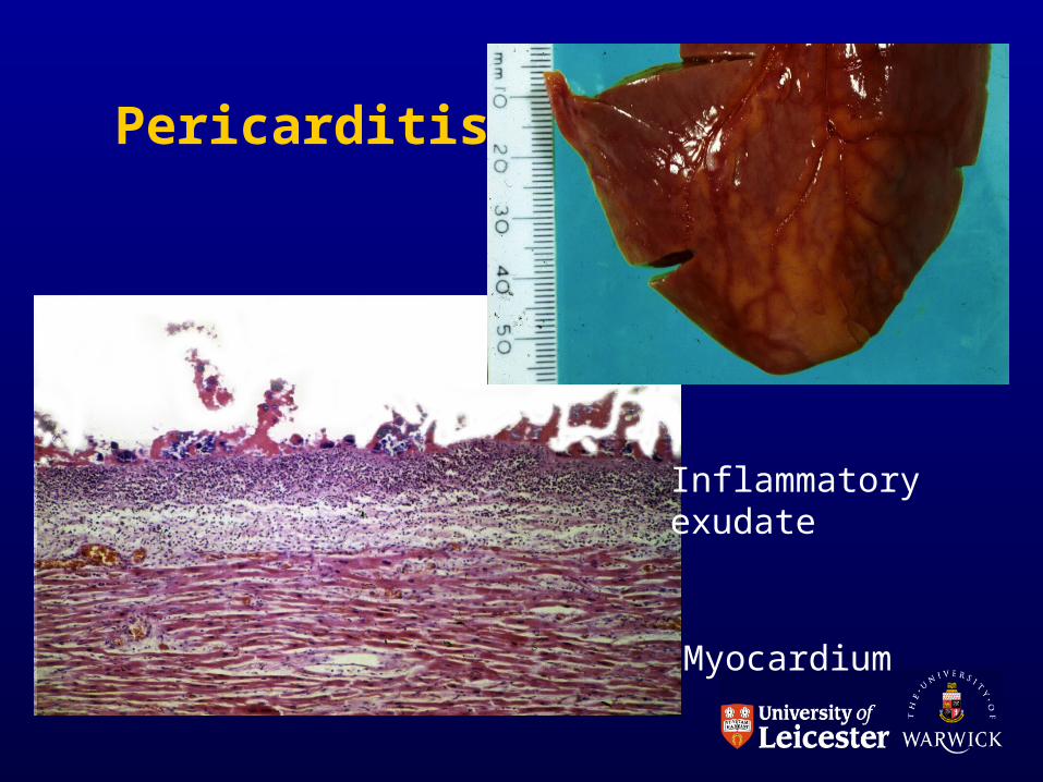
Pericarditis
Inflammatoryexudate
Myocardium
DISORDERS OF ACUTE INFLAMMATION
• These are rare diseases (natural selection ensures that!) but illustrate the importance of apparently small parts of this complex web of mechanisms.
A few examples:
• Hereditary angio-oedema (‘angioneurotic oedema’)
• Alpha-1 antitrypsin deficiency.• Inherited complement deficiencies.• Defects in neutrophil function.• Defects in neutrophil numbers.