Embed Size (px)
Citation preview

Leica VibratomeTM SeriesVibrating Blade Microtomes

2
Cutting Edge PrecisionVibrating blade microtomes are used to produce monolayer or thick sections of fixed or fresh tissue under
physiological conditions without freezing or embedding. Sectioning fresh tissue specimens with
Leica Biosystems’ VT Series maintains the morphology, enzyme activity and cell viability of the tissue.
Their use also minimizes artifacts, compression distortion, cell destruction and other inherent deleterious
effects of sectioning.
applications for these instruments include immunohistochemistry, cell culturing of different organs,
sections for patchclamping, electrophysiology, free floating sections and many other applications in
neuroscience.
in order to maintain physiological conditions while sectioning, it is common to use chilled buffer and
minimize the vertical deflection of the blade holder as well as the blade. During operation, the blade
vibrates laterally and advances forward through the specimen. Section thickness is determined by
motorized or manual vertical feeding of the specimen stage. Other parameters that influence section
quality are amplitude, frequency, knife travel speed and blade angle. The Leica VT Series of instruments
offers a complete product range that control some or all of these parameters.
Leica Biosystems offers a wide variety of vibrating blade
microtomes that have been developed in collaboration with
renowned scientists throughout the world. There is an instrument
for every researcher’s application and budget. The features of each
instrument vary in the degree of automation, ranging from the Leica
VT2000 to the fully automated Leica VT1000 S and VT1200 S with
optional Vibrocheck, for measuring and minimizing vertical
blade deflection
Lean principles are revolutionizing the way laboratories operate.
Leica Biosystems prides itself on providing high quality, reliable and
durable instruments. The Vibratome Series microtomes can greatly
improve productivity and reduce costs in the laboratory by
producing high quality sections with viable cells without the need of
replicating experiments.
Leica Microsystems expresses its thanks to Prof. Dr. Peter Jonas, Francisco Javier García Ladona, Ph.D., Shawn Hochman, Ph.D., and Dr. andreas Schober for granting their permission to use the application photographs contained in this brochure.

3
Leica VT1000 S
Leica VT1200 S
Leica VT1200
Specifications Leica VT1000 S Leica VT1200 Leica VT1200 S
Vibrocheck (measurement device for vertical deflection of the blade)
• •
Fully automated cut mode • •
Specimen retraction • •
adjustable amplitude • • •
adjustable frequency •
Blade travel speed 0.025 - 2.5mm/s 0.01 - 1.5 mm/s 0.01 - 1.5 mm/s
adjustable cutting window electronic individually programable front and rear position
Maximum specimen size 70 x 40 x 15 mm 33 x 50 x 20 mm 33 x 50 x 20 mm
Total vertical specimen stroke 15 mm 20 mm 20 mm
Selection of buffer trays • • •
cooling options chiller chiller chiller
Memory capability for storing section thickness •
Multiple user settings 8 different user settings
adjustable return stroke 1- 5 mm/s
adjustable forward speed in manual mode 1- 5 mm/s
Magnification options 2x magnifier 2x magnifier, microscope 2x magnifier, microscope

4
The classic design of the Leica VT1000 S makes working with the instrument a pleasure. ergonomic hand rests and direct access
to all functional elements provide exceptional comfort and added safety. The VT1000 S features very fine adjustable knife
advance speed, a freely programmable cutting window, and accelerated return knife speed to minimize overall sectioning time of
even the smallest specimens. The VT1000 S vibrating blade microtome is designed to consistently produce thin sections of fixed
tissue specimens, even non-homogeneous specimens that are difficult to section. it is also used for some industrial applications
related to structural analysis of foam and other very soft materials and botanical specimens such as plants and roots.
Key FeaTureS• ergonomic design for comfortable working conditions
• 5 different amplitude settings from 0.2 - 1 mm
• Linear sectioning speed adjustment from 0.025 mm - 2.5 mm
• Linear sectioning frequency adjustment from 0 - 100 Hz
• Programmable specimen retraction
• Freely programmable sectioning window
• Single and continuous stroke options for ultimate versatility
• easy mounting and removing of knife holder and buffer tray for efficient workflow
• Dark buffer tray provides excellent contrast to the specimen
The optional double-walled buffer tray is available in two different sizes which allows sectioning of specimens 33 x 40 mm or as large as 70 x 40 mm.
Standard knife holder S and buffer tray S with optional magnetic specimen holder.
The Leica VT1000 S sections specimens under physiological conditions, which protects tissue, enzymes
and antigens. For that purpose, the specimen is mounted directly onto a specimen plate, using
cyanoacrylate adhesive, and placed into a buffer tray filled with cooled physiological buffer solution. The
buffer provides a flotation medium for the sections. To maintain stable, cold buffer temperature, the
integrated ice bath can be filled with crushed ice or the optional double-walled buffer tray can be
connected with a circulation cooling device. Both the knife holder and buffer tray are easily removed to
reduce the risk of reagent carryover or contamination when sectioning.
Leica VT1000 S Vibrating Blade Microtome

5
The variable frequency allows the VT1000 S to adapt to a variety of applications. accurate control of the knife or blade
movement is integral in the design of this instrument. The visual clarity provided by the wide large-field magnifier, supplied as
standard delivery, can be enhanced with a fiber optic lighting system (optional). Together, these features provide exact,
individually adjustable illumination of the entire sectioning range, and prevents surface reflection of the buffer solution for
accurate sectioning.
Leica Design by Werner Hölbl
Labeling of cholinergic septal neurons in rat basal forebrain by using a polyclonal antiserum against choline acetyltransferase (ChAT).
Transversal section through rat brain cortex at the forebrain level. A large single neuron was labeled by NADPH-diaphorase histochemistry. The small axon and some branching dendrites are visible.
CA3 field of rat hippocampus. Syntaxin positive axon terminals over pyramidal cells. 40 μm section. 400x.
Epipremnum pinnatum (ivy).50 µm section.

6
Leica VT1200 and VT1200 S Vibrating Blade MicrotomesFresh nervous tissues, brain and spinal cord are soft, fragile and extremely susceptible to mechanical
damage. The Leica VT1200 and VT1200 S vibrating blade microtomes are designed to meet the highest
demands for cutting fresh and fixed tissue in Neuropathology, Neurophysiology (patch-clamping) and
electrophysiology. These robust instruments feature a new blade holder design with the possibility to
measure vertical deflection using the optional Vibrocheck device. Negative mechanical effects on the
tissue are reduced to a minimum. This produces the highest quality sections that retain viable cells on the
section surface/s.
Key FeaTureS• Vertical deflection of the blade can be measured with the optional Vibrocheck device and minimized
below 1µm
• Blade holder can be rotated through 90° to permit accurate insertion of a whole double edged razor
blade, sapphire knife or injector blade.
• Optimized blade holder designed for minimum buffer spillage.
• Motorized blade holder sectioning speed adjustable between 0.01 to 1.5 mm/sec.
• Palm rests on the ice bath or double-walled buffer tray allow a relaxed, ergonomic working position.
• Built-in LeD illumination provides natural, comfortable lighting during sectioning without adding heat,
which could deteriorate the tissue. The Leica VT1200 S features 5-step adjustable light intensity.
These instruments were designed in collaboration with Prof. Dr. Peter Jonas and his team, Physiology Department, university of Freiburg, Germany.

7
The Semi-auTomaTed Leica VT1200 has been designed for users who prefer
to manually control sectioning parameters such as section thickness and cutting
stroke for each individual section. The VT1200 offers straightforward, intuitive
operation, fast sectioning and a full range of accessories at an attractive price.
The fuLLy-auTomaTed Leica VT1200 S is recommended for multi user
laboratories where users of both semi-automated vibrating blade microtomes and
fully automated instruments work together. VT1200 S can be used in both semi- or
fully-automated sectioning modes depending on the users’ requirements. The fully
automated mode of the VT1200 S offers automatic feeding, specimen
retraction, and a cutting window. The mode of operation can be individu-
ally selected, and settings can be stored for up to 8 users. automatic
feeding, specimen retraction and use of a cutting window are
designed to minimize sectioning time.
MoDuLAr FuNCTioNALiTy Both instrument versions can be enhanced with an optional magnifier (2x) or microscope to improve visual clarity.
FLExiBLE PErForMANCEThe removable ice bath and buffer tray allow working under physiological conditions and away from the instrument, e.g., under a microscope.
Leica Design by Werner Hölbl
OPTiONaL MeaSureMeNT DeVice: ViBrOcHecK™The vertical deflection of the blade can be measured by the Vibrocheck measurement
device. Both vertical deflection (in μm) and rotation direction of the adjustment screw
are displayed on the separate, foil-protected control panel. The adjustment screw
on the blade holder allows minimization of the vertical deflection to below 1 μm,
which significantly increases the number of viable cells.
CuSToMizED CoMForTThe separate, foil-protected control panel can be placed on either side of the instrument depending on the personal preference of the user
95.8539 rev B - Order no. 1495.8539 ∙ 12/2012 ∙ copyright © by Leica Biosystems,
Nussloch, Germany, 2012. Subject to modifications. Leica and the Leica Logo are
registered trademarks of Leica Microsystems ir GmbH.
Leica VT1000 S
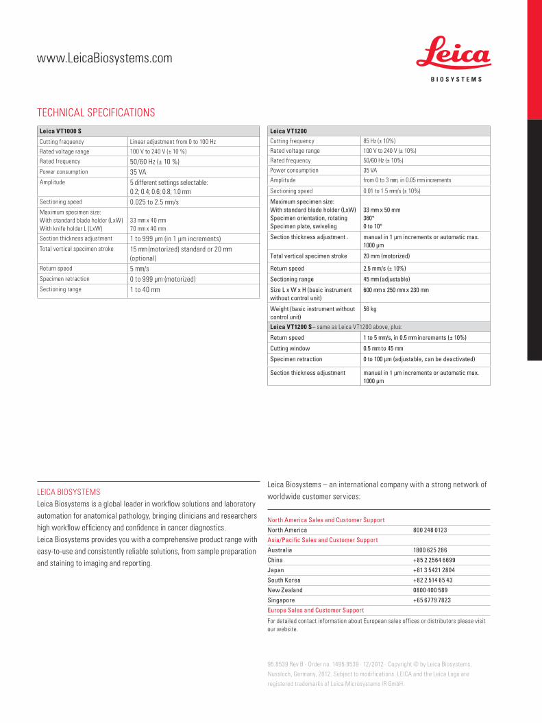
cutting frequency Linear adjustment from 0 to 100 Hz
rated voltage range 100 V to 240 V (± 10 %)
rated frequency 50/60 Hz (± 10 %)Power consumption 35 Vaamplitude 5 different settings selectable:
0.2; 0.4; 0.6; 0.8; 1.0 mmSectioning speed 0.025 to 2.5 mm/sMaximum specimen size:With standard blade holder (LxW) With knife holder L (LxW)
33 mm x 40 mm70 mm x 40 mm
Section thickness adjustment 1 to 999 μm (in 1 μm increments)Total vertical specimen stroke 15 mm (motorized) standard or 20 mm
(optional)return speed 5 mm/sSpecimen retraction 0 to 999 μm (motorized)Sectioning range 1 to 40 mm
Leica VT1200
cutting frequency 85 Hz (± 10%)
rated voltage range 100 V to 240 V (± 10%)
rated frequency 50/60 Hz (± 10%)
Power consumption 35 Va
amplitude from 0 to 3 mm, in 0.05 mm increments
Sectioning speed 0.01 to 1.5 mm/s (± 10%)
Maximum specimen size:With standard blade holder (LxW)Specimen orientation, rotating Specimen plate, swiveling
33 mm x 50 mm360°0 to 10°
Section thickness adjustment . manual in 1 μm increments or automatic max. 1000 μm
Total vertical specimen stroke 20 mm (motorized)
Return speed 2.5 mm/s (± 10%)
Sectioning range 45 mm (adjustable)
Size L x W x H (basic instrument without control unit)
600 mm x 250 mm x 230 mm
Weight (basic instrument without control unit)
56 kg
Leica VT1200 S– same as Leica VT1200 above, plus:
Return speed 1 to 5 mm/s, in 0.5 mm increments (± 10%)
Cutting window 0.5 mm to 45 mm
Specimen retraction 0 to 100 μm (adjustable, can be deactivated)
Section thickness adjustment manual in 1 μm increments or automatic max. 1000 μm
TecHNicaL SPeciFicaTiONS
Leica BiOSySTeMSLeica Biosystems is a global leader in workflow solutions and laboratory automation for anatomical pathology, bringing clinicians and researchers high workflow efficiency and confidence in cancer diagnostics. Leica Biosystems provides you with a comprehensive product range with easy-to-use and consistently reliable solutions, from sample preparation and staining to imaging and reporting.
Leica Biosystems – an international company with a strong network of worldwide customer services:
North America Sales and Customer Support
North America 800 248 0123
Asia/Pacific Sales and Customer Support
Australia 1800 625 286
China +85 2 2564 6699
Japan +81 3 5421 2804
South Korea +82 2 514 65 43
New zealand 0800 400 589
Singapore +65 6779 7823
Europe Sales and Customer Support
For detailed contact information about european sales offices or distributors please visit our website.
www.LeicaBiosystems.com