Embed Size (px)
Citation preview

ORIGINAL ARTICLE
Left Atrial and Left Ventricular Diastolic FunctionAfter the Maze Procedure for Atrial Fibrillation in MitralValve Disease: Degenerative Versus Rheumatic
Hwan Wook Kim & Mi Hyoung Moon & Keon Hyun Jo & Hyun Song & Jae Won Lee
Received: 21 January 2012 /Accepted: 3 September 2012# Association of Surgeons of India 2012
Abstract The present study was aimed to compare the leftatrial and left ventricular diastolic functions amongst therheumatic and degenerative mitral valve disease patients inatrial fibrillation who reverted to normal sinus rhythm fol-lowing Cox-maze procedure. We prospectively investigatedthe left atrial and left ventricular function with Dopplerechocardiography, by dividing into the rheumatic (N0105)and the degenerative group (N047). Over the follow-upperiod (mean: 4.4±1.2 years in the rheumatic group, 4.8±1.3 years in the degenerative group), the rheumatic groupshowed statistically significant decrease in A' velocity andE' velocity, on contrary to degenerative group (A' velocity:mean decrease of 0.43±0.13 cm/s in the rheumatic group,mean increase of 0.57±0.11 cm/s in the degenerative group,p00.029, E' velocity: mean decrease of 0.23±0.17 cm/s inthe rheumatic group, mean increase of 0.21±0.15 cm/s inthe degenerative group, p00.031). In addition, the rheumat-ic group showed statistically significant increase in E/E'ratio than the degenerative group (mean increase of 4.49±1.98 in the rheumatic group, mean increase of 1.74±1.52 inthe degenerative group, p00.047). Despite successful sinusrhythm restoration, the progressive loss of LA function aswell as LV diastolic function is more prominent in therheumatic group than the degenerative group. Therefore,
differentiated strategies for postoperative surveillance areneeded according to the pathology of mitral valve disease.
Keywords Left atrial function . Left ventricular diastolicfunction . Maze . Degenerative . Rheumatic
Introduction
As atrial fibrillation is observed in 30 % to 50 % of patientswho require operation for mitral valve disease [1], the Cox-maze procedure has been performed concomitantly with mi-tral valve surgery as a standard surgical treatment for atrialfibrillation. Although high efficiency of conversion into sinusrhythm has been demonstrated after the Cox-maze procedure,irrespective of the origin of valve pathology [2], the changesin size and function of left atrium (LA) have been poorlyelucidated, especially in long-term periods.
In fact, in terms of sinus conversion rate and existence ofLA electrical activity, some authors reported unsatisfactoryresults of the Cox-maze procedure in patients more withrheumatic disease than other causes. The left atrial contractil-ity determined as the peak velocity of the atrial filling wave topeak velocity of early filling wave ratio for the LA in therheumatic group was significantly reduced comparedwith thatin the nonrheumatic group [3]. On the contrary, others includ-ing our group demonstrated equally effective results, eventhough those were short-term outcomes [4, 5].
Along with the sinus conversion rate and electrical activ-ity, one of the issues of debate in the Cox-maze procedure isthat whether the etiology of mitral valve disease has aneffect on the change of LA function and left ventricular(LV) function over a long-term follow-up, in patients withrestoration to normal sinus rhythm after surgery. Althoughsome studies dealt with the improvement of LA and LVfunctions after the successful maze procedure, these were
H. W. Kim :M. H. Moon :K. H. Jo :H. SongDepartment of Thoracic and Cardiovascular Surgery,Seoul St. Mary’s Hospital, School of Medicine,The Catholic University of Korea,505 Banpo-dong, Seocho-gu,Seoul 137-701, Republic of Korea
J. W. Lee (*)Department of Thoracic and Cardiovascular Surgery, Seoul AsanHospital, School of Medicine, The University of Ulsan,388-1 Pungnap-dong, Songpa-gu,Seoul 138-736, Republic of Koreae-mail: [email protected]
Indian J SurgDOI 10.1007/s12262-012-0724-0

separately performed only according to the etiology of mi-tral valve diseases, having degenerative or rheumatic origins[6, 7], not for the comparison between the two etiologies.Furthermore, most of these studies focused predominantlyon the LV dimension and systolic function (ejection frac-tion) [5–8], and no prospective studies have been performed,to our knowledge. In Korea, a significant number of patientswith mitral valve disease accompanied by atrial fibrillationhad rheumatic origin.
The objective of this study was to compare LA size andmechanical function, and LV diastolic function after the Cox-maze procedure for atrial fibrillation accompanied by rheu-matic and degenerative mitral valve disease. We have per-formed the Cox-maze III procedure concomitantly with mitralvalve operations since 1997, and we prospectively investigat-ed serial changes in LA dimension, LA function, and LVdiastolic function with Doppler echocardiography.
Materials and Method
Patients
Between July 1997 and July 2008, 647 consecutive patientsunderwent surgery for atrial fibrillation associated with mi-tral valve disease. The Cox-maze procedure and mitral valveoperation were performed concomitantly, with or withoutother cardiac procedures. A total of 332 patients who had anoperation within the last 3 years (N0284) or were lostduring follow-up (N048) were excluded. From the total of315 patients with a follow-up period of more than 3 years,patients with intractable atrial fibrillation (N042) were alsoexcluded, as were patients who maintained normal sinusrhythm with intermittent antiarrhythmic medication orsynchronized electrical cardioversion (N061). During thefollow-up, 60 patients were newly diagnosed with valvularregurgitation greater than grade II or mild grade valvularstenosis. Therefore, 152 patients that were able to be fol-lowed up more than 3 years after surgery had sustainednormal sinus rhythm without antiarrhythmic medicationsand did not have valvular regurgitation greater than gradeII or valvular stenosis more than mild grade were selectedfor evaluation. The patients were divided into the rheumaticgroup (N0105) and the degenerative group (N047).
The clinical characteristics for each group were summa-rized in Table 1. Results showed that patients in both groupswere similar in terms of age, sex, preoperative cardiothoracicratio (CT ratio), and the nature of atrial fibrillation.
Rheumatic mitral valve disease was determined by pre-operative echocardiography and gross findings of valvemorphology, as previously reported [5]. In brief, irrespectiveof mitral stenosis or regurgitation, the lesions combinedwith immobility of the thickened leaflet, calcification of
subvalvular apparatus, commissural fusion, and contractionof the chordae tendineae were defined as rheumatic. Inaddition, all stenotic lesions of the mitral valve combinedwith aortic valve stenosis were considered rheumatic.
After the Cox-maze procedure, other cardiac procedureswere performed with the mitral valve surgery. To access valvecompetence in mitral valvuloplasty, the entire coaptation lineparallel to the mural part of the annulus was checked aftersaline injection forcibly through the valve to fill the leftventricle using a 20-Fr red rubber catheter attached to a bulbsyringe. Once the heart was beating and the patient had beenweaned from cardiopulmonary bypass, less than mild regur-gitations were confirmed by intraoperative transesophagealechocardiography. The most common concomitant proce-dures were tricuspid annuloplasty (N054). Other proceduresincluded aortic valve replacement in 19 patients, coronaryartery bypass grafting in 6 patients, atrial septal defect closurein 6 patients, ventricular septal defect closure in 1 patient, andascending aortic graft replacement in 1 patient.
This cohort study was approved by the institutional reviewboard and the ethics committee of our hospital. Informedconsent forms were obtained.
Surgical Procedures
We modified the classic Cox-maze procedure to preserve andenhance the LA function. The details of our Cox-maze proce-dure have been reported previously in detail [5]. A standardLA incision was made and extended inferiorly to the orifice ofthe left inferior pulmonary vein. To reduce LA tissue inclusionon the pulmonary vein isolation, we tightly encircled thepulmonary vein orifices. To minimize the number of incisions,cut and sew lesions were replaced with either cryoablation(N036) or microwave ablation (N0116). The lesion set of theLA side included a single box lesion for pulmonary veinisolation (two round lesions for the isolation of pulmonaryveins in pairs, two parallel lesions for connecting betweenboth the superior and inferior pulmonary veins) and two linearablations from the pulmonary isolation lesion to the posteriormitral valve annulus and LA appendage, respectively, withadditional epicardial coronary sinus ablation. To reduce theLA dimension, the parallel resection of LAwall to the poste-rior mitral annulus was performed after exclusion of the leftatrial appendage from within, especially in all patients withLA size greater than 60 mm preoperatively. Our modificationof the maze procedure was illustrated in Fig. 1. For cryoabla-tion, the cryoprobe (Frigitronics Cardiac Cryosurgical System200; Frigitronics, Inc, Coopersurgical, Shelton, Conn) wasapplied endocardially at −60 °C. The duration of ablationvaried depending on the thickness of the atrium, usuallybetween 90 s and 2 min after ice crystals were observedtransmurally. For microwave ablation, the FLEX microwaveprobe (Afx Inc, Fremont, Calif) was applied endocardially
Indian J Surg

with an energy level of 65 W for 2 min uniformly because ofthe lacking of transmural feedback sensor system. In addition,to prevent postoperative atrial flutter, the right atrial isthmuswas isolated completely with cryoablation after two ablation
lines on the intercaval and free wall of right atrium. The endpoint of ablation was the conversion to regular atrial tachy-cardias or sinus rhythm during the weaning process of cardio-pulmonary bypass.
Table 1 Preoperative andoperative data of the patients
ACC aortic cross clamp, Af atrialfibrillation, ASD atrial septal de-fect, AVR aortic valve replace-ment, CABG coronary arterybypass graft, CPB cardiopulmo-nary bypass, CT ratio cardiotho-racic ratio, LA left atrium, LVEDDleft ventricular end diastolic di-mension, LVEF left ventricularejection fraction, LVESD leftventricular end systolic dimen-sion, MVP mitral valvuloplasty,MVR mitral valve replacement,TAP tricuspid annuloplasty,VSD ventricular septal defect
Characteristic Degenerative group Rheumatic group p value
No 47 105
Age (yr) 50.6±13.1 47.2±10.5 0.089
Male/Female 25/22 69/36 0.142
Mitral valve disease
Stenosis/Regugitation 1/45 64/33 < 0.001
Stenoregurgitation 1 9
Af profile
CT ratio (%) 58.7±7.6 58.7±10.5 0.132
Af duration (yr) 3.6±4.6 5.8±6.7 0.018
Paroxysmal/Continuous 12/35 5/100 < 0.001
Fine/Coarse 20/27 49/56 0.638
Myocardial function
LVEF(%) 57.8±9.0 53.3±10.7 0.008
LA dimension (mm) 58.8±10.9 60.8±9.7 0.260
LVESD (mm) 41.5±7.9 40.3±7.7 0.390
LVEDD (mm) 62.3±11.2 56.7±9.0 < 0.001
Prev. Cardiac OP history 2 (4.3 %) 6 (5.7 %)
Operative data
CPB (min) 169±41 158±41 0.132
ACC (min) 125±35 115±32 0.090
Microwave/Cryo 31/16 85/20 0.118
MVR/MVP 6/41 62/43 < 0.001
LA volume reduction procedure 31 85 0.118
TAP 10 44 0.032
AVR 1 18
CABG 0 6
ASD or VSD 6 1
Other 0 1
Follow-up period (yr) 4.4±1.2 4.8±1.3 0.079
Fig. 1 Schematic illustrationof the modified mazeprocedure
Indian J Surg
Postoperative Follow-Up
To evaluate cardiac rhythm after surgery, standard 12-channelsurface electrocardiography and 24-hour Holter monitoringwere performed at 6 month intervals during the first 2 yearsand then repeated every year after.
To evaluate LA dimension, LA function, and mitral annu-lus movement postoperatively, transthoracic echocardiogra-phy with tissue Doppler imaging (Hewlett-Packard Sonos2500 or 5500 imaging system with a 2.5-MHz transducer)were performed at 6-month intervals during the first 2 yearsand then repeated every year after. Echocardiographic varia-bles on 1 year after surgery were regarded as the data of baseline. Ejection fraction of the LV was calculated using thebiplane Simpson method. The LV end diastolic and end sys-tolic dimensions were measured from parasternal M-modeacquisitions. LA dimension was measured at end systole asthe largest distance between the posterior aortic wall and thecenter of the line denoting the posterior LA wall. Using thesample volume on the tip of the mitral valve in an apical fourchamber view, mean peak velocity of transmitral E wave andAwave was obtained. Particularly, E/A ratio was calculated bythe mean values of five consecutive beats. Mean peak early(E’) and late (A’) diastolic annulus velocity and the ratio ofearly to late peak velocities (E’/A’) were calculated with thesample volume located at the septal side of the mitral annulus.For later assessment, all data were collected prospectively andstored in a specially organized and controlled database.
For patients with early atrial fibrillation recurrences, amio-darone was prescribed at 1200 mg/d after the Cox-mazeprocedure and tapered after conversion into normal sinusrhythm. For patients with valve repair (N084), or replacementwith biological prosthesis (N013), warfarin was prescribedfor 6 months postoperatively with international normalizedratio (INR) of 1.5 to 2.0. For patients with mechanical valvereplacement (N055), warfarin was prescribed for life-longperiods with INR of 2.0 to 3.0.
Statistics
All continuous variables were expressed asmean±S.D andweretested using a student t test. For categorical variables, Chi-squarestatistics were used. For comparison of repeated data betweentwo sets of data within a group at different time periods, thepaired t test was used. A p value of 0.05 or less was consideredstatistically significant in all cases. The SPSS software package14.0 (SPSS Inc, Chicago, IL) was used for statistical analysis.
Results
Preoperative echocardiographic findings of the degenerativeand rheumatic groups are shown in Table 1. Mean LA
dimension did not significantly differ between the twogroups. However, in the degenerative group, a preoperativemean duration of atrial fibrillation was longer (p00.018),the continuous nature of atrial fibrillation was more oftenfound (p<0.001), and the LV end diastolic dimension waslarger (p<0.001). Compared to the rheumatic group, almostall patients in the degenerative group had mitral valve sur-gery for regurgitation (p<0.001).
Operative information is summarized in Table 1. Comparedto the rheumatic group, a significantly greater number ofpatients in the degenerative group received mitral valvulo-plasty, p<0.001. However, tricuspid annuloplasty was moreoften used in the rheumatic group than the degenerative group(p00.032). Nevertheless, there was no difference in statisticalresults according to the cardiopulmonary bypass time, aorticcross clamp time, and energy source for the maze procedurebetween the two groups, respectively. In addition, postopera-tive follow-up periods were a little longer in the rheumaticgroup, but there was no significant difference (p00.079).
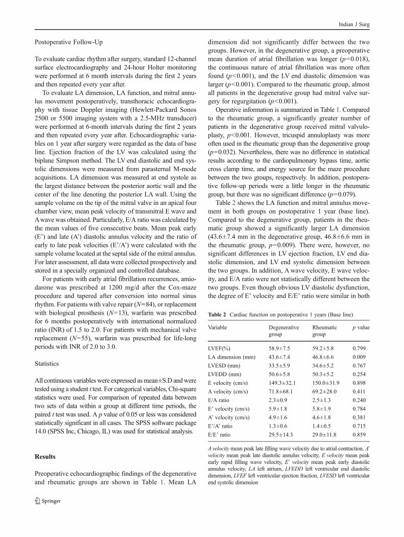
Table 2 shows the LA function and mitral annulus move-ment in both groups on postoperative 1 year (base line).Compared to the degenerative group, patients in the rheu-matic group showed a significantly larger LA dimension(43.6±7.4 mm in the degenerative group, 46.8±6.6 mm inthe rheumatic group, p00.009). There were, however, nosignificant differences in LV ejection fraction, LV end dia-stolic dimension, and LV end systolic dimension betweenthe two groups. In addition, Awave velocity, E wave veloc-ity, and E/A ratio were not statistically different between thetwo groups. Even though obvious LV diastolic dysfunction,the degree of E’ velocity and E/E’ ratio were similar in both
Table 2 Cardiac function on postoperative 1 years (Base line)
Variable Degenerativegroup
Rheumaticgroup
p value
LVEF(%) 58.9±7.5 59.2±5.8 0.799
LA dimension (mm) 43.6±7.4 46.8±6.6 0.009
LVESD (mm) 33.5±5.9 34.6±5.2 0.767
LVEDD (mm) 50.6±5.8 50.3±5.2 0.254
E velocity (cm/s) 149.3±32.1 150.0±31.9 0.898
A velocity (cm/s) 71.8±68.1 69.2±28.0 0.411
E/A ratio 2.3±0.9 2.5±1.3 0.240
E’ velocity (cm/s) 5.9±1.8 5.8±1.9 0.784
A’ velocity (cm/s) 4.9±1.6 4.6±1.8 0.381
E’/A’ ratio 1.3±0.6 1.4±0.5 0.715
E/E’ ratio 29.5±14.3 29.0±11.8 0.859
A velocitymean peak late filling wave velocity due to atrial contraction, A’velocity mean peak late diastolic annulus velocity, E velocity mean peakearly rapid filling wave velocity, E’ velocity mean peak early diastolicannulus velocity, LA left atrium, LVEDD left ventricular end diastolicdimension, LVEF left ventricular ejection fraction, LVESD left ventricularend systolic dimension
Indian J Surg
groups (E’ velocity: 5.9±1.8 cm/s in the degenerative group,5.8±1.9 cm/s in the rheumatic group, p00.784; E/E’: 29.5±14.3 in the degenerative group, 29,0±11.8 in the rheumaticgroup, p00.859).
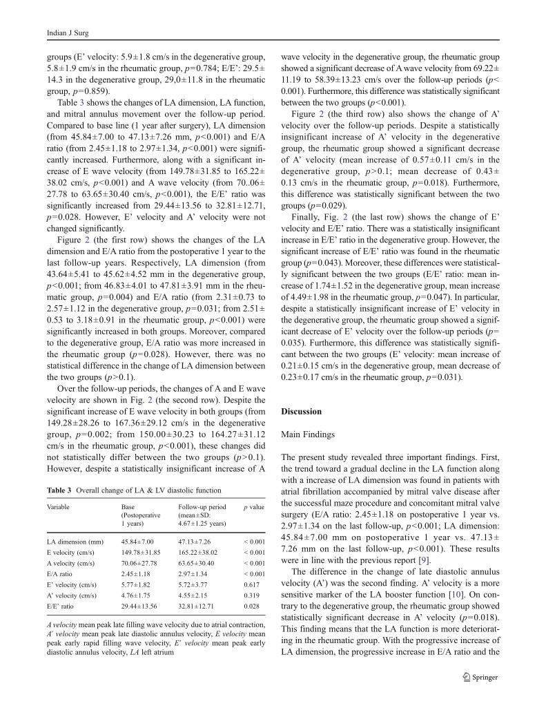
Table 3 shows the changes of LA dimension, LA function,and mitral annulus movement over the follow-up period.Compared to base line (1 year after surgery), LA dimension(from 45.84±7.00 to 47.13±7.26 mm, p<0.001) and E/Aratio (from 2.45±1.18 to 2.97±1.34, p<0.001) were signifi-cantly increased. Furthermore, along with a significant in-crease of E wave velocity (from 149.78±31.85 to 165.22±38.02 cm/s, p<0.001) and A wave velocity (from 70..06±27.78 to 63.65±30.40 cm/s, p<0.001), the E/E’ ratio wassignificantly increased from 29.44±13.56 to 32.81±12.71,p00.028. However, E’ velocity and A’ velocity were notchanged significantly.
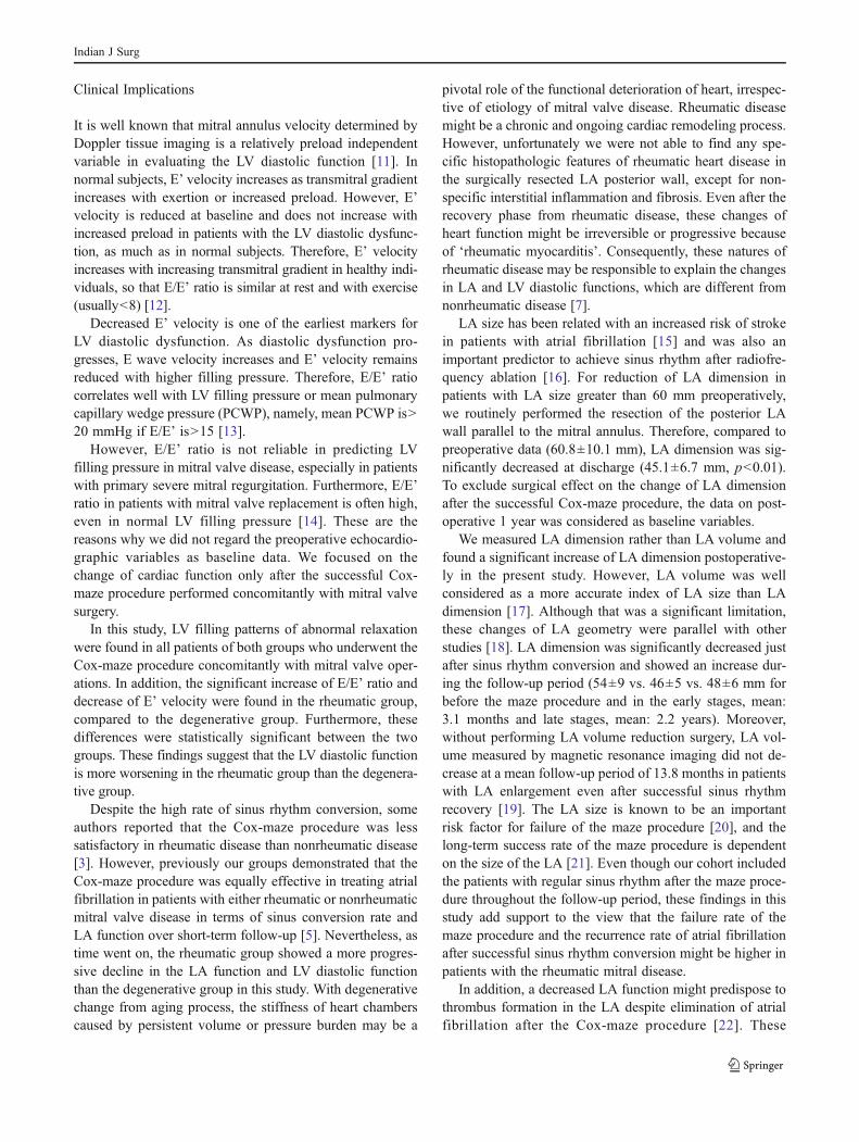
Figure 2 (the first row) shows the changes of the LAdimension and E/A ratio from the postoperative 1 year to thelast follow-up years. Respectively, LA dimension (from43.64±5.41 to 45.62±4.52 mm in the degenerative group,p<0.001; from 46.83±4.01 to 47.81±3.91 mm in the rheu-matic group, p00.004) and E/A ratio (from 2.31±0.73 to2.57±1.12 in the degenerative group, p00.031; from 2.51±0.53 to 3.18±0.91 in the rheumatic group, p<0.001) weresignificantly increased in both groups. Moreover, comparedto the degenerative group, E/A ratio was more increased inthe rheumatic group (p00.028). However, there was nostatistical difference in the change of LA dimension betweenthe two groups (p>0.1).
Over the follow-up periods, the changes of A and E wavevelocity are shown in Fig. 2 (the second row). Despite thesignificant increase of E wave velocity in both groups (from149.28±28.26 to 167.36±29.12 cm/s in the degenerativegroup, p00.002; from 150.00±30.23 to 164.27±31.12cm/s in the rheumatic group, p<0.001), these changes didnot statistically differ between the two groups (p>0.1).However, despite a statistically insignificant increase of A
wave velocity in the degenerative group, the rheumatic groupshowed a significant decrease of Awave velocity from 69.22±11.19 to 58.39±13.23 cm/s over the follow-up periods (p<0.001). Furthermore, this difference was statistically significantbetween the two groups (p<0.001).
Figure 2 (the third row) also shows the change of A’velocity over the follow-up periods. Despite a statisticallyinsignificant increase of A’ velocity in the degenerativegroup, the rheumatic group showed a significant decreaseof A’ velocity (mean increase of 0.57±0.11 cm/s in thedegenerative group, p>0.1; mean decrease of 0.43±0.13 cm/s in the rheumatic group, p00.018). Furthermore,this difference was statistically significant between the twogroups (p00.029).
Finally, Fig. 2 (the last row) shows the change of E’velocity and E/E’ ratio. There was a statistically insignificantincrease in E/E’ ratio in the degenerative group. However, thesignificant increase of E/E’ ratio was found in the rheumaticgroup (p00.043). Moreover, these differences were statistical-ly significant between the two groups (E/E’ ratio: mean in-crease of 1.74±1.52 in the degenerative group, mean increaseof 4.49±1.98 in the rheumatic group, p00.047). In particular,despite a statistically insignificant increase of E’ velocity inthe degenerative group, the rheumatic group showed a signif-icant decrease of E’ velocity over the follow-up periods (p00.035). Furthermore, this difference was statistically signifi-cant between the two groups (E’ velocity: mean increase of0.21±0.15 cm/s in the degenerative group, mean decrease of0.23±0.17 cm/s in the rheumatic group, p00.031).
Discussion
Main Findings
The present study revealed three important findings. First,the trend toward a gradual decline in the LA function alongwith a increase of LA dimension was found in patients withatrial fibrillation accompanied by mitral valve disease afterthe successful maze procedure and concomitant mitral valvesurgery (E/A ratio: 2.45±1.18 on postoperative 1 year vs.2.97±1.34 on the last follow-up, p<0.001; LA dimension:45.84±7.00 mm on postoperative 1 year vs. 47.13±7.26 mm on the last follow-up, p<0.001). These resultswere in line with the previous report [9].
The difference in the change of late diastolic annulusvelocity (A’) was the second finding. A’ velocity is a moresensitive marker of the LA booster function [10]. On con-trary to the degenerative group, the rheumatic group showedstatistically significant decrease in A’ velocity (p00.018).This finding means that the LA function is more deteriorat-ing in the rheumatic group. With the progressive increase ofLA dimension, the progressive increase in E/A ratio and the
Table 3 Overall change of LA & LV diastolic function
Variable Base(Postoperative1 years)
Follow-up period(mean±SD:4.67±1.25 years)
p value
LA dimension (mm) 45.84±7.00 47.13±7.26 < 0.001
E velocity (cm/s) 149.78±31.85 165.22±38.02 < 0.001
A velocity (cm/s) 70.06±27.78 63.65±30.40 < 0.001
E/A ratio 2.45±1.18 2.97±1.34 < 0.001
E’ velocity (cm/s) 5.77±1.82 5.72±3.77 0.617
A’ velocity (cm/s) 4.76±1.75 4.55±2.15 0.319
E/E’ ratio 29.44±13.56 32.81±12.71 0.028
A velocitymean peak late filling wave velocity due to atrial contraction,A’ velocity mean peak late diastolic annulus velocity, E velocity meanpeak early rapid filling wave velocity, E’ velocity mean peak earlydiastolic annulus velocity, LA left atrium
Indian J Surg

progressive decrease in A’ velocity raises have concernedabout the effects of the Cox-maze procedure on the LAfunction, especially in the rheumatic group.
The last interesting finding was the difference in thechange of the LV diastolic function, which was found byDoppler tissue imaging. At first, we found that despite alittle decrease of E’ velocity (5.77±1.82 cm/s on postoper-ative 1 year vs. 5.72±3.77 cm/s on the last follow-up, p>0.1), E/E’ ratio was significantly increased on the whole( 29.44±13.56 on postoperative 1 year vs. 32.81±12.71 onthe last follow-up, p00.028). In addition, after analyzing by
dividing into two groups, namely degenerative and rheu-matic group, different patterns of the change of the LVdiastolic function were revealed according to mitral valvepathology. On contrary to the degenerative group, the rheu-matic group showed statistically significant decrease in E’velocity (p00.031). In addition, the rheumatic group showedstatistically significant increase in E/E’ ratio compared to thedegenerative group (p00.047). This finding means that al-though the LV diastolic dysfunction is apparent in bothgroups, the LV diastolic function is more worsening in therheumatic group than the degenerative group.
Fig. 2 Change of LAdimension, E/A ratio,transmitral A wave velocity,transmitral E wave velocity, latediastolic annulus velocity (A’),early diastolic annulusvelocity (E’), and E/E’ratio in both groups
Indian J Surg
Clinical Implications
It is well known that mitral annulus velocity determined byDoppler tissue imaging is a relatively preload independentvariable in evaluating the LV diastolic function [11]. Innormal subjects, E’ velocity increases as transmitral gradientincreases with exertion or increased preload. However, E’velocity is reduced at baseline and does not increase withincreased preload in patients with the LV diastolic dysfunc-tion, as much as in normal subjects. Therefore, E’ velocityincreases with increasing transmitral gradient in healthy indi-viduals, so that E/E’ ratio is similar at rest and with exercise(usually<8) [12].
Decreased E’ velocity is one of the earliest markers forLV diastolic dysfunction. As diastolic dysfunction pro-gresses, E wave velocity increases and E’ velocity remainsreduced with higher filling pressure. Therefore, E/E’ ratiocorrelates well with LV filling pressure or mean pulmonarycapillary wedge pressure (PCWP), namely, mean PCWP is>20 mmHg if E/E’ is>15 [13].
However, E/E’ ratio is not reliable in predicting LVfilling pressure in mitral valve disease, especially in patientswith primary severe mitral regurgitation. Furthermore, E/E’ratio in patients with mitral valve replacement is often high,even in normal LV filling pressure [14]. These are thereasons why we did not regard the preoperative echocardio-graphic variables as baseline data. We focused on thechange of cardiac function only after the successful Cox-maze procedure performed concomitantly with mitral valvesurgery.
In this study, LV filling patterns of abnormal relaxationwere found in all patients of both groups who underwent theCox-maze procedure concomitantly with mitral valve oper-ations. In addition, the significant increase of E/E’ ratio anddecrease of E’ velocity were found in the rheumatic group,compared to the degenerative group. Furthermore, thesedifferences were statistically significant between the twogroups. These findings suggest that the LV diastolic functionis more worsening in the rheumatic group than the degenera-tive group.
Despite the high rate of sinus rhythm conversion, someauthors reported that the Cox-maze procedure was lesssatisfactory in rheumatic disease than nonrheumatic disease[3]. However, previously our groups demonstrated that theCox-maze procedure was equally effective in treating atrialfibrillation in patients with either rheumatic or nonrheumaticmitral valve disease in terms of sinus conversion rate andLA function over short-term follow-up [5]. Nevertheless, astime went on, the rheumatic group showed a more progres-sive decline in the LA function and LV diastolic functionthan the degenerative group in this study. With degenerativechange from aging process, the stiffness of heart chamberscaused by persistent volume or pressure burden may be a
pivotal role of the functional deterioration of heart, irrespec-tive of etiology of mitral valve disease. Rheumatic diseasemight be a chronic and ongoing cardiac remodeling process.However, unfortunately we were not able to find any spe-cific histopathologic features of rheumatic heart disease inthe surgically resected LA posterior wall, except for non-specific interstitial inflammation and fibrosis. Even after therecovery phase from rheumatic disease, these changes ofheart function might be irreversible or progressive becauseof ‘rheumatic myocarditis’. Consequently, these natures ofrheumatic disease may be responsible to explain the changesin LA and LV diastolic functions, which are different fromnonrheumatic disease [7].
LA size has been related with an increased risk of strokein patients with atrial fibrillation [15] and was also animportant predictor to achieve sinus rhythm after radiofre-quency ablation [16]. For reduction of LA dimension inpatients with LA size greater than 60 mm preoperatively,we routinely performed the resection of the posterior LAwall parallel to the mitral annulus. Therefore, compared topreoperative data (60.8±10.1 mm), LA dimension was sig-nificantly decreased at discharge (45.1±6.7 mm, p<0.01).To exclude surgical effect on the change of LA dimensionafter the successful Cox-maze procedure, the data on post-operative 1 year was considered as baseline variables.
We measured LA dimension rather than LA volume andfound a significant increase of LA dimension postoperative-ly in the present study. However, LA volume was wellconsidered as a more accurate index of LA size than LAdimension [17]. Although that was a significant limitation,these changes of LA geometry were parallel with otherstudies [18]. LA dimension was significantly decreased justafter sinus rhythm conversion and showed an increase dur-ing the follow-up period (54±9 vs. 46±5 vs. 48±6 mm forbefore the maze procedure and in the early stages, mean:3.1 months and late stages, mean: 2.2 years). Moreover,without performing LA volume reduction surgery, LA vol-ume measured by magnetic resonance imaging did not de-crease at a mean follow-up period of 13.8 months in patientswith LA enlargement even after successful sinus rhythmrecovery [19]. The LA size is known to be an importantrisk factor for failure of the maze procedure [20], and thelong-term success rate of the maze procedure is dependenton the size of the LA [21]. Even though our cohort includedthe patients with regular sinus rhythm after the maze proce-dure throughout the follow-up period, these findings in thisstudy add support to the view that the failure rate of themaze procedure and the recurrence rate of atrial fibrillationafter successful sinus rhythm conversion might be higher inpatients with the rheumatic mitral disease.
In addition, a decreased LA function might predispose tothrombus formation in the LA despite elimination of atrialfibrillation after the Cox-maze procedure [22]. These
Indian J Surg

findings mean that the possibility of thrombus formationwithin the LA was high in spite of the successful Cox-mazeprocedure, especially in the rheumatic mitral disease. Further-more, the risk of thromboembolic events might also be a trendtoward increase over the periods of follow-up in the rheumaticmitral disease compared to the degenerative mitral disease.Therefore, according to the pathology of mitral valve disease,different guidelines on anticoagulation management after thesuccessful Cox-maze procedure deserve consideration, butfurther studies are necessary. In fact, we experienced twocases of minor intracerebral thromboembolic events in therheumatic group during the follow-up periods, in spite ofnormal sinus rhythm.
This study has several limitations. First, the majority ofpatients in this study had abnormal LV diastolic functionpreoperatively. We do not guarantee the same results of theCox-maze procedure in patients with a normal LV diastolicfunction. Second, like LA function, LV diastolic functionusually depends on other factors such as the degree of residualvalvular disease, volume status, and so on. Consequently,echocardiographic data may vary according to the status ofpatients just at that time. Third, the current study involvedpatients who received the Cox-maze procedure with concom-itant mitral valve operation. Therefore, these changes are notinterpreted as the effect of only the Cox-maze procedure on LAor LV diastolic functions. The change ofmyocardial function isusually an ongoing process despite mitral valve operations.Fourth, many patients had concomitant cardiac interventions(aortic valve replacement, tricuspid annuloplasty, coronaryartery bypass grafting, or correction of congenital heart dis-eases) with the Cox-maze procedure, which may have influ-enced the cardiac diastolic functions. Another limitation wasthe nonhomogeneous pathology of mitral valve disease. Notonly the etiology of mitral valve disease (rheumatic diseaseversus degenerative disease), but also the pathology of mitralvalve disease (mitral regurgitation versus mitral stenosis) maybe significant determinants of the outcome of the patients inthis study. Finally, a relatively small sample size may affect theresults of the Cox-maze procedure in patients with atrial fibril-lation. Nevertheless, this study was encouraging because itshowed distinct results in respect of etiology, differently fromthe other studies previously discussed.
In conclusion, there was a steady enlargement of the LAsize and decrease of LA function during the follow-up periodin patients sustaining normal sinus rhythm conversion afterthe Maze procedure with mitral valve operation. Particularly,the progressive loss of the LA function as well as LV diastolicfunction was more prominent in the rheumatic group than thedegenerative group. Therefore, differentiated strategies forpostoperative follow-up and anticoagulation treatment areneeded according to the pathology of mitral valve disease.
References
1. Brodell GK, Cosgrove D, Schiavone W, Underwood DA, Loop FD(1991) Cardiac rhythm and conduction disturbances in patientsundergoing mitral valve surgery. Cleve Clin J Med 58:397–9
2. Cox JL, Schuessler RB, D’Agostino JH Jr et al (1991) The surgicaltreatment of atrial fibrillation. III. Development of a definitivesurgical procedure. J Thorac Cardiovasc Surg 101:569–83
3. Fukada J, Morishita K, Komatsu K et al (1998) Is atrial fibrillationresulting from rheumatic valve disease a proper indication for themaze procedure? Ann Thorac Surg 65:1566–9
4. Jatene MB, Marcial MB, Tarasoutchi F, Cardoso RA, PomerantzeffP, Jatene AD (2000) Influence of themaze procedure on the treatmentof rheumatic atrial fibrillation evaluation of rhythm control andclinical outcome in a comparative study. Eur J Cardiothorac Surg17:117–24
5. Lee JW, Park NH, Choo SJ, Jo MS, Song H, Song MG (2003)Surgical outcome of the maze procedure for atrial fibrillation inmitral valve disease: rheumatic versus degenerative. Ann ThoracSurg 75:57–61
6. Fujita T, Kobayashi J, Toda K et al (2010) Long-term outcome ofcombined valve repair and maze procedure for nonrheumatic mi-tral regurgitation. J Thorac Cardiovasc Surg 140:1332–7
7. Kim KC, Cho KR, Kim YJ, Sohn DW, Kim KB (2007) Long-termresults of the Cox-Maze III procedure for persistent atrial fibrillationassociated with rheumatic mitral valve disease: 10-year experience.Eur J Cardiothorac Surg 31:261–6
8. Kobayashi J, Sasako Y, Bando K et al (2002) Eight-year experi-ence of combined valve repair for mitral regurgitation and mazeprocedure. J Heart Valve Dis 11:165–72
9. Lönnerholm S, Blomström P, Nilsson L, Blomström-Lundqvist C(2008) Long-term effects of the maze procedure on atrial size andmechanical function. Ann Thorac Surg 85:916–20
10. Hesse B, Schuele SU, Thamilasaran M, Thomas J, Rodriguez L(2004) A rapid method to quantify left atrial contractile function:Doppler tissue imaging of the mitral annulus during atrial systole.Eur J Echocardiogr 5:86–92
11. Sohn DW, Chai IH, Lee DJ et al (1997) Assessment of mitralannulus velocity by Doppler tissue imaging in the evaluation ofleft ventricular diastolic function. J Am Coll Cardiol 30:474–80
12. Yu CM, Sanderson JE, Marwick TH, Oh JK (2007) Tissue Dopplerimaging a new prognosticator for cardiovascular disease. J AmColl Cardiol 49:1903–14
13. Ommen SR, Nishimura RA, Appleton CP et al (2000) Clinicalutility of Doppler echocardiography and tissue Doppler imaging inthe estimation of left ventricular filling pressure: a comparativesimultaneous Doppler-catheterization study. Circulation 102:1788–94
14. Bruch C, Stypmann J, Gradaus R, Breithrdt G, Wichter T (2004)Usefulness of tissue Doppler imaging for estimation of filling pres-sures in patients with primary or secondary pure mitral regurgitation.Am J Cardiol 93:324–8
15. Dittrich HC, Pearce LA, Asinger RW et al (1999) LA diameter innonvalvular AF: an echocardiographic study. Am Heart J 137:494–9
16. Chen MC, Chang JP, Guo GB, Chang HW (2001) Atrial sizereduction as a predictor of the success of radiofrequencymaze procedure for chronic atrial fibrillation in patients un-dergoing concomitant valvular surgery. J Cadiovasc Electrophysiol12:867–74
17. Tsang TS, Abhayaratna WP, Barnes ME et al (2006) Prediction ofcardiovascular outcomes with left atrial size; is volume superior toarea or diameter? J Am Coll Cardiol 47:1018–23
Indian J Surg

18. Yuda S, Nakatani S, Kosakai Y, Yamagishi M, Miyatake K(2001) Long-term follow-up of atrial contraction after themaze procedure in patients with mitral valve disease. J Am CollCardiol 37:1622–7
19. Marui A, Nishina T, Tadamura E et al (2008) Impact of left atrialvolume reduction concomitant with atrial fibrillation surgery onleft atrial geometry and mechanical function. J Thorac CardiovascSurg 135:1297–305
20. Kamata J, Kawazoe K, Izumoto H et al (1997) Predictors of sinusrhythm restoration after Cox maze procedure concomitant withother cardiac operations. Ann Thorac Surg 64:394–8
21. Schuessler R (2001) Does size matter? J Cardiovasc Electrophysiol12:875–6
22. Lemola K, Desjardins B, Sneider M et al (2005) Effect of left atrialcircumferential ablation for atrial fibrillation on left atrial transportfunction. Heart Rhythm 2:923–8
Indian J Surg