Embed Size (px)
Citation preview

Neuropsychology2001, Vol. 15, No. 4, 597-606
In the public domainDOI: 10.1037//0894-4105.15.4.597
Left and Right Hemisphere Contributions to Physiognomicand Verbal Discrimination
Henry A. BuchtelUniversity of Michigan and Veterans Administration Ann Arbor Healthcare System
The relative contributions of the right- and left-temporal lobes in rapid recognition of facesand letters were studied in patients with anterior right- or left-temporal lobe excisions and amatched control group. On the basis of findings in patients with unilateral and bilateral braindamage, it was hypothesized that left hemisphere damage would not change the reaction timeof letters analyzed by the right hemisphere and that right hemisphere damage would notchange the reaction time of faces analyzed by the left hemisphere. The hypothesis wassupported for letters but not for faces. Patients in the right-temporal group, particularly thosewith large hippocampal removals, were slow to recognize faces in both visual fields. Twopossible explanations for the findings with faces are explored: One holds that right hemi-sphere mechanisms are involved even when a face is presented to the left hemisphere forrapid recognition; the other holds that specialized encoding is carried out by the righthemisphere during learning, with the encoded template then being used by each hemisphereindependently.
Descriptions of face recognition disorders can be foundas early as in nineteenth century neurological writings (e.g.,Charcot, 1883), but it was only in the mid-1900s thatpatients were described in whom a disproportionate loss offace recognition existed in the absence of other visualproblems (Bodamer, 1947). Bodamer (1947) named thecondition prosopagnosia (literally, loss of person knowl-edge; from Greek prosopon meaning person and agnosiameaning ignorance). Subsequent work has shown that theright cerebral hemisphere plays a predominant role in ana-lyzing and learning complex visual material such as familiar(but not famous—see below) faces. The most completetheory to date of face recognition (Bruce & Young, 1986)posits an initial structural encoding followed by severalanalytical processes (carried out by face recognition units),which probably act in parallel, followed by recognition and
Henry A. Buchtel, Neuropsychology Program, University ofMichigan, and Psychology Service, Veterans Administration AnnArbor Healthcare System.
The experiments described here were carried out in the Neuro-psychology Laboratories of the Montreal Neurological Instituteand Hospital, Montreal, Quebec, Canada. The research was sup-ported in part by Medical Research Council of Canada GrantMT2624. Preparation of this article was aided by the VeteransAdministration Ann Arbor Healthcare System Medical Center andthe Department of Psychiatry of the University of Michigan Med-ical School. I am grateful to Brenda Milner for advice and facilitiesand to Theodore Rasmussen, William Feindel, and Andre Olivierfor the opportunity to study patients under their care. I also thankGiacomo Rizzolatti and Giovanni Berlucchi for their valuablecomments on an earlier version of this article.
Correspondence concerning this article should be addressed toHenry A. Buchtel, Neuropsychology Program, 480 Med Inn, Uni-versity of Michigan, Ann Arbor, Michigan 48109-0840. Electronicmail may be sent to [email protected].
naming. The Bruce and Young (1986) model focuses onfunctional requirements in facial recognition and does notattempt to designate which cerebral hemisphere is respon-sible for a particular analytical component, though many ofthe units (e.g., analysis of emotional expression) are likelyto be organized by the right hemisphere (e.g., Anderson,Spencer, Fulbright & Phelps, 2000; Buchtel, Campari, DeRisio, & Rota, 1978), even in chimpanzees (Morris &Hopkins, 1993).
Evidence that the right cerebral hemisphere has a majorresponsibility for learning and remembering familiar facescomes from studying patients with lateralized brain damage(e.g., Benton & Van Allen, 1968; De Renzi & Spinnler,1966; Milner, 1968; Warrington & James, 1967) and fromtachistoscopic studies in which normal participants viewlateralized visual stimuli that are sent initially to one or theother cerebral hemisphere (e.g., Geffen, Bradshaw, & Wal-lace, 1971; Rizzolatti, Umilta, & Berlucchi, 1971). Recentwork showing activation of the right fusiform gyrus in facetasks supports these conclusions (e.g., Kanwisher, McDer-mott, & Chun, 1997; Kim et al., 1999; O'Craven & Kan-wisher, 2000). It is noteworthy that individuals with defectsin face recognition do not show this fusiform activation(Schultz et al., 2000). Right-hemisphere activation with facestimuli has been seen in nonhuman species as well (Broad,Mimmack, & Kendrick, 2000).
Hypotheses about the origin of this right-hemispheresuperiority in face perception have ranged from classifyingfaces as a special category of visual-spatial stimuli, whichwould fit with the lateralization of mechanisms to the righthemisphere along with other spatial skills involved in ana-lyzing stimuli that are difficult to translate into a verbal code(e.g., De Renzi, 1982). For some time it was thought that theanalysis and recognition of faces and other complex nonfacestimuli (e.g., buildings) were subserved by the same mech-anisms because deficits in one were almost invariably asso-
597
This
doc
umen
t is c
opyr
ight
ed b
y th
e A
mer
ican
Psy
chol
ogic
al A
ssoc
iatio
n or
one
of i
ts a
llied
pub
lishe
rs.
This
arti
cle
is in
tend
ed so
lely
for t
he p
erso
nal u
se o
f the
indi
vidu
al u
ser a
nd is
not
to b
e di
ssem
inat
ed b
road
ly.

598 BUCHTEL
elated with deficits in the others (for a recent treatment ofthis topic, see Gauthier, Behrmann, & Tarr, 1999); newfunctional magnetic resonance imaging evidence has shownthat the areas activated in remembering faces and buildingsare within millimeters of each other, but clearly dissociable(O'Craven & Kanwisher, 2000). The question of why theright hemisphere might be dedicated to analyzing this kindof stimulus is beyond the scope of this article, but it ispossible that spatial skills were pushed into the right hemi-sphere because this part of the brain had not already becomededicated to speech processes. Finally, Sergent (1982) hasdemonstrated that the right hemisphere is superior to the lefthemisphere in the analysis of stimuli with low spatial fre-quencies. Because face stimuli are richer in low spatialfrequencies than in high spatial frequencies (which are morecharacteristic of letters and words), Sergent proposed thatthe right hemisphere superiority for faces could simply besecondary to the lateralization of spatial frequency sensitiv-ity. Although the explanation for face lateralization basedon spatial frequencies has now been called into question,even by Sergent herself (1987), it has served the purpose ofhighlighting the fact that the two hemispheres are differen-tially sensitive to spatial frequencies and that studies ofvisual-field differences need to take this potential confoundinto account.
Although there is a convergence of findings that the righthemisphere is superior to the left hemisphere in face anal-ysis and face memory, both the left and right cerebralhemispheres appear to possess mechanisms that allow them,in isolation, to recognize certain classes of faces. In the caseof unilateral right-hemispheric lesions in adulthood, patientsmay be extremely impaired in the learning of new, previ-ously unknown faces (Benton & Van Allen, 1968; Milner,1968; Warrington & James, 1967), but they are usually stillable to recognize, for example, family members, perhaps byusing mechanisms located in the intact left hemisphere. Itshould be noted that clinical observations and CT scanfindings in living patients have suggested that right hemi-sphere damage alone may be capable of producing a com-plete loss of memory for faces, including family members(e.g., De Renzi, 1986; Landis, Cummings, Christen, Bogen,& Imhof, 1986; Sergent & Poncet, 1988; see also Sergent &Villemure, 1989, for a case of prosopagnosia after righthemispherectomy at the age of 13 years), but cases ofprosopagnosia that have come to autopsy have shown dam-age in both cerebral hemispheres (Meadows, 1974) or haveshown right-hemisphere damage plus an interruption ofcritical pathways to posterior left cortical areas (Damasio,Damasio, & Van Hoesen, 1982). Ettlin et al. (1992) havedocumented a case in which progressive infarcts of the righthemisphere did not cause prosopagnosia until the additionof a left-hemisphere infarct.
Further evidence of a left-hemisphere contribution to facerecognition in normal individuals comes from findings withcertain kinds of face stimuli presented tachistoscopically.For example, there is a right visual field (left hemisphere)superiority when the stimuli consist of famous faces (Marzi& Berlucchi, 1977), discrimination of expression in faceswith a single salient feature (presence of visible teeth in
judging emotional expressions; Buchtel et al., 1978), over-learned faces (Umilta, Brizzolara, Tabossi, & Fairweather,1978), and line drawings of faces with a few discriminativefeatures (Patterson & Bradshaw, 1975; Sergent, 1982).Findings of this kind have led some authors to suggest thatthe lateralization of face analysis and discrimination de-pends on the degree of familiarity of the stimuli, withmerely familiar faces being analyzed in a holistic manner,appropriate to the right hemisphere, and very familiar (fa-mous) faces being analyzed in a more analytic and feature-oriented manner, appropriate to the left hemisphere (e.g.,Umilta et al., 1978; Ross-Kossack & Turkewitz, 1984,1986). Data in support of this characterization have comefrom studies on faces (e.g., Ross & Turkewitz, 1982; Ross-Kossack & Turkewitz, 1984; Turkewitz & Ross, 1983) andcomplex nonface stimuli (e.g., Japanese ideograms viewedby non-Japanese individuals; Kittler, Turkewitz, & Gold-berg, 1989). Other authors have argued that figural andconfigurational information is usually interdependent inface recognition (Tanaka & Sengco, 1997).
Although mechanisms located in the left hemisphere maybe capable of contributing to the analysis and recognition offaces, it has not been easy to determine what actuallyhappens when a familiar face is presented in the right visualfield (projecting to the left hemisphere). It is clear that leftvisual field (right hemisphere) presentation of familiar facesusually results in faster (Geffen, Bradshaw, & Wallace,1971; Rizzolatti et al., 1971) or more accurate (Hilliard,1973; Jones, 1979) recognition. However, it is not clearwhether the slower, less accurate performance associatedwith right visual field (left hemisphere) presentation repre-sents the outcome of relatively inefficient activities of theleft hemisphere or rather the loss of time and precisioncaused by a transfer of information from the left to the righthemisphere for analysis. If Meadows (1974) was correctthat severe face recognition deficits are seen only withbilateral brain dysfunction, one is led to conclude that theleft hemisphere possesses face recognition mechanisms (thefindings with famous faces are consistent with this conclu-sion). Therefore, one could hypothesize that right-hemi-sphere damage would not necessarily affect the time that theleft hemisphere typically takes to recognize faces. The in-dividuals in the present study have had temporal lobe re-sections for the control of intractable epilepsy. Becausereading ability is affected in only a minor way, if at all, afterleft-temporal lobe resections (Milner, 1974), and becauseletter classification does not appear to be lateralized to onehemisphere or the other (Bruyer, 1986), one may analo-gously hypothesize that left-hemisphere damage would notsubstantially affect the time that the right hemisphere typi-cally takes to recognize letters. The present study wasdesigned to test these hypotheses by studying the time takenby the less competent hemisphere to recognize stimuli thatare more appropriately handled by the other hemisphere,with the expectation that whereas reaction times associatedwith presentation to the damaged hemisphere may be ele-vated, reaction times associated with presentation to anintact incompetent hemisphere will change little or not atall. If, on the other hand, lateralized damage to the compe-
This
doc
umen
t is c
opyr
ight
ed b
y th
e A
mer
ican
Psy
chol
ogic
al A
ssoc
iatio
n or
one
of i
ts a
llied
pub
lishe
rs.
This
arti
cle
is in
tend
ed so
lely
for t
he p
erso
nal u
se o
f the
indi
vidu
al u
ser a
nd is
not
to b
e di
ssem
inat
ed b
road
ly.

LEFT AND RIGHT HEMISPHERE CONTRIBUTIONS 599
tent hemisphere were found to disturb the abilities of boththe damaged and intact hemispheres, the most parsimoniousinterpretation would be that the competent hemisphere playsa role in the rapid analysis of appropriate stimuli regardlessof which hemisphere receives the information directly.
skilled/professional (one lawyer, one policeman, etc). The IQ andeducational level of the control group were not obtained, but thecharacteristics of the patient groups (IQ values within one standarddeviation of the mean of the general population; educational levelsin the high school range) are such that a demographic differencesbetween the control group and the patient groups are very unlikely.
Method
Participants
Participants were 76 right-handed individuals consisting of 62patients who had undergone temporal lobectomy at the MontrealNeurological Hospital for surgical relief of intractable temporallobe epilepsy and 14 matched controls. All patients signed aninformed consent form and control participants gave their oralinformed consent. All patients were left-hemisphere dominant forspeech according to the intracarotid amobarbital procedure and/orsurgical stimulation. Thirty-three had right-temporal lobe exci-sions (16 women, 17 men), and 29 had left-temporal lobe excisions(13 women, 16 men). The general characteristics of patients havebeen described in previous publications (see Milner, 1975). Duringthe operation, varying amounts of the hippocampus and parahip-pocampal gyrus were removed; the entire amygdaloid complexwas removed in all cases. The extent of hippocampal removal wasrecorded by the surgeon at the time of surgery and was later codedfor data analysis according to a method developed by Corsi (1972;Milner, 1974). If less than 2 cm of hippocampus or hippocampalgyrus were removed (Corsi's Groups I and II), the patient wasplaced in the Small Removal subgroup; if 2 cm or more wereremoved (Corsi's Groups III and IV), the patient was placed in theLarge Removal subgroup. Some of the patients had upper-quadrantvisual defects contralateral to the temporal lobe removal, but thesedefects never impinged on vision within 10° of the fovea andtherefore did not interfere with the visual discrimination tasks usedin this study. A control group of 14 individuals (6 women, 8 men)was equivalent in age to the patients (for the 12 individuals whocompleted the testing, mean age was 28.4 years ± 7.6 comparedwith 28.2 ± 7.6 for the patients who completed the testing) andconsisted of relatives of the patients and ancillary staff of thehospital. Patient characteristics for the 54 patients who were ableto learn the task and perform at adequate levels of accuracy are asfollows: 25 patients were seen 2 weeks postsurgery (11 left tem-poral, 14 right temporal), and 29 patients were seen from 6 monthsto 16 years postsurgery (17 left temporal, 12 right temporal). Timesince surgery was also equally distributed among the patients withsmall mesial excisions (18 seen at 2 weeks postsurgery, 21 seen atleast 6 months postsurgery) or large mesial excisions (7 seen at 2weeks postsurgery, 8 seen at least 6 months postsurgery). Theright-temporal group contained 18 patients with small excisionsand 8 patients with large excisions. The left-temporal group con-tained 21 patients with small excisions and 7 patients with largeexcisions. Patient groups were equivalent in terms of age (left-temporal group mean age = 28.5 years ± 8.2, right-temporalgroup mean age = 27.9 ±7.1) and Full Scale IQ (left-temporalgroup mean = 110.7 ± 11.7, right-temporal group mean =110.3 ± 14.8). There were nonsignificant trends (p > .2) forVerbal IQ to be slightly lower in the left-temporal group than inthe right-temporal group (107.3 ± 13.4 vs. 110.5 ± 15.4) and forthe Performance IQ to be slightly lower in the right-temporalgroup than in the left temporal group (108.5 ± 15.5 vs.112.3 ± 10.5). The educational levels of the patient groups weresimilar (left-temporal group mean = 12.1 years ± 3.3, right-temporal group mean = 11.8 ± 2.4) and occupations varied fromunemployed (n = 11), through student status (« = 10), and
Stimuli and Apparatus
Stimuli consisted of four faces from the set described by Riz-zolatti et al. (1971) and four vertically oriented letter pairs (AN, BS,AS, BN). Letters were 24 point in size, similar in style to HelveticaLight, and written in India black ink on white stimulus cards, oneletter above the other.
The face stimuli subtended 1.3° X 2.0° of visual angle. Duringthe test session, the center of the face appeared 2.3° of visual angleto the left or right of a central fixation spot, which consisted of ared spot of light subtending 0.2° of visual angle. The near edge ofthe face was approximately 1.6° of visual angle from the fixationspot. Both light and dark versions of the faces were used to ensurethat the discrimination could not be made on the basis of simplebrightness differences between the stimuli (see Reynolds & Jeeves,1978). The faces were centered on a rectangular black backgroundsubtending 1.79° X 2.55° of visual angle. The letter pairs sub-tended 0.26° X 0.9° of visual angle, with the center of the stim-ulus 2.3° of visual angle from the fixation spot. Stimuli werepresented in a three-field tachistoscope (Scientific Prototype Man-ufacturing Co., Waltham, MA), and between stimuli the partici-pants looked at a blank white field. Pilot work with the patientgroups had shown that the addition of a secondary task to check forcentral fixation (e.g., McKeever & Huling, 1971; Sperry, 1974)interfered with the patients' ability to identify the faces, so it wasdecided not to use such a task. However, the participants could notpredict the visual field of the next stimulus and the distribution ofreaction times is consistent with central fixation (i.e., reactiontimes to lateralized stimuli were slower than when stimuli wereshown in central fixation during training trials as described below).
Procedure
Participants within each group were assigned alternatively toone of two subgroups. One subgroup first learned and responded tothe faces and then learned and responded to the pairs of letters; theother group learned the letter pairs and then the faces. In both casesthe stimuli were first shown to the participants under normallighting conditions with approximately 5-10 s of exposure to eachstimulus. In the case of faces, the participants were told that theyshould press a button when they saw either Face 1 or 2 (Figure 1 A,first two faces in upper row) but not when they saw Face 3 or 4(Figure 1 A, last two faces in upper row). In the case of letters, theywere told to respond to AN and BS but not to AS and BN. For thepurpose of this article, we refer to these stimuli as "positivestimuli" and "negative stimuli," but when they were introduced tothe patients, they were simply referred to as "stimuli to be re-sponded to" or "stimuli not to be responded to." In each case,during this introductory phase, the two positive stimuli wereshown both singly and together, as were the negative stimuli.Positive and negative stimuli were never shown simultaneously inorder to discourage the participants from trying to base the recog-nition on single feature differences between the faces or headcoverings.
The participant was then given a microswitch to hold in his orher right hand and instructed how to respond by pressing with hisor her thumb. The room lights were dimmed and the participant
This
doc
umen
t is c
opyr
ight
ed b
y th
e A
mer
ican
Psy
chol
ogic
al A
ssoc
iatio
n or
one
of i
ts a
llied
pub
lishe
rs.
This
arti
cle
is in
tend
ed so
lely
for t
he p
erso
nal u
se o
f the
indi
vidu
al u
ser a
nd is
not
to b
e di
ssem
inat
ed b
road
ly.

600 BUCHTEL
1
B
Figure I. A: The four faces used in face discrimination task. Faces 1 and 2 were responded to bythe participants; Faces 3 and 4 were not to be responded to. B: Same faces as in A but with the eyesand mouth exchanged. Participants relying on a single detail for their discrimination would beexpected to have less trouble identifying these "scrambled" faces than a participant who has learnedto recognize the face as a complex visual stimulus. The faces in A are from "Opposite Superioritiesof the Right and Left Cerebral Hemispheres in Discriminative Reaction Time to Physiognomical andAlphabetic Material," by G. Rizzolatti, C. Umilta, and G. Berlucchi, 1971, Brain, 94, p. 432.Copyright 1971 by Oxford University Press. Reprinted with permission.
was instructed to look into the tachistoscope where the stimuliwere shown in central vision for 150 ms. A 400-ms tone precededthe stimulus by 50 ms, and the central fixation spot disappearedduring the visual presentations. Stimuli requiring or not requiringa response were still identified by the experimenter for 4-10further exposures per stimulus, until the participant reported thatthe recognition had been mastered. An incorrectly identified stim-ulus was repeated, for a longer duration if necessary, until theparticipant recognized it. With completion of this phase of train-ing, the durations were set permanently to those of the test con-ditions (150 ms), lateralized stimuli replaced the centrally pre-sented stimuli, and final criterion training was begun. Stimuli werepresented either in the left or right visual field according topseudorandom sequences (Gellerman, 1933).
Participants made button press responses to the positive stimuli,and feedback as to the accuracy of the response was given until theparticipant completed 12 consecutive responses with no errors(button presses to the negative faces were counted as errors).Participants were given a maximum of 100 trials to learn torecognize the stimuli. Then a sequence of test trials was begunwith 101 trials total (5 practice and 96 test trials; 12 test trials pervisual field per stimulus). Halfway through the sequence the par-ticipant was given an opportunity to rest for 3-5 min. If a rest wastaken, the second half was preceded by another set of five practicetrials, which were not analyzed. Time between trials was approx-
imately 5 s. Reaction times from the onset of the stimulus to theinitiation of the button press were recorded in milliseconds. Errorsof omission (false negatives) were noted but the trial was notreplaced, and trials on which the patient erroneously responded toa negative stimulus (errors of commission, false positives) werecounted but the reaction times were not analyzed. Pilot work witha pilot group of neurologically intact participants indicated that acutoff of 40% errors (false positives + false negatives) would beappropriate for distinguishing between individuals who could learnthe task and those who could not.
Testing with the second type of material (faces or letters)followed immediately upon completion of the first type, using thesame procedure. When both tasks were finished, the participantswere asked if they found one kind of material easier or harder thanthe other, and whether they had used any particular strategies toremember which were the positive and negative stimuli. Responsesto these questions were recorded for later qualitative analysis.Finally, participants were shown, one at a time, the rearrangedversions of the faces (Figure IB, lower four faces) and were askedto identify which were the negative and positive faces. Latency tomake this decision was recorded to the nearest 0.5 s. This task wasadministered because it was hoped that it might reveal whether aparticipant had learned to use a single feature or a small number offeatures for identifying the faces. Such participants might beexpected to have less trouble identifying the scrambled faces than
This
doc
umen
t is c
opyr
ight
ed b
y th
e A
mer
ican
Psy
chol
ogic
al A
ssoc
iatio
n or
one
of i
ts a
llied
pub
lishe
rs.
This
arti
cle
is in
tend
ed so
lely
for t
he p
erso
nal u
se o
f the
indi
vidu
al u
ser a
nd is
not
to b
e di
ssem
inat
ed b
road
ly.

LEFT AND RIGHT HEMISPHERE CONTRIBUTIONS 601
those using the total configuration of the face, because the scram-bling disturbed the spatial relations between face parts (distancebetween eyes and nose, for example). It was predicted that astrategy based on single details might be seen more frequently inthe right-temporal group than in the left-temporal group and thecontrol group.
Results
Statistical analyses were carried out with analysis ofvariance (ANOVA), with repeated measures (BMD-P2Vand StatView), and t tests, with Tukey-Kramer post hocanalyses and p < .05 to indicate statistical significance. TheMann-Whitney U Test was used when assumptions under-lying the parametric tests were violated. All participantslearned the letter task within 100 trials. Six participantswere dropped from the study because they were unable tolearn the faces within 100 trials (four from the right-tem-poral group: one with a small removal, three with largeremovals; one from the left-temporal group: large removal;and one from the control group). Mean trials to learn theletter task were 12.6 ± 1.5 for the control group, 14.1 ± 2.9for the left-temporal group, and 14.5 ± 3.9 for the right-temporal group (group differences were not significant byMann-Whitney U Test). Mean trials to learn the faces tocriterion were 25.0 ± 10.5 for the control group, 20.5 ±11.3 for the left-temporal group, and 26.1 ± 14.6 for theright-temporal group. Although there was a trend for theright-temporal group to take longer to learn the faces, thedifference did not reach statistical significance, F(2,63) = 1.23, ns. Even for the slowest patient, who made 14errors in 84 trials before achieving 12 correct trials in a row,performance during the criterion trials was far better thanchance (p < .00001). For those who learned the tasks,median reaction times to the two positive faces and the twopositive letter pairs during the test session were calculated.Error rates during the test phase were also calculated forfalse positives (responses to negative stimuli), false nega-tives (lack of response to positive stimuli), and total errors(sum of false positives and false negatives). Because theinteraction of error type and group was not significant foreither letters or faces, only the total error score was used insubsequent analyses. Four further participants, three fromthe right-temporal group (one with a small removal, twowith large removals) and one from the control group, wereexcluded because of high error rates with the face stimuli.This left 26 patients with right-temporal lobe excisions (11women, 15 men; mean age = 27.7 years), 28 patients withleft-temporal lobe excisions (12 women, 16 men; meanage = 28.5 years), and 12 participants in the control group(5 women, 7 men; mean age = 28.4 years).
Error rates during the test phase were considerably higherfor faces than for letters, r(65) = 15.1, p < .0001. Forletters, total errors ranged from 0% to 14.8% among theright-temporal group (mean error rate = 4.1% ± 3.5), 0%to 18.8% among the left-temporal group (mean errorrate = 4.9% ± 5.2), and 0% to 10.2% among the controlgroup (mean error rate = 2.9% ± 3.7). Error rates were notstatistically different for the three groups, F(2, 63) = 0.89,ns, but the two patient groups differ in the variance of their
scores; a nonparametric analysis showed no difference be-tween the patients groups (U — 363.5, Z = .009, ns). Forfaces, total errors ranged from 5% to 38.8% among theright-temporal group (mean error rate = 23.1% ± 8.1),4.8% to 34.5% among the left-temporal group (mean errorrate = 18.0% ± 7.2), and 6.7% to 29.2% among the controlgroup (mean error rate = 16.4% ±7.1). Although there wasconsiderable overlap between the groups, there was anoverall effect of group, F(2, 63) = 4.41, p < .017, with theright-temporal group making significantly more errors thanthe left-temporal group (mean difference = 5.1, criticaldifference = 4.96) and the control group (mean differ-ence = 6.7, critical difference = 6.4), but no differencebetween the left-temporal and control groups (mean differ-ence = 1.6, critical difference = 6.3). The relatively highererror rate in the face task among the right-temporal groupshows that their slower reaction times were not caused by aspeed-accuracy trade-off.
Because the gender of the participant has occasionallybeen found to be relevant in research involving reactiontimes to faces (see McGlone, 1980; Rizzolatti & Buchtel,1977), this factor was initially included as a grouping vari-able. However, because the only effect of gender was atrend for the women to respond faster than the men, F ( l ,60) = 3.51, p < .069, this factor was dropped from subse-quent analyses.
The main ANOVA factors were group (two groups oftemporal lobe patients and one control group), task (faces,letters), and visual field (left vs. right). A separate four-wayANOVA was also carried out for the patients alone withextent of hippocampal removal (size) as a second groupingvariable. Higher variance within the reaction times of theright-temporal group in the face task requires caution ininterpreting the results of the following parametric analysesbut, as shown later, the differences are confirmed and clar-ified by focused nonparametric analyses. Mean reactiontimes are shown in Table 1. It should be noted that becauseof the small amount of pretest training, the relatively fewtrials administered in a single session, and the smallness ofthe groups, it was not expected that the usual visual field
Table 1Means and Standard Errors of Reaction Times to Lettersand Faces in Control Participants and Patients WithSmall and Large Mesial Temporal Resections
Letters Faces
LVF RVF LVF RVF
Group M SE M SE M SE M SE
Control 620 41 615 40 705 37 682 38Left temporal
Small 703 38 736 45 749 28 790 25Large 719 35 745 57 766 50 761 36
Righttemporal
Small 648 24 628 21 751 54 819 66Large 711 47 742 34 970 45 1,027 93
Note. LVF = left visual field; RVF = right visual field.
This
doc
umen
t is c
opyr
ight
ed b
y th
e A
mer
ican
Psy
chol
ogic
al A
ssoc
iatio
n or
one
of i
ts a
llied
pub
lishe
rs.
This
arti
cle
is in
tend
ed so
lely
for t
he p
erso
nal u
se o
f the
indi
vidu
al u
ser a
nd is
not
to b
e di
ssem
inat
ed b
road
ly.

602 BUCHTEL
differences with verbal and physiognomic stimuli wouldnecessarily emerge in the control group or in the patientgroups. As can be seen in Table 1, the reaction times dependon the kind of material being presented, with responses toletter pairs being significantly faster than responses to facestimuli, F(l, 63) = 15.51, p < .0003. The group effectapproached significance, F(2, 63) = 2.80, p < .069 (655 msfor the control group vs. 745 ms for the left-temporal groupand 758 ms for the right-temporal group). The controlstended to be faster than both the left-temporal group (meandifference = 89.8, critical difference = 72.9) and the right-temporal group (mean difference = 102.7, critical differ-ence = 73.7), whereas the two patient groups were equiv-alent (mean difference = 12.9, critical difference = 57.5).Finally, responses to left visual field (LVF) stimuli tended tobe faster than responses to right visual field (RVF) stimuli(722 ms vs. 745 ms), F(l, 63) = 3.05, p < .086. The onlysignificant interaction was of group and task, F(2,63) = 3.65, p < .032, which derived from a reversal ofthe right- and left-temporal groups depending on the task:The rank order of reaction times from fastest to slowest forthe face task was controls, left temporals, right temporals;the order for the letter task was controls, right temporals,left temporals.
The next finding of significance is found when only thereaction times of the two patient groups are considered. It isclear from Table 1 that the patients with right-temporal lobeexcisions tended to respond more slowly to the face stimulithan the patients with the left-temporal lobe excisions. Ascan be seen in Table 1, the slowness in face recognition seenin the right-temporal lobe group derives specifically fromslow reaction times in the patients with large removals. Theeffect of size of removal is weakly significant when the dataare collapsed across groups and tasks, F(l, 50) = 4.23, p <.046 (729 ms for patients with small removals, 809 ms forpatients with large removals), and the interaction betweenthe size of the lesion and the side of removal approachessignificance, F(l, 50) = 3.87, p < .055 (reaction times ofthe small and large left temporal removals are similar: 744ms and 748 ms, respectively; small and large right temporalremovals are associated with significantly different reactiontimes: 712 ms and 862 ms, respectively). Other statisticallysignificant differences come from the factors of task (reac-tion times to faces were generally slower than to letterstimuli: 808 ms vs. 696 ms), F(l, 50) = 15.67, p < .0003,visual field (LVF times faster than RVF times: 736 ms vs.767 ms), F(l, 50) = 6.44, p < .015, and the interaction oftask and group, F(l, 50) = 7.15, p < .011. The latterinteraction derives from the left-temporal group havingroughly equivalent reaction times in the two tasks (723 msfor letters and 768 ms for faces), f(27) = 1.34, p < .10,whereas the right-temporal group was considerably fasterwith the letters than with the faces (665 ms for letters vs.851 ms for faces), ?(25) = 4.02, p < .0003, critical p =.025. The interaction of Visual Field X Task was notsignificant. However, although the trends were generally inthe expected direction, the number of trials was too few toobtain the usual significant visual field differences appro-priate for the different tasks. Nor did visual field and task
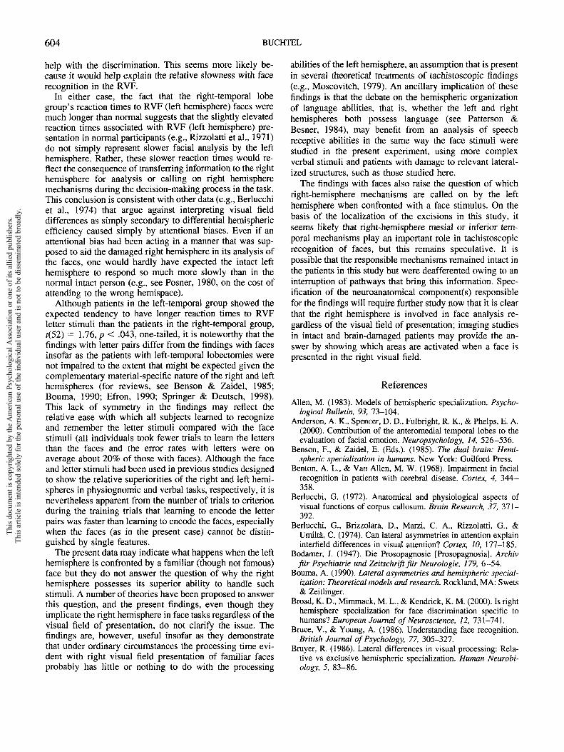
interact significantly with group, as might have occurred ifthe left hemisphere in the right-temporal lobe group hadbeen faster than the right hemisphere in the face task. Asseen in Table 1, the slowest reaction times were associatedwith RVF presentation of faces in the right-temporal group.However, the three-way interaction suggested by this ob-servation (Task X Group X Size) was not significant, F(l,50) = 1.27, ns, nor was the four-way interaction includingvisual field, F(l, 50) = 0.06, ns. It is noteworthy, however,that the variance associated with these slow reaction times isalso considerably higher than in the other subgroups. Usinga nonparametric test, the reaction times for faces shown inthe RVF do indeed tend to be slower for the right-temporalgroup with large removals than for the equivalent left-temporal group (U = 8.0, Z = 2.32, p < .021).
There was no significant effect of time since surgery ineither reaction times or errors, nor did this variable interactsignificantly with side or size of the removal. The reactiontimes associated with left and right visual fields of theright-temporal lobe patients are shown in Figure 2. Noconsistent differences between groups emerged in an anal-ysis of the conscious strategy (or lack of strategy) in carry-ing out the tasks, nor were there any consistent trends in theparticipants' reaction times or accuracies in recognizing thescrambled faces (Figure IB).
Discussion
Several explanations have been proposed to account forthe presence of hemispheric asymmetries in cognitive andperceptual tasks (see Allen, 1983, for a review of most ofthese approaches). In attempting to explain the faster andmore accurate recognition of faces in the left visual field,
MEDIAN REACTION TIME TO FACES
RIGHT-TEMPORAL GROUP
1400 r
1200
tc.a 1000
800
600
400
400 600 800 IOOO I2OO 1400
RIGHT VISUAL FIELD RT (msec)
Figure 2. Relationship between reaction times (RTs) to facestimuli presented in the right visual field and left visual field for thepatients with right-temporal lobe excisions. Each point representsa single patient. Points below the line are from patients with fasterRTs with left visual field presentation than with right visual fieldpresentation. Solid circles = patients with large excisions of me-sial temporal lobe tissue; open circles = patients with smallexcisions of mesial temporal lobe tissue.
This
doc
umen
t is c
opyr
ight
ed b
y th
e A
mer
ican
Psy
chol
ogic
al A
ssoc
iatio
n or
one
of i
ts a
llied
pub
lishe
rs.
This
arti
cle
is in
tend
ed so
lely
for t
he p
erso
nal u
se o
f the
indi
vidu
al u
ser a
nd is
not
to b
e di
ssem
inat
ed b
road
ly.
LEFT AND RIGHT HEMISPHERE CONTRIBUTIONS 603
two dimensions have been explored. One dimension con-cerns whether visual field differences are a direct or only anindirect consequence of the physiological substrate acti-vated in the task. The possibility that large visual fielddifferences arise because of an attentional bias has beenproposed by Kinsbourne (1970), who argued that withoutsuch an attentional bias the difference between visual fieldswould only be in the order of a few milliseconds, reflectingcommissural transmission time. Other authors (e.g., Berluc-chi, 1972; Rizzolatti, 1979) have proposed that the visualfield differences directly reflect the relative competencies ofthe two hemispheres, and they have obtained clear experi-mental support for their conclusion by randomly mixingtrials of face and letter discrimination (Berlucchi, Brizzo-lara, Marzi, Rizzolatti, & Umilta, 1974; see also the discus-sion of face familiarity in the introduction to the presentarticle). The second dimension focuses on the nature of thedifferences in competency between the two hemispheres.One approach is to conclude that a difference of a certainnumber of milliseconds between discrimination times ofright and left visual field presentation results from onehemisphere taking that number of milliseconds more thanthe other in carrying out the analysis. That is, both hemi-spheres are "competent," but one is more competent thanthe other (Umilta, Rizzolatti, Anzola, Luppino, & Porro,1985; see also Zaidel, 1983, for a discussion of the possiblecontinuum between competency and noncompetency). Theresults of work with commissurotimized patients have con-tributed to the popularity of the notion of bilateral compe-tency (see Sperry, 1974; Zaidel, 1994); work with chimericfaces (faces made up of two different faces split along thevertical meridian) has shown that split-brain patients canname faces shown in the right visual field and point with theleft hand to a face that had been shown in the left visual fieldamong a set of distractors (Levy, Trevarthen, & Sperry,1972; see also Levy 1974). Although these split-brain stud-ies show a potential role of the left hemisphere in faceperception and discrimination, the most accurate perfor-mance was seen with left visual field (right hemisphere)presentation.
Alternatively, one of the hemispheres may be so incom-petent in the particular analysis required that it never con-tributes to the final decision but rather transfers the infor-mation that it has to the other, competent hemisphere foranalysis. In this case, visual field differences would reflectthe time lost in transferring information plus any addedanalysis time caused by the degraded nature of the infor-mation that is received after transfer (poor quality would beattributed to distortions caused by the transfer process itselfand perhaps to an inappropriate initial elaboration carriedout by the incompetent hemisphere), plus any additionaltime taken for transfer of the motor output to the hemispherecontrolling the response if this is different from the onemaking the decision to respond. This pattern appears to beappropriate for certain verbal tasks (Bruyer, 1986; Umilta etal., 1985). Finally, there may be tasks in which the twohemispheres are able to carry out the necessary analyses inparallel; which hemisphere is ultimately responsible fororganizing the response would depend on whether the time
lost in transfer of the motor command from the slowerhemisphere to the hemisphere that controls the response ismore or less than the difference in analysis times of the twohemispheres (Bruyer, 1986; Umilta et al., 1985).
The present results with faces support the idea that right-hemisphere mechanisms are used in tachistoscopic facerecognition tasks regardless of the visual field of presenta-tion, and the findings therefore do not support the hypoth-esis that the left hemisphere can act alone in recognizingfaces in the tachistoscopic situation tested in this study.Before rejecting the hypothesis of independent left-hemi-sphere analysis, however, an alternative explanation needsto be evaluated. It is conceivable that the very slow reactiontimes to stimuli in the right visual field stimuli in theright-temporal group represent the usual time taken byleft-hemisphere mechanisms to carry out the task (e.g.,using "concrete structural codes"; Polster & Rapcsak,1996), with these long reaction times not normally beingseen, because in the intact brain the more efficient righthemisphere mechanisms "win" in the race between inter-hemispheric communication and intrahemispheric analysisas described above. However, if this were the case onemight have expected to see a visual field difference in favorof the right visual field, whereas the opposite was found.Furthermore, the reaction times in the right visual field arevery highly correlated with the reaction times in the leftvisual field (Figure 2; r = .832, p < .001), increasing thelikelihood that the parallel increases in reaction times inboth visual fields reflect the consequence of a disruption ofmechanism(s) ordinarily subserved by the damaged righthemisphere. (Although it is true that high correlations be-tween reaction times within an individual could reflect asubject variable rather than the activation of a single mech-anism, it can be argued that independent activities of the leftand right hemispheres in the task in these individuals wouldhave led to less concordance between the LVF and RVFreaction times.)
Thus, we are left with interpretations that implicate theright hemisphere in the performance of this face discrimi-nation task regardless of the field of presentation of thestimulus. Interestingly, this means that although the studywas designed to test the abilities of the noncompetent hemi-sphere, the findings presumably reflect the functions of thecompetent hemisphere. There are at least two ways the righthemisphere damage may be involved in producing the pat-tern of findings. First, under normal circumstances the righthemisphere may form a recognition template, presumablythe outcome of the initial structural encoding of Bruce andYoung (1986), which is stored bilaterally and can be usedby each hemisphere independently when a stimulus arrivesfrom the associated visual field (though if this occurs onewould still need to explain why the left hemisphere remainsslower than the right hemisphere using the template). In theabsence of such a template, as in the right-temporal lobegroup in this study, responses to both LVF and RVF stimuliwould be slowed. The second possibility is that the righthemisphere may be called on in all phases of the task for
This
doc
umen
t is c
opyr
ight
ed b
y th
e A
mer
ican
Psy
chol
ogic
al A
ssoc
iatio
n or
one
of i
ts a
llied
pub
lishe
rs.
This
arti
cle
is in
tend
ed so
lely
for t
he p
erso
nal u
se o
f the
indi
vidu
al u
ser a
nd is
not
to b
e di
ssem
inat
ed b
road
ly.
604 BUCHTEL
help with the discrimination. This seems more likely be-cause it would help explain the relative slowness with facerecognition in the RVF.
In either case, the fact that the right-temporal lobegroup's reaction times to RVF (left hemisphere) faces weremuch longer than normal suggests that the slightly elevatedreaction times associated with RVF (left hemisphere) pre-sentation in normal participants (e.g., Rizzolatti et al., 1971)do not simply represent slower facial analysis by the lefthemisphere. Rather, these slower reaction times would re-flect the consequence of transferring information to the righthemisphere for analysis or calling on right hemispheremechanisms during the decision-making process in the task.This conclusion is consistent with other data (e.g., Berlucchiet al., 1974) that argue against interpreting visual fielddifferences as simply secondary to differential hemisphericefficiency caused simply by attentional biases. Even if anattentional bias had been acting in a manner that was sup-posed to aid the damaged right hemisphere in its analysis ofthe faces, one would hardly have expected the intact lefthemisphere to respond so much more slowly than in thenormal intact person (e.g., see Posner, 1980, on the cost ofattending to the wrong hemispace).
Although patients in the left-temporal group showed theexpected tendency to have longer reaction times to RVFletter stimuli than the patients in the right-temporal group,f(52) = 1.76, p < .043, one-tailed, it is noteworthy that thefindings with letter pairs differ from the findings with facesinsofar as the patients with left-temporal lobectomies werenot impaired to the extent that might be expected given thecomplementary material-specific nature of the right and lefthemispheres (for reviews, see Benson & Zaidel, 1985;Bouma, 1990; Efron, 1990; Springer & Deutsch, 1998).This lack of symmetry in the findings may reflect therelative ease with which all subjects learned to recognizeand remember the letter stimuli compared with the facestimuli (all individuals took fewer trials to learn the lettersthan the faces and the error rates with letters were onaverage about 20% of those with faces). Although the faceand letter stimuli had been used in previous studies designedto show the relative superiorities of the right and left hemi-spheres in physiognomic and verbal tasks, respectively, it isnevertheless apparent from the number of trials to criterionduring the training trials that learning to encode the letterpairs was faster than learning to encode the faces, especiallywhen the faces (as in the present case) cannot be distin-guished by single features.
The present data may indicate what happens when the lefthemisphere is confronted by a familiar (though not famous)face but they do not answer the question of why the righthemisphere possesses its superior ability to handle suchstimuli. A number of theories have been proposed to answerthis question, and the present findings, even though theyimplicate the right hemisphere in face tasks regardless of thevisual field of presentation, do not clarify the issue. Thefindings are, however, useful insofar as they demonstratethat under ordinary circumstances the processing time evi-dent with right visual field presentation of familiar facesprobably has little or nothing to do with the processing
abilities of the left hemisphere, an assumption that is presentin several theoretical treatments of tachistoscopic findings(e.g., Moscovitch, 1979). An ancillary implication of thesefindings is that the debate on the hemispheric organizationof language abilities, that is, whether the left and righthemispheres both possess language (see Patterson &Besner, 1984), may benefit from an analysis of speechreceptive abilities in the same way the face stimuli werestudied in the present experiment, using more complexverbal stimuli and patients with damage to relevant lateral-ized structures, such as those studied here.
The findings with faces also raise the question of whichright-hemisphere mechanisms are called on by the lefthemisphere when confronted with a face stimulus. On thebasis of the localization of the excisions in this study, itseems likely that right-hemisphere mesial or inferior tem-poral mechanisms play an important role in tachistoscopicrecognition of faces, but this remains speculative. It ispossible that the responsible mechanisms remained intact inthe patients in this study but were deafferented owing to aninterruption of pathways that bring this information. Spec-ification of the neuroanatomical component(s) responsiblefor the findings will require further study now that it is clearthat the right hemisphere is involved in face analysis re-gardless of the visual field of presentation; imaging studiesin intact and brain-damaged patients may provide the an-swer by showing which areas are activated when a face ispresented in the right visual field.
References
Allen, M (1983). Models of hemispheric specialization. Psycho-logical Bulletin, 93, 73-104.
Anderson, A. K., Spencer, D. D., Fulbright, R. K., & Phelps, E. A.(2000). Contribution of the anteromedial temporal lobes to theevaluation of facial emotion. Neuropsychology, 14, 526-536.
Benson, F., & Zaidel, E. (Eds.). (1985). The dual brain: Hemi-spheric specialization in humans. New York: Guilford Press.
Benton, A. L., & Van Allen, M. W. (1968). Impairment in facialrecognition in patients with cerebral disease. Cortex, 4, 344-358.
Berlucchi, G. (1972). Anatomical and physiological aspects ofvisual functions of corpus callosum. Brain Research, 37, 371-392.
Berlucchi, G., Brizzolara, D., Marzi, C. A., Rizzolatti, G., &Umilta, C. (1974). Can lateral asymmetries in attention explaininterfield differences in visual attention? Cortex, 10, 177-185.
Bodamer, J. (1947). Die Prosopagnosie [Prosopagnosia]. Archivfur Psychiatrie und Zeitschrift fur Neurologic, 179, 6-54.
Bouma, A. (1990). Lateral asymmetries and hemispheric special-ization: Theoretical models and research. Rockland, MA: Swets& Zeitlinger.
Broad, K. D., Mimmack, M. L., & Kendrick, K. M. (2000). Is righthemisphere specialization for face discrimination specific tohumans? European Journal of Neuroscience, 12, 731-741.
Bruce, V., & Young, A. (1986). Understanding face recognition.British Journal of Psychology, 77, 305-327.
Bruyer, R. (1986). Lateral differences in visual processing: Rela-tive vs exclusive hemispheric specialization. Human Neurobi-ology, 5, 83-86.
This
doc
umen
t is c
opyr
ight
ed b
y th
e A
mer
ican
Psy
chol
ogic
al A
ssoc
iatio
n or
one
of i
ts a
llied
pub
lishe
rs.
This
arti
cle
is in
tend
ed so
lely
for t
he p
erso
nal u
se o
f the
indi
vidu
al u
ser a
nd is
not
to b
e di
ssem
inat
ed b
road
ly.

LEFT AND RIGHT HEMISPHERE CONTRIBUTIONS 605
Buchtel, H., Campari, F., De Risio, C, & Rota, R. (1978). Hemi-spheric differences in discriminative reaction time to facialexpressions. Italian Journal of Psychology, 5, 159-169.
Charcot, J. M. (1883). Un cas de suppression brusque et iso!6e dala vision mentale del signes et des objets (formes et couleurs) [Acase of sudden and isolated suppression of mental images ofsigns and of objects (forms and colors)]. Progres Medical, 11,568-571.
Corsi, P. M. (1972). Human memory and the medial temporalregion of the brain. Unpublished doctoral thesis, McGillUniversity.
Damasio, A. R., Damasio, H., & Van Hoesen, G. W. (1982).Prosopagnosia: Anatomical basis and behavioral mechanisms.Neurology, 32, 331-341.
De Renzi, E. (1982). Disorders of space exploration and cogni-tion. New York: Wiley.
De Renzi, E. (1986). Prosopagnosia in two patients with CT scanevidence of damage confined to the right hemisphere. Neuro-psychologia, 24, 385-389.
De Renzi, E., & Spinnler, H. (1966). Visual recognition in patientswith unilateral cerebral disease. Journal of Nervous and MentalDisease, 142, 513-525.
Efron, R. (1990). The decline and fall of hemispheric specializa-tion. Hillsdale, NJ: Erlbaum.
Ettlin, T. M., Beckson, M., Benson, D. F., Langfitt, J. T., Amos,E. C., & Pineda, G. S. (1992). Prosopagnosia: A bihemisphericdisorder. Cortex, 28, 129-134.
Gauthier, I., Behrmann, M., & Tarr, M. J. (1999). Can facerecognition really be dissociated from object recognition? Jour-nal of Cognitive Neuroscience, 11, 349-370.
Geffen, G., Bradshaw, J. L., & Wallace, G. (1971). Interhemi-spheric effects on reaction time to verbal and non-verbal visualstimuli. Journal of Experimental Psychology, 87, 415-422.
Gellerman, L. W. (1933). Chance orders of alternating stimuli invisual discrimination experiments. Journal of Genetic Psychol-ogy, 42, 206-208.
Hilliard, R. D. (1973). Hemispheric laterality effects on a facialrecognition task in normal subjects. Cortex, 9, 246-258.
Jones, B. (1979). Lateral asymmetry in testing long-term memoryfor faces. Cortex, 15, 183-186.
Kanwisher, N., McDermott, J., & Chun, M. M. (1997). The fusi-form face area: A module in human extrastriate cortex special-ized for face perception. Journal of Neuroscience, 17, 4302-4311.
Kim, J. J., Andreasen, N. C., O'Leary, D. S., Wiser, A. K., Ponto,L. L., Watkins, G. L., & Hichwa, R. D. (1999). Direct compar-ison of the neural substrates of recognition memory for wordsand faces. Brain, 122, 1069-1083.
Kinsbourne, M. (1970). The cerebral basis of lateral asymmetriesin attention. Acta Psychologica, 33, 193-201.
Kittler, P., Turkewitz, G., & Goldberg, E. (1989). Shifts in hemi-spheric advantage during familiarization with complex visualpatterns. Cortex, 25, 27-32.
Landis, T., Cummings, J. G., Christen, L., Bogen, J. E., & Imhof,H. G. (1986). Are unilateral right posterior cerebral lesionssufficient to cause prosopagnosia? Clinical and radiologicalfindings in six additional patients. Cortex, 22, 243-252.
Levy, J. (1974). Psychological implications of bilateral asymme-try. In S. J. Dimond & J. G. Beaumont (Eds.), Hemispherefunction in the human brain (pp. 121-183). New York: Wiley.
Levy, J., Trevarthen, C., & Sperry, R. W. (1972). Perception ofbilateral chimeric figures following hemispheric deconnexion.Brain, 95, 61-78.
Marzi, C. A., & Berlucchi, G. (1977). Right visual field superiorityfor accuracy of recognition of famous faces in normals. Neuro-psychologia, 15, 751-756.
McGlone, J. (1980). Sex differences in human brain asymmetries:A critical survey. Behavioral and Brain Sciences, 3, 215-263.
McKeever, W. F., & Huling, M. D. (1971). Bilateral tachistoscopicword recognition as a function of hemisphere stimulated andinterhemispheric transfer time. Neuropsychologia, 9, 281-288.
Meadows, J. C. (1974). The anatomical basis of prosopagnosia.Journal of Neurology, Neurosurgery and Psychiatry, 37, 489-501.
Milner, B. (1968). Visual recognition and recall after right tem-poral-lobe excision in man. Neuropsychologia, 6, 191-209.
Milner, B. (1974). Hemispheric specialization: Scope and limits.In F. O. Schmitt & F. G. Worden (Eds.), The neurosciences:Third study program (pp. 75-89). Cambridge, MA: MIT Press.
Milner, B. (1975). Psychological aspects of focal epilepsy and itsneurosurgical management. In D. P. Purpura, J. K. Penry &R. D. Walter (Eds.), Advances in neurology, Vol. 8 (pp. 299-321). New York: Raven.
Morris, R. D., & Hopkins, W. D. (1993). Perception of humanchimeric faces by chimpanzees: Evidence for a right hemisphereadvantage. Brain and Cognition, 21, 111-122.
Moscovitch, M. (1979). Information processing and the cerebralhemispheres. In M. S. Gazzaniga (Ed.), Handbook of behavioralneurobiology. Vol. 2: Neuropsychology (pp. 379-446). NewYork: Plenum.
O'Craven, K. M., & Kanwisher, N. (2000). Mental imagery offaces and places activates corresponding stimulus-specific brainregions. Journal of Cognitive Neuroscience, 12, 1013-1023.
Patterson, K., & Besner, D. (1984). Is the right hemisphere liter-ate? Cognitive Neuropsychology, 1, 315-341.
Patterson, R., & Bradshaw, J. L. (1975). Differential hemisphericmediation of nonverbal visual stimuli. Journal of ExperimentalPsychology: Human Perception and Performance, 1. 246-252.
Polster, M. R., & Rapcsak, S. Z. (1996). Representations in learn-ing new faces: Evidence from prosopagnosia. Journal of theInternational Neuropsychological Society, 2, 240-248.
Posner, M. I. (1980). Orienting of attention. Quarterly Journal ofExperimental Psychology, 32, 3-25.
Reynolds, D. M., & Jeeves, M. A. (1978). A developmental studyof hemisphere specialization for recognition of faces in normalsubjects. Cortex, 14, 511-520.
Rizzolatti, G. (1979). Interfield differences in reaction times tolateralized visual stimuli in normal subjects. In I. Steel-Russell,M. W. van Hof & G. Berlucchi (Eds.), Structure and function ofcerebral hemispheres (pp. 390-399). London: Macmillan.
Rizzolatti, G., & Buchtel, H. A. (1977). Hemispheric superiority inreaction time to faces: A sex difference. Cortex, 13, 300-305.
Rizzolatti, G., Umilta, C., & Berlucchi, G. (1971). Opposite supe-riorities of the right and left cerebral hemispheres in discrimi-native reaction time to physiognomical and alphabetic material.Brain, 94, 431-442.
Ross, P., & Turkewitz, G. (1982). Changes in hemispheric advan-tage in processing facial information with increasing stimulusfamiliarization. Cortex, 18, 489-499.
Ross-Kossack, P., & Turkewitz, G. (1984). Relationship betweenchanges in hemispheric advantage during familiarization tofaces and proficiency in facial recognition. Neuropsycholo-gia, 22, 471-477.
Ross-Kossack, P., & Turkewitz, G. (1986). A micro and macrodevelopmental view of the nature of changes in complex infor-mation processing: A consideration of changes in hemispheric
This
doc
umen
t is c
opyr
ight
ed b
y th
e A
mer
ican
Psy
chol
ogic
al A
ssoc
iatio
n or
one
of i
ts a
llied
pub
lishe
rs.
This
arti
cle
is in
tend
ed so
lely
for t
he p
erso
nal u
se o
f the
indi
vidu
al u
ser a
nd is
not
to b
e di
ssem
inat
ed b
road
ly.

606 BUCHTEL
advantage during familiarization. In R. Bruyer (Ed.), Neuropsy-chology of face perception facial expression (pp. 125-145).Hillsdale, NJ: Erlbaum.
Schultz, R. T., Gauthier, I., Klin, A., Fulbright, R. K., Anderson,A. W., Volkmar, F., Skudlarski, P., Lacadie, C, Cohen, D. J., &Gore, J. C. (2000). Abnormal ventral temporal cortical activityduring face discrimination among individuals with autism andAsperger syndrome. Archives of General Psychiatry, 57, 331-340.
Sergent, J. (1982). About face: Left-hemisphere involvement inprocessing physiognomies. Journal of Experimental Psychol-ogy: Human Perception and Performance, 8, 1-14.
Sergent, J. (1987). Failures to confirm the spatial-frequency hy-pothesis: Fatal blow or healthy complication. Canadian Journalof Psychology, 41, 412-428.
Sergent, J., & Poncet, M. (1988). Patterns of perceptual impair-ments in two prosopagnosic patients. Journal of Clinical andExperimental Neuropsychology, 10, 49-51.
Sergent, J., & Villemure, J. G. (1989). Prosopagnosia in a righthemispherectomized patient. Brain, 112, 975-995.
Sperry, R. W. (1974). Lateral specialization in the surgicallyseparated hemispheres. In F. O. Schmitt & F. G. Worden (Eds.),The neurosciences: Third study program (pp. 5-19). Cam-bridge, MA: MIT Press.
Springer, S. P., & Deutsch, G. (1998). Left brain, right brain:Perspectives from cognitive neuroscience. New York: Freeman.
Tanaka, J. W., & Sengco, J. A. (1997). Features and their configura-tion in face recognition. Memory & Cognition, 25, 583-592.
Turkewitz, G., & Ross, P. (1983). Changes in visual field advan-tage for facial recognition: The development of a general pro-cessing strategy. Cortex, 19, 179-185.
Umilta, C., Brizzolara, D., Tabossi, P., & Fairweather, H.(1978). Factors affecting face recognition in the cerebralhemispheres: Familiarity and naming. In J. Requin (Ed.),Attention and performance, VII (pp. 363-374). Hillsdale, NJ:Erlbaum.
Umilta, C., Rizzolatti, G., Anzola, G. P., Luppino, G., & Porro, C.(1985). Evidence of interhemispheric transmission in lateralityeffects. Neuropsychologia, 23, 203-213.
Warrington, E. K., & James, M. (1967). An experimental investi-gation of facial recognition in patients with unilateral cerebrallesions. Cortex, 3, 317-326.
Zaidel, D. W. (1994). View of the world from a split-brain per-spective. In D. M. R. Critchley (Ed.), Neurological boundariesof reality (pp. 161-174). London: Farrand Press.
Zaidel, E. (1983). Advances and retreats in laterality research.Behavioral and Brain Sciences, 6, 523-528.
Received November 24, 2000Revision received May 17, 2001
Accepted May 18, 2001
Wanted: Your Old Issues!
As APA continues its efforts to digitize journal issues for the PsycARTICLESdatabase, we are finding that older issues are increasingly unavailable in our inventory.We are turning to our long-time subscribers for assistance. If you would like to donateany back issues toward this effort (preceding 1982), please get in touch with us atjournals @apa.org and specify the journal titles, volumes, and issue numbers that youwould like us to take off your hands. (Your donation is of course tax deductible.)Th
is d
ocum
ent i
s cop
yrig
hted
by
the
Am
eric
an P
sych
olog
ical
Ass
ocia
tion
or o
ne o
f its
alli
ed p
ublis
hers
.Th
is a
rticl
e is
inte
nded
sole
ly fo
r the
per
sona
l use
of t
he in
divi
dual
use
r and
is n
ot to
be
diss
emin
ated
bro
adly
.