Embed Size (px)
Citation preview

Lundy: Chapter 10/11 Lundy: Chapter 12 (traumatic injury to
peripheral nerves)
Reading
Lundy-Ekman. Neuroscience: Fundamentals for Rehabilitation, 4th Edition. W.B. Saunders Company, 2013.
Kandel et al. Principles of Neural Science, 5th Edition. McGraw Hill, 2012.
Tortura & Derrickson. Principles of anatomy and physiology, 13th Edition. Wiley. 2012.

Disorders of LMNs (Polio) Disorders of UMNs Basal Nuclei Movement Disorders
◦ Hypokinetic Disorders Parkinson’s Disease - seminar
◦ Hyperkinetic Disorders Huntington’s Disease - seminar
Cerebellar Disorders Cerebral Palsy
Overview

Have an understanding of different types of disorders that can affect the motor system
Gain an appreciation for difficulties experienced by people with CP and how, as an osteo, these can be mitigated
Learning Objectives

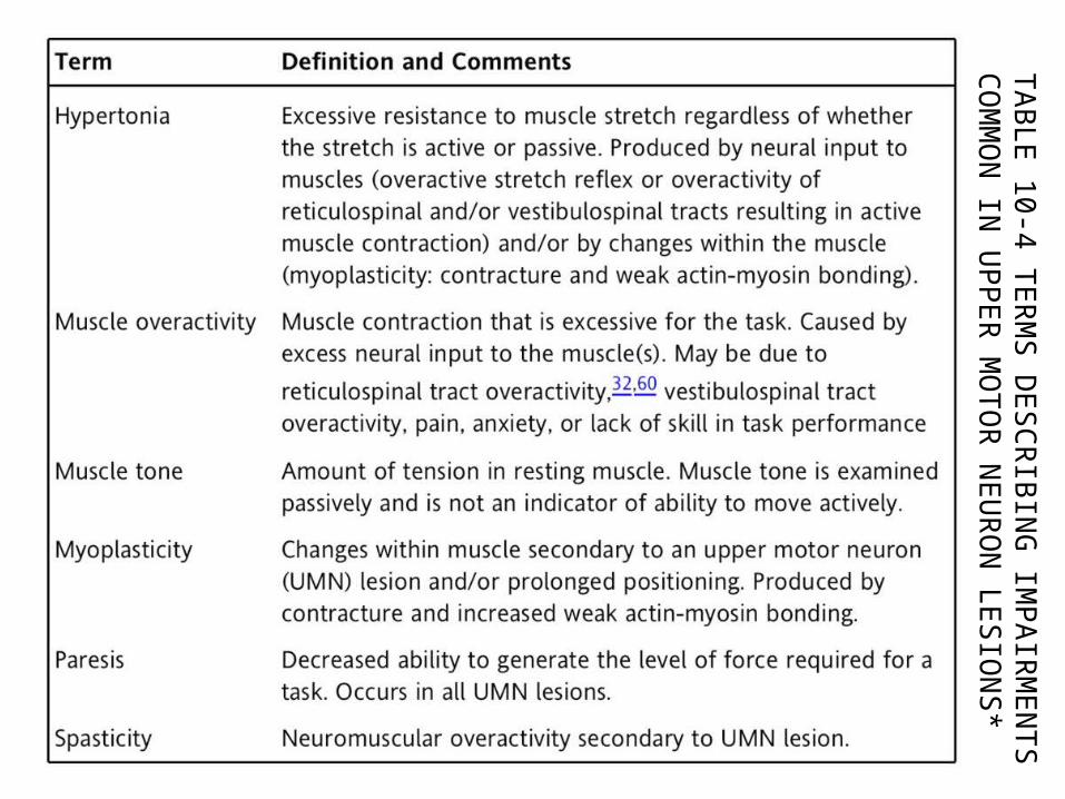
Paresis or paralysis◦ Decreased ability or inability to generate muscle force
Muscle atrophy◦ Loss of muscle bulk as pattern of muscle protein
production changes Involuntary muscle contraction
◦ Spasms, cramps, fasciculations (eye twitch), tremors Abnormal muscle tone
◦ Muscle tone: resistance to stretch in resting muscle◦ Hypotonia and flaccidity are abnormally low resistance to,
or lack of resistance to passive stretch◦ Hypertonia is abnormally strong resistance to passive
stretch
Signs of motor neuron lesions

Caused by: traumatic injuries, infections like Poliomyelitis (next), degenerative or vascular disorders, tumors
Interrupting LMN signals to muscle decreases or prevents muscle contraction, leading to:◦ Loss of reflexes◦ Atrophy◦ Flaccid paralysis◦ Fibrillations
Disorders of LMNs

Poliovirus affects only LMNs Onset marked by fever, severe
headache, stiff neck/back, deep muscle pain/weakness
Enters body from faeces-contaminated water (pool)
Destroys cell bodies of motor neurons (ventral horn) and, therefore, denervates muscle fibers (Wallerian degeneration – plasticity lecture)
Unilateral Victim may die from paralysis of
respiratory muscles or cardiac arrest (if neurons in medulla oblongata are destroyed)
Poliomyelitis

Horizontal section of a spinal cord post polio. The section has been stained for myelin, so that the white matter appears dark. Loss of cell bodies is visible in the anterior (ventral) horn.

Spinal cord section
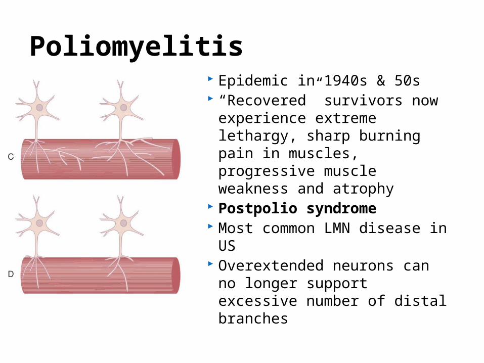
Epidemic in 1940s & 50s “Recovered” survivors now
experience extreme lethargy, sharp burning pain in muscles, progressive muscle weakness and atrophy
Postpolio syndrome Most common LMN disease
in US Overextended neurons can
no longer support excessive number of distal branches
Poliomyelitis

UMNs damaged by spinal cord injury, spastic CP, MS, trauma, loss of blood supply (stroke)
Changes in movement control include:◦ Paresis or paralysis◦ Loss of fractionation of movement◦ Abnormal reflexes◦ Velocity-dependent hypertonia
Disorders of UMNs

Paresis or paralysis◦ Paresis common following stroke, CP, trauma◦ Leads to muscle disuse -> secondary changes in muscles
and nervous system (causes adaptive muscle contracture and atrophy, and decreases motor cortex representation, further exacerbating paresis)
◦ Paralysis occurs from complete spinal cord lesion◦ Loss of all somatosensory and motor function below level
of lesion Loss of fractionation of movement
◦ Fractionation is ability to move individual muscles independently (piano)
◦ Lower limbs: interferes with dorsiflexion of ankle – plantarflexion instead
Changes with UMN Lesions

Abnormal reflexes◦ Babinski’s sign (usual for infants
under 7mo – WHY?)◦ Extension of big toe in response to
stroking sole of foot – mechanism not understood
Velocity-dependent hypertonia◦ Limits joint range of motion,
interfering with function E.g., toe walking due to lack of
ankle dorsiflexion (prevents heels from touching floor)
Changes with UMN Lesions

Disease destroys only somatic motor neurons UMNs and LMNs bilaterally resulting in UMN and
LMN signs concurrently Paresis, myoplasticity (changes in the muscles),
hyperreflexia (overactive reflexes), Babinski’s sign, atrophy, fasciculations, fibrillations
Loss LMNs in cranial nerves causes difficulty breathing, swallowing, speaking
Death occurs ~5yrs, onset 50yrs+ Astrocytes fail to clean up excessive glutamate,
causing excitotoxicity (more on this in plasticity lecture)
Amyotrophic Lateral Sclerosis (ALS)


Spinal cord section, stained for myelin, showing loss of upper motor neurons (UMNs) in amyotrophic lateral sclerosis (ALS). The loss is visible dorsolaterally, where the lateral corticospinal and rubrospinal axons should be, and ventromedially, where the medial UMNs should be
Amyotrophic Lateral Sclerosis (ALS)

Extrapyramidal Tracts
www.baileybio.com/plogger/images/biology/powerpoint_-nervous_system/spinal_cord_tracts.jpg


TAB
LE 1
0-4
TE
RM
S D
ES
CR
IBIN
G IM
PAIR
ME
NTS
C
OM
MO
N IN
UPPE
R M
OTO
R N
EU
RO
N LE
SIO
NS
*
TAB
LE 1
0-4
TE
RM
S D
ES
CR
IBIN
G IM
PAIR
ME
NTS
C
OM
MO
N IN
UPPE
R M
OTO
R N
EU
RO
N LE
SIO
NS
*

Range from hypokinetic (too little movt) to hyperkinetic (too much movt) disorders
BN INHIBIT motor thalamus, PPN and MLR Excessive inhibition -> hypokinetic disorder (PD) Inadequate inhibition -> hyperkinetic disorder (HD)
Basal Nuclei Disorders
Red facilitationBlack inhibition

Seminar topic: Emma, Chelsea, Amanda, Russell
Hypokinetic disorder Akinetic/rigid 50% Tremor-dominant 40% Mixed 10% Let’s cover pathology:
◦ Death of dopaminergic cells in substantia nigra compacta (cell death occurs to 80% before signs of PD become apparent)
Parkinson’s Disease


Loss of dopamine to putamen reduces level of inhibition provided to GP internus
Therefore GP internus provides excessive inhibition to 3 pathways
Downstream effects are:
Pathology of PD
Decreased voluntary movts
Excessive contraction of postural muscles
Loss of automatic gait

Pathology of PD


Seminar topic: Kaspara, Matthew, Bryan Hyperkinetic disorder Autosomal dominant hereditary disorder Degeneration of striatum and cerebral cortex Characterised by chorea (dance) – jerky,
involuntary movts that interfere with daily functioning ad cognitive decline
Let’s cover pathology:◦ Death of cells in striatum lead to excessive
involuntary movt (chorea)
Huntington’s Disease

Degeneration (HD disease) decreases signals from GP internus
Leading to disinhibition (not enough inhibition!) of motor thalamus and PPN
Downstream effects are:
Pathology of HD
Insufficient activity to girdle muscles
hyperkinesia
Not shown: Subthalamic nucleus output decr. due to enhanced activity of Gpe.MLR omitted due to lack of data of function in HD.


Ataxia: inability to coordinate muscular movements
Symptoms: unable to close eyes touch nose, staggered walking, changed speech pattern (ringing any alcohol-related bells???!)
Alcohol inhibits activity of cerebellum http://www.hhmi.org/biointeractive/
neuroscience/spinocerebellar_ataxia.html
Cerebellar Disorders

ICD-10, WHO classification of CP:◦ “difficult to define but is conventionally described
as a group of motor disorders with or without associated sensory and intellectual deficits where the cause is injury to the immature brain”
Movement and postural disorder caused by permanent, non-progressive damage to developing brain (in utero 80% of time)
Cerebral Palsy

Brain damage may have been:◦ Asphyxia (oxygen deprivation during birth)◦ Infection◦ Trauma◦ Metabolic disorder◦ Unknown
Type classification:◦ Spastic◦ Dyskinetic◦ Ataxic◦ Hypotonic◦ Mixed
Cerebral Palsy

Type classification:◦ Spastic
excessive involuntary skeletal muscle contraction (high muscle tone, stiff muscles -> toe walking)
Damage is to axons adjacent to lateral ventricles In developmental spastic CP: Lesion affects corticospinal and corticobulbar tracts During normal development corticospainal axon may
synapse with agonist muscle as well as synergists and antagonists. Syngerists/antagonists normally eliminated by age 4
Inappropriate connections not eliminated during dev. CP cause co-contraction of antagonist muscles, interfering with performance
Also disinhibition of reticulospinal tract causing abnormal synergies, and overactivation of neck reflexes (collicular signals to reticulospianl tract)
Cerebral Palsy Types

Type classification:◦ Spastic◦ Dyskinetic
muscle tone fluctuates from hypotonia to hypertonia Choreoathetosis – involuntary choreiform (jerky,
abrupt, irregular movts, lack of coordination) Athetosis – slow, writhing movts Dystonia – involuntary sustained muscle contraction Damage is in basal nuclei and ventrolateral thalamus
Cerebral Palsy Types

Type classification:◦ Spastic◦ Dyskinetic◦ Ataxic
Incoordination, weakness, shaking during voluntary movt
Damage is in the cerebellum
Cerebral Palsy Types

Type classification:◦ Spastic◦ Dyskinetic◦ Ataxic◦ Hypotonic
Low muscle tone (floppy), little or no ability to move Damage site unknown
Cerebral Palsy Types

Type classification:◦ Spastic◦ Dyskinetic◦ Ataxic◦ Hypotonic◦ Mixed
If more than one type of abnormal movement coexist Philip has choreoathetosis and spasticity
CP also classified as:◦ Hemiplegic, quadriplegic, diplegic (upper limbs
less severely affected)
Cerebral Palsy Types

Problems with fine motor skills (writing) Walking or balance (or both) Involuntary movements (some types of CP) Difficulties with speech, eating, swallowing Cognitive impairment Visual or auditory impairment
Cerebral Palsy symptoms