Embed Size (px)
DESCRIPTION
Lecture XIII. Brain Diseases I - Parkinsonism. Bio 3411 Wednesday October 13, 2010. Brain Diseases I. NEUROSCIENCE THE BRAIN ATLAS 3 rd ed PageFigureFeature 46518.10Substantia Nigra in Parkinsonism 466Box 18AParkinson’s Disease: An Opportunity… - PowerPoint PPT Presentation
Citation preview
Lecture XIII. Brain Diseases I -
ParkinsonismBio 3411 Wednesday
October 13, 2010
October 13, 2010 1Lecture XIII. Brain Diseases - I.
October 13, 2010 Lecture XIII. Brain Diseases - I. 2
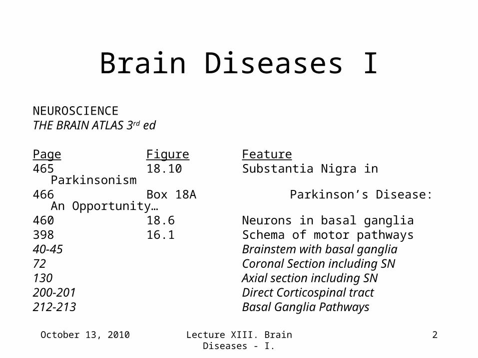
Brain Diseases I
NEUROSCIENCETHE BRAIN ATLAS 3rd ed
Page Figure Feature465 18.10 Substantia Nigra in Parkinsonism466 Box 18A Parkinson’s Disease: An Opportunity…460 18.6 Neurons in basal ganglia398 16.1 Schema of motor pathways40-45 Brainstem with basal ganglia72 Coronal Section including SN130 Axial section including SN200-201 Direct Corticospinal tract212-213 Basal Ganglia Pathways
October 13, 2010 Lecture XIII. Brain Diseases - I. 3
References†Barker RA, Dunnett SB 1999 Functional integration of neural grafts in
Parkinson’s disease. Nature Neuroscience 2:1047-1048.†Gulie S 2007 A shock to the system: to slow the progress of Parkinson’s
disease, doctors planted electrodes deep in my brain. Then they turned on the juice. [http://www.wired.com/wired/archive/15.03/brainsurgery.html?pg=2&topic=brainsurgery&topic_set=] (check out the video!!)
†Perlmutter JS 2006 [http:/www.Harrisonline.Com/audio/parkinsons.Mp3]†Starr PA, Vitek JL, Bakay RAE 1998 Ablative surgery and deep brain
stimulation for Parkinson’s disease. Neurosurgery 43:989-1015.†Wichmann T, DeLong MR 1998 Models of basal ganglia function and
pathophysiology of movement disorders. Neurosurgery Clinics of North America 9:223-236.
_______
†Articles/Abstract/Audio posted on website.
October 13, 2010 Lecture XIII. Brain Diseases - I. 4
What this lecture is about:
• Motor Systems - Reprise
• Pyramidal and Extrapyramidal (Basal
ganglia)
• Parkinsonism a Movement Disorder
• Mechanisms and Treatment Strategies
October 13, 2010 Lecture XIII. Brain Diseases - I. 5
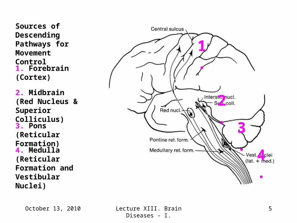
Sources of Descending Pathways for Movement Control
4.
3.
2.
1.
4. Medulla (Reticular Formation and Vestibular Nuclei)
3. Pons (Reticular Formation)
2. Midbrain (Red Nucleus & Superior Colliculus)
1. Forebrain (Cortex)
October 13, 2010 Lecture XIII. Brain Diseases - I. 6
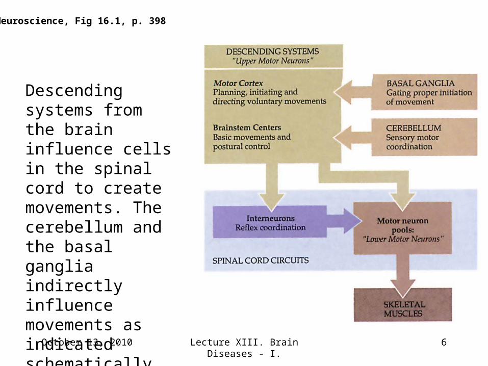
Descending systems from the brain influence cells in the spinal cord to create movements. The cerebellum and the basal ganglia indirectly influence movements as indicated schematically here.
Neuroscience, Fig 16.1, p. 398
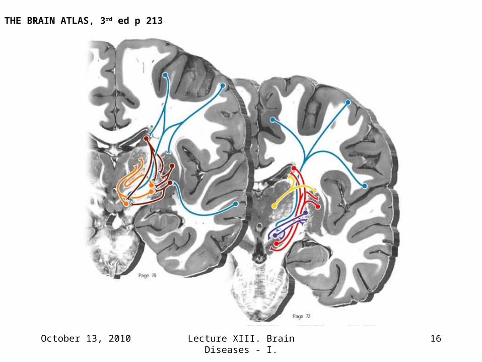
The basal ganglia inhibit unwanted movement patterns
and permit selected ones. They may also inhibit
unwanted mental activities such as inappropriate
utterances, and permit selected ones, such as proper
speech.
Basal Ganglia (Extrapyramidal) Pathways.
October 13, 2010 7Lecture XIII. Brain Diseases - I.
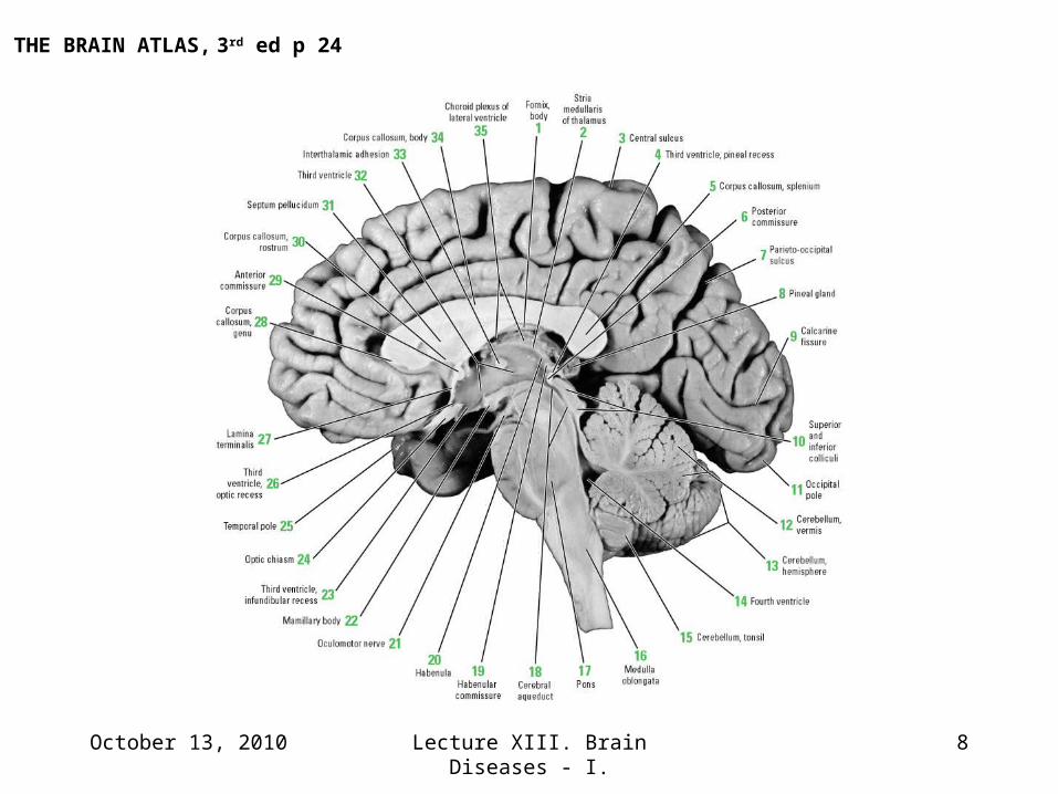
October 13, 2010 Lecture XIII. Brain Diseases - I. 8
THE BRAIN ATLAS, 3rd ed p 24
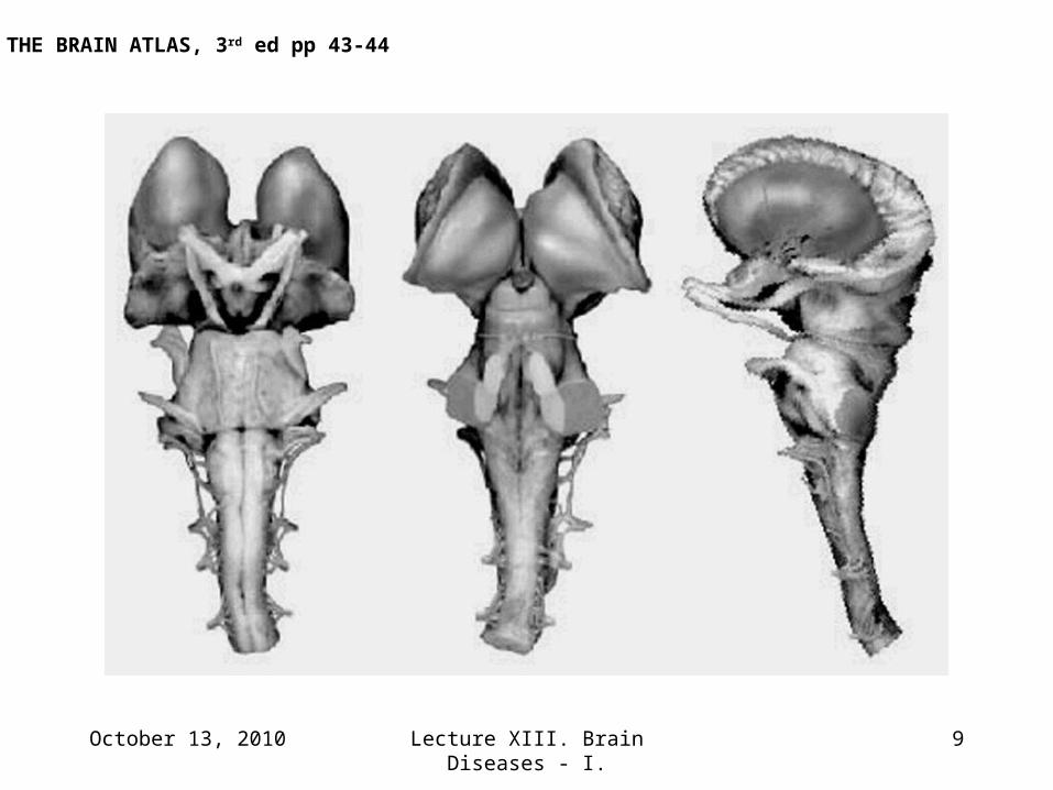
October 13, 2010 Lecture XIII. Brain Diseases - I. 9
THE BRAIN ATLAS, 3rd ed pp 43-44
October 13, 2010 Lecture XIII. Brain Diseases - I. 10
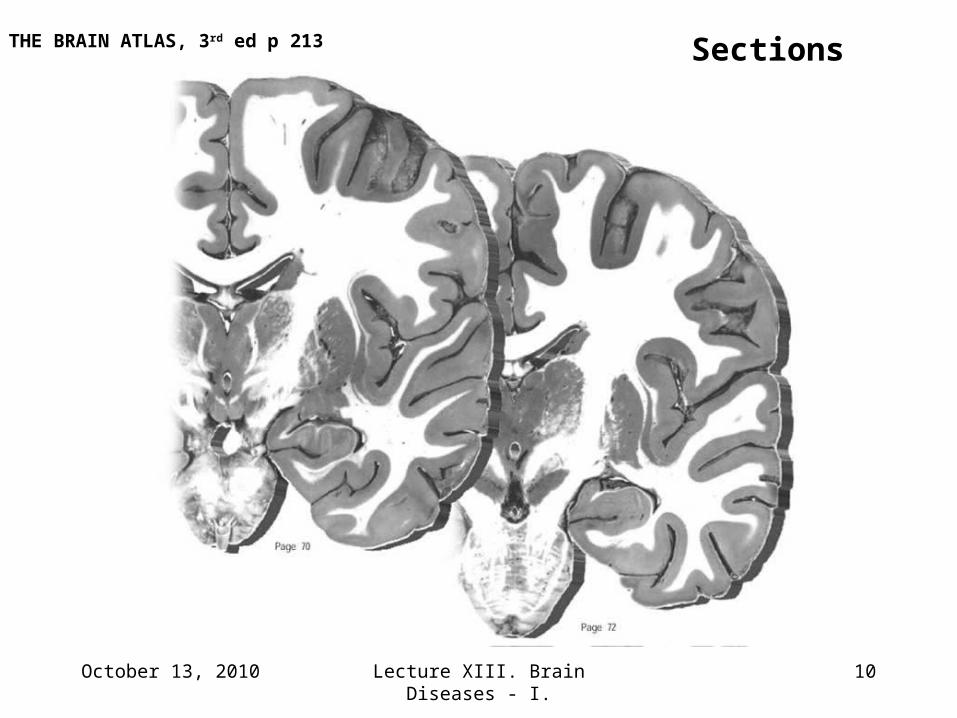
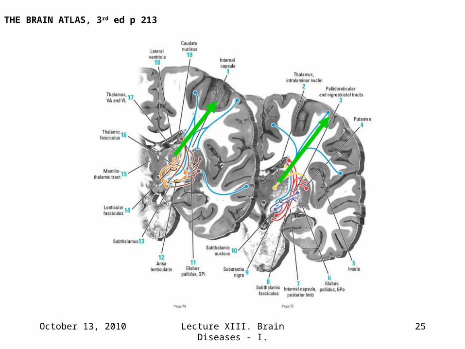
SectionsTHE BRAIN ATLAS, 3rd ed p 213
October 13, 2010 Lecture XIII. Brain Diseases - I. 11
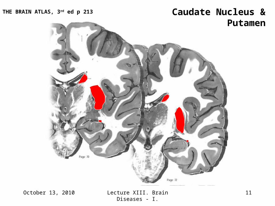
Caudate Nucleus & PutamenTHE BRAIN ATLAS, 3rd ed p 213
October 13, 2010 Lecture XIII. Brain Diseases - I. 12
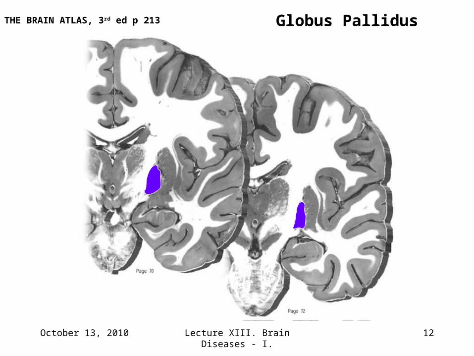
Globus PallidusTHE BRAIN ATLAS, 3rd ed p 213
October 13, 2010 Lecture XIII. Brain Diseases - I. 13
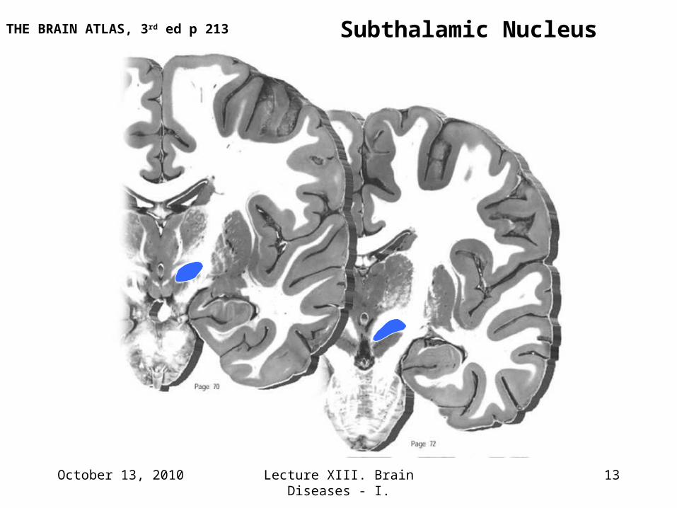
Subthalamic NucleusTHE BRAIN ATLAS, 3rd ed p 213
October 13, 2010 Lecture XIII. Brain Diseases - I. 14
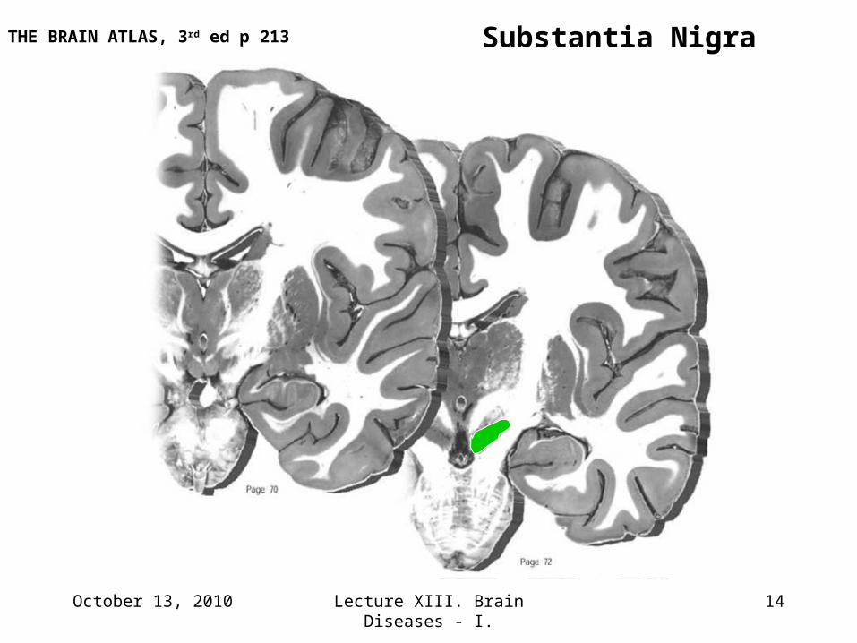
Substantia NigraTHE BRAIN ATLAS, 3rd ed p 213
October 13, 2010 Lecture XIII. Brain Diseases - I. 15
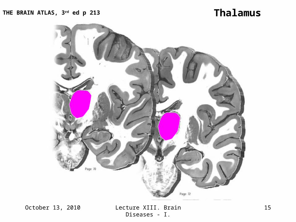
ThalamusTHE BRAIN ATLAS, 3rd ed p 213
October 13, 2010 Lecture XIII. Brain Diseases - I. 16
THE BRAIN ATLAS, 3rd ed p 213
October 13, 2010 Lecture XIII. Brain Diseases - I. 17
Movie Clip # 1
Patient(s) with tremor and paralysis
October 13, 2010 Lecture XIII. Brain Diseases - I. 18
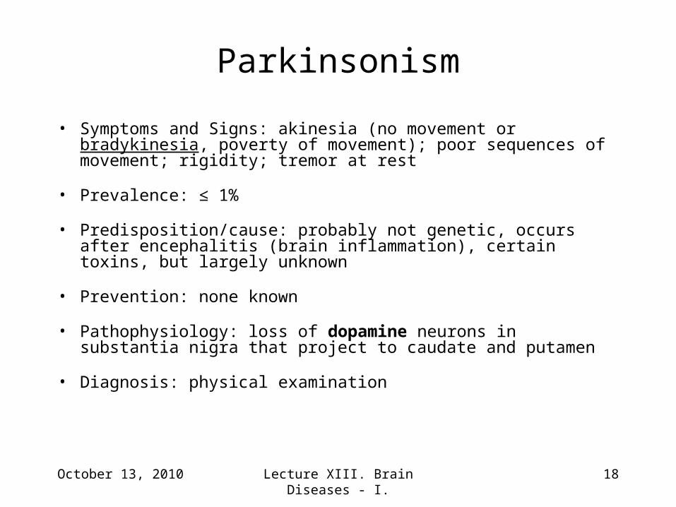
• Symptoms and Signs: akinesia (no movement or bradykinesia, poverty of movement); poor sequences of movement; rigidity; tremor at rest
• Prevalence: ≤ 1%
• Predisposition/cause: probably not genetic, occurs after encephalitis (brain inflammation), certain toxins, but largely unknown
• Prevention: none known
• Pathophysiology: loss of dopamine neurons in substantia nigra that project to caudate and putamen
• Diagnosis: physical examination
Parkinsonism
October 13, 2010 Lecture XIII. Brain Diseases - I. 19
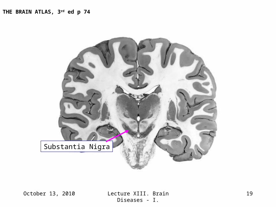
Substantia Nigra
THE BRAIN ATLAS, 3rd ed p 74
October 13, 2010 Lecture XIII. Brain Diseases - I. 20
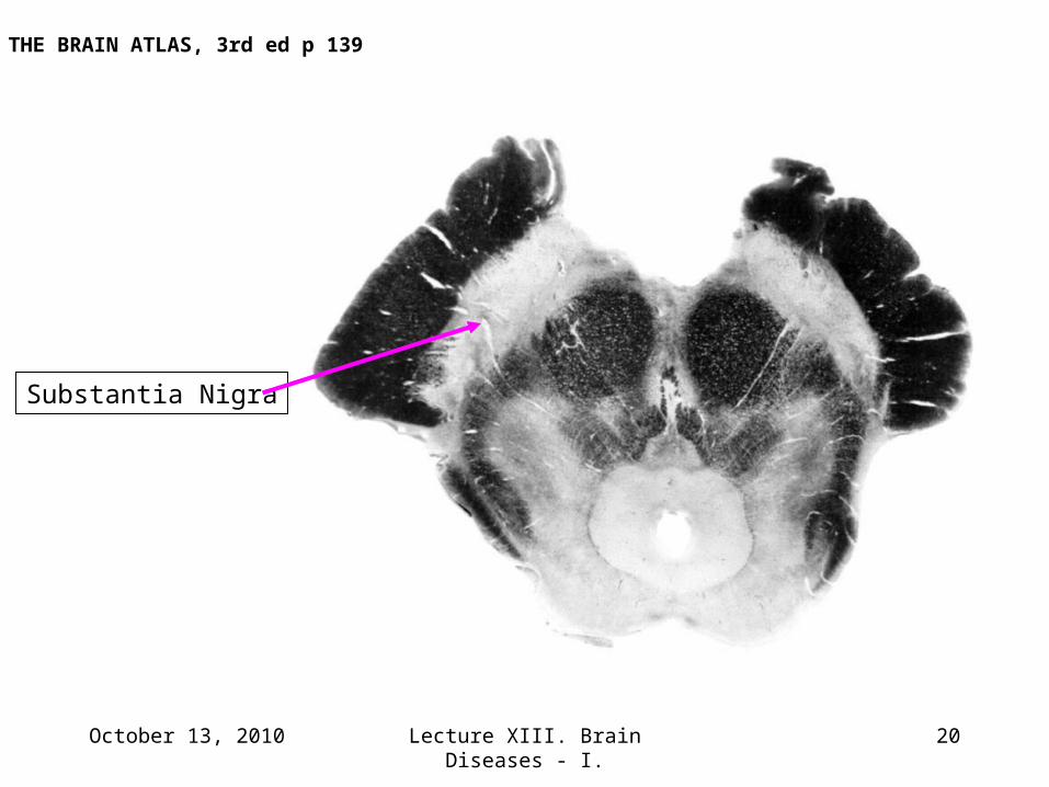
THE BRAIN ATLAS, 3rd ed p 139
Substantia Nigra
October 13, 2010 Lecture XIII. Brain Diseases - I. 21
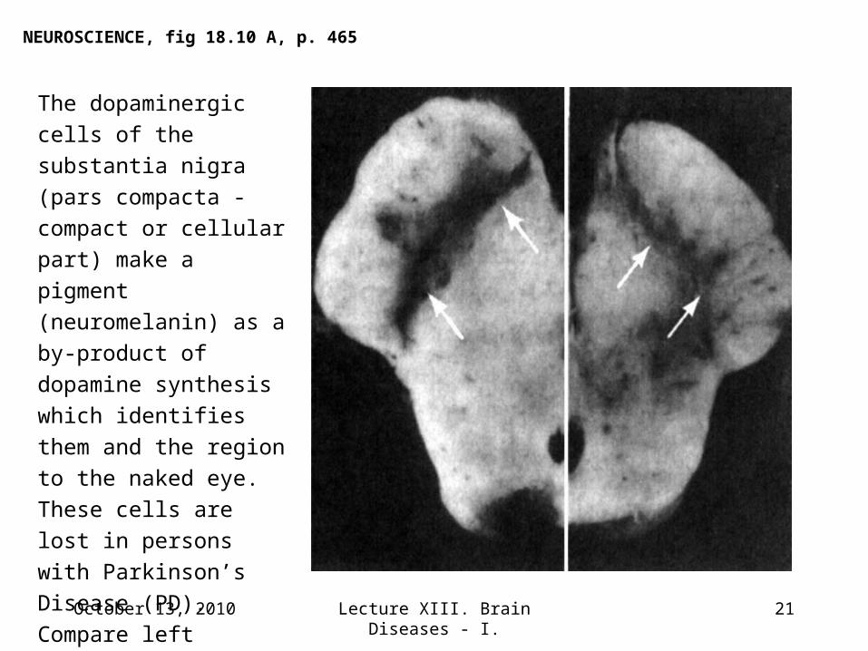
The dopaminergic cells of
the substantia nigra (pars
compacta - compact or
cellular part) make a
pigment (neuromelanin)
as a by-product of
dopamine synthesis
which identifies them and
the region to the naked
eye. These cells are lost
in persons with
Parkinson’s Disease
(PD). Compare left
(normal) to right (PD) in
these sections through
the midbrain.
NEUROSCIENCE, fig 18.10 A, p. 465
October 13, 2010 Lecture XIII. Brain Diseases - I. 22
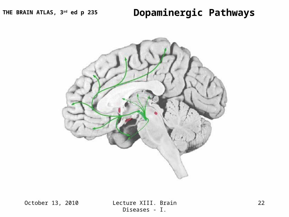
Dopaminergic PathwaysTHE BRAIN ATLAS, 3rd ed p 235
October 13, 2010 Lecture XIII. Brain Diseases - I. 23
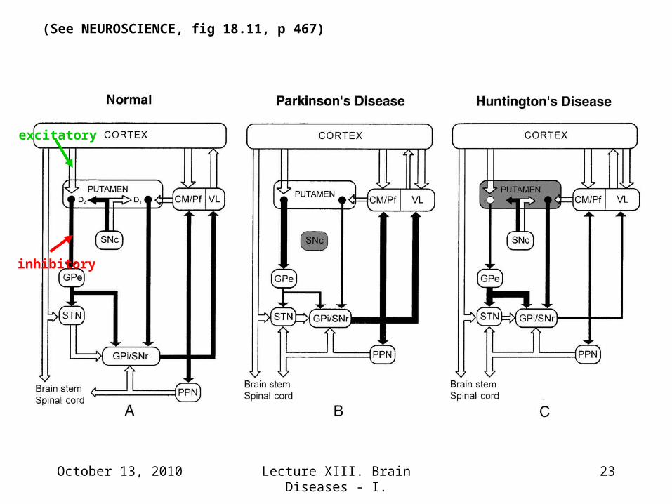
excitatory
inhibitory
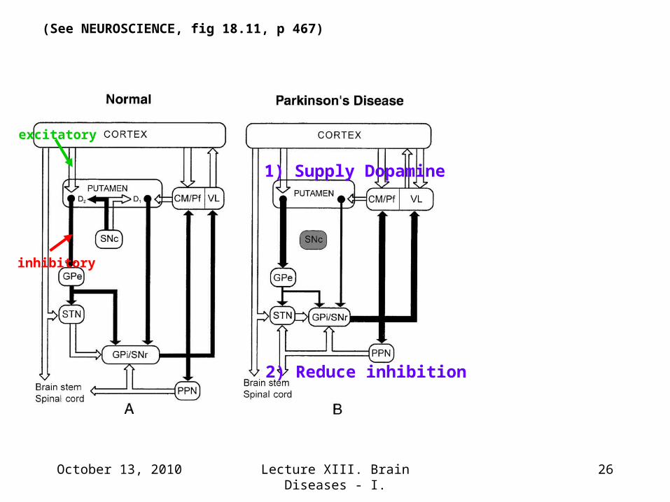
(See NEUROSCIENCE, fig 18.11, p 467)
October 13, 2010 Lecture XIII. Brain Diseases - I. 24
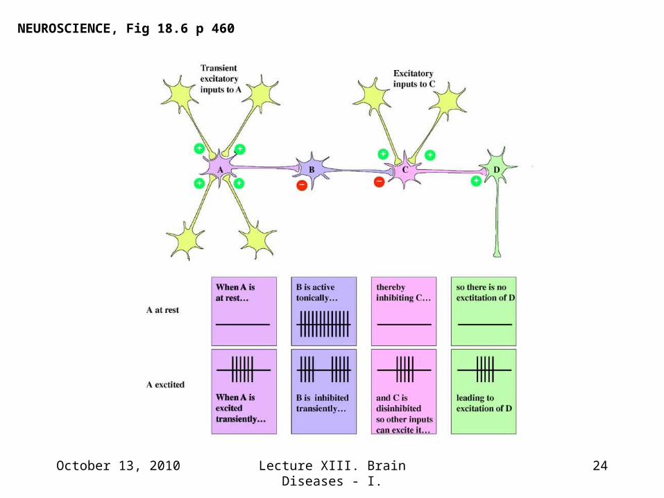
NEUROSCIENCE, Fig 18.6 p 460
October 13, 2010 Lecture XIII. Brain Diseases - I. 25
THE BRAIN ATLAS, 3rd ed p 213
October 13, 2010 Lecture XIII. Brain Diseases - I. 26
(See NEUROSCIENCE, fig 18.11, p 467)
excitatory
inhibitory
1) Supply Dopamine
2) Reduce inhibition

October 13, 2010 Lecture XIII. Brain Diseases - I. 27
Movie Clip # 2
L-DOPA relieves the tremors and paralysis but can produce involuntary (choreiform) movements
October 13, 2010 Lecture XIII. Brain Diseases - I. 28
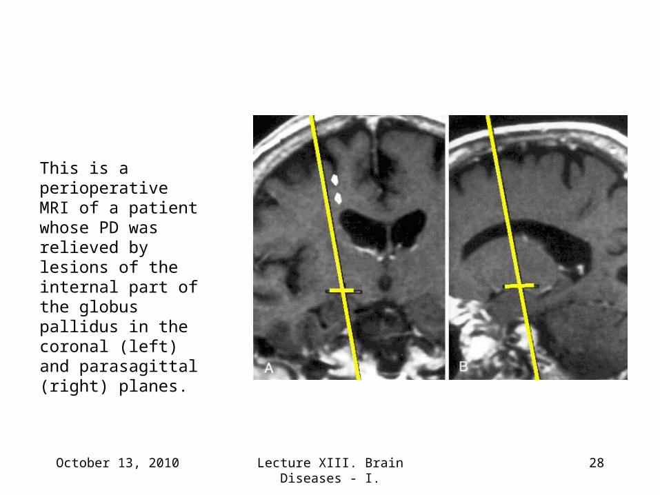
This is a perioperative MRI of a patient whose PD was relieved by lesions of the internal part of the globus pallidus in the coronal (left) and parasagittal (right) planes.
October 13, 2010 Lecture XIII. Brain Diseases - I. 29
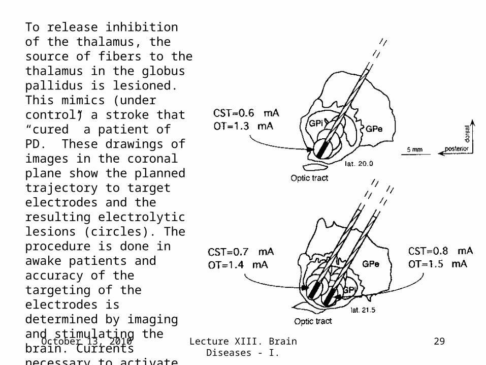
To release inhibition of the thalamus, the source of fibers to the thalamus in the globus pallidus is lesioned. This mimics (under control) a stroke that “cured” a patient of PD. These drawings of images in the coronal plane show the planned trajectory to target electrodes and the resulting electrolytic lesions (circles). The procedure is done in awake patients and accuracy of the targeting of the electrodes is determined by imaging and stimulating the brain. Currents necessary to activate nearby structures the optic tract (OT) and the cortical spinal tract (CST) are indicated.
October 13, 2010 Lecture XIII. Brain Diseases - I. 30
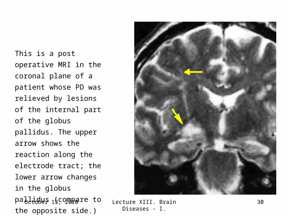
This is a post operative
MRI in the coronal plane of
a patient whose PD was
relieved by lesions of the
internal part of the globus
pallidus. The upper arrow
shows the reaction along
the electrode tract; the
lower arrow changes in the
globus pallidus (compare to
the opposite side.)
October 13, 2010 Lecture XIII. Brain Diseases - I. 31
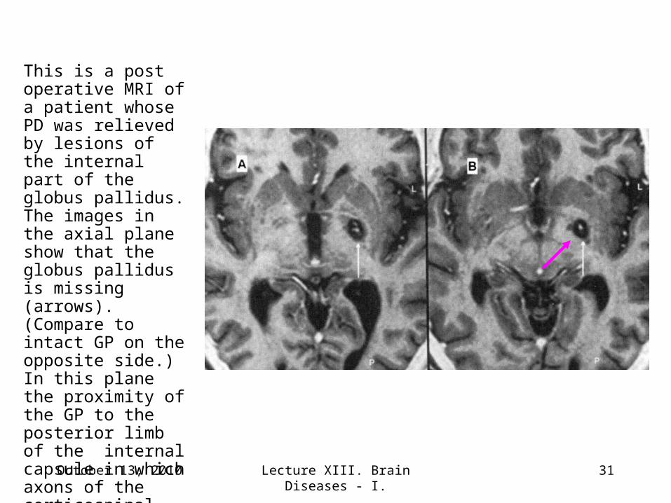
This is a post operative MRI of a patient whose PD was relieved by lesions of the internal part of the globus pallidus. The images in the axial plane show that the globus pallidus is missing (arrows). (Compare to intact GP on the opposite side.) In this plane the proximity of the GP to the posterior limb of the internal capsule in which axons of the corticospinal tract travel is apparent (arrow).
October 13, 2010 Lecture XIII. Brain Diseases - I. 32
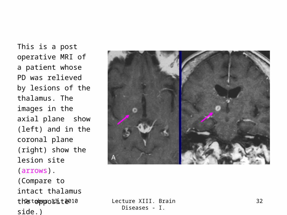
This is a post
operative MRI of a
patient whose PD was
relieved by lesions of
the thalamus. The
images in the axial
plane show (left) and
in the coronal plane
(right) show the lesion
site (arrows).
(Compare to intact
thalamus the opposite
side.)
October 13, 2010 Lecture XIII. Brain Diseases - I. 33
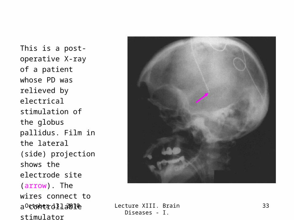
This is a post-operative
X-ray of a patient
whose PD was relieved
by electrical stimulation
of the globus pallidus.
Film in the lateral (side)
projection shows the
electrode site (arrow).
The wires connect to a
controllable stimulator
usually implanted under
the skin of the chest.
October 13, 2010 Lecture XIII. Brain Diseases - I. 34
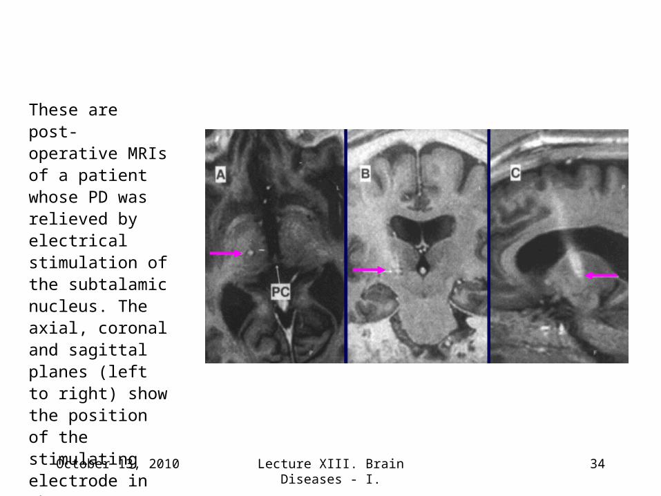
These are post- operative MRIs of a patient whose PD was relieved by electrical stimulation of the subtalamic nucleus. The axial, coronal and sagittal planes (left to right) show the position of the stimulating electrode in the target.
October 13, 2010 Lecture XIII. Brain Diseases - I. 35
Movie Clip # 3
When the electrode is targeted the tremors cease
October 13, 2010 Lecture XIII. Brain Diseases - I. 36
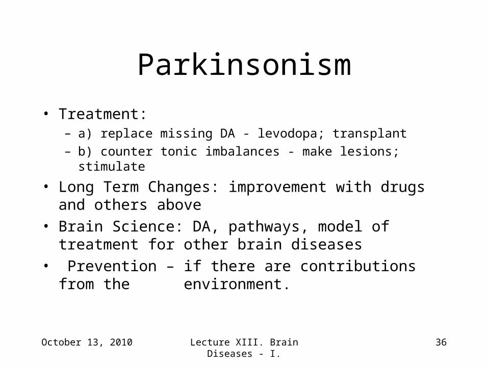
Parkinsonism
• Treatment: – a) replace missing DA - levodopa; transplant– b) counter tonic imbalances - make lesions; stimulate
• Long Term Changes: improvement with drugs and others above
• Brain Science: DA, pathways, model of treatment for other brain diseases
• Prevention – if there are contributions from the environment.
October 13, 2010 Lecture XIII. Brain Diseases - I. 37
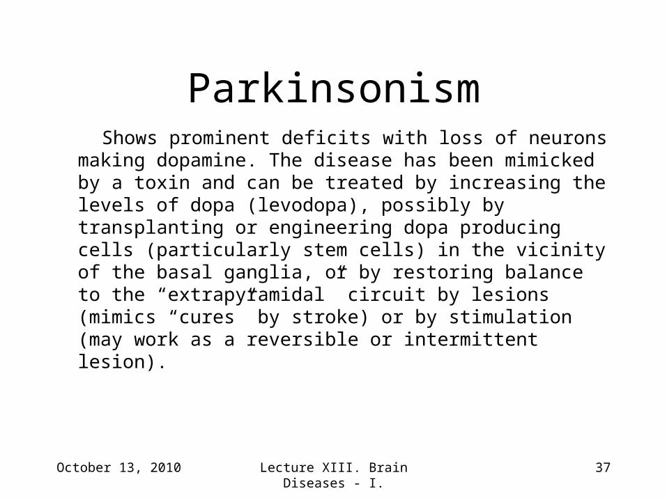
Shows prominent deficits with loss of neurons making dopamine. The disease has been mimicked by a toxin and can be treated by increasing the levels of dopa (levodopa), possibly by transplanting or engineering dopa producing cells (particularly stem cells) in the vicinity of the basal ganglia, or by restoring balance to the “extrapyramidal” circuit by lesions (mimics “cures” by stroke) or by stimulation (may work as a reversible or intermittent lesion).
Parkinsonism
October 13, 2010 Lecture XIII. Brain Diseases - I. 38
Movie Clip # 4
Two weeks later with the stimulator off the tremor returns; with the stimulator on it ceases
October 13, 2010 Lecture XIII. Brain Diseases - I. 39
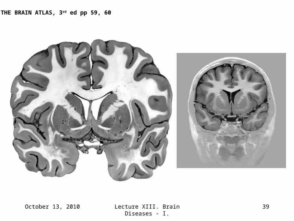
THE BRAIN ATLAS, 3rd ed pp 59, 60
October 13, 2010 Lecture XIII. Brain Diseases - I. 40
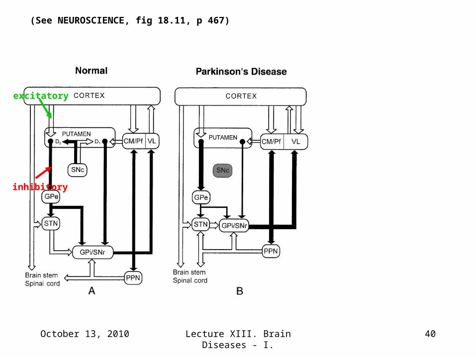
excitatory
inhibitory
(See NEUROSCIENCE, fig 18.11, p 467)
October 13, 2010 Lecture XIII. Brain Diseases - I. 41
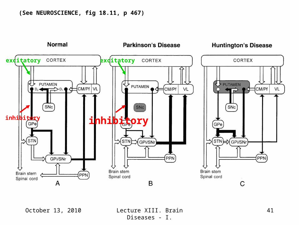
excitatory
inhibitory
excitatory
inhibitory
(See NEUROSCIENCE, fig 18.11, p 467)
October 13, 2010 Lecture XIII. Brain Diseases - I. 42
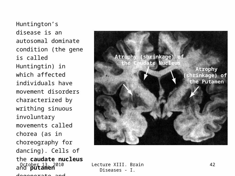
Atrophy (shrinkage) of the Caudate Nucleus
Atrophy(shrinkage) of the Putamen
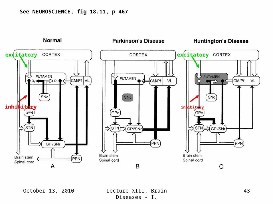
Huntington’s disease is
an autosomal dominate
condition (the gene is
called Huntingtin) in
which affected individuals
have movement disorders
characterized by writhing
sinuous involuntary
movements called chorea
(as in choreography for
dancing). Cells of the
caudate nucleus and
putamen degenerate and
these nuclei atrophy
(shrink).
October 13, 2010 Lecture XIII. Brain Diseases - I. 43
See NEUROSCIENCE, fig 18.11, p 467
excitatory
inhibitory
excitatory
inhibitory
October 13, 2010 Lecture XIII. Brain Diseases - I. 44
Movie Clip # 2
L-DOPA relieves the tremors and paralysis but can produce involuntary (choreiform) movements
October 13, 2010 Lecture XIII. Brain Diseases - I. 45
Movie Clip # 5
Stimulators allow modulation of Rx in real time. Here the patient walks out of the hospital on her way home.
October 13, 2010 Lecture XIII. Brain Diseases - I. 46
Science, medicine ≠ ignorance, politics
October 13, 2010 Lecture XIII. Brain Diseases - I. 47
What this lecture was about:
• Motor Systems a Reprise
• Pyramidal and Extrapyramidal (Basal
ganglia)
• Parkinsonism a Movement Disorder
• Mechanisms and Treatment Strategies
END
October 13, 2010 48Lecture XIII. Brain Diseases - I.


![Vascular parkinsonism · Vascular parkinsonism – REVIEW future science groupfuture science group 239 20%) suffered from parkinsonism with strong evidence of CVD [23]](https://img.pdfslide.us/doc/110x75/5c12e69c09d3f208438bb500/vascular-parkinsonism-vascular-parkinsonism-review-future-science-groupfuture.jpg)