Embed Size (px)
Citation preview

Lecture Title: Lecture Title: Regional Anaesthesia
Lecturer name: Dr .Lecturer name: Dr .
Lecture Date:Lecture Date:
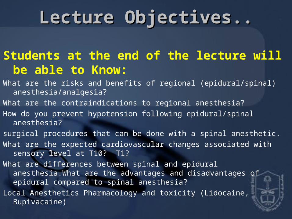
Lecture Objectives..Lecture Objectives..
Students at the end of the lecture will be able to Know:
What are the risks and benefits of regional (epidural/spinal) anesthesia/analgesia?What are the contraindications to regional anesthesia?How do you prevent hypotension following epidural/spinal anesthesia?surgical procedures that can be done with a spinal anesthetic.What are the expected cardiovascular changes associated with sensory level at T10?
T1?What are differences between spinal and epidural anesthesia.What are the
advantages and disadvantages of epidural compared to spinal anesthesia?Local Anesthetics Pharmacology and toxicity (Lidocaine, Bupivacaine)

DEFINITION OF REGIONAL ANESTHESIA
❏ Local anesthetic applied around a peripheral nerve at any point along the length of the nerve
(from spinal cord up to, but not including, the nerve endings) for the purposes of reducing or preventing impulse transmission ❏ No CNS depression (unless overdose (OD) of local anesthetic); patient conscious ❏ Regional anesthetic techniques categorized as follows
• Epidural and spinal anesthesia• Peripheral nerve blockades• IV regional anesthesia

Physiology:
• Physiologic response to central blockade is determined by the effects of interrupting the afferent and efferent innervation of somatic (sensory and motor innervation) and visceral (autonomic nervous system).

Physiology:
• Somatic blockade:– Prevention of pain.– Skeletal muscle relaxation.
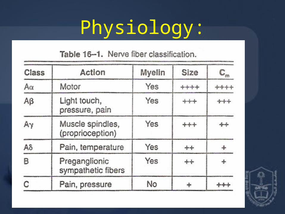
Physiology:

Physiology:
• Fibers blocked more easily small and myleinated.
• Less easily – large – unmylinated.
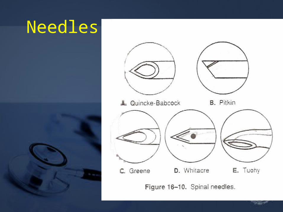
Needles

PREPARATION FOR REGIONAL ANESTHESIAPatient Preparation
❏ Thorough pre-op evaluation and assessment of patient ❏ Technique explained to patient ❏ IV sedation may be indicated before block ❏ Monitoring should be as extensive as for general anesthesia

Nerve Localization
❏ Anatomical landmarks, local anatomy, e.g. line joining iliac crests cross L3-L4 interspace; axillary artery as guide to brachial plexus ❏ Paresthesias and peripheral nerve stimulation used as a guide to proper needle placement
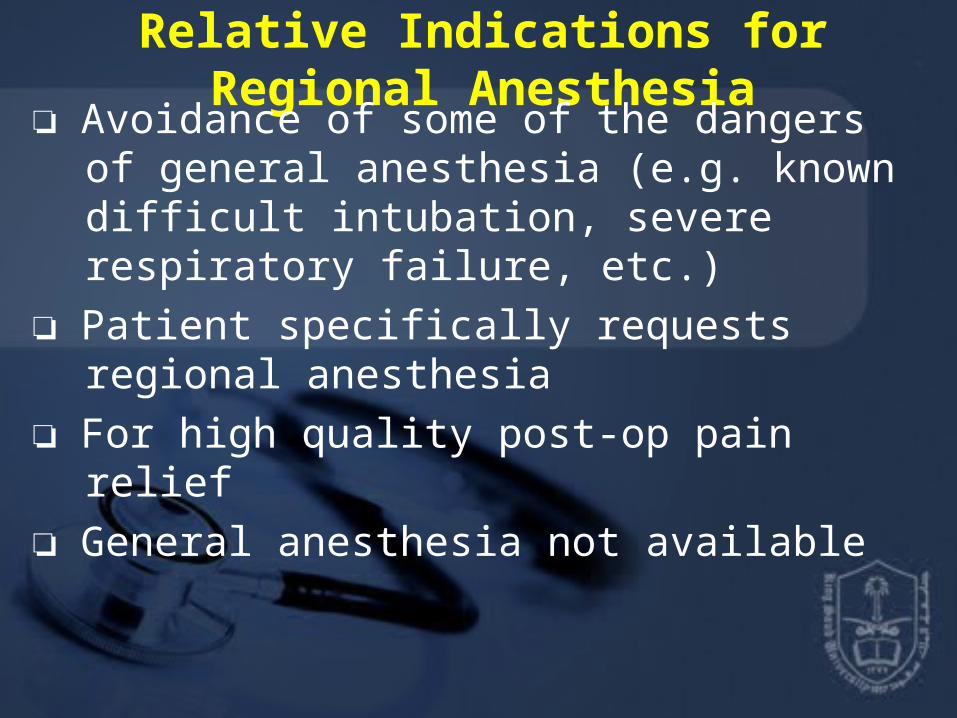
Relative Indications for Regional Anesthesia ❏ Avoidance of some of the dangers of general anesthesia (e.g. known difficult intubation, severe respiratory failure, etc.)
❏ Patient specifically requests regional anesthesia
❏ For high quality post-op pain relief ❏ General anesthesia not available

Contraindications to Regional Anesthesia
❏ Allergy to local anesthetic ❏ Patient refusal, lack of cooperation ❏ Lack of resuscitation equipment ❏ Lack of IV access
❏Coagulopathy ❏ Certain types of preexisting neurological dysfunction ❏ Local infection at block site

Complications of Regional Anesthesia
❏ Failure of technique ❏ Systemic drug toxicity due to overdose or intravascular injection
❏ Peripheral neuropathy due to intraneural injection
❏ Pain or hematoma at injection site

EPIDURAL AND SPINAL ANESTHESIA

anatomy
• The vertebrae are 33 number, divided by structural into five region: cervical 8, thoracic 12, lumber5, sacral 5, coccygeal3.

EPIDURAL AND SPINAL ANESTHESIAAnatomy of Spinal/Epidural AreaThe spinal cord lies within the spinal canal. Surround by meanings, dura mater sub archnoid
space, then piamatter, end by hoarse tail (Couda equina)
The spinal cord receives blood supply from ant spinal artery, and posterior spinal artery.
Spinal cord extends to L2, dural sac to S2 Nerve roots (cauda equina) from L2 to S2Needle inserted below L2 should not encounter cord, thus
L3-L4, L4-L5 interspace commonly used

Structures penetrated
• Skin, subcutaneous fat• Supraspinous ligament• Interspinous ligament • ligamentum flavum (last layer before epidural
space) • dura + arachnoid for spinal anesthesia

anatomy

anatomy
• The spine double “C” curve with cervical and lumbar. Structural of the vertebra:

anatomy
• The vertebrae are joined together by intervertebral disc by strong ant and post longitudinal ligaments.
Spinal Anesthesia ❏ Relatively small LA dose injected into subarachnoid space in the dural sac surrounding the spinal cord + nerve roots ❏ LA solution may be made hyperbaric (of greater specific gravity (SG) than the cerebrospinal fluid (CSF) by mixing with 10% dextrose, thus increasing spread of LA to the dependent (low) areas of the subarachnoid space

Epidural Anesthesia ❏ LA deposited in epidural space (potential space between ligamentum flavum and dura) ❏ Solutions injected here spread in all directions of the potential space; SG of solution does not affect spread ❏ Initial blockade is at the spinal roots followed by some degree of spinal cord anesthesia as LA diffuses into the subarachnoid space through the dura
• ❏ Larger dose of LA used

Spinal vs. Epidural Anesthesia ❏ spinal
•Easier to perform Smaller dose of LA required (usually < toxic IV dose)• Rapid blockade (onset in 2-5 minutes)• Very effective blockade• Hyperbaric LA solution - position of patient important
❏ epidural• Technically more difficult; greater failure rate• Larger volume/doses of LA (usually > toxic IV dose)• Significant blockade requires 10-15 minutes• Effectiveness of blockade can be variable• Use of catheter allows for continuous infusion or repeat injections• Slower onset of side effects• Position of patient not as important• SG of LA solution not as important

Complications of Spinal/Epidural Anesthesia ❏ Spinal anesthesia
– Failure of technique– Hypotension, bradycardia if block reaches T2-4
(sympathetic nervous system (SNS) block)– Post-spinal headache– Extensive spread of anesthetic ("high spinal")– Persistent paresthesias (usually transient)– Epidural or subarachnoid hematoma– Spinal cord trauma, infection

❏ Epidural anesthesia– Failure of technique– Hypotension - common– Bradycardia if cardiac sympathetics blocked (only if
T2-4 block)– Systemic toxicity of LA (accidental intravenous)– Accidental subarachnoid injection can lead to total
spinal anesthesia– Catheter complications (shearing, kinking, vascular
or subarachnoid placement)– Epidural or subarachnoid hematoma

Contraindications to Spinal/Epidural Anesthesia ❏ Absolute contraindications include – lack of proper equipment or properly trained personnel– patient refusal– lack of IV access– allergy to LA– infection at puncture site or underlying tissues– uncorrected hypovolemia– coagulation abnormalities– raised ICP
❏ Relative contraindications include – Bacteremia– preexisting neurological disease– aortic/mitral valve stenosis, – previous spinal surgery– other back problems– severe/unstable psychiatric disease or emotional instability

Peripheral nerve blockades

Utrasound for regional anesthesia• The use of ultrasound for regional anesthesia is relatively
new, was first described as early as 1978, but it was not until the advent of advanced ultrasound technology in the 1990's that interest in this field grew.
• Published reports of ultrasound guided regional anesthesia have largely focused on brachial plexus blockade in the interscalene, supraclavicular, infraclavicular and axillary regions.
• Recent studies examining the efficacy of ultrasound guidance for femoral, sciatic, psoas compartment, celiac plexus and stellate ganglion blocks are promising, while ultrasound visualization of the epidural space can facilitate neuraxial blockade in children, adults and parturients.
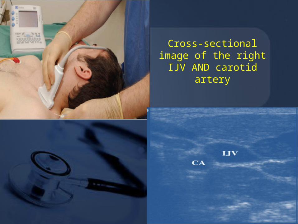
Cross-sectional image of the right IJV AND carotid artery


Evidence regarding the use of ultrasound in regional anaesthesia indicates that the use of US may:-
• Decrease the time taken to perform a block• Decrease the time for a block to onset• Improve the duration of the block• Improve patient satisfaction in the block process• Allow a successful block without the use of a nerve stimulator• Lower the dose of local anaesthetic required for a block• Allow regional anaesthesia in otherwise difficult circumstances• Allow the detection of an intraneural injection• Allow confirmation of local anaesthetic spread• Allow confirmation of catheter placement.

Local Anesthetics

Definition and Mode of Action ❏ LA are drugs that block the generation and propagation of impulses in excitable tissues: nerves, skeletal muscle, cardiac muscle, brain ❏ LA substances bind to a Na+ channel receptor on the cytosolic side of the Na+ channel (i.e. must be lipid soluble), inhibiting Na+ flux and thus blocking impulse conduction ❏ LA must convert to an ionized form to properly bind to receptor ❏ Different types of nerve fibres undergo blockade at different rates (see Regional Anesthesia section)

Absorption, Distribution, Metabolism ❏ LA readily crosses the blood-brain barrier (BBB) once absorbed into the blood stream ❏ Ester-type LA (procaine, tetracaine) broken down by plasma and hepatic esterases; metabolites excreted via kidneys ❏ Amide-type LA (lidocaine, bupivicaine) broken down by hepatic mixed function oxidases (P450 system); metabolites excreted via kidney

Choice of LA depends on:Onset of action –influenced by pKa (lower the pKa, the higher
the concentration of the base form of the LA and the faster the onset of action)
Duration of desired effects – influenced by protein binding (long duration of action when the protein binding of LA is strong)
Potency – influenced by lipid solubility (agents with high lipid solubility will penetrate the nerve membrane more easily)
Unique needs (e.g. sensory blockade with relative preservation of motor function, for pain management)
Potential for toxicity
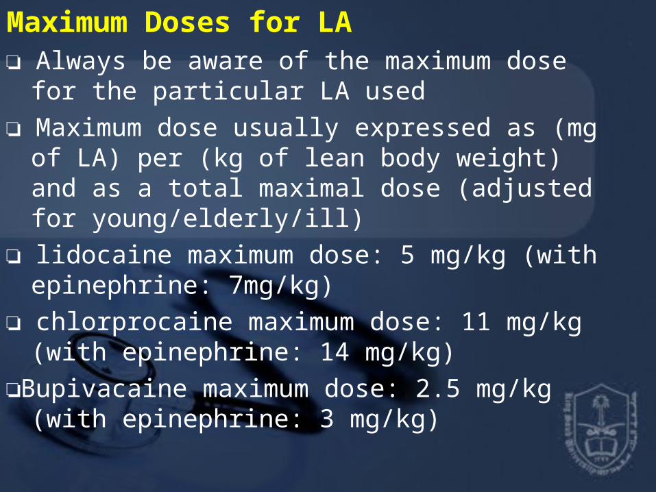
Maximum Doses for LA ❏ Always be aware of the maximum dose for the particular LA used ❏ Maximum dose usually expressed as (mg of LA) per (kg of lean body weight) and as a total maximal dose (adjusted for young/elderly/ill) ❏ lidocaine maximum dose: 5 mg/kg (with epinephrine: 7mg/kg) ❏ chlorprocaine maximum dose: 11 mg/kg (with epinephrine: 14 mg/kg)
❏Bupivacaine maximum dose: 2.5 mg/kg (with epinephrine: 3 mg/kg)

Systemic Toxicity ❏ Occurs by accidental intravascular injection, LA overdose, or unexpectedly rapid absorption
❏ Systemic toxicity manifests itself mainly at CNS and CVS ❏ CNS effects first appear to be excitatory due to initial block of inhibitory fibres; subsequently, block of excitatory fibres
❏ CNS effects (in approximate order of appearance)• Numbness of tongue, perioral tingling ,disorientation, drowsiness• Tinnitus• Visual disturbances• Muscle twitching, tremors• Convulsions, seizures• Generalized CNS depression, coma, respiratory arrest

❏ CVS effects–Vasodilatation, hypotension–Decreased myocardial contractility–Dose-dependent delay in cardiac impulse
transmission–Prolonged PR, QRS intervals– Sinus bradycardia–CVS collapse

❏ Treatment of systemic toxicity
1.Early recognition of signs2.100% O2, manage ABCs3. Diazepam may be used to increase seizure
threshold4.If the seizures are not controlled by diazepam,
consider using:5.Thiopental, Possible ETT.


Suggested Reading•Campagna JA, Miller KW, Forman SA. Mechanisms of actions of inhaled anesthetics. N Engl J Med 2003;348:2110-2124.•Eger EI. Uptake and distribution. In: Miller RD, ed. Anesthesia, 6th ed. New York: Churchill Livingstone, 2005;131-153.•Kennedy, SK. Pharmacology of intravenous anesthetic agents. In: Longnecker DE, •Clinical Anesthesia Procedures of the Massachusetts General Hospital, 7th Edition•Copyright ©2007آ Lippincott Williams & Wilkins

DrDr..
Date: Date:
TThank You hank You