Embed Size (px)
Citation preview
Chapter 16*Lecture Outline
Copyright © The McGraw-Hill Companies, Inc. Permission required for reproduction or display.
*See separate Image PowerPoint slides for
all figures and tables pre-inserted into
PowerPoint without notes.
PowerPoints prepared by
Melanie Waite-AltringerBiology Faculty Member of
Anoka-Ramsey Community College
2
Chapter 16
Respiratory System

3
Introduction
A. The respiratory system consists of tubes that
filter incoming air and transport it into the
microscopic alveoli where gases are
exchanged.
CopyrightThe McGraw-Hill Companies, Inc. Permission required for reproduction or display.
4
B. The entire process of exchanging gases
between the atmosphere and body cells
is called respiration and consists of the
following: ventilation, gas exchange between
blood and lungs, gas transport in the
bloodstream, gas exchange between the blood
and body cells, and cellular respiration.
CopyrightThe McGraw-Hill Companies, Inc. Permission required for reproduction or display.
5
Organs of the Respiratory System
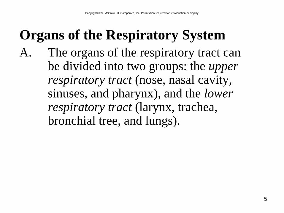
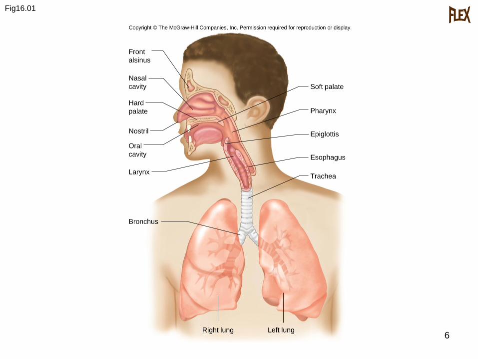
A. The organs of the respiratory tract can be divided into two groups: the upper respiratory tract (nose, nasal cavity, sinuses, and pharynx), and the lower respiratory tract (larynx, trachea, bronchial tree, and lungs).
CopyrightThe McGraw-Hill Companies, Inc. Permission required for reproduction or display.
6
Fig16.01
Front
alsinus
Nasal
cavity
Oral
cavity
Larynx
Bronchus
Hard
palate
Nostril
Right lung Left lung
Trachea
Soft palate
Pharynx
Epiglottis
Esophagus
Copyright © The McGraw-Hill Companies, Inc. Permission required for reproduction or display.
7
B. Nose
1. The nose, supported by bone and
cartilage, provides an entrance for air in
which air is filtered by coarse hairs
inside the nostrils.
CopyrightThe McGraw-Hill Companies, Inc. Permission required for reproduction or display.
8
C. Nasal Cavity
1. The nasal cavity is a space posterior tothe nose that is divided medially by thenasal septum.
2. Nasal conchae divide the cavity intopassageways that are lined with mucousmembrane, and help increase thesurface area available to warm and filterincoming air.
CopyrightThe McGraw-Hill Companies, Inc. Permission required for reproduction or display.
9
3. Particles trapped in the mucus are
carried to the pharynx by ciliary action,
swallowed, and carried to the stomach
where gastric juice destroys any
microorganisms in the mucus.
CopyrightThe McGraw-Hill Companies, Inc. Permission required for reproduction or display.
10
D. Paranasal Sinuses
1. Sinuses are air-filled spaces within themaxillary, frontal, ethmoid, andsphenoid bones of the skull.
2. These spaces open to the nasal cavityand are lined with mucus membranethat is continuous with that lining thenasal cavity.
3. The sinuses reduce the weight of theskull and serve as a resonant chamber toaffect the quality of the voice.
CopyrightThe McGraw-Hill Companies, Inc. Permission required for reproduction or display.
11
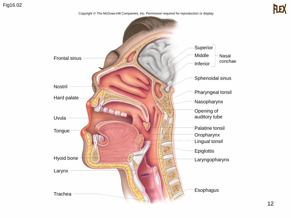
E. Pharynx
1. The pharynx is a common passageway
for air and food.
2. The pharynx aids in producing sounds
for speech.
CopyrightThe McGraw-Hill Companies, Inc. Permission required for reproduction or display.
12
Fig16.02
Frontal sinus
Nostril
Hard palate
Uvula
Tongue
Epiglottis
Hyoid bone
Larynx
Trachea
Superior
Middle
Inferior
Sphenoidal sinus
Pharyngeal tonsil
Nasopharynx
Opening of
auditory tube
Palatine tonsil
Oropharynx
Lingual tonsil
Laryngopharynx
Esophagus
Nasal
conchae
Copyright © The McGraw-Hill Companies, Inc. Permission required for reproduction or display.
13
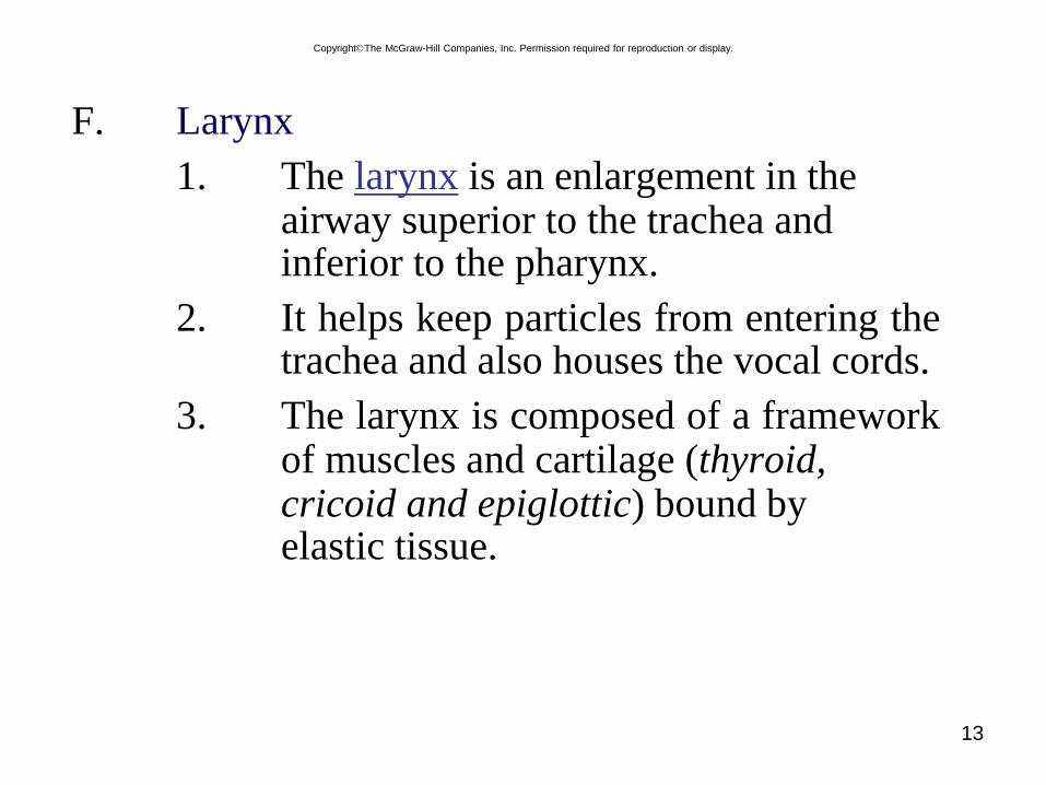
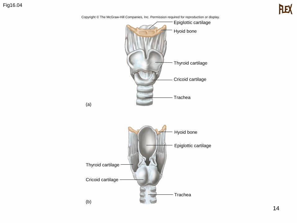
F. Larynx
1. The larynx is an enlargement in theairway superior to the trachea andinferior to the pharynx.
2. It helps keep particles from entering thetrachea and also houses the vocal cords.
3. The larynx is composed of a frameworkof muscles and cartilage (thyroid,cricoid and epiglottic) bound byelastic tissue.
CopyrightThe McGraw-Hill Companies, Inc. Permission required for reproduction or display.
14
Fig16.04
Trachea
Epiglottic cartilage
Hyoid bone
Thyroid cartilage
Cricoid cartilage
Hyoid bone
Epiglottic cartilage
Thyroid cartilage
Cricoid cartilage
(b)
Trachea
(a)
Copyright © The McGraw-Hill Companies, Inc. Permission required for reproduction or display.
15
4. Inside the larynx, two pairs of folds of muscle and connective tissue covered with mucous membrane make up the vocal cords.
a. The upper pair is the false vocal cords.
b. The lower pair is the true vocalcords.
c. Changing tension on the vocal cords controls pitch, while increasing the loudness depends upon increasing the force of air vibrating the vocal cords.
CopyrightThe McGraw-Hill Companies, Inc. Permission required for reproduction or display.
16
5. During normal breathing, the vocal
cords are relaxed and the glottis is a
triangular slit.
6. During swallowing, the false vocal
cords and epiglottis close off the glottis.
CopyrightThe McGraw-Hill Companies, Inc. Permission required for reproduction or display.
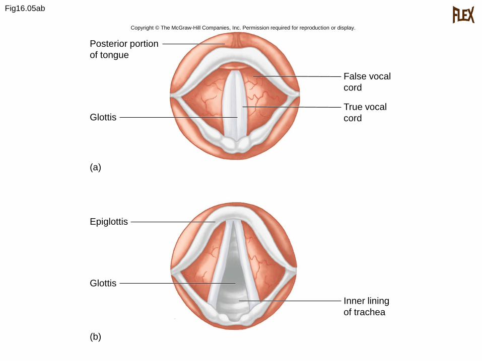
Fig16.05ab
Posterior portion
of tongue
Inner lining
of trachea
Glottis
(a)
(b)
Epiglottis
Glottis
True vocal
cord
False vocal
cord
Copyright © The McGraw-Hill Companies, Inc. Permission required for reproduction or display.
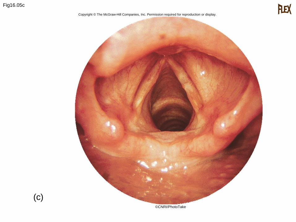
Fig16.05c
Copyright © The McGraw-Hill Companies, Inc. Permission required for reproduction or display.
(c)©CNRI/PhotoTake
19
G. Trachea
1. The trachea extends downward anterior to the esophagus and into the thoracic cavity, where it splits into right and left bronchi.
2. The inner wall of the trachea is linedwith ciliated mucous membrane withmany goblet cells that serve to trapincoming particles.
3. The tracheal wall is supported by 20incomplete cartilaginous rings.
CopyrightThe McGraw-Hill Companies, Inc. Permission required for reproduction or display.
20
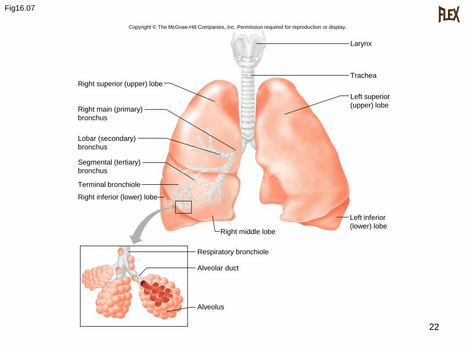
H. Bronchial Tree
1. The bronchial tree consists of branched
tubes leading from the trachea to the
alveoli.
2. The bronchial tree begins with the two
main (primary) bronchi, each leading to
a lung.
CopyrightThe McGraw-Hill Companies, Inc. Permission required for reproduction or display.
21
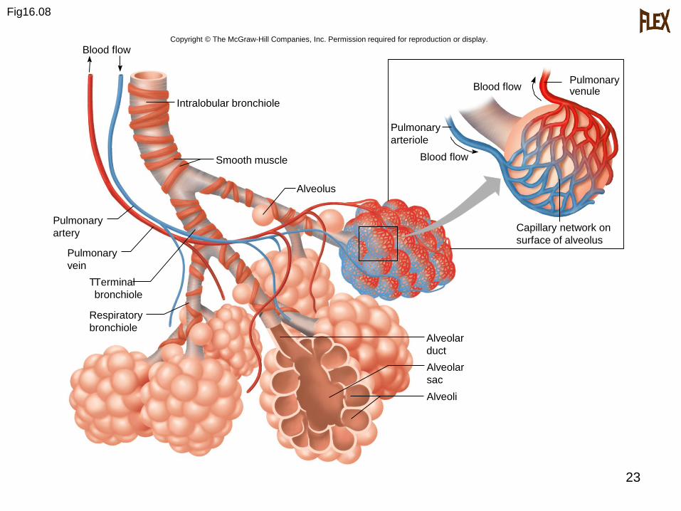
3. The branches of the bronchial tree from
the trachea are right and left primary
bronchi; these further subdivide until
bronchioles give rise to alveolar ducts ,
then alveolar sacs which terminate in
alveoli.
4. It is through the thin epithelial cells of
the alveoli that gas exchange between
the blood and air occurs.
CopyrightThe McGraw-Hill Companies, Inc. Permission required for reproduction or display.
22
Fig16.07
Larynx
Trachea
Left superior
(upper) lobe
Left inferior
(lower) lobeRight middle lobe
Right superior (upper) lobe
Right main (primary)
bronchus
Lobar (secondary)
bronchus
Segmental (tertiary)
bronchus
Right inferior (lower) lobe
Alveolar duct
Alveolus
Terminal bronchiole
Respiratory bronchiole
Copyright © The McGraw-Hill Companies, Inc. Permission required for reproduction or display.
23
Fig16.08
Pulmonary
vein
Pulmonary
artery
Pulmonary
arteriole
Pulmonaryvenule
Intralobular bronchiole
Blood flow
Alveolus
TTerminal
bronchiole
Smooth muscle
Alveolar
duct
Alveolar
sac
Alveoli
Capillary network on
surface of alveolus
Blood flow
Blood flow
Respiratory
bronchiole
Copyright © The McGraw-Hill Companies, Inc. Permission required for reproduction or display.
24
I. Lungs
1. The right and left soft, spongy, cone-
shaped lungs are separated medially by
the mediastinum and are enclosed by
the diaphragm and thoracic cage.
2. The bronchus and large blood vessels
enter each lung.
CopyrightThe McGraw-Hill Companies, Inc. Permission required for reproduction or display.
25
3. A layer of serous membrane, the
visceral pleura, folds back to form the
parietal pleura.
4. The visceral pleura is attached to the
lung, and the parietal pleura lines the
thoracic cavity; serous fluid lubricates
the pleural cavity between these two
membranes.
CopyrightThe McGraw-Hill Companies, Inc. Permission required for reproduction or display.
26
5. The right lung has three lobes, the left
has two.
6. Each lobe is composed of lobules that
contain air passages, alveoli, nerves,
blood vessels, lymphatic vessels, and
connective tissues.
CopyrightThe McGraw-Hill Companies, Inc. Permission required for reproduction or display.
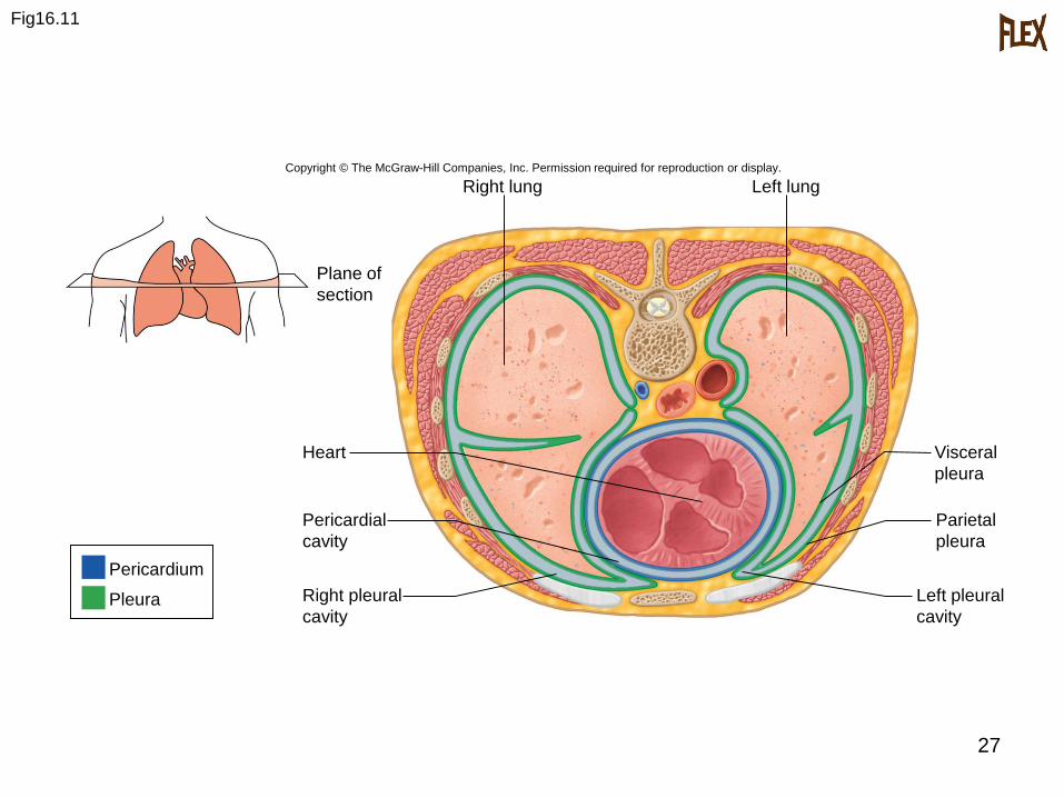
27
Fig16.11
Right lung
Pericardial
cavity
Heart
Left pleural
cavity
Parietal
pleura
Visceral
pleura
Left lung
Plane of
section
Right pleural
cavity
Pericardium
Pleura
Copyright © The McGraw-Hill Companies, Inc. Permission required for reproduction or display.
28
Breathing Mechanism
A. Ventilation (breathing), the movement of air in and out of the lungs, is composed of inspiration and expiration.
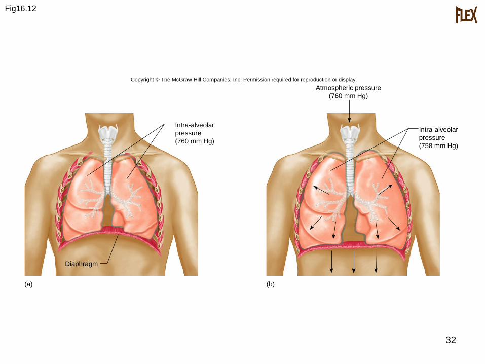
B. Inspiration
1. Atmospheric pressure is the force thatmoves air into the lungs.
2. When pressure on the inside of thelungs decreases, higher pressure airflows in from the outside.
CopyrightThe McGraw-Hill Companies, Inc. Permission required for reproduction or display.
29
3. Air pressure inside the lungs isdecreased by increasing the size of thethoracic cavity; due to surface tensionbetween the two layers of pleura, thelungs follow with the chest wall andexpand.
4. Muscles involved in expanding the thoracic cavity include the diaphragm and the external intercostal muscles.
CopyrightThe McGraw-Hill Companies, Inc. Permission required for reproduction or display.
30
5. As the lungs expand in size, surfactant
keeps the alveoli from sticking to each
other so they do not collapse when
internal air pressure is low.
CopyrightThe McGraw-Hill Companies, Inc. Permission required for reproduction or display.
31
C. Expiration
1. The forces of expiration are due to the
elastic recoil of lung and muscle tissues
and from the surface tension within the
alveoli.
2. Forced expiration is aided by thoracic
and abdominal wall muscles that
compress the abdomen against the
diaphragm.
CopyrightThe McGraw-Hill Companies, Inc. Permission required for reproduction or display.
32
Fig16.12
Diaphragm
Atmospheric pressure
(760 mm Hg)
Intra-alveolar
pressure
(758 mm Hg)
Intra-alveolar
pressure
(760 mm Hg)
(a) (b)
Copyright © The McGraw-Hill Companies, Inc. Permission required for reproduction or display.
33
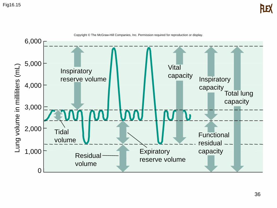
D. Respiratory Air Volumes and Capacities
1. The measurement of different air volumes is called spirometry, and it describes four distinct respiratoryvolumes.
2. One inspiration followed by expiration is called a respiratory cycle; the amount of air that enters or leaves the lungs during one respiratory cycle is the tidal volume.
CopyrightThe McGraw-Hill Companies, Inc. Permission required for reproduction or display.
34
3. During forced inspiration, an additional volume, the inspiratory reserve volume, can be inhaled into the lungs. IRV + TV gives us the inspiratory capacity.
4. During a maximal forced expiration, an expiratory reserve volume can be exhaled, but there remains a residual volume in the lungs. Adding the two together gives us the functional reservecapacity.
CopyrightThe McGraw-Hill Companies, Inc. Permission required for reproduction or display.
35
5. Vital capacity is the tidal volume plus
inspiratory reserve and expiratory
reserve volumes combined.
6. Vital capacity plus residual volume is
the total lung capacity.
7. Anatomic dead space is air remaining in
the bronchial tree.
CopyrightThe McGraw-Hill Companies, Inc. Permission required for reproduction or display.
36
Fig16.15
6,000
5,000
4,000
3,000
2,000
1,000
0
Total lung
capacity
Inspiratory
capacity
Functional
residual
capacityExpiratory
reserve volumeResidual
volume
Tidal
volume
Vital
capacityInspiratory
reserve volume
Copyright © The McGraw-Hill Companies, Inc. Permission required for reproduction or display.
Lu
ng
vo
lum
e in m
illili
ters
(m
L)
37
Control of Breathing
A. Normal breathing is a rhythmic, involuntary
act even though the muscles are under
voluntary control.
CopyrightThe McGraw-Hill Companies, Inc. Permission required for reproduction or display.
38
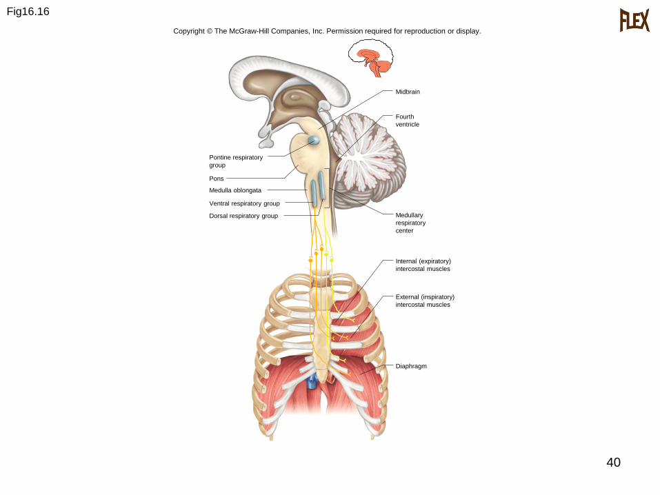
B. Respiratory Areas
1. Groups of neurons in the brain stem comprise the respiratory areas, which controls breathing by causing inspiration and expiration and by adjusting the rate and depth of breathing.
CopyrightThe McGraw-Hill Companies, Inc. Permission required for reproduction or display.
39
3. The medullary rhythmicity centerincludes two groups of neurons: the dorsal respiratory group and the ventral respiratory group.
a. The dorsal respiratory group is responsible for the basic rhythm of breathing.
b. The ventral respiratory group is active when more forceful breathing is required.
4. Neurons in the pons form the pontine respiratory group. They may contribute to the rhythm of breathing by limiting inspiration.
CopyrightThe McGraw-Hill Companies, Inc. Permission required for reproduction or display.
40
Fig16.16
Internal (expiratory)
intercostal muscles
External (inspiratory)
intercostal muscles
Diaphragm
Fourth
ventricle
Medullary
respiratory
center
Ventral respiratory group
Medulla oblongata
Pons
Pontine respiratory
group
Midbrain
Dorsal respiratory group
Copyright © The McGraw-Hill Companies, Inc. Permission required for reproduction or display.
41
C. Factors Affecting Breathing
1. Chemicals, lung tissue stretching, andemotional state affect breathing.
2. Chemosensitive areas (centralchemoreceptors) are associated with therespiratory center and are sensitive tochanges in the blood concentration ofcarbon dioxide and hydrogen ions.
CopyrightThe McGraw-Hill Companies, Inc. Permission required for reproduction or display.
42
a. If either carbon dioxide or
hydrogen ion concentrations rise,
the central chemoreceptors signal
the respiratory center, and
breathing rate increases.
CopyrightThe McGraw-Hill Companies, Inc. Permission required for reproduction or display.
43
3. Peripheral chemoreceptors in
carotid bodies and aortic bodies sense
changes in blood oxygen concentration,
transmit impulses to the respiratory
center, and breathing rate and tidal
volume increase.
CopyrightThe McGraw-Hill Companies, Inc. Permission required for reproduction or display.
44
4. An inflation reflex, triggered by stretch
receptors in the visceral pleura,
bronchioles, and alveoli, helps to
prevent overinflation of the lungs
during forceful breathing.
5. Hyperventilation lowers the amount of
carbon dioxide in the blood.
CopyrightThe McGraw-Hill Companies, Inc. Permission required for reproduction or display.
45
Alveolar Gas Exchanges
A. The alveoli are the only sites of gas exchange
between the atmosphere and the blood.
B. Alveoli
1. The alveoli are tiny sacs clustered at the
distal ends of the alveolar ducts.
CopyrightThe McGraw-Hill Companies, Inc. Permission required for reproduction or display.
46
C. Respiratory Membrane
1. The respiratory membrane consists of
the epithelial cells of the alveolus, the
endothelial cells of the capillary, and
the two fused basement membranes of
these layers.
2. Gas exchange occurs across this
respiratory membrane.
CopyrightThe McGraw-Hill Companies, Inc. Permission required for reproduction or display.
47
D. Diffusion Across the Respiratory Membrane
1. Gases diffuse from areas of higher
pressure to areas of lower pressure.
2. In a mixture of gases, each gas accounts
for a portion of the total pressure; the
amount of pressure each gas exerts is
equal to its partial pressure.
CopyrightThe McGraw-Hill Companies, Inc. Permission required for reproduction or display.
48
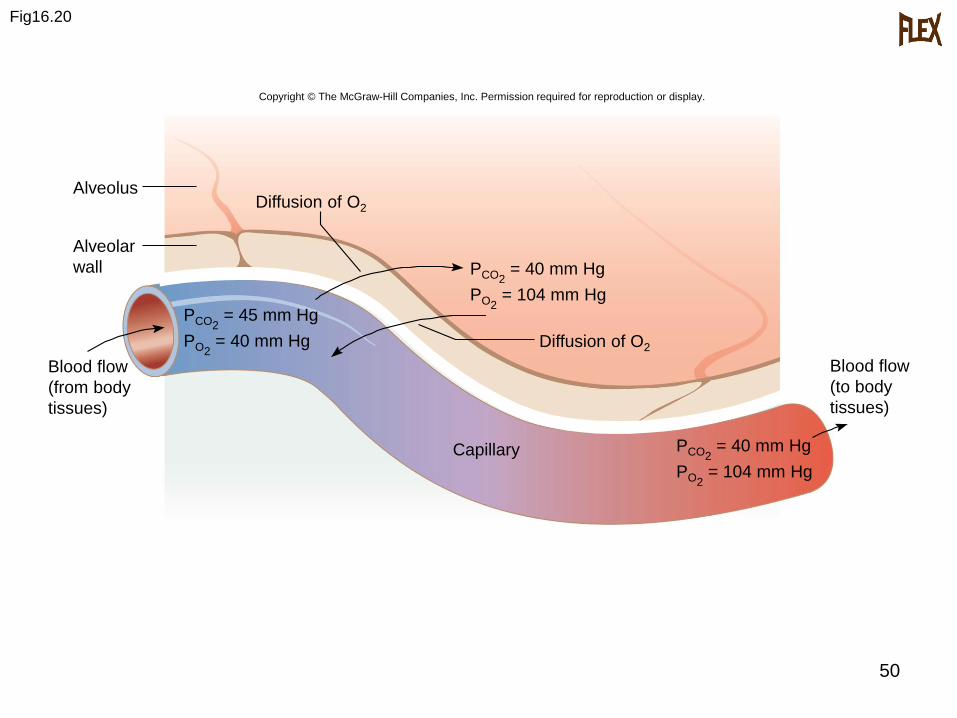
3. When the partial pressure of oxygen is
higher in the alveolar air than it is in the
capillary blood, oxygen will diffuse into
the blood.
4. When the partial pressure of carbon
dioxide is greater in the blood than in
the alveolar air, carbon dioxide will
diffuse out of the blood and into the
alveolus.
CopyrightThe McGraw-Hill Companies, Inc. Permission required for reproduction or display.
49
5. A number of factors favor increased
diffusion; more surface area, shorter
distance, greater solubility of gases, and
a steeper partial pressure gradient.
CopyrightThe McGraw-Hill Companies, Inc. Permission required for reproduction or display.
50
Fig16.20
Blood flow
(to body
tissues)
AlveolusDiffusion of O2
Diffusion of O2
PCO2= 40 mm Hg
PO2= 104 mm Hg
Capillary
Alveolar
wall
Blood flow
(from body
tissues)
Copyright © The McGraw-Hill Companies, Inc. Permission required for reproduction or display.
PCO2= 40 mm Hg
PO2= 104 mm Hg
PCO2= 45 mm Hg
PO2= 40 mm Hg
51
Gas Transport
A. Gases are transported in association with molecules in the blood or dissolved in the plasma.
B. Oxygen Transport
1. Over 98% of oxygen is carried in theblood bound to hemoglobin of redblood cells, producing oxyhemoglobin.
2. Oxyhemoglobin is unstable in areaswhere the concentration of oxygen islow, and gives up its oxygen molecules
in those areas.
CopyrightThe McGraw-Hill Companies, Inc. Permission required for reproduction or display.
52
3. More oxygen is released as the blood
concentration of carbon dioxide
increases, as the blood becomes more
acidic, and as blood temperature
increases.
4. A deficiency of oxygen reaching the
tissues is called hypoxia and has a
variety of causes.
CopyrightThe McGraw-Hill Companies, Inc. Permission required for reproduction or display.
53
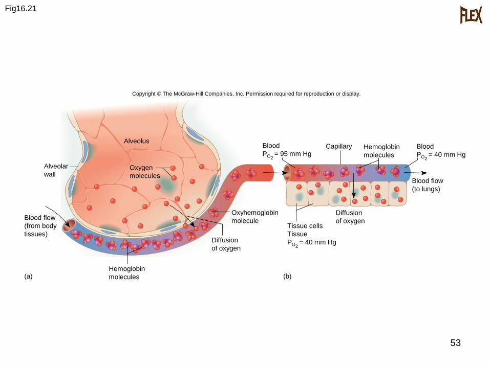
Fig16.21
Oxygen
molecules
Alveolus
Diffusion
of oxygen
Alveolar
wall
Oxyhemoglobin
molecule
Hemoglobin
molecules
Capillary Hemoglobin
molecules
Blood flow
(from body
tissues)
Blood flow
(to lungs)
(a) (b)
Copyright © The McGraw-Hill Companies, Inc. Permission required for reproduction or display.
Tissue cells
Tissue
PO2= 40 mm Hg
Diffusion
of oxygen
Blood
PO2= 95 mm Hg
Blood
PO2= 40 mm Hg

54
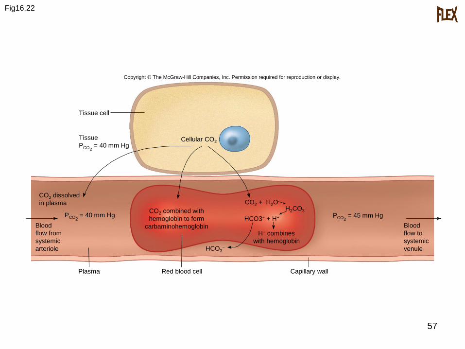
C. Carbon Dioxide Transport
1. Carbon dioxide may be transported
dissolved in blood plasma, as
carbaminohemoglobin, or as
bicarbonate ions.
2. Most carbon dioxide is transported in
the form of bicarbonate.
CopyrightThe McGraw-Hill Companies, Inc. Permission required for reproduction or display.
55
3. When carbon dioxide reacts with water
in the plasma, carbonic acid is formed
slowly, but instead much of the carbon
dioxide enters red blood cells, where the
enzyme carbonic anhydrase speeds this
reaction.
CopyrightThe McGraw-Hill Companies, Inc. Permission required for reproduction or display.
56
4. The resulting carbonic acid dissociates
immediately, releasing bicarbonate and
hydrogen ions.
5. Carbaminohemoglobin also releases its
carbon dioxide which diffuses out of the
blood into the alveolar air.
CopyrightThe McGraw-Hill Companies, Inc. Permission required for reproduction or display.
57
Fig16.22
Tissue cell
Tissue
PCO2= 40 mm Hg
Cellular CO2
CO2 dissolved
in plasma
PCO2= 40 mm Hg
CO2 combined with
hemoglobin to form
carbaminohemoglobinBlood
flow from
systemic
arteriole
Plasma
CO2 + H2OH2CO3
HCO3− + H+
HCO3−
H+ combines
with hemoglobin
Red blood cell Capillary wall
Blood
flow to
systemic
venule
PCO2= 45 mm Hg
Copyright © The McGraw-Hill Companies, Inc. Permission required for reproduction or display.