Embed Size (px)
DESCRIPTION
Microbiology, Lecture, Medical stuff
Citation preview
Morphology and structure of bacteria
Prof. Marianna Murdjeva, MD, PhD
Department of Microbiology and ImmunologyMedical University-Plovdiv
Lecture course – micro 2013/2014winter term

Bacterial Morphology
• Shape
• Size
• Cell arangement
• Gram staining

Bacterial shape• Round (cocci)
– staphylococci, streptococci
– diplococci (pneumococci, meningococci)
• Rod-shaped (rods)– non-spore forming (M. tuberculosis,
C. diphtheriae)
– spore-forming (bacilli) - clostridia, B. anthracis
• Spiral (spirilli)
– vibrios (V. cholerae) – 1 curve
– spirilli (Helicobacter) – 2 curves
– spirochaetes: Treponema, Leprospira, Borrelia – many curves

Cell arrangement • Single – monococci
• Pairs– diplococci (pneumococci,
meningococci)
– diplobacteria (Klebsiella)
• Tetrads (sarcina)
• Chains (streptococci, B. anthracis)
• Clusters (staphylococci)

Bacterial size
Measurement – in micrometers (m)
• Small (0.2-0.3 m).
haemophillus, brucella
• Medium (0.5-2 m).
staphylococci, streptococci, E. coli
• Large (3-10 m).
Clostridia, B. anthracis

Procaryotic cell structures

Bacterial structure• Cell envelope = cell wall (CW)+cytoplasmic membrane (CM)
• Cytoplasmic components:
- core material (nucleoid) –N
- ribosomes (Ri)
- inclusions
• External structures
- capsule
- flagella and pilli
- spores
• Essential (obligatory) organels - CW, CM, N, Ri
• Non-essential (additional) organels – capsule, flagella, pilli, spores

Cell envelope =CW+ CM
Cell wall (CW)
Peptidoglycan:
1. glycan part:
N-acetyl glucosamine
N-acetyl muramic acid
-1,4 glycoside link
2. peptide part

CW in Gram (+) bacteria• Thicker PG layer
• Presence of teichoic acids
Variety of aminoacids
• Lack or small amount of lipids
• Surface proteins– protein А (S. aureus)
– M protein (S. pyogenes)

CW in Gram (-) bacteria
• Thinner
• Absence of teichoic acids
• More lipids
• Less aminoacids
• Presence of outer membrane (OM) – О Ag, LPS and core oligosacharide.
– LPS – Ag and toxic properties. Lipid А (endotoxin). Endotoxic shock.
– Periplasmic space. Porins.
– S and R (w/o О Аg) colonies.

The three major, covalently linked regions
that form the typical LPS

Function of CW in B
• Protection – protects CM from destruction (the high intracellular pressure)
• Gives the shape (skeletal structure) of B – due to PG
• Osmotic barrier
• Ag and pathogenic properties
• Target for lyzozyme, penicillins and cephalosporins

Atypical CW
• Some bacterial groups lack typical cell wall structure i.e. Mycobacterium and Nocardia
– Gram-positive cell wall structure with lipid mycolic acid (cord factor)
• pathogenicity and high degree of resistance to certain chemicals and dyes
• basis for acid-fast stain used for diagnosis of infections caused by these microorganisms
• Some have no cell wall i.e. Mycoplasma– cell structure is stabilized by sterols
– pleomorphic

Damage to Cell Wall• Lysozyme digests
disaccharide in peptidoglycan.
• Penicillin inhibits peptide bridges in peptidoglycan.
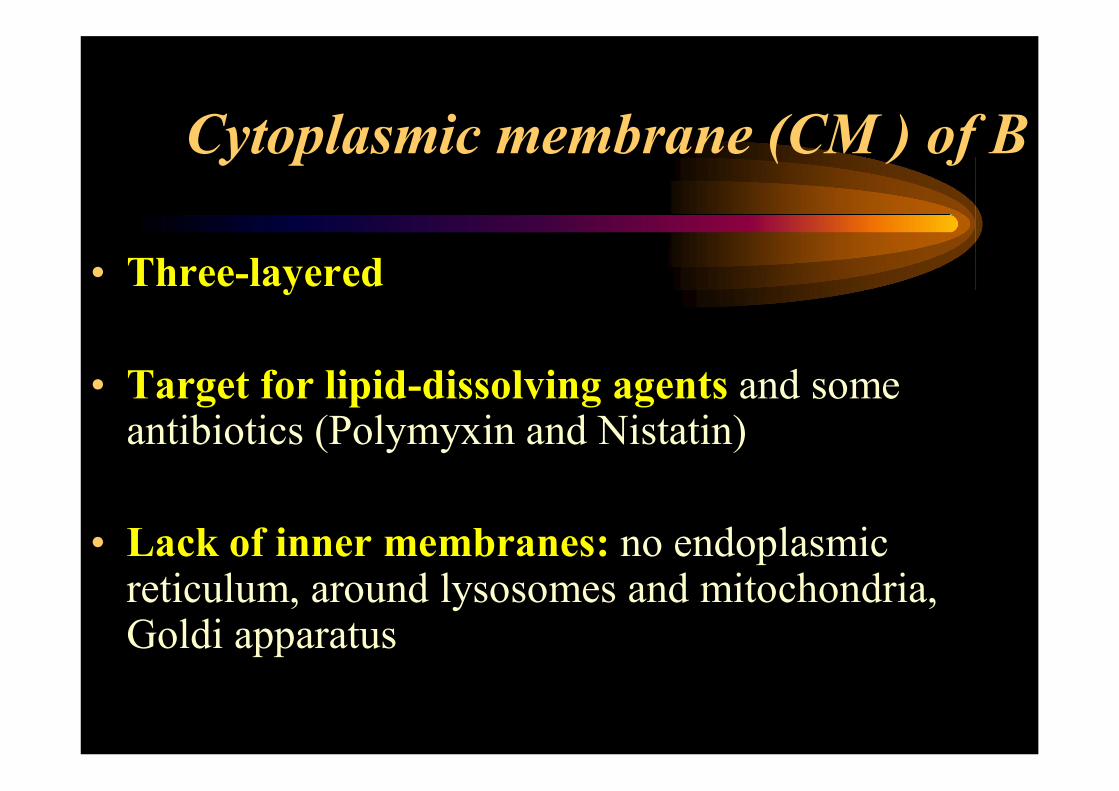
Cytoplasmic membrane (CM ) of B
• Three-layered
• Target for lipid-dissolving agents and some antibiotics (Polymyxin and Nistatin)
• Lack of inner membranes: no endoplasmic reticulum, around lysosomes and mitochondria, Goldi apparatus

Function of bacterial CM
• Respiratory (mitochondrial) equivalent
• Selective permeability
• Particpation in peptidoglycan synthesis and formation of penicillin-binding proteins, necessary for linkage with some antibiotics
• Participation in chromosome replication and large plasmids

Bacterial mesosomes
• Organels, formed by CM invagination
• Functions:
– respiratory (mitochondrial) equivalent
– coordination of core material division and cytoplasm during binary fission of bacteria

Cytoplasm and cytoplasmiccomponents of B
• Ribosomes – difference with Eu
• Inclusions:– volutine (diphtherial bacteria)
– glycogen (enteric bacteria)
– others
• Core (nucleoid)- a single DNA molecule
• Extra-chromosomal genetic elements (plasmids, bacteriophages)

Non-essential structures
• Capsule
• Flagella
• Pilli
• Spores

Capsule
• Real (S. рneumoniae, B. anthracis), slime (S. mutans), microcapsule (S. typhi)
• Structure
– polysaccharide (S. pneumoniae)
– polypeptide (B. anthracis)
• Staining (Klett, Neuffeld)
• Function:
– protection (virulence factor)
– Ag properties (K Ag)

Flagella• Structure – 3 parts:
- filament – long, thin, helical structure composed of protein flagellin
- hook- curved sheath
- basal body – stack of rings firmly anchored in cell wall
• Composition – protein (flagellin)
• Function
– motion
– virulence factor
– antigenic
– receptor
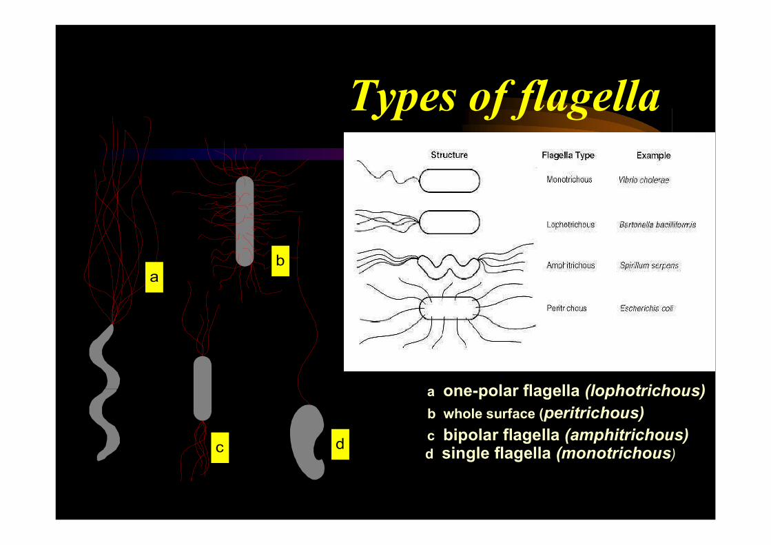
Types of flagella
a
a one-polar flagella (lophotrichous)
b
b whole surface (peritrichous)
c bipolar flagella (amphitrichous)c d
d single flagella (monotrichous)

Pilli (fimbriae)
• Types
– common (adhesins)
– sex (participate in conjugation)
Composition - protein (pillin=fimbrillin)
• Function:
– adhesion to cells (gonococci, E. coli)
– participation in conjugative transfer
– Ag properties (F Ag)
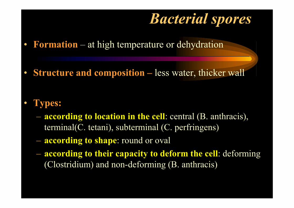
Bacterial spores
• Formation – at high temperature or dehydration
• Structure and composition – less water, thicker wall
• Types:
– according to location in the cell: central (B. anthracis), terminal(С. tetani), subterminal (C. perfringens)
– according to shape: round or oval
– according to their capacity to deform the cell: deforming(Clostridium) and non-deforming (B. anthracis)

Sporulation

Methods for studying bacterial structure
Microscopic
• Native
• Staining
– simple (Loffler, Pfeiffer)
– complex (Gram, Neisser, Zhiel-Neelsen, Moller, Klett)
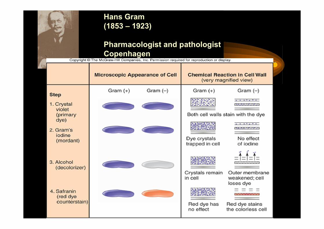
Hans Gram(1853 – 1923)
Pharmacologist and pathologistCopenhagen

Acid-fast Cell Walls
• Genus Mycobacterium and Nocardia
• mycolic acid (waxy lipid) covers thin peptidoglycan layer
• Do not stain well with Gram stain use acid-fast stain

Brightfield Microscopy
• Simplest of all the optical microscopy illumination. techniques
• Dark objects are visible against a bright background.
Darkfield Microscopy• Light objects visible
against dark background.
• used to enhance the contrast in unstained samples.
• Instrument of choice for spirochetes
Microscopy: The Instruments

Electron Microscopy: Detailed Images of Cell Parts
Uses electrons, electromagnetic lenses, and fluorescent screens
Electron wavelength ~ 100,000 x smaller than visible light wavelength
Specimens may be stained with heavy metal salts
Two types of EMs:?

Recommended literature
• Todar’s Online Textbook of Bacteriology
site: textbookofbacteriology.net
• Medical Microbiology
Edited by Samuel Baron
http://www.ncbi.nlm.nih.gov/books/NBK7627/
• Microbiology and Immunology On-line
http://www.microbiologybook.org