Embed Size (px)
Citation preview
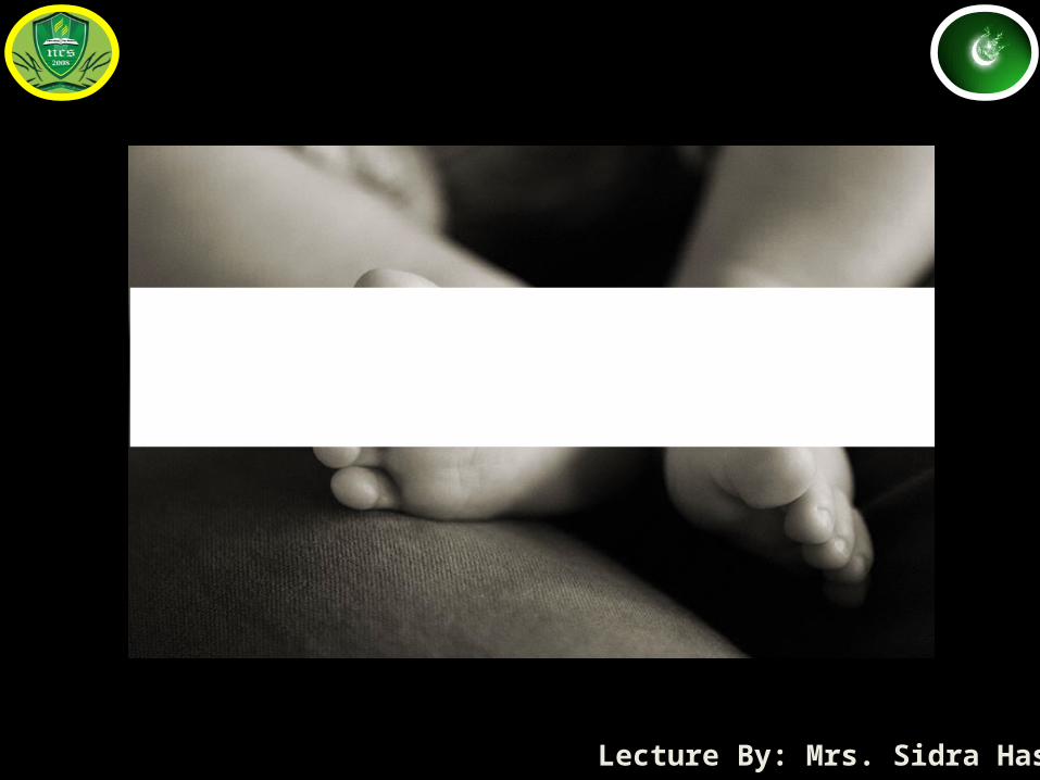
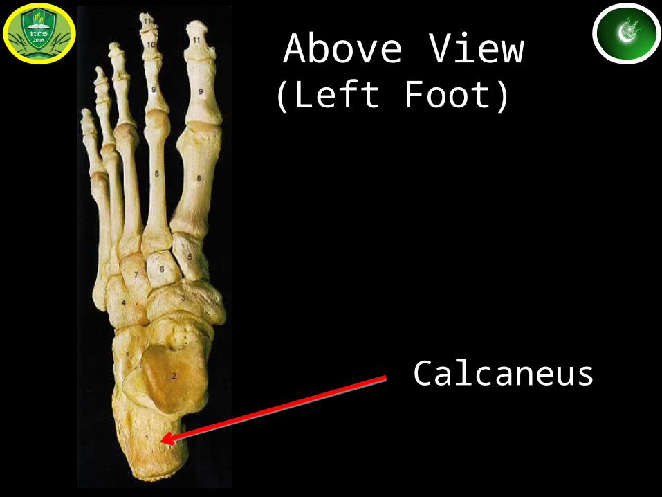
Anatomy of Foot
Lecture By: Mrs. Sidra Hasan
Calcaneus
Above View(Left Foot)
Talus
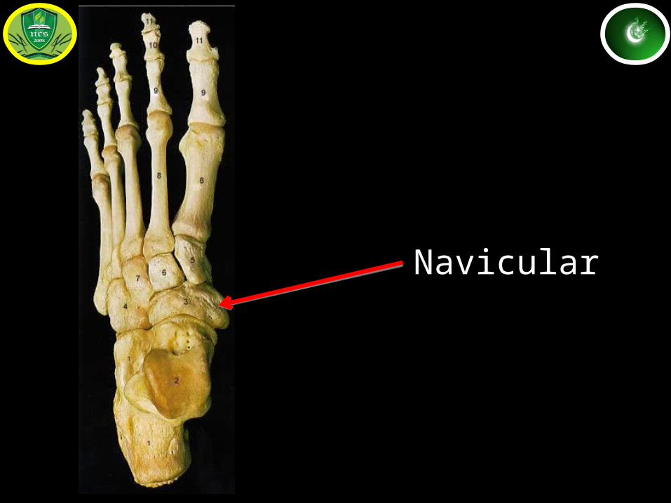
Navicular
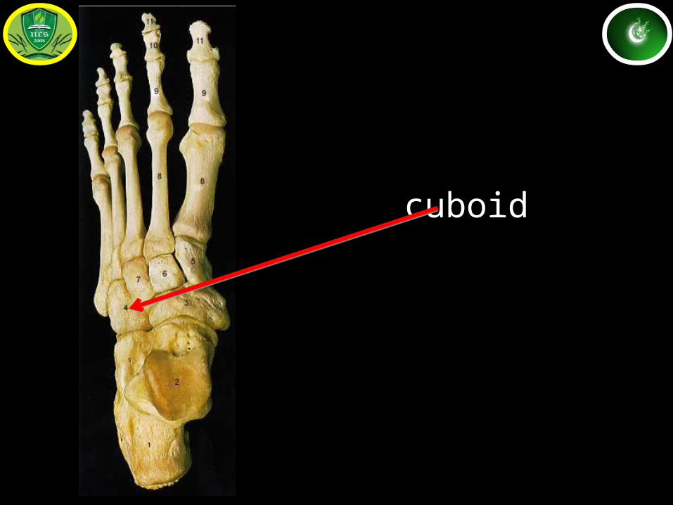
cuboidCuboid
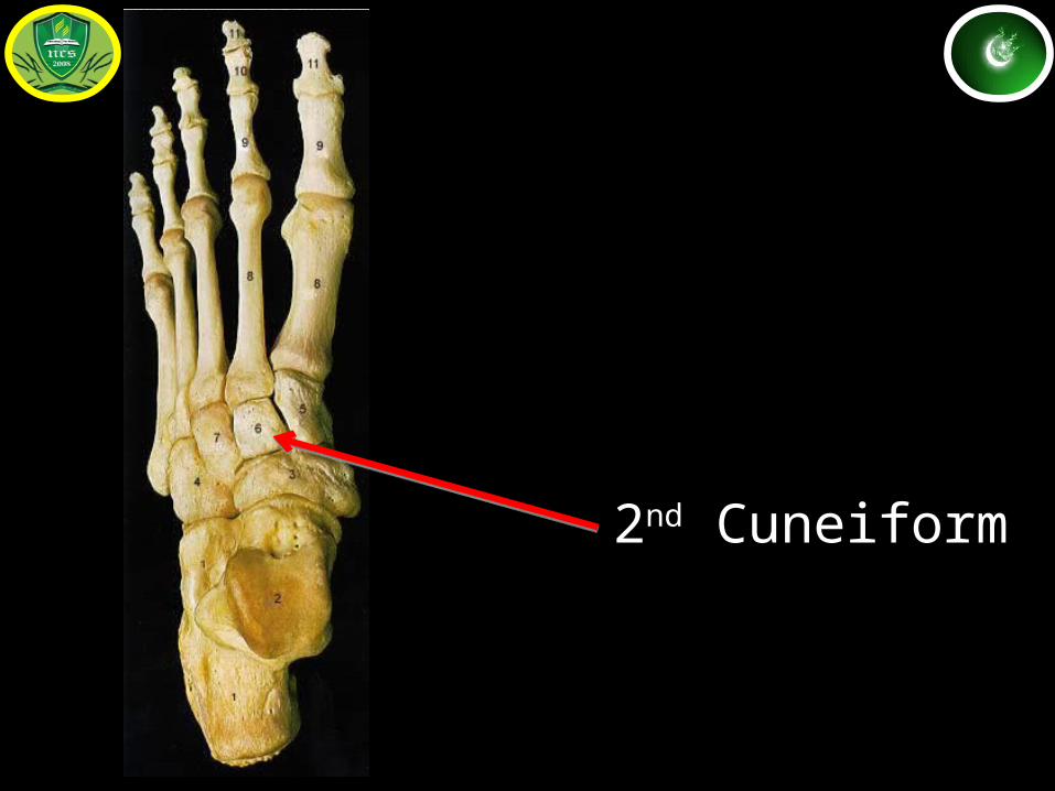
2nd Cuneiform
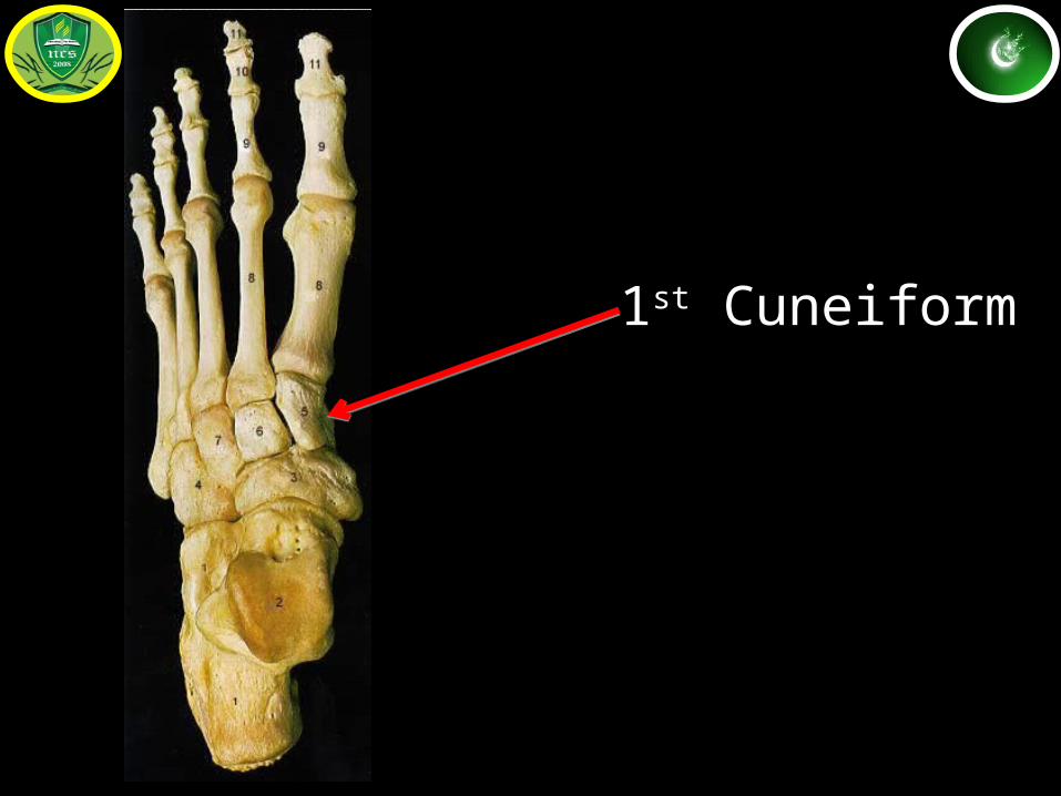
1st Cuneiform
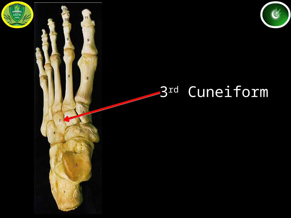
3rd Cuneiform
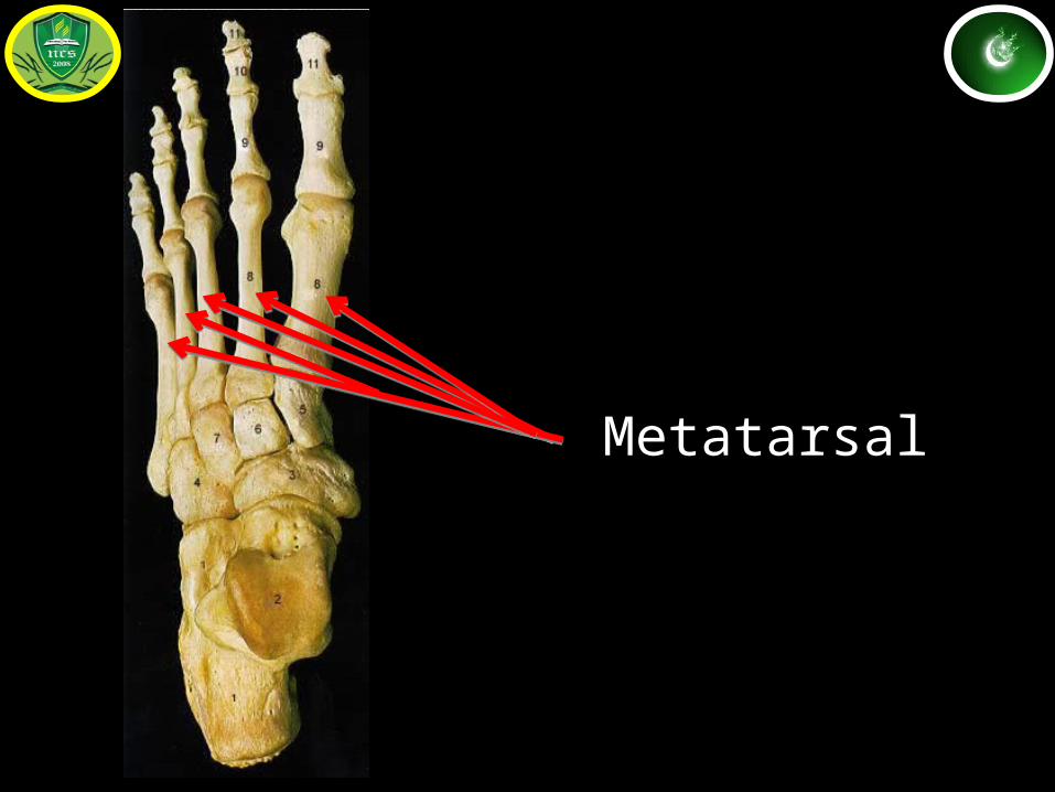
Metatarsal
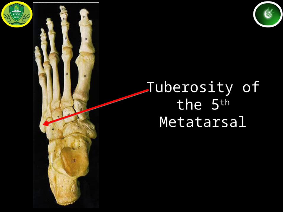
Tuberosity of the 5th
Metatarsal
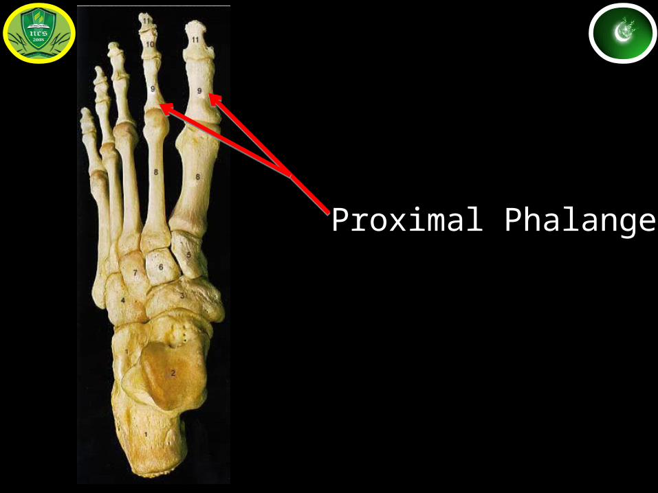
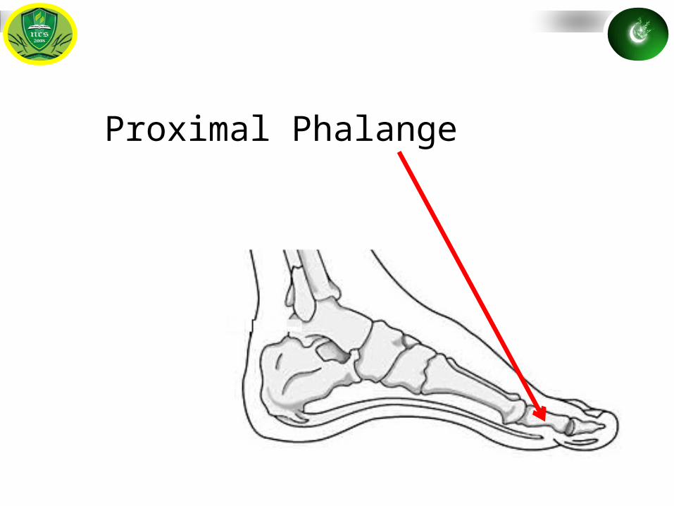
Proximal Phalange
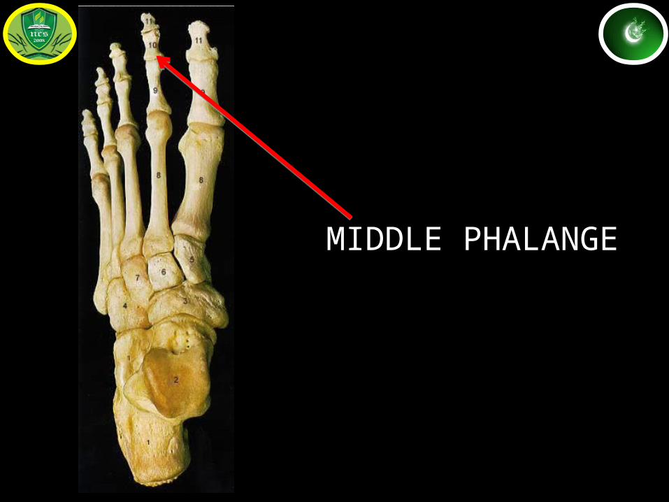
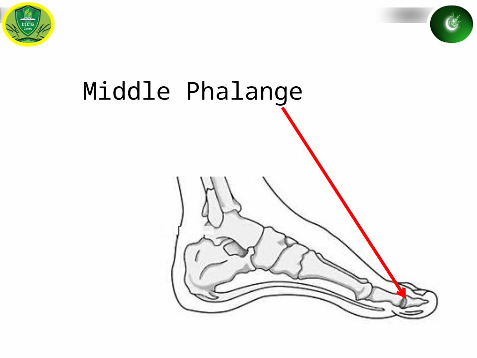
MIDDLE PHALANGE
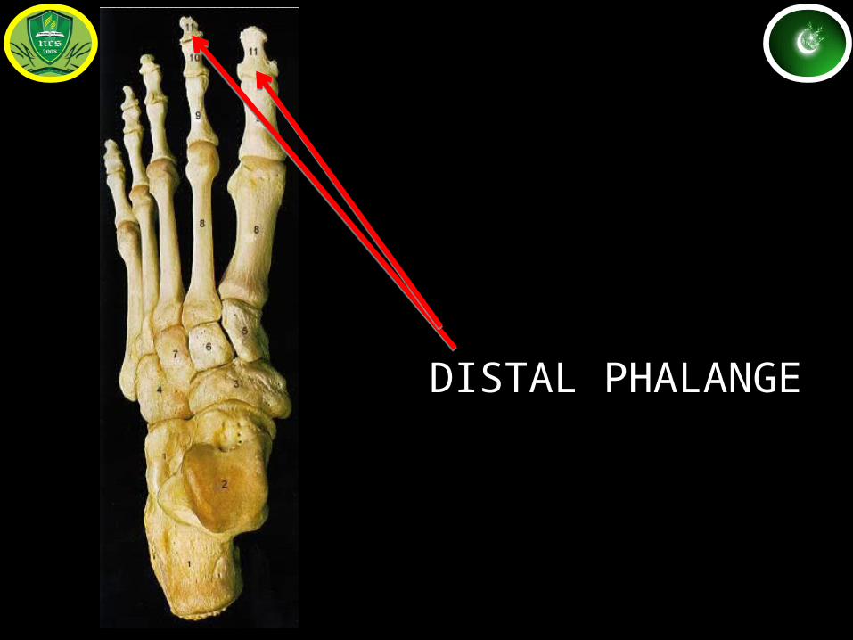
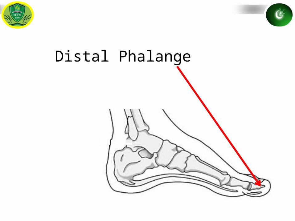
DISTAL PHALANGE
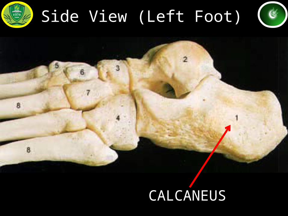
CALCANEUS
Side View (Left Foot)
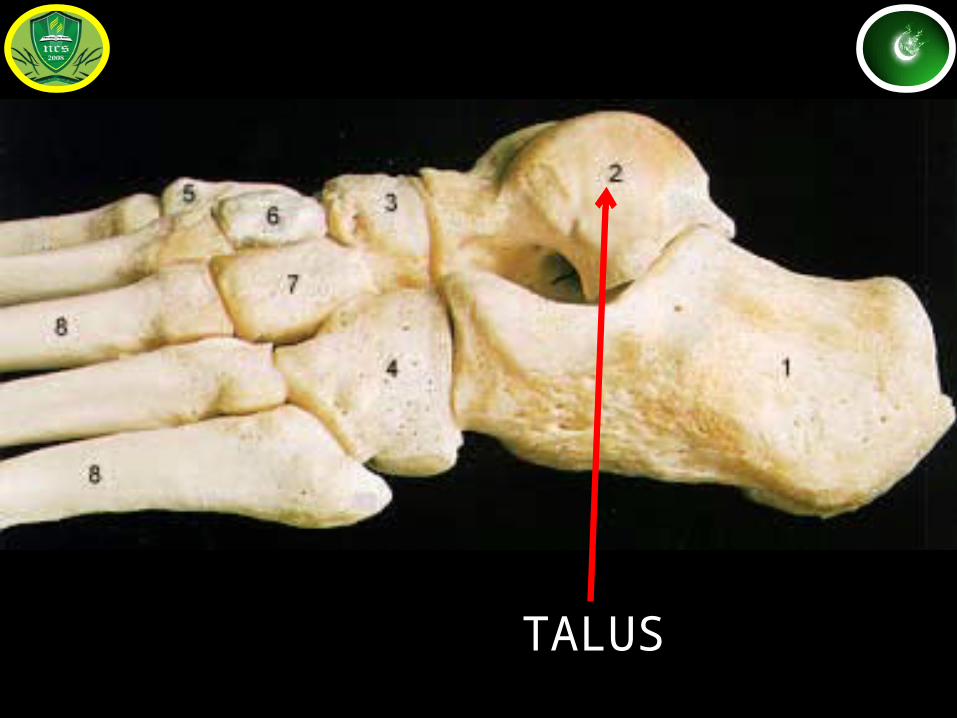
TALUS
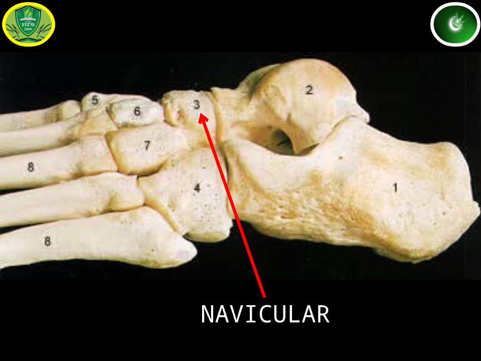
NAVICULAR
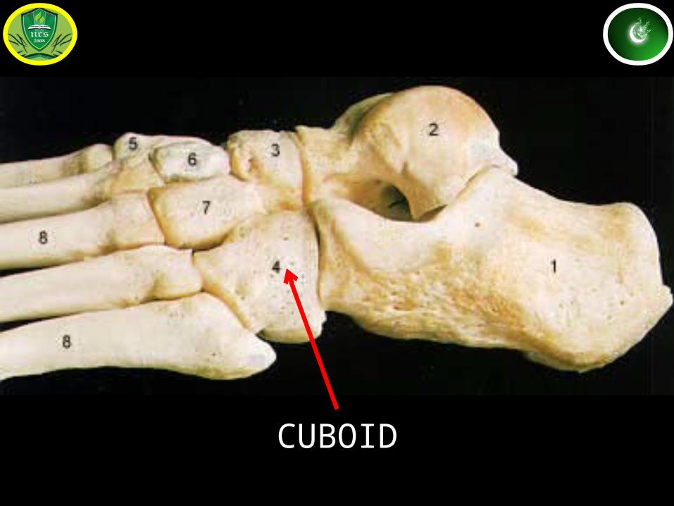
CUBOID
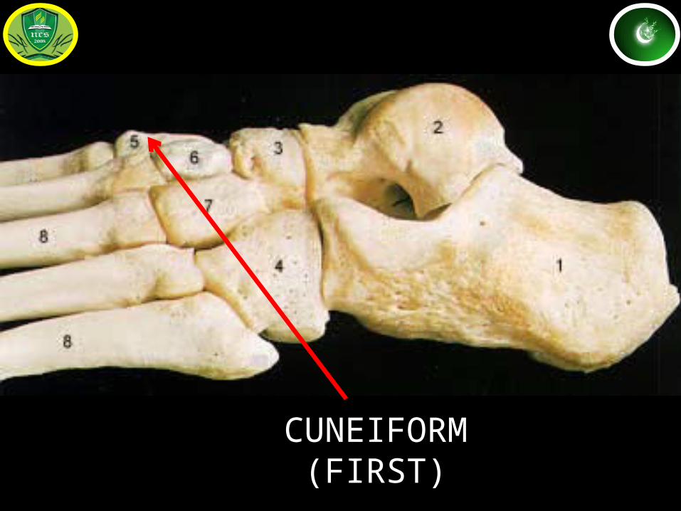
CUNEIFORM (FIRST)
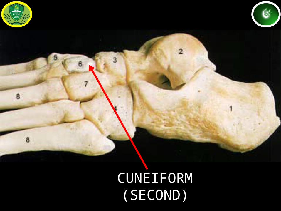
CUNEIFORM (SECOND)
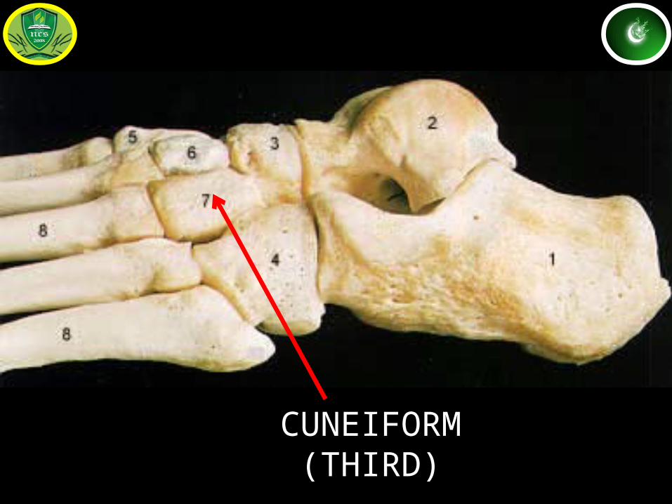
CUNEIFORM (THIRD)
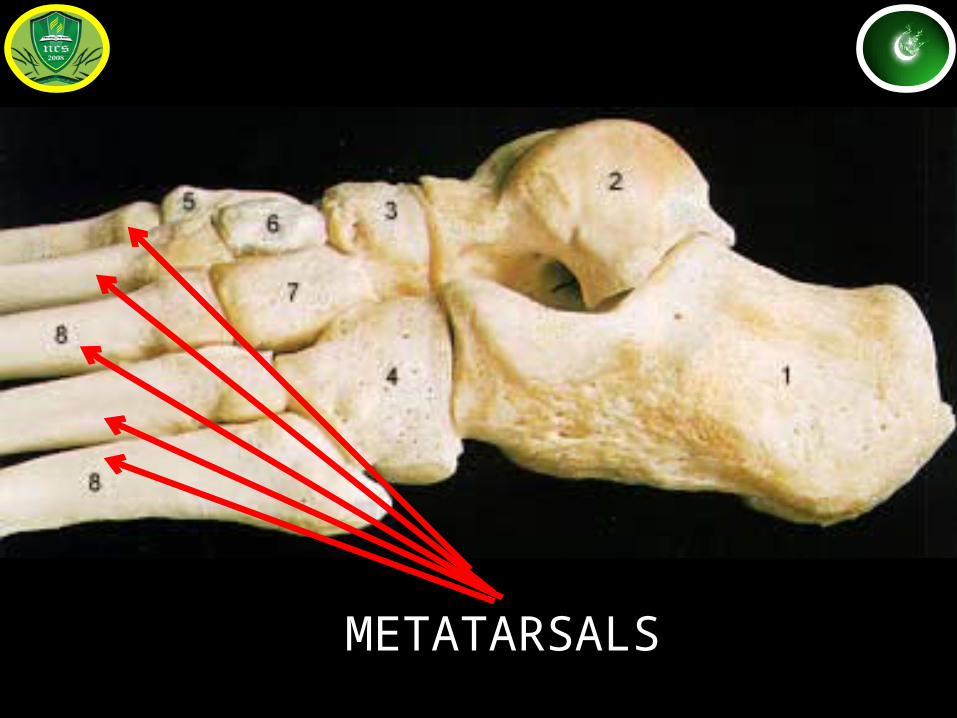
METATARSALS
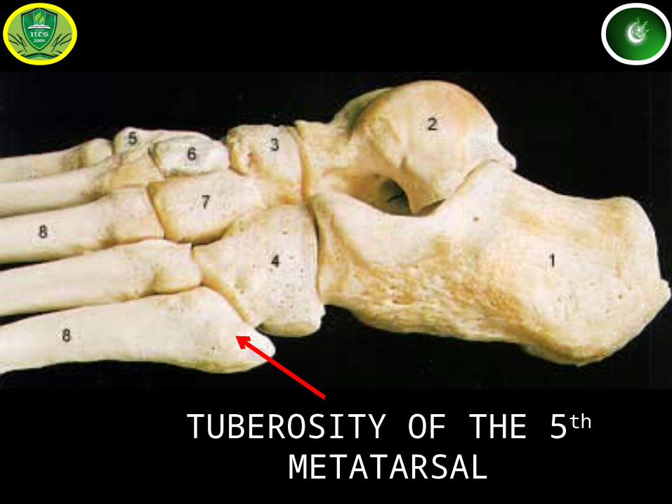
TUBEROSITY OF THE 5th METATARSAL
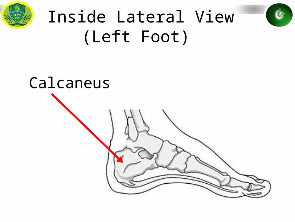
Calcaneus
Inside Lateral View (Left Foot)
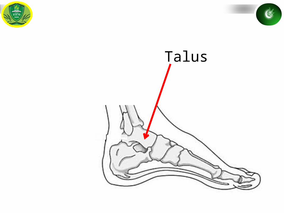
Talus
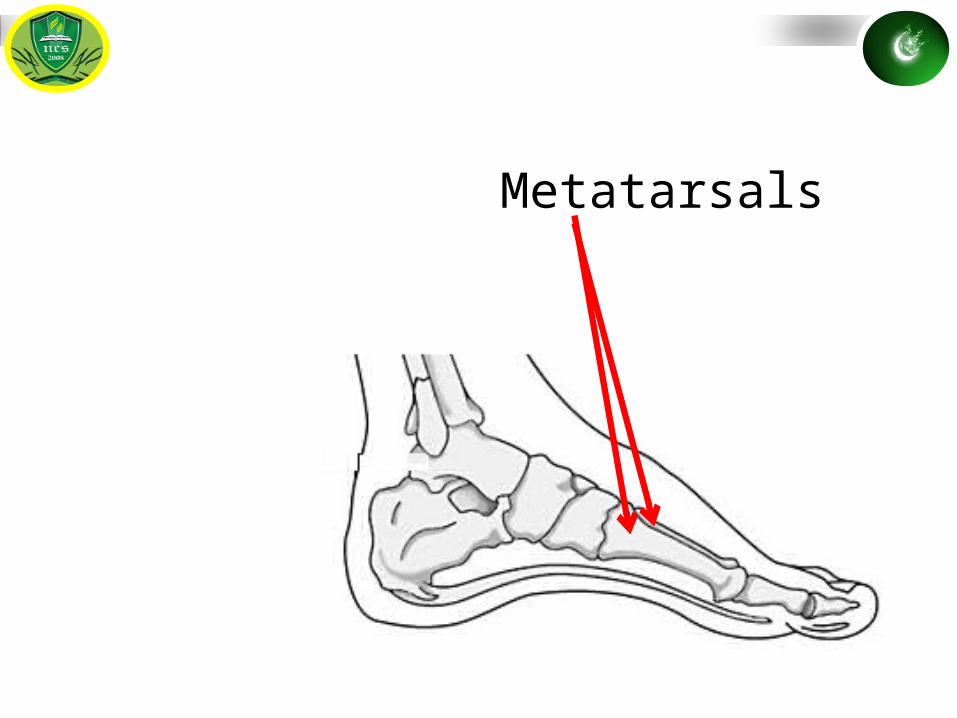
Metatarsals
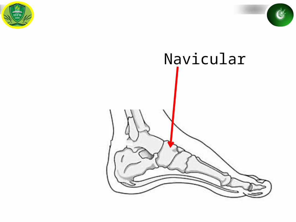
Navicular
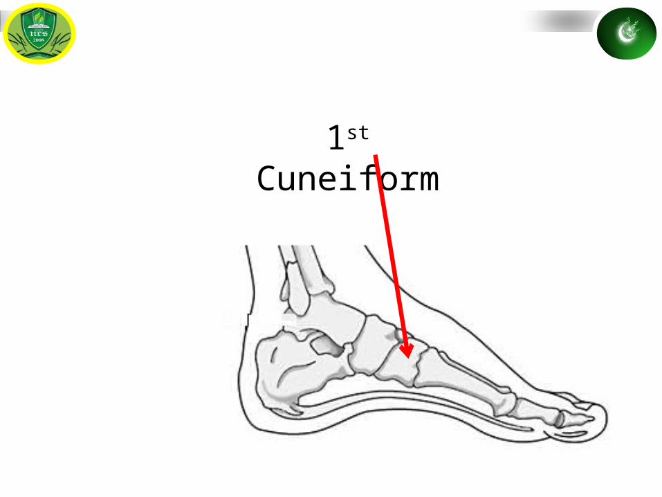
1st Cuneiform
Distal Phalange
Middle Phalange
Proximal Phalange
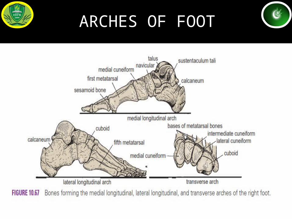
ARCHES OF FOOT
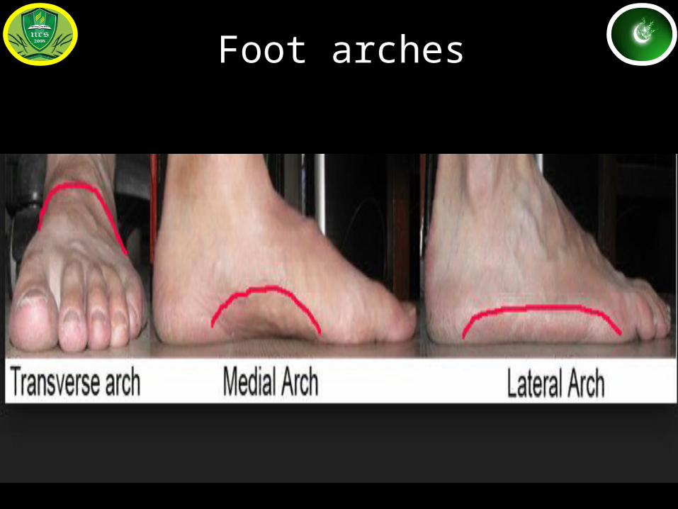
Foot arches
CLASSIFICATION OF ARCHES
1)Medial longitudinal arch2)Lateral longitudinal arch3)Transverse arches
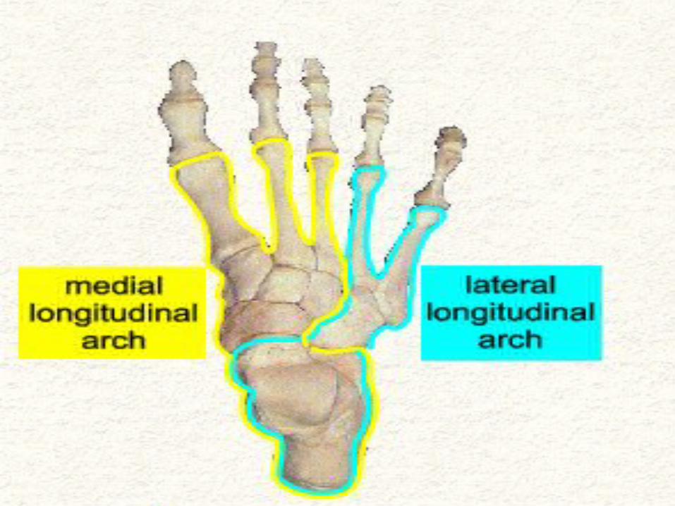
Medial longitudinal arch
• More higher, more mobile, more resilient than lateral arch.
• Formed by the calcaneum, the talus, the navicular,3 cuneiforms & 1st 3 metatarsal bones.
ENDS:• ANTERIOR END: FORMED BY heads of 1st,2nd.3rd
metatarsals.• POSTERIOR END: FORMED BY medial tubercle of
calcaneum. SUMMIT: FORMED BY sup. Articular surface of talus
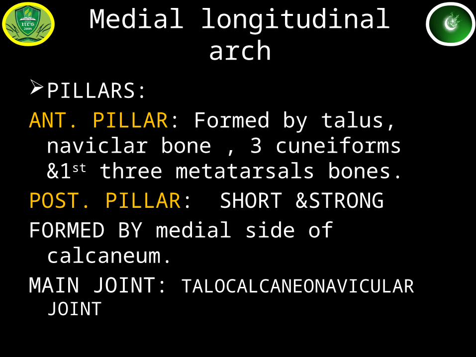
Medial longitudinal arch
PILLARS: ANT. PILLAR: Formed by talus, naviclar bone , 3
cuneiforms &1st three metatarsals bones.POST. PILLAR: SHORT &STRONGFORMED BY medial side of calcaneum.MAIN JOINT: TALOCALCANEONAVICULAR JOINT
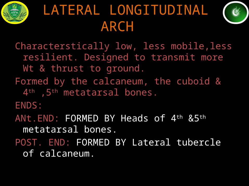
LATERAL LONGITUDINAL ARCHLL
Characterstically low, less mobile,less resilient. Designed to transmit more Wt & thrust to ground.
Formed by the calcaneum, the cuboid & 4th ,5th metatarsal bones.
ENDS:ANt.END: FORMED BY Heads of 4th &5th metatarsal
bones.POST. END: FORMED BY Lateral tubercle of
calcaneum.
.
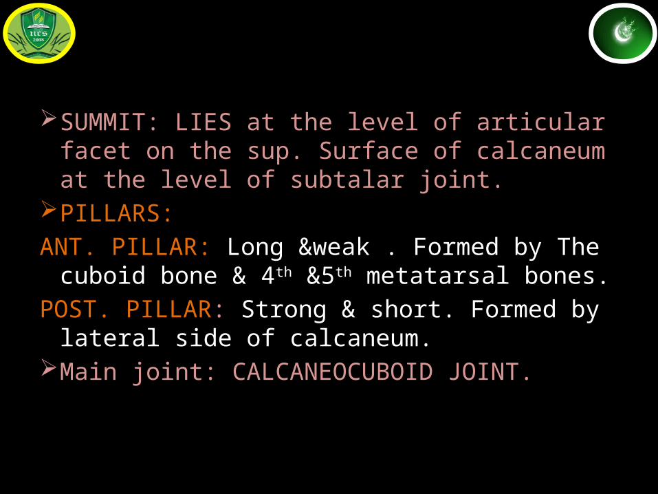
SUMMIT: LIES at the level of articular facet on the sup. Surface of calcaneum at the level of subtalar joint.
PILLARS: ANT. PILLAR: Long &weak . Formed by The cuboid
bone & 4th &5th metatarsal bones.POST. PILLAR: Strong & short. Formed by lateral
side of calcaneum.Main joint: CALCANEOCUBOID JOINT.
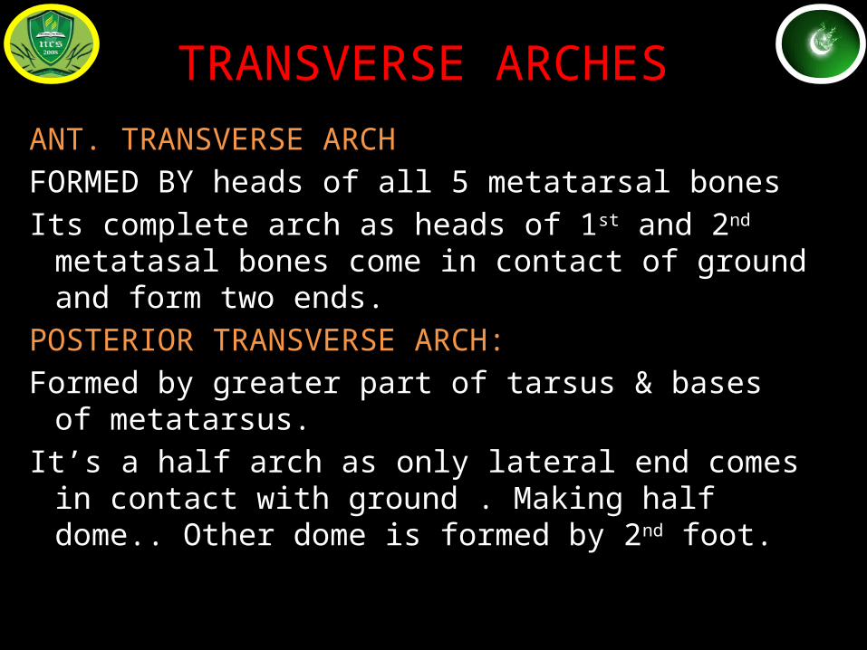
TRANSVERSE ARCHES
ANT. TRANSVERSE ARCHFORMED BY heads of all 5 metatarsal bonesIts complete arch as heads of 1st and 2nd metatasal
bones come in contact of ground and form two ends.
POSTERIOR TRANSVERSE ARCH:Formed by greater part of tarsus & bases of
metatarsus.It’s a half arch as only lateral end comes in contact
with ground . Making half dome.. Other dome is formed by 2nd foot.
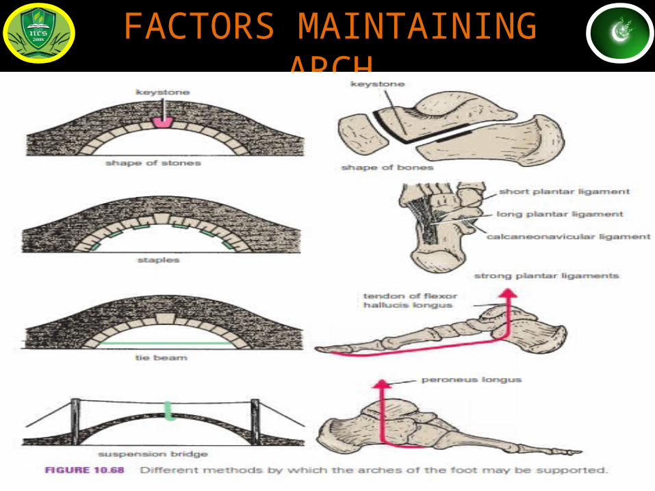
FACTORS MAINTAINING ARCH
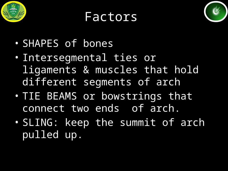
Factors
• SHAPES of bones• Intersegmental ties or ligaments & muscles
that hold different segments of arch• TIE BEAMS or bowstrings that connect two
ends of arch.• SLING: keep the summit of arch pulled up.
Factors
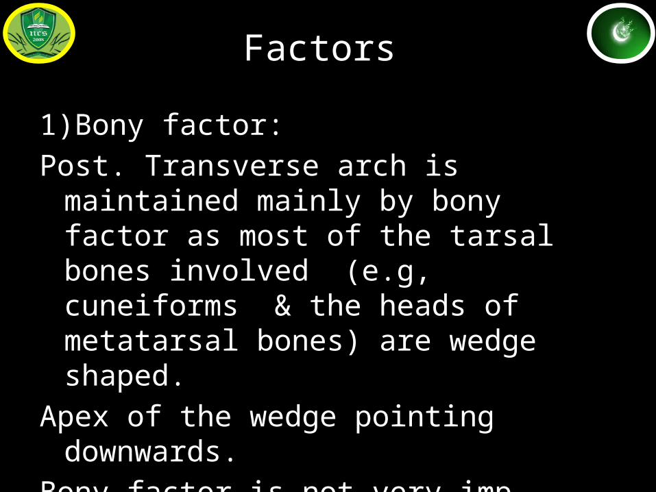
1)Bony factor: Post. Transverse arch is maintained mainly by
bony factor as most of the tarsal bones involved (e.g, cuneiforms & the heads of metatarsal bones) are wedge shaped.
Apex of the wedge pointing downwards.Bony factor is not very imp incase of other
arches.
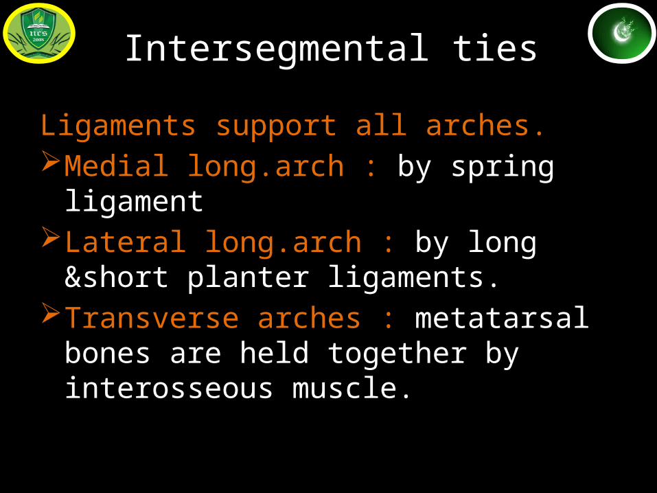
Intersegmental ties
Ligaments support all arches.Medial long.arch : by spring ligamentLateral long.arch : by long &short planter
ligaments.Transverse arches : metatarsal bones are held
together by interosseous muscle.
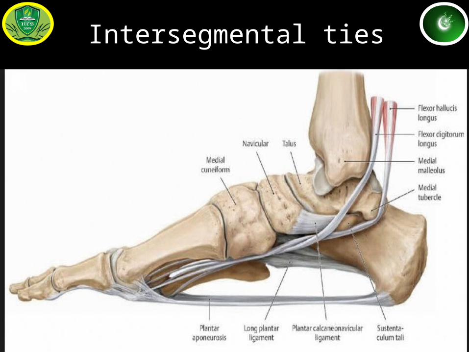
Intersegmental ties
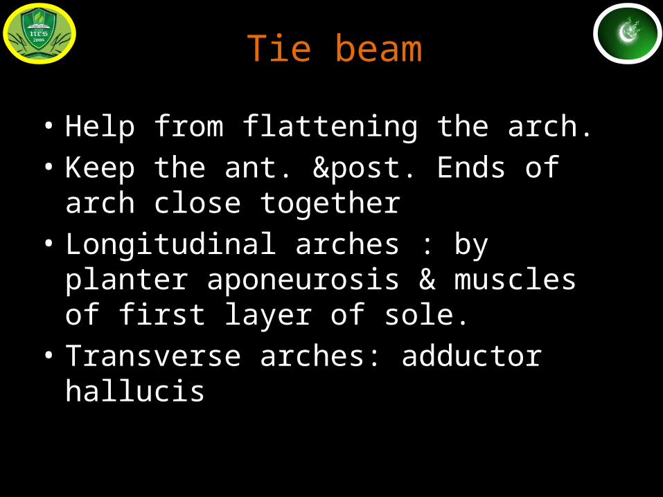
Tie beam
• Help from flattening the arch.• Keep the ant. &post. Ends of arch close
together• Longitudinal arches : by planter aponeurosis &
muscles of first layer of sole.• Transverse arches: adductor hallucis
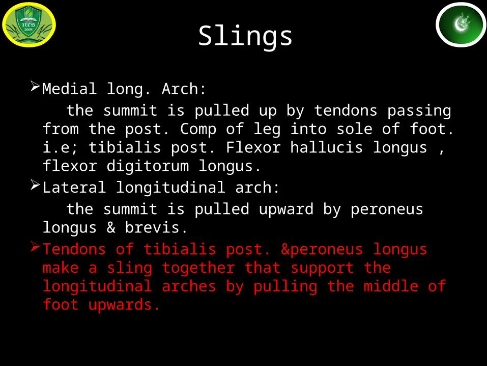
Slings
Medial long. Arch: the summit is pulled up by tendons passing from the
post. Comp of leg into sole of foot. i.e; tibialis post. Flexor hallucis longus , flexor digitorum longus.
Lateral longitudinal arch: the summit is pulled upward by peroneus longus &
brevis.Tendons of tibialis post. &peroneus longus make a
sling together that support the longitudinal arches by pulling the middle of foot upwards.
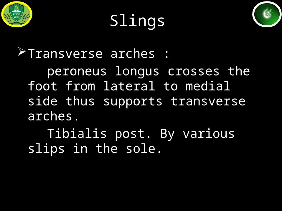
Slings
Transverse arches : peroneus longus crosses the foot from lateral
to medial side thus supports transverse arches.
Tibialis post. By various slips in the sole.
FUNCTIONS OF ARCHES
• Distribution of body wt• Spring action during walking &running• Shock absorber• Protection of soft tissues of sole
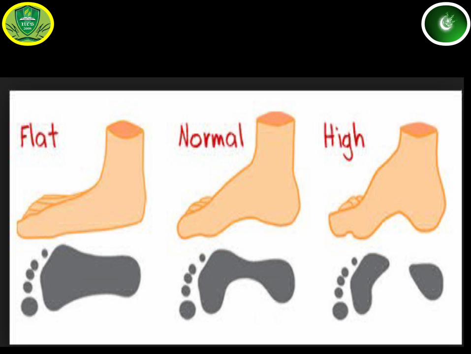
Clinical anatomy of arches
Pes planusPes cavusClaw footTalipes equanusTalipes calcaneusTalipes varusTalipes valgusTalipes equanuvarus (club foot)Talipes calcaneovalgus
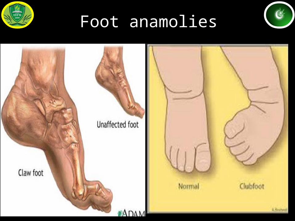
Foot anamolies
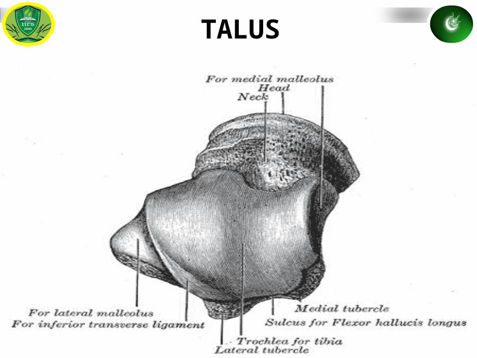
TALUS
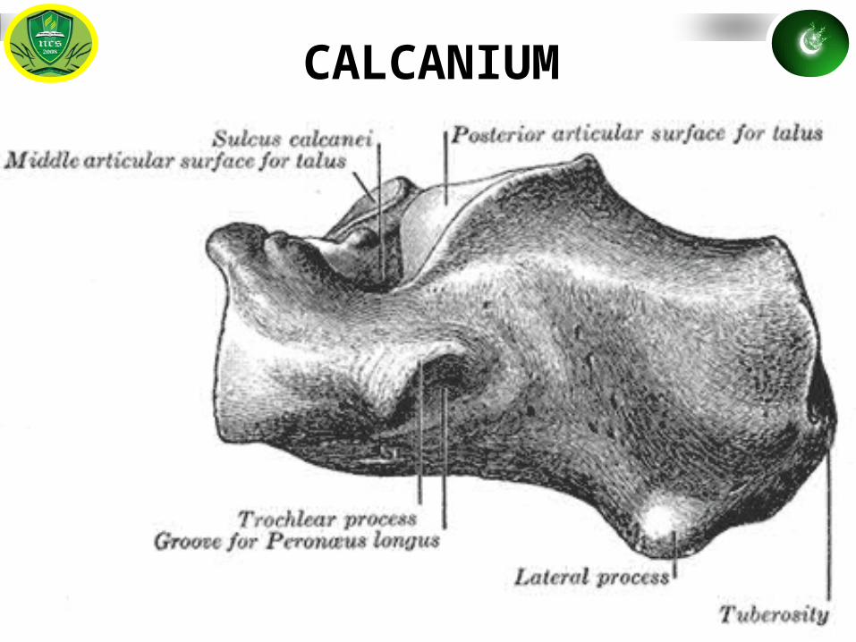
CALCANIUM
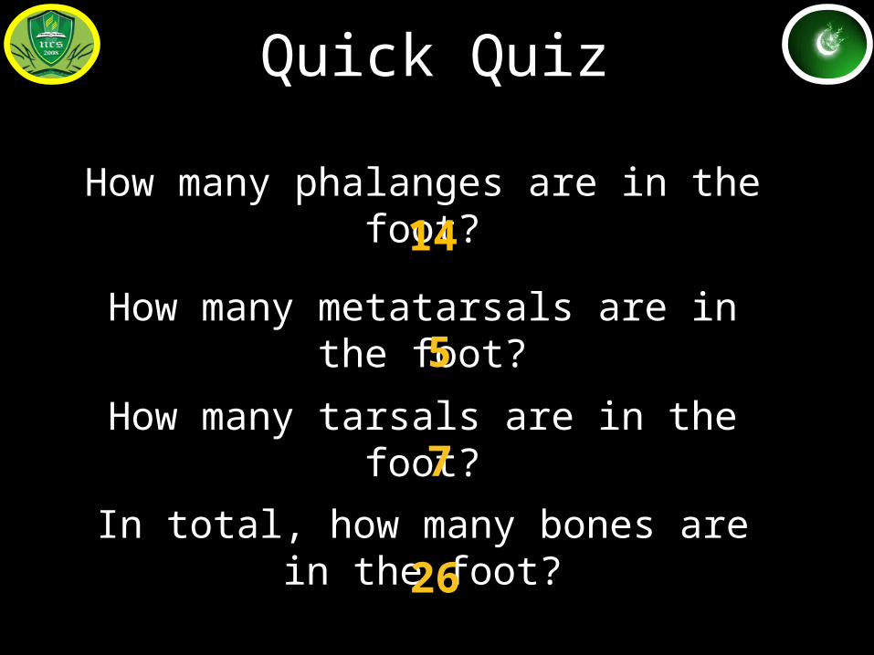
Quick Quiz
How many phalanges are in the foot?
14
How many metatarsals are in the foot?5
How many tarsals are in the foot?7
In total, how many bones are in the foot?
26