Embed Size (px)
Citation preview
Lecture B4 - Biosurface EngineeringPractical aspects of bioactive coatings
Faculty of Mechanical Engineering, Institute of Materials Science, Max Bergmann Center of biomaterials
Teaching Gdansk October 2012
Cornelia Wolf-Brandstetter
Teaching Gdansk 2012 - C. Wolf-Brandstetter Slide 2 of 65
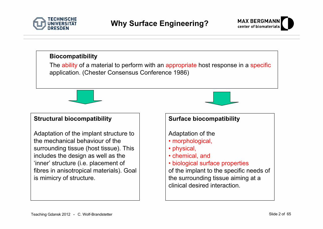
Why Surface Engineering?
BiocompatibilityThe ability of a material to perform with an appropriate host response in a specificapplication. (Chester Consensus Conference 1986)
Structural biocompatibility
Adaptation of the implant structure to the mechanical behaviour of the surrounding tissue (host tissue). This includes the design as well as the ‘inner’ structure (i.e. placement of fibres in anisotropical materials). Goal is mimicry of structure.
Surface biocompatibility
Adaptation of the • morphological,• physical,• chemical, and • biological surface propertiesof the implant to the specific needs of the surrounding tissue aiming at a clinical desired interaction.

Teaching Gdansk 2012 - C. Wolf-Brandstetter Slide 3 of 65
Biomaterials and cells
All healing is due to cells: from blood cells (coagulation, inflammation) to stem cells (source of undifferentiated cells that form new tissue specificcells).
The ideal biomaterials has to give the right cues for cellbehavior and differentiation!
OH-Groups+ -
Ions Proteins
Cells
Tissue
0,1 1 10 100 1000 104 105 106
1 10d ds s s s
time
Implant / Biomaterial
Teaching Gdansk 2012 - C. Wolf-Brandstetter Slide 4 of 65
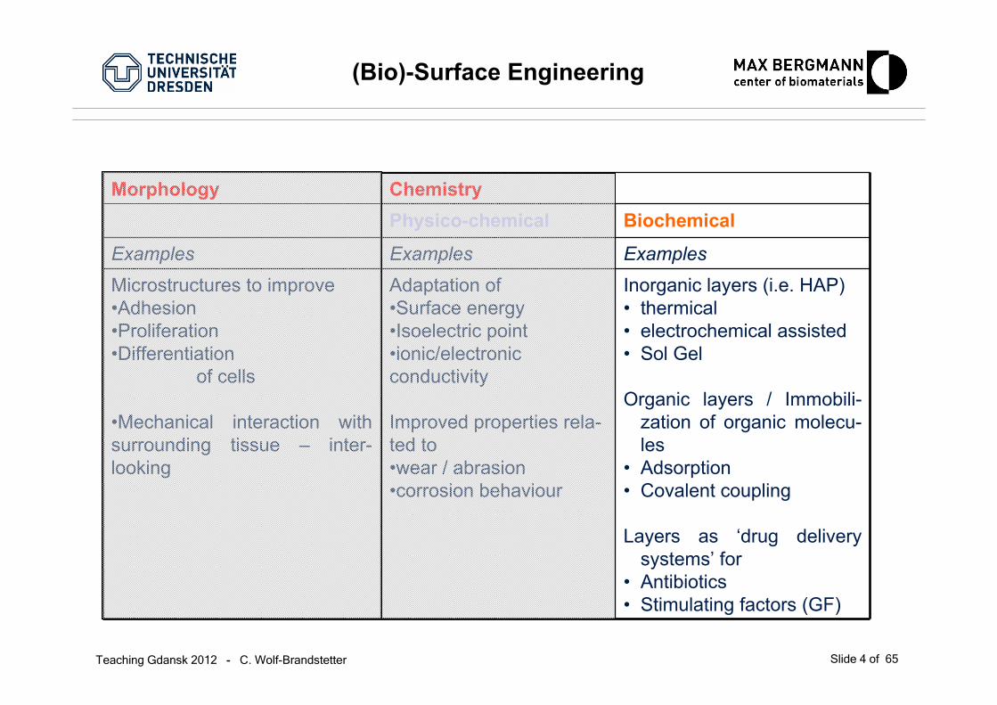
(Bio)-Surface Engineering
Morphology ChemistryPhysico-chemical Biochemical
Examples Examples ExamplesMicrostructures to improve•Adhesion•Proliferation•Differentiation
of cells
•Mechanical interaction with surrounding tissue – inter-looking
Adaptation of•Surface energy•Isoelectric point•ionic/electronic conductivity
Improved properties rela-ted to•wear / abrasion•corrosion behaviour
Inorganic layers (i.e. HAP)• thermical• electrochemical assisted• Sol Gel
Organic layers / Immobili-zation of organic molecu-les
• Adsorption• Covalent coupling
Layers as ‘drug delivery systems’ for
• Antibiotics• Stimulating factors (GF)

Teaching Gdansk 2012 - C. Wolf-Brandstetter Slide 5 of 65
(Bio)-Surface Engineering for Bone
What does this mean in thespecial case of bone?
Aim: Engineer the surface to resemble the microenvironment of cells! Method: By using the main components of their surroundings!
Mineral (HAp) Protein* H2O
0 20 40 60 80 100 %
Structural protein collagen type I
Other collagens, proteoglycans, glycosaminoglycans, growth factors,…

Teaching Gdansk 2012 - C. Wolf-Brandstetter Slide 6 of 65
Adhesion and the biomaterial surface
Aim: Engineer the surface to resemble the microenvironment of cells! Method: By using the main components of their surroundings!
Possible approaches: Inorganic coatings (bone)
hydroxy apatiteOrganic coatings
Peptides (i.e. RGD)ECM proteins (i.e. collagen)Synthetic matrices mimicingthe ECM (ie. )
Aim to incorporate ECM signals into biomaterialsby using motifs of thenatural ECM
ECM motifson thebiomaterial
Influence: • cell adhesion/ migration• matrix remodelling• cytokine signalling•….
biomaterial
Teaching Gdansk 2012 - C. Wolf-Brandstetter Slide 7 of 65
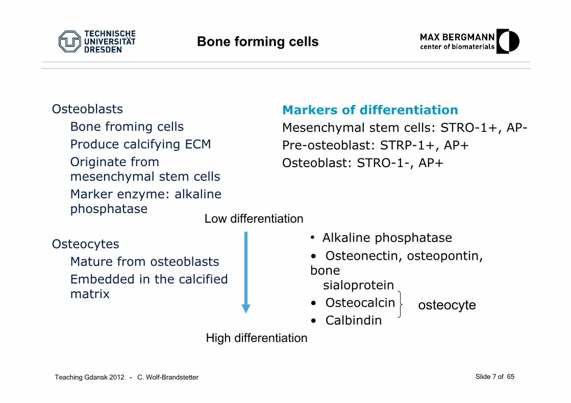
Bone forming cells
OsteoblastsBone froming cellsProduce calcifying ECMOriginate frommesenchymal stem cellsMarker enzyme: alkalinephosphatase
OsteocytesMature from osteoblastsEmbedded in the calcifiedmatrix
Markers of differentiationMesenchymal stem cells: STRO-1+, AP-Pre-osteoblast: STRP-1+, AP+Osteoblast: STRO-1-, AP+
• Alkaline phosphatase• Osteonectin, osteopontin, bone
sialoprotein• Osteocalcin• Calbindin
osteocyte
Low differentiation
High differentiation

Teaching Gdansk 2012 - C. Wolf-Brandstetter Slide 8 of 65
Bone degrading cells
OsteoclastsBone degrading cellsLysate and phagocyte boneOriginate from heamatopoeticcells (macrophage line)Marker enzyme: tartrateresistant acid phosphatase(TRAP) Characteristics
• 4-20 nuclei• Ruffled border and sealing zone• Calcitonin receptor positive• Vitronectin receptor positive• Cathepsin K positive• Matrixmetalloproteinase 9 (MMP-9)
Activity test• Resorption pit formation• Collagen degradation products(hydroxyproline)

Teaching Gdansk 2012 - C. Wolf-Brandstetter Slide 9 of 65
Inorganic coatings –for boneimplants
Teaching Gdansk 2012 - C. Wolf-Brandstetter Slide 10 of 65
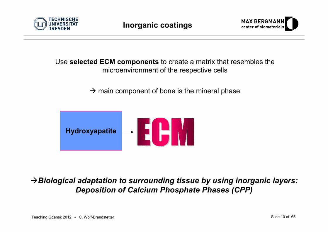
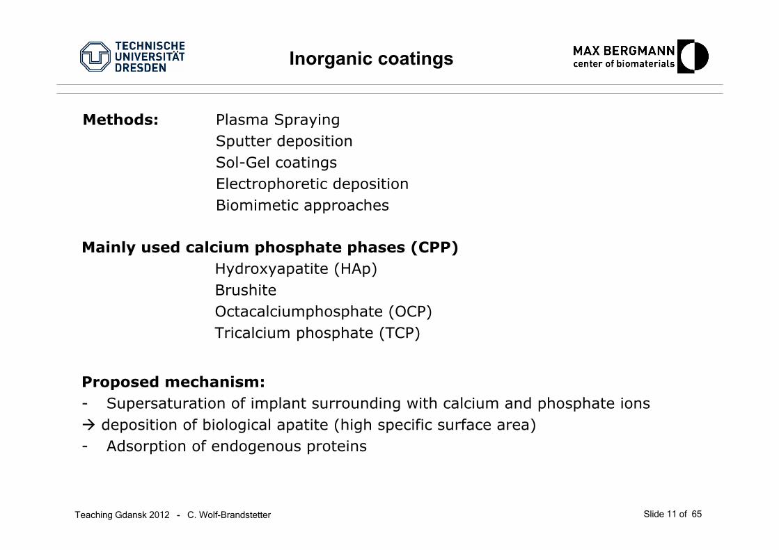
Inorganic coatings
Hydroxyapatite
main component of bone is the mineral phase
Biological adaptation to surrounding tissue by using inorganic layers:Deposition of Calcium Phosphate Phases (CPP)
Use selected ECM components to create a matrix that resembles the microenvironment of the respective cells
Teaching Gdansk 2012 - C. Wolf-Brandstetter Slide 11 of 65
Proposed mechanism:- Supersaturation of implant surrounding with calcium and phosphate ions
deposition of biological apatite (high specific surface area)- Adsorption of endogenous proteins
Methods: Plasma SprayingSputter depositionSol-Gel coatingsElectrophoretic depositionBiomimetic approaches
Inorganic coatings
Mainly used calcium phosphate phases (CPP)Hydroxyapatite (HAp)BrushiteOctacalciumphosphate (OCP)Tricalcium phosphate (TCP)
Teaching Gdansk 2012 - C. Wolf-Brandstetter Slide 12 of 65
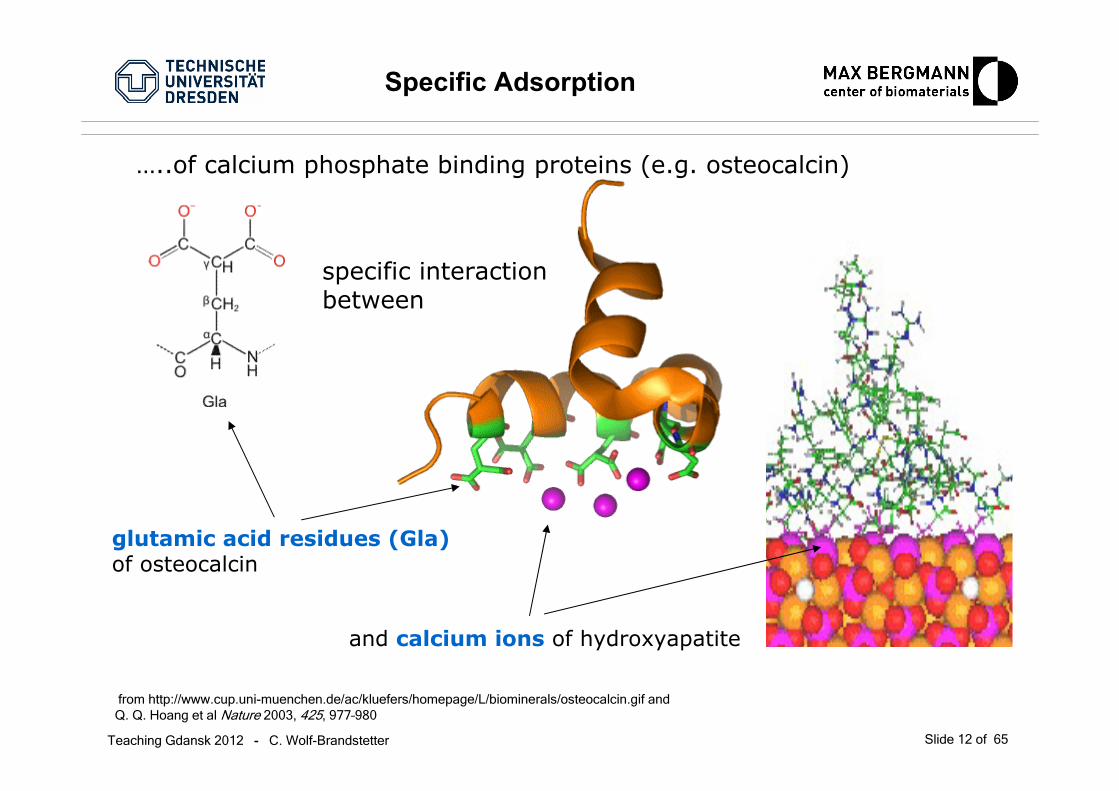
Specific Adsorption
from http://www.cup.uni-muenchen.de/ac/kluefers/homepage/L/biominerals/osteocalcin.gif and Q. Q. Hoang et al Nature 2003, 425, 977–980
glutamic acid residues (Gla)of osteocalcin
…..of calcium phosphate binding proteins (e.g. osteocalcin)
and calcium ions of hydroxyapatite
specific interactionbetween
Teaching Gdansk 2012 - C. Wolf-Brandstetter Slide 13 of 65
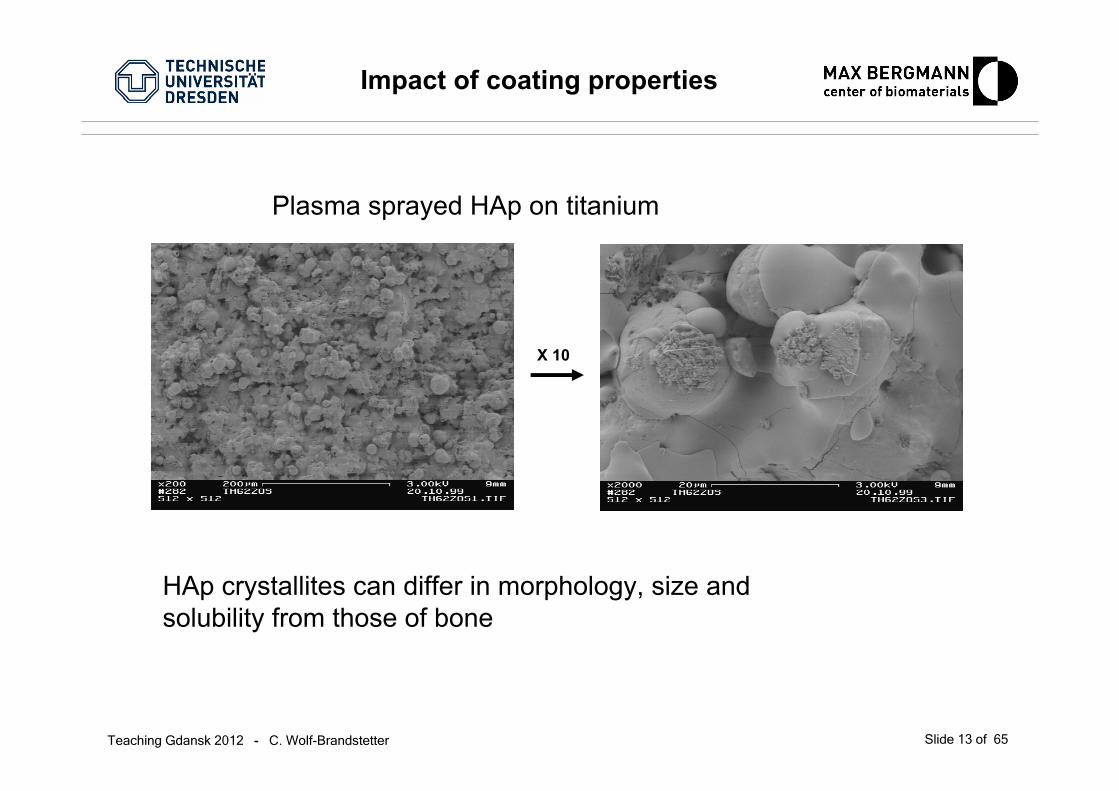
Impact of coating properties
HAp crystallites can differ in morphology, size and solubility from those of bone
X 10
Plasma sprayed HAp on titanium
Teaching Gdansk 2012 - C. Wolf-Brandstetter Slide 14 of 65
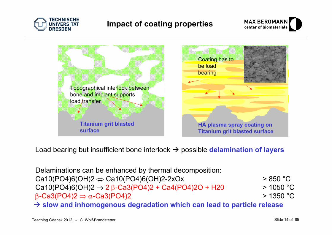
Load bearing but insufficient bone interlock possible delamination of layers
Delaminations can be enhanced by thermal decomposition:Ca10(PO4)6(OH)2 ⇔ Ca10(PO4)6(OH)2-2xOx > 850 °CCa10(PO4)6(OH)2 ⇒ 2 β-Ca3(PO4)2 + Ca4(PO4)2O + H20 > 1050 °Cβ-Ca3(PO4)2 ⇒ α-Ca3(PO4)2 > 1350 °C
HA plasma spray coating on Titanium grit blasted surface
Coating has to be load bearing
Titanium grit blasted surface
Topographical interlock between bone and implant supports load transfer
slow and inhomogenous degradation which can lead to particle release
Impact of coating properties
Teaching Gdansk 2012 - C. Wolf-Brandstetter Slide 15 of 65
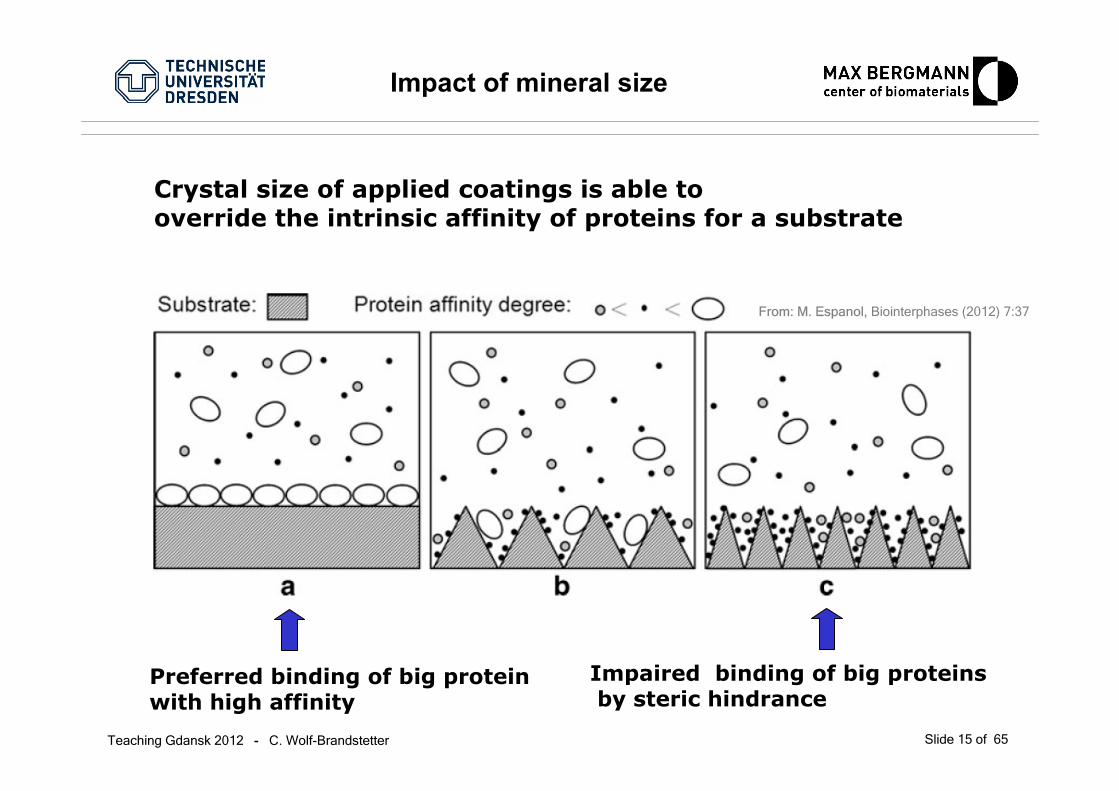
Impact of mineral size
From: M. Espanol, Biointerphases (2012) 7:37
Crystal size of applied coatings is able to override the intrinsic affinity of proteins for a substrate
Preferred binding of big proteinwith high affinity
Impaired binding of big proteinsby steric hindrance
Teaching Gdansk 2012 - C. Wolf-Brandstetter Slide 16 of 65
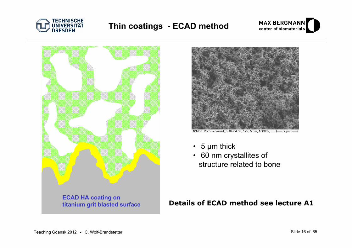
Thin coatings - ECAD method
ECAD HA coating on titanium grit blasted surface
• 5 µm thick• 60 nm crystallites of
structure related to bone
Details of ECAD method see lecture A1
Teaching Gdansk 2012 - C. Wolf-Brandstetter Slide 17 of 65
• Deposition of thin, not monolithic layers with a composition, crystal size and chemical history close to that of bone mineral possible
• Thickness and chemical composition of coatings can be controlled to the sub-µm level• Generation of coatings with high specific surface area possible - specific surface of about
50 g/cm2, thus for 100 µg/cm2 coatings surface enlargement by factor of about 40• Use of appropriate conditions results in CPP with a composition and structure close to
mineral phase of bone• Low energy process• Low cost process (with respect to production costs as well as waste management)• No adverse effect of heat on substrate material• Compared to incubation in SBF more defined and higher relative supersaturation at the
interface, resulting in shorter processing times• Due to possible physiological processing parameters (pH, temperature, aqueous solution)
the deposition of CPP can be combined with the immobilisation/incorporation of organic components like proteins (peptides)
• Excellent homogeneity of coatings on structured and porous surfaces as well as on irregularly formed structures as it is no line of sight process.
Advantages of the ECAD method

Teaching Gdansk 2012 - C. Wolf-Brandstetter Slide 18 of 65
Organic coatings
PeptidesODN
NH
O
O
OH
O
OH
NHNH
NH2
NH
NH
NH
NH
O
O
O
NH
O
O
N
OHO
Teaching Gdansk 2012 - C. Wolf-Brandstetter Slide 19 of 65
Peptides
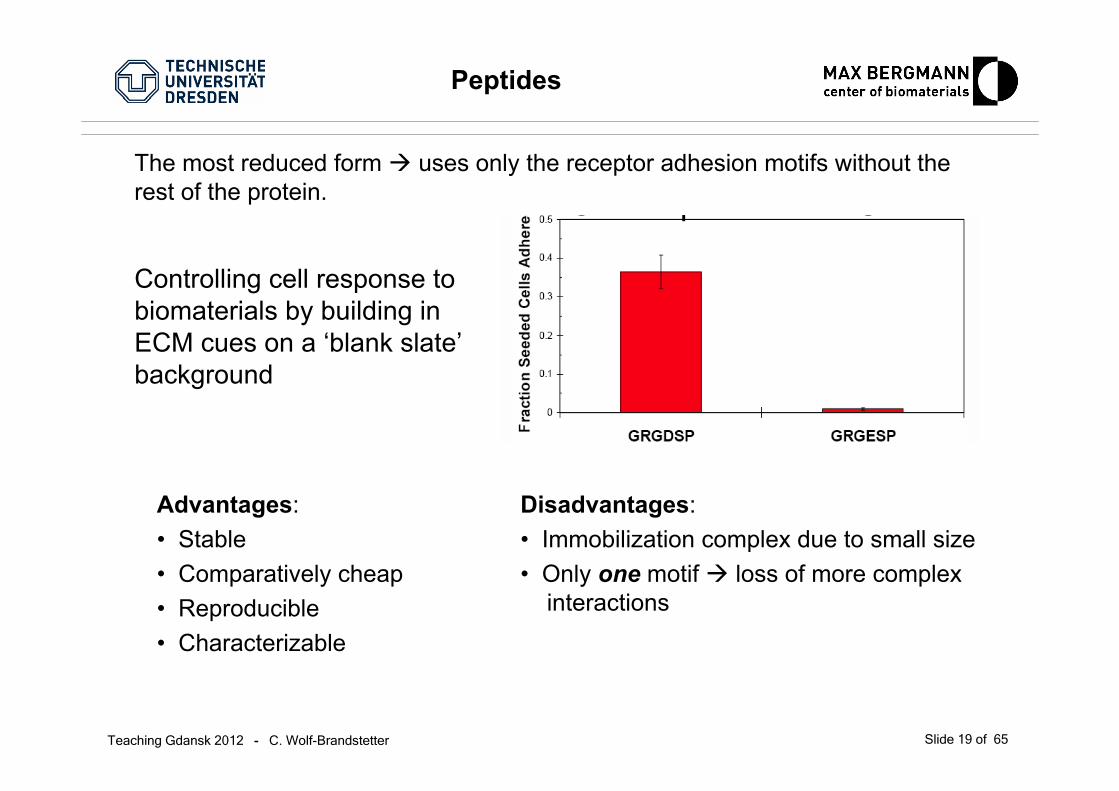
The most reduced form uses only the receptor adhesion motifs without therest of the protein.
Advantages:• Stable• Comparatively cheap• Reproducible• Characterizable
Disadvantages:• Immobilization complex due to small size• Only one motif loss of more complex
interactions
Controlling cell response to biomaterials by building in ECM cues on a ‘blank slate’background

Teaching Gdansk 2012 - C. Wolf-Brandstetter Slide 20 of 65
RGD peptide as ligand for osteoblasts
GR
KNH
Df
BioactiveRGD
component
R= ArgininG= GlycinD= AspartatK= Lysinf= D-Phenylalanin
A
A
A
ASurface anchor
A= Phosphonat
(AHX)3
Spacer
K
K
KAHX= Aminohexoic acid
Osteoblast
Integrin
Teaching Gdansk 2012 - C. Wolf-Brandstetter Slide 21 of 65
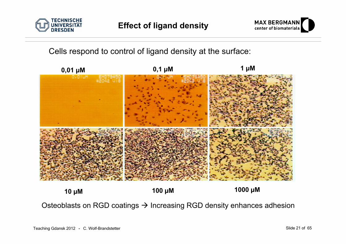
Effect of ligand density
0,01 µM 0,1 µM 1 µM
10 µM 100 µM 1000 µM
Osteoblasts on RGD coatings Increasing RGD density enhances adhesion
Cells respond to control of ligand density at the surface:

Teaching Gdansk 2012 - C. Wolf-Brandstetter Slide 22 of 65
BUT: too much RGD reduces cell motility!
Effect of ligand density

Teaching Gdansk 2012 - C. Wolf-Brandstetter Slide 23 of 65
Other peptide adhesion motifs
aMb2fibrinogen, C3bQKRLDGS
αvβ1, α4β7fibronectin, VCAMEILDV
β3 integrinsfibrinogenKQAGDV
Elastase receptorelastaseVAPG
α2β1collagenDGEA
N-cadherinN-cadherinHAV
?lamininRNIAEJIKDI
α1β1, α3β1lamininYIGSR
α3β1, α4β1, αvβ1, αIIbβ3, αvβ5, αvβ3, ..
fibronectin, laminin, collagen, vitronectin, vWF, bone sialo protein osteopontin, thrombospondin
RGD
LB110lamininIKVAV
ReceptorOriginPeptide
Teaching Gdansk 2012 - C. Wolf-Brandstetter Slide 24 of 65
Collagen
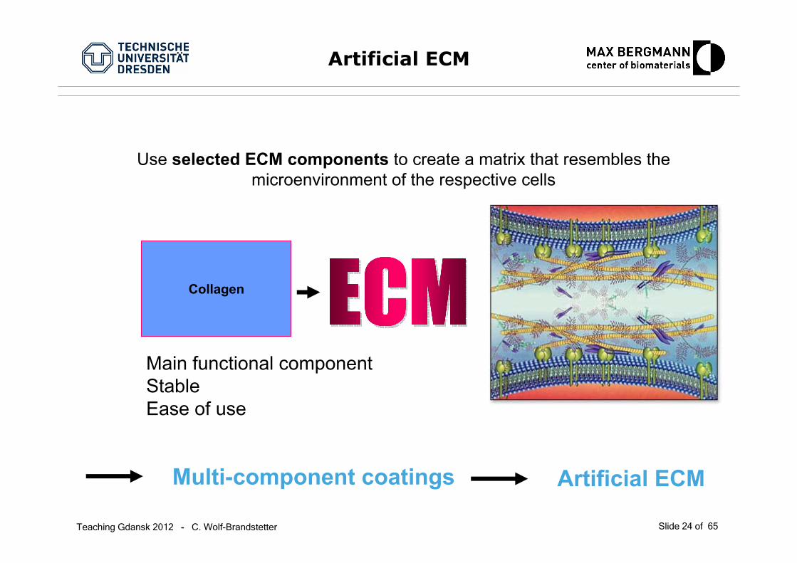
Artificial ECM
Use selected ECM components to create a matrix that resembles the microenvironment of the respective cells
Multi-component coatings Artificial ECM
Main functional componentStableEase of use

Teaching Gdansk 2012 - C. Wolf-Brandstetter Slide 25 of 65
Basic idea – what is the biological target? What mechanism of response do I anticipate?
Example: coating of titanium implants for use as bone substitute
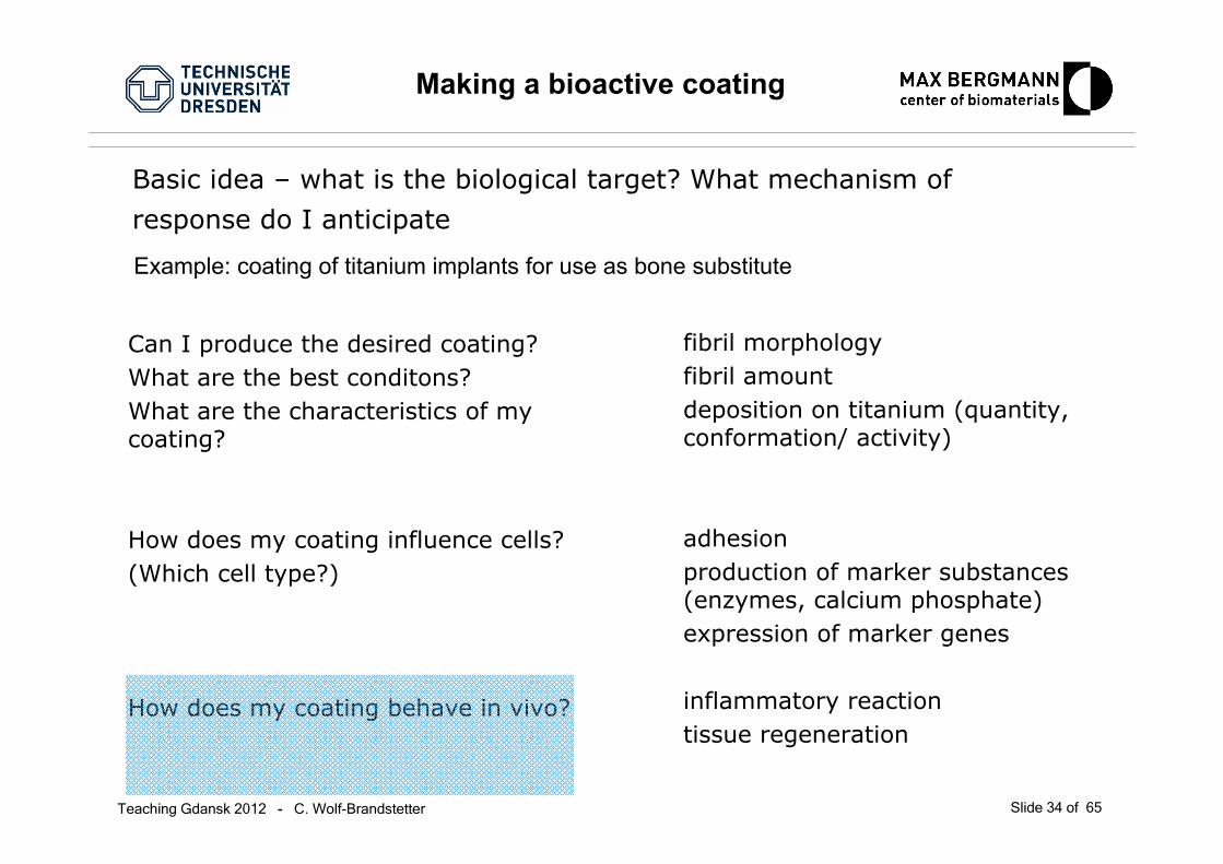
Making a bioactive coating
Teaching Gdansk 2012 - C. Wolf-Brandstetter Slide 26 of 65
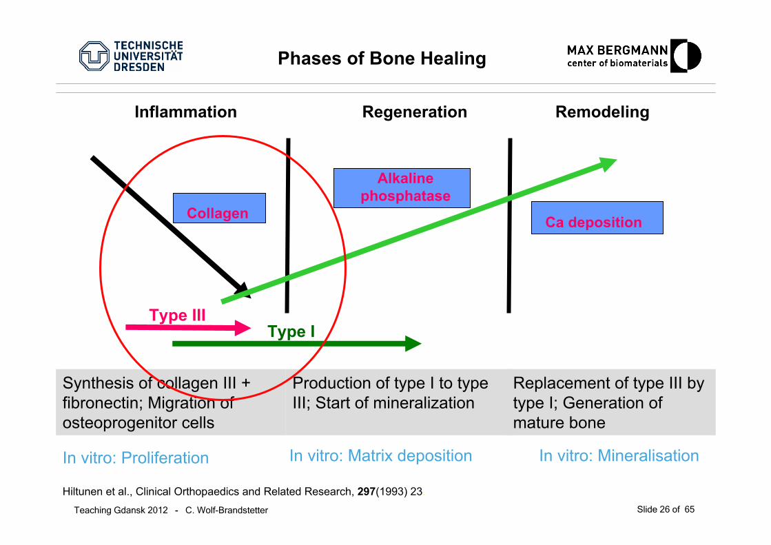
Replacement of type III by type I; Generation of mature bone
Phases of Bone Healing
Hiltunen et al., Clinical Orthopaedics and Related Research, 297(1993) 23.
In vitro: Proliferation In vitro: Matrix deposition In vitro: Mineralisation
Synthesis of collagen III + fibronectin; Migration of osteoprogenitor cells
Production of type I to type III; Start of mineralization
Ca deposition
Alkalinephosphatase
Collagen
Type IIIType I
Inflammation Regeneration Remodeling

Teaching Gdansk 2012 - C. Wolf-Brandstetter Slide 27 of 65
Basic idea – what is the biological target? What mechanism of response do I anticipate
Can I produce the desired coating?What are the best conditons?What are the characteristics of mycoating?
Example: coating of titanium implants for use as bone substitute
Making a bioactive coating
fibril morphologyfibril amountdeposition on titanium (quantity, conformation/ activity)

Teaching Gdansk 2012 - C. Wolf-Brandstetter Slide 28 of 65
0 200 400 600 8000.0
0.2
0.4
0.6
0.8
OD
313
time [min]
100% I
20% III
50% III
100% III
0 % 2.5 % 5 % 10 % 20 % 50 %100 %0
20
40
60
80
100
depo
sitio
n in
per
cent
percentage type III
fibrillogenesis collagen deposition in fibrils
Fibrillogenesis of heterotypic fibrils
Collagen I/III fibrils can be formed, but …
Teaching Gdansk 2012 - C. Wolf-Brandstetter Slide 29 of 65
2,5 µm 2,5 µm
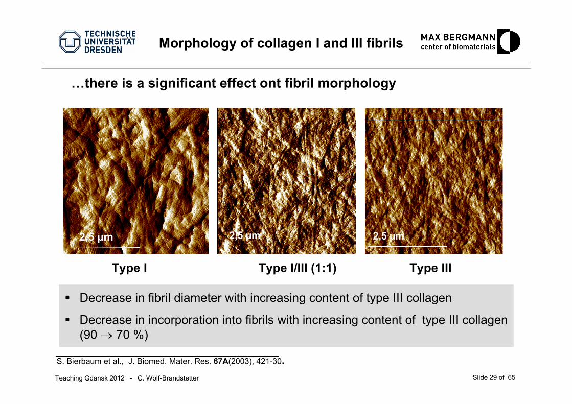
Morphology of collagen I and III fibrils
Type I Type I/III (1:1) Type III
S. Bierbaum et al., J. Biomed. Mater. Res. 67A(2003), 421-30.
2,5 µm
Decrease in fibril diameter with increasing content of type III collagen
Decrease in incorporation into fibrils with increasing content of type III collagen (90 → 70 %)
…there is a significant effect ont fibril morphology

Teaching Gdansk 2012 - C. Wolf-Brandstetter Slide 30 of 65
Basic idea – what is the biological target? What mechanism of response do I anticipate
Can I produce the desired coating?What are the best conditons?What are the characteristics of mycoating?
How does my coating influence cells?(Which cell type?)
Example: coating of titanium implants for use as bone substitute
Making a bioactive coating
fibril morphologyfibril amountdeposition on titanium (quantity, conformation/ activity)
adhesionproduction of marker substances(enzymes, calcium phosphate)expression of marker genes
Teaching Gdansk 2012 - C. Wolf-Brandstetter Slide 31 of 65
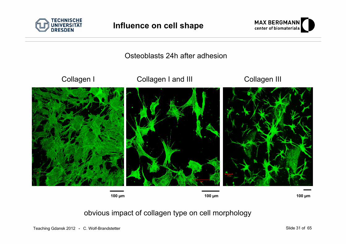
Influence on cell shape
Collagen I Collagen I and III Collagen III
100 µm 100 µm 100 µm
Osteoblasts 24h after adhesion
obvious impact of collagen type on cell morphology
Teaching Gdansk 2012 - C. Wolf-Brandstetter Slide 32 of 65
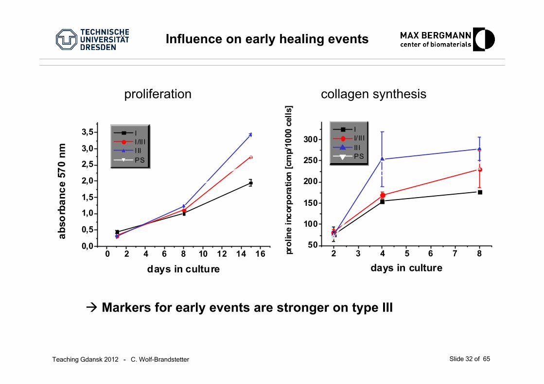
collagen synthesisproliferation
0 2 4 6 8 10 12 14 160,0
0,5
1,0
1,5
2,0
2,5
3,0
3,5 I I /II I I II PS
abso
rban
ce 5
70 n
m
days in culture2 3 4 5 6 7 8
50
100
150
200
250
300 I I/ II I II I PS
prol
ine
inco
rpor
atio
n [c
mp/
1000
cel
ls]
days in culture
Influence on early healing events
Markers for early events are stronger on type III

Teaching Gdansk 2012 - C. Wolf-Brandstetter Slide 33 of 65
activity of ALP extracellular calcium
21 22 23 24 25 26 27 28 29 30
1
2
3
4
5
6
7
8
Ca
- µm
ol/ c
m²
I I /II I I II PS
days in culture3 4 5 6 7 8 9 10 11 12
0,05
0,10
0,15
0,20
0,25 I I/I II III PS
ALP
act
ivity
[U/m
g pr
otei
n]
days in culture
Markers for late events are stronger on type I
In vitro cell behavior on different matrices differs and reflects eventsassociated with such a matrix in vivo
Influence on late healing events
Teaching Gdansk 2012 - C. Wolf-Brandstetter Slide 34 of 65
Basic idea – what is the biological target? What mechanism of response do I anticipate
Can I produce the desired coating?What are the best conditons?What are the characteristics of mycoating?
How does my coating influence cells?(Which cell type?)
How does my coating behave in vivo?
Example: coating of titanium implants for use as bone substitute
Making a bioactive coating
fibril morphologyfibril amountdeposition on titanium (quantity, conformation/ activity)
adhesionproduction of marker substances(enzymes, calcium phosphate)expression of marker genes
inflammatory reactiontissue regeneration

Teaching Gdansk 2012 - C. Wolf-Brandstetter Slide 35 of 65
Collagen
Artificial ECM
Use selected ECM components to create a matrix that resembles the microenvironment of the respective cells
Multi-component coatings Artificial ECM
Syntheticmatrices
Teaching Gdansk 2012 - C. Wolf-Brandstetter Slide 36 of 65
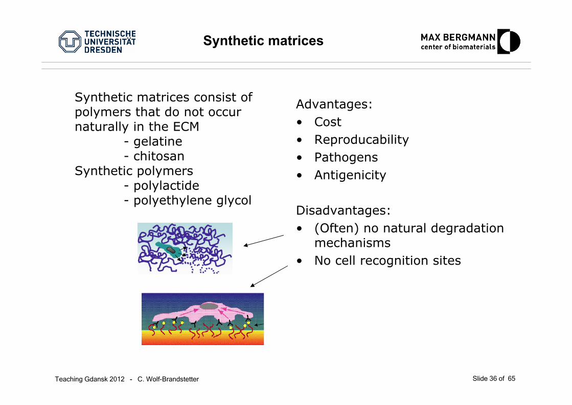
Synthetic matrices
Synthetic matrices consist of polymers that do not occurnaturally in the ECM
- gelatine- chitosan
Synthetic polymers- polylactide- polyethylene glycol
Advantages:• Cost• Reproducability• Pathogens• Antigenicity
Disadvantages:• (Often) no natural degradation
mechanisms• No cell recognition sites
Teaching Gdansk 2012 - C. Wolf-Brandstetter Slide 37 of 65
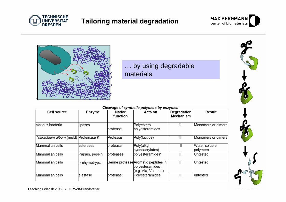
Tailoring material degradation
… by using degradablematerials
Teaching Gdansk 2012 - C. Wolf-Brandstetter Slide 38 of 65
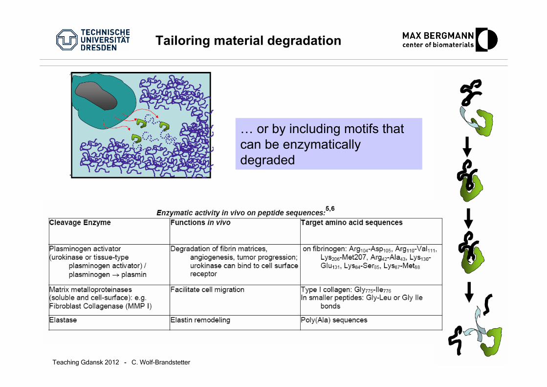
… or by including motifs thatcan be enzymaticallydegraded
Tailoring material degradation

Teaching Gdansk 2012 - C. Wolf-Brandstetter Slide 39 of 65
Cross-linking the material
For stability, many biomaterials, both synthetic and natural, arecross-linked
Advantages: • higher stability• lower degradation rates
Disadvantages:• lower degradation rates
(depends on application!)• Usually chemical treatment
necessary– changes proteins– can be cytotoxic– …
Use enzyme-sensitivecrosslinks
Use enzymatic cross-linking

Teaching Gdansk 2012 - C. Wolf-Brandstetter Slide 40 of 65
Enzym cross-linking and enzyme-sensitive cross-links

Teaching Gdansk 2012 - C. Wolf-Brandstetter Slide 41 of 65
Collagen
Artificial ECM
Use selected ECM components to create a matrix that resembles the microenvironment of the respective cells
Type I
FibronectinDecorinBiglycan
Chondroitin sulfateHeparin
Heparan sulfate
(Type II)
Multi-component coatings Artificial ECM
Type III

Teaching Gdansk 2012 - C. Wolf-Brandstetter Slide 42 of 65
• dimeric protein of 240.000 kD
• occurs in plasma and on the cell surface
• important role in woundhealing
• binds cells primarily via RGD-sequence
• binds native and denaturedcollagen
collagenbinding site
cellbinding
site
heparinbinding site
Glycoproteins: Fibronectin
Teaching Gdansk 2012 - C. Wolf-Brandstetter Slide 43 of 65
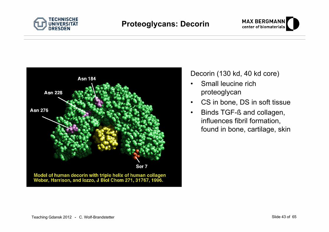
Decorin (130 kd, 40 kd core)• Small leucine rich
proteoglycan• CS in bone, DS in soft tissue• Binds TGF-ß and collagen,
influences fibril formation, found in bone, cartilage, skin
Proteoglycans: Decorin
Teaching Gdansk 2012 - C. Wolf-Brandstetter Slide 44 of 65
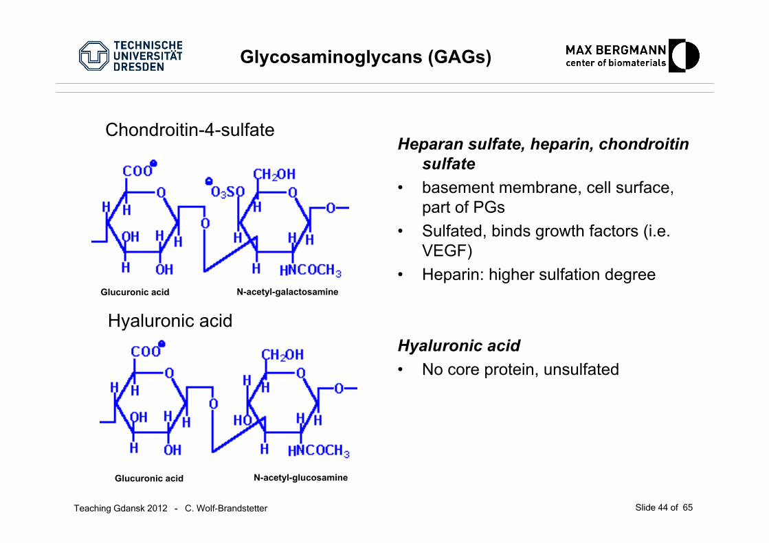
Heparan sulfate, heparin, chondroitinsulfate
• basement membrane, cell surface, part of PGs
• Sulfated, binds growth factors (i.e. VEGF)
• Heparin: higher sulfation degree
Hyaluronic acid• No core protein, unsulfated
Glycosaminoglycans (GAGs)
Chondroitin-4-sulfate
Hyaluronic acid
Glucuronic acid
Glucuronic acid N-acetyl-glucosamine
N-acetyl-galactosamine
Teaching Gdansk 2012 - C. Wolf-Brandstetter Slide 45 of 65
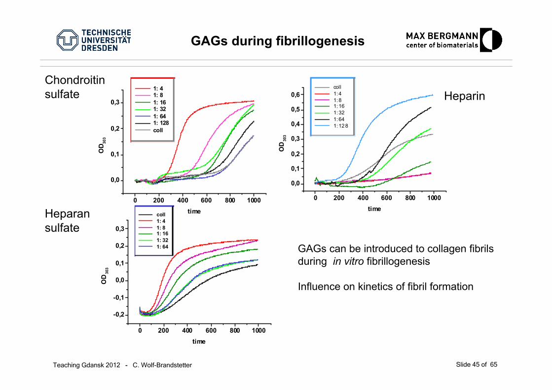
GAGs during fibrillogenesis
0 200 400 600 800 1000
0,0
0,1
0,2
0,3
1: 4 1: 8 1: 16 1: 32 1: 64 1: 128 coll
OD 30
3
time
0 200 400 600 800 1000
-0,2
-0,1
0,0
0,1
0,2
0,3
coll 1: 4 1: 8 1: 16 1: 32 1: 64
OD
303
time
Chondroitinsulfate
Heparansulfate
Heparin
0 200 400 600 800 1000
0,0
0,1
0,2
0,3
0,4
0,5
0,6 coll 1: 4 1: 8 1: 16 1: 32 1: 64 1: 12 8
OD
303
time
GAGs can be introduced to collagen fibrilsduring in vitro fibrillogenesis
Influence on kinetics of fibril formation
Teaching Gdansk 2012 - C. Wolf-Brandstetter Slide 46 of 65
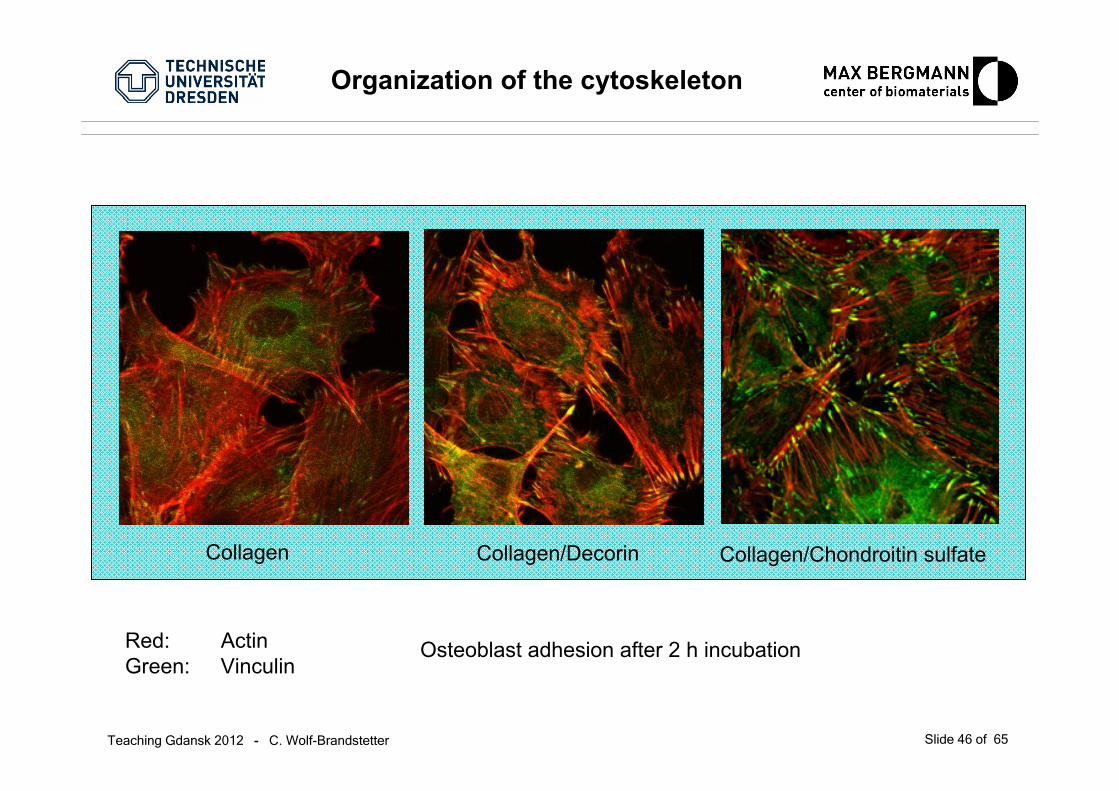
Organization of the cytoskeleton
Collagen Collagen/Chondroitin sulfateCollagen/Decorin
Red: ActinGreen: Vinculin
Osteoblast adhesion after 2 h incubation
Teaching Gdansk 2012 - C. Wolf-Brandstetter Slide 47 of 65
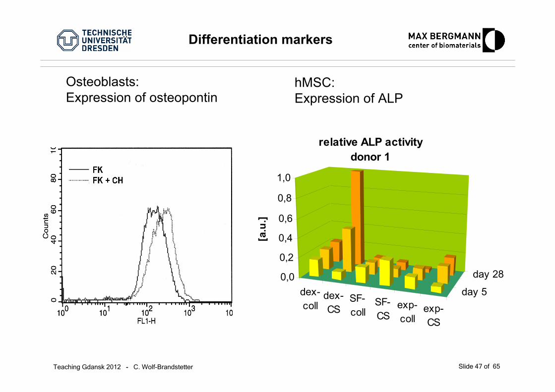
Differentiation markers
Osteoblasts: Expression of osteopontin
dex-coll
dex-CS
SF-coll
SF-CS
exp-coll
exp-CS
day 5
day 280,0
0,2
0,4
0,6
0,8
1,0
[a.u
.]
relative ALP activitydonor 1
hMSC: Expression of ALP
Teaching Gdansk 2012 - C. Wolf-Brandstetter Slide 48 of 65
dex-coll dex-CS SF-coll SF-CS exp-coll exp-CS0
250
500
750
4000
5000
6000
7000
OC
rela
tive
to G
APD
H
donor 1 day 5 day 15 day 28
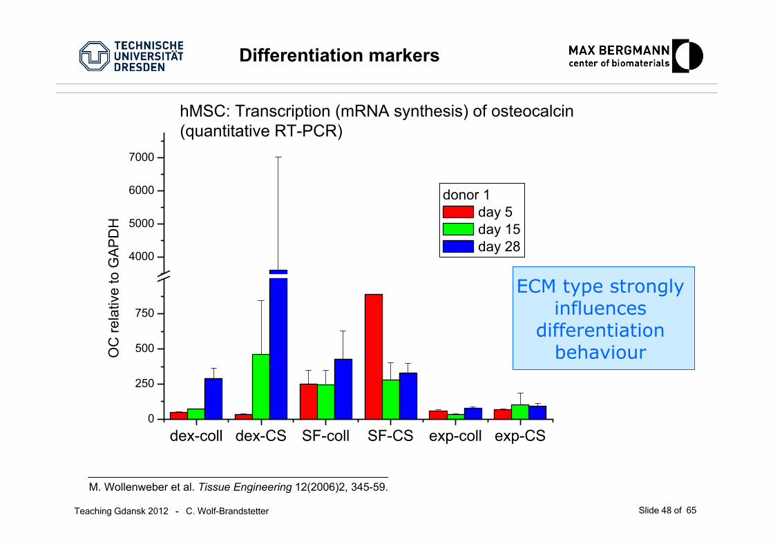
M. Wollenweber et al. Tissue Engineering 12(2006)2, 345-59.
ECM type stronglyinfluences
differentiationbehaviour
hMSC: Transcription (mRNA synthesis) of osteocalcin(quantitative RT-PCR)
Differentiation markers
Teaching Gdansk 2012 - C. Wolf-Brandstetter Slide 49 of 65
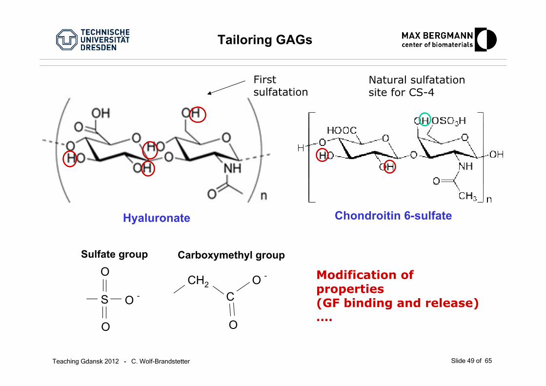
Tailoring GAGs
CH2
C
O
O -
S
O
O
O -
Chondroitin 6-sulfateHyaluronate
Carboxymethyl groupSulfate group
First sulfatation
Modification of properties(GF binding and release)….
Natural sulfatationsite for CS-4

Teaching Gdansk 2012 - C. Wolf-Brandstetter Slide 50 of 65
Cell response to tailored GAGs
Cell culture wihout any osteogenic supplements - upper rowAddition of dexamethasone (corticoal growth hormone) - lower row
Increased amount of sulfate groupsleads to increased activity of ALP (even without osteogenic induction)
Teaching Gdansk 2012 - C. Wolf-Brandstetter Slide 51 of 65
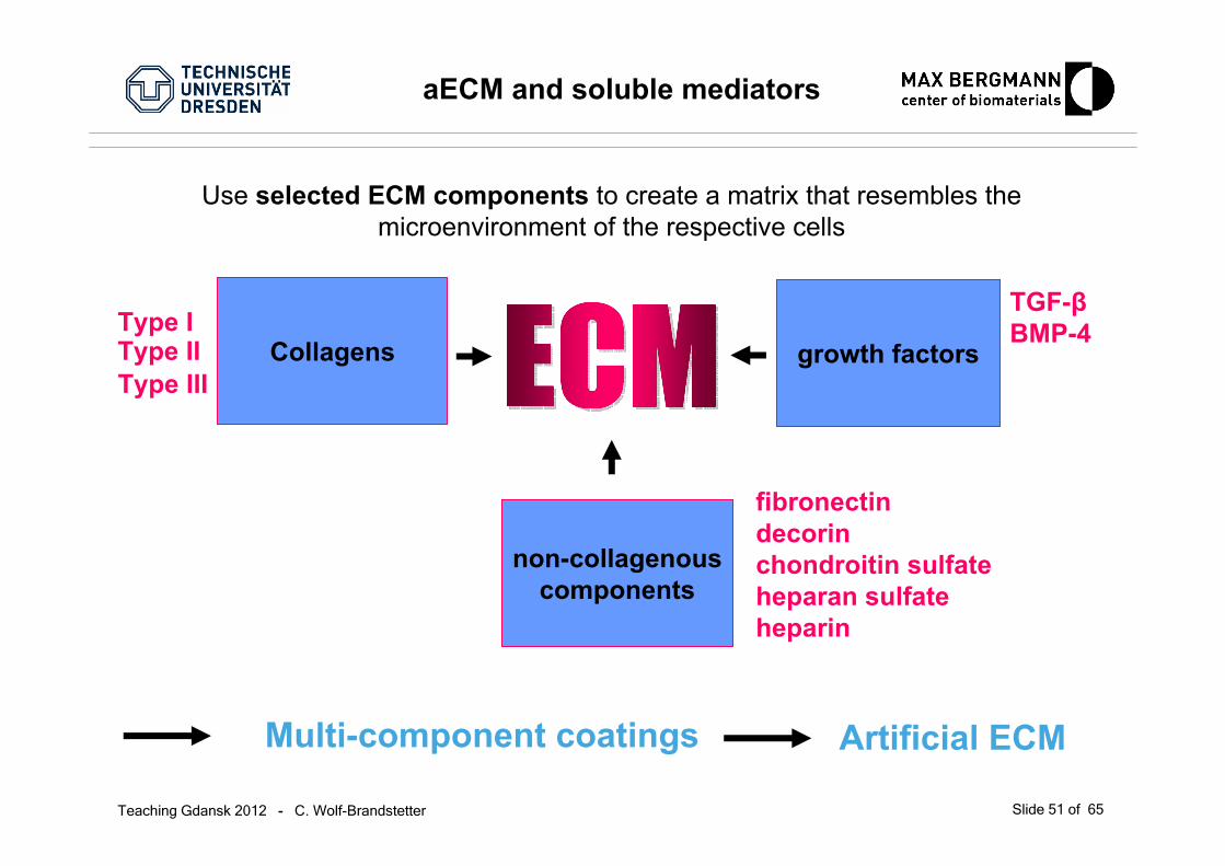
Collagens
aECM and soluble mediators
Use selected ECM components to create a matrix that resembles the microenvironment of the respective cells
Type IType II
Multi-component coatings Artificial ECM
Type III
non-collagenouscomponents
fibronectindecorinchondroitin sulfateheparan sulfateheparin
growth factors
TGF-βBMP-4
Teaching Gdansk 2012 - C. Wolf-Brandstetter Slide 52 of 65
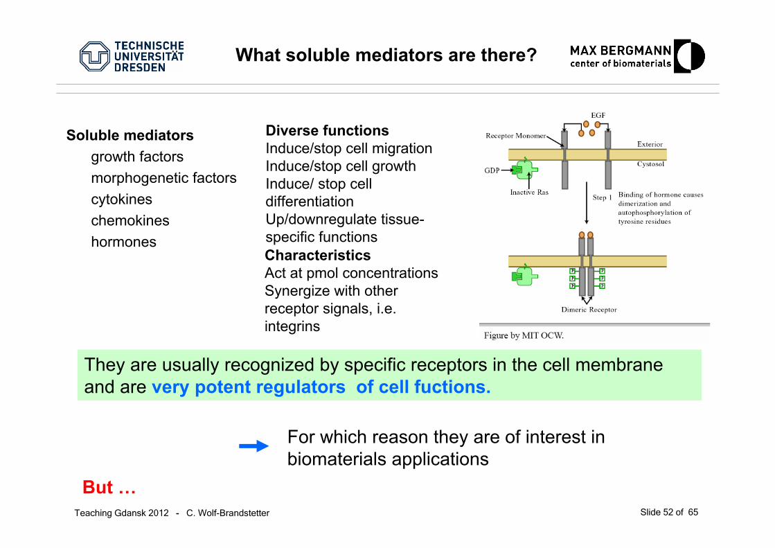
Soluble mediatorsgrowth factorsmorphogenetic factorscytokineschemokineshormones
They are usually recognized by specific receptors in the cell membraneand are very potent regulators of cell fuctions.
For which reason they are of interest in biomaterials applications
But …
Diverse functionsInduce/stop cell migrationInduce/stop cell growthInduce/ stop celldifferentiationUp/downregulate tissue-specific functionsCharacteristicsAct at pmol concentrationsSynergize with otherreceptor signals, i.e. integrins
What soluble mediators are there?
Teaching Gdansk 2012 - C. Wolf-Brandstetter Slide 53 of 65
What soluble mediators are there?
… they are:
expensiveunstable (both in vivo and in vitro)recombinant growth factors often far less effectivethan their physiological counterpartssoluble
Soluble mediators are used in combination with carriers toprevent quick diffusionprotect them from degradation
Teaching Gdansk 2012 - C. Wolf-Brandstetter Slide 54 of 65
Strategies for Biomaterial Presentation of Growth Factors
1. Chemical conjugation of GFs to scaffold materialsa) non-covalent incorporation
direct adsorption via direct charge-charge or hydrophobic interactions
indirect interaction via specific biological sites that are coated on the surface(heparin, fibronectin, gelatin, small oligopeptides)
b) covalent incorporation
conjugation for the surface via functional groups
2. Physical encapsulation of GFs with pre-programmed release, and diffusioninto surrounding tissue
Adapted from Lee, K et al., 2010
Teaching Gdansk 2012 - C. Wolf-Brandstetter Slide 55 of 65
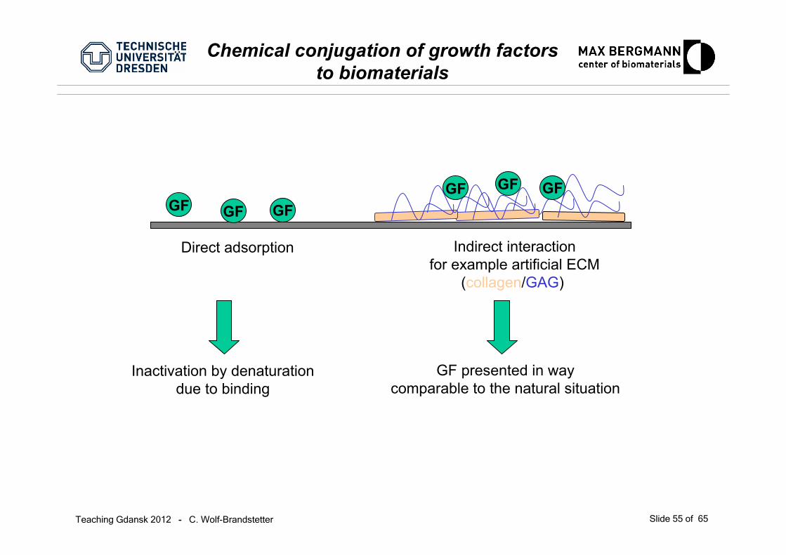
Chemical conjugation of growth factors to biomaterials
Direct adsorption Indirect interactionfor example artificial ECM
(collagen/GAG)
GF GF GFGF GF GF
Inactivation by denaturationdue to binding
GF presented in waycomparable to the natural situation
Teaching Gdansk 2012 - C. Wolf-Brandstetter Slide 56 of 65
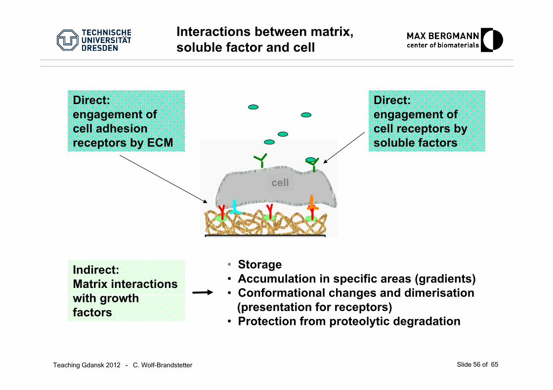
Interactions between matrix, soluble factor and cell
cell
Direct:engagement of cell adhesion receptors by ECM
Indirect: Matrix interactionswith growth factors
• Storage• Accumulation in specific areas (gradients)• Conformational changes and dimerisation
(presentation for receptors)• Protection from proteolytic degradation
Direct:engagement of cell receptors bysoluble factors
Teaching Gdansk 2012 - C. Wolf-Brandstetter Slide 57 of 65
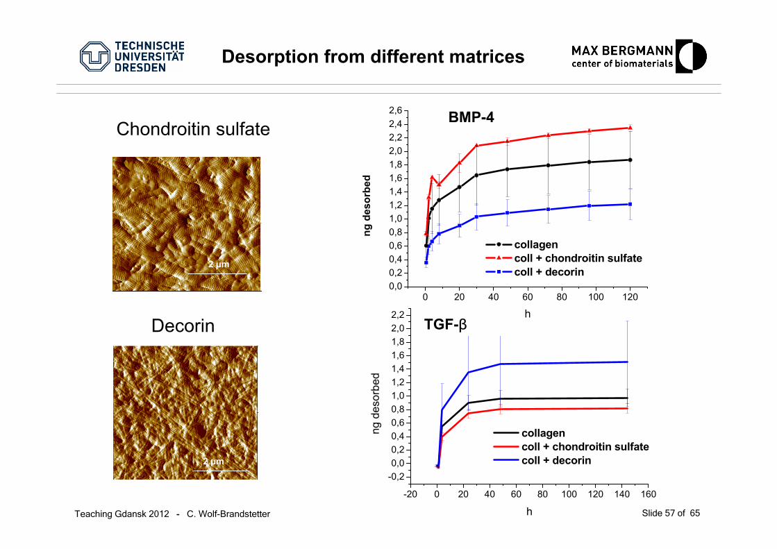
BMP-4
Desorption from different matrices
-20 0 20 40 60 80 100 120 140 160
-0,20,00,20,40,60,81,01,21,41,61,82,02,2
collagen coll + chondroitin sulfate coll + decorin
ng d
esor
bed
h
0 20 40 60 80 100 1200,00,20,40,60,81,01,21,41,61,82,02,22,42,6
collagen coll + chondroitin sulfate coll + decorin
ng d
esor
bed
hTGF-βDecorin
2 µm
Chondroitin sulfate
2 µm

Teaching Gdansk 2012 - C. Wolf-Brandstetter Slide 58 of 65
Using 3d matrix structure
osteoblast
collagen/ decorin/ (TGF-ß)
collagen/ chondroitinsulfat/ (BMP-4)
titanium
A
collagen/ collagen-chondroitin sulfate (collagenred, CS green).
Modulation of growth factor activity through influencing the temporal release pattern

Teaching Gdansk 2012 - C. Wolf-Brandstetter Slide 59 of 65
Osteoblastic cells on layered aECM
Message: ‚construction principle‘ of aECM stronglyinfluences cell reactions
0
10
20
30
40
50
coll/CS/BMPcoll/dec/TGF
coll/CScoll/dec/TGF
coll/CS/BMPcoll/dec
coll/CScoll/dec
prol
in in
tegr
atio
n [c
pm/1
000
cells
]
0
50
100
150
200
250
300
350
coll/CS/BMPcoll/dec/TGF
coll/CScoll/dec/TGF
coll/CS/BMPcoll/dec
coll/CScoll/dec
ALP
act
ivity
[mU
/mg
prot
ein]
Collagen synthesis ALP activityday 4

Teaching Gdansk 2012 - C. Wolf-Brandstetter Slide 60 of 65
Control of GF-binding
Highest amount of bound GF for high sulfatated GAGsMethyl group decreases affinity for GF
… by suitable modification of GAGs (sulfatation degree and pattern)
From: Hintze et al.
Teaching Gdansk 2012 - C. Wolf-Brandstetter Slide 61 of 65
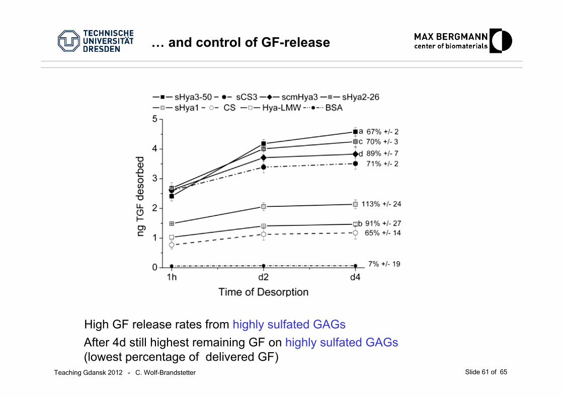
… and control of GF-release
High GF release rates from highly sulfated GAGsAfter 4d still highest remaining GF on highly sulfated GAGs(lowest percentage of delivered GF)
Teaching Gdansk 2012 - C. Wolf-Brandstetter Slide 62 of 65
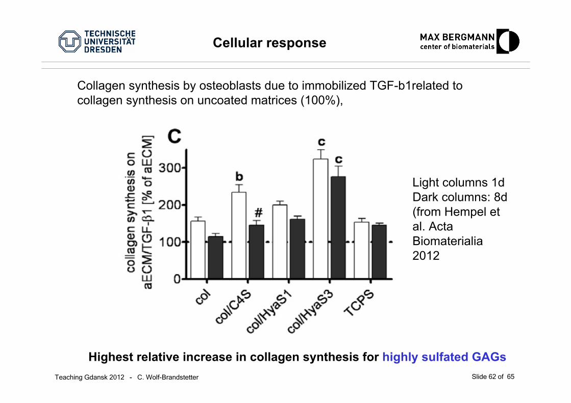
Cellular response
Highest relative increase in collagen synthesis for highly sulfated GAGs
Collagen synthesis by osteoblasts due to immobilized TGF-b1related to collagen synthesis on uncoated matrices (100%),
Light columns 1dDark columns: 8d(from Hempel et al. Acta Biomaterialia2012
Teaching Gdansk 2012 - C. Wolf-Brandstetter Slide 63 of 65
Increase in complexity of the artificial matricesallows for better adaptation to natural systems,
but …..
…..increases also complexity of the in vitro characterization of thea-ECM coatings:
• Interference of several quantification methods by othercompounds (e.g. collagen quantification is strongly affected bypresence of GAGs)
• Desorption of growth factors can be accompanied by simultaneousdelivery of GAGs from a-ECM, which mayimpair the detectability of the GFas well as influence the activity of the mixture free GF/GAG-GF
Challenges in a-ECM characterization

Teaching Gdansk 2012 - C. Wolf-Brandstetter Slide 64 of 65
Summary
Within certain limits growth factors release can be influenced byinclusion of GF binding components, which also influences cellbehavior
Using modified GAGs, growth factor binding can be increased(and their presentation altered)
A matrix can be/should be „tailored“ for different growth factors
Layering differently composed matrices can allow for time differences in growth factor release, which influences cellresponses
Teaching Gdansk 2012 - C. Wolf-Brandstetter Slide 65 of 65
Finished !!! …