Embed Size (px)
Citation preview

Lecture 9
Jan Żeromski
Lecture 9
Jan Żeromski
HYPERSENSITIVITYHYPERSENSITIVITY
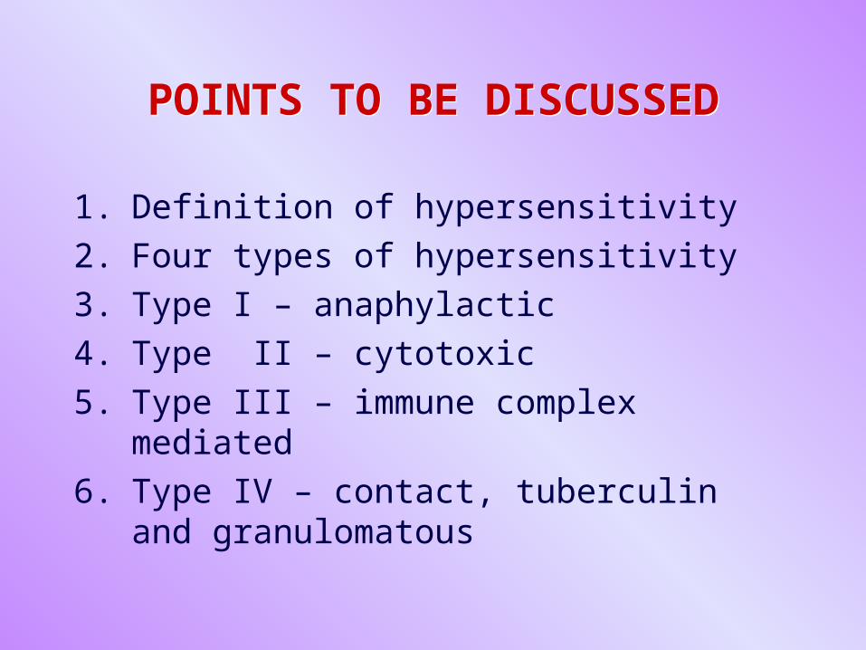
POINTS TO BE DISCUSSEDPOINTS TO BE DISCUSSED
1. Definition of hypersensitivity
2. Four types of hypersensitivity
3. Type I – anaphylactic
4. Type II – cytotoxic
5. Type III – immune complex mediated
6. Type IV – contact, tuberculin and granulomatous
HYPERSENSITIVITY TYPE I – KEY POINTS
HYPERSENSITIVITY TYPE I – KEY POINTS
• IgE antibody response directed against innocuous environmental Ag’s such as animal dander, house-dust mites, pollen, industrial pollutants, etc.
• IgE becomes coated on mast cells or basophiles via their Fc receptors
HYPERSENSITIVITY TYPE I – KEY POINTS - 2
HYPERSENSITIVITY TYPE I – KEY POINTS - 2
• Binding of allergen by IgE results in degranulation of mast cells and mediator release
• Allergic manifestations ensue such as hay fever, asthma, skin eczema, anaphylaxis
TYPE I HYPERSENSITIVITY IN HOST DEFENSE IN THE
AFFERENT IMMUNE RESPONSE
TYPE I HYPERSENSITIVITY IN HOST DEFENSE IN THE
AFFERENT IMMUNE RESPONSE
• IgE-mediated allergen capture
• IgE-mediated allergen processing
• IgE-mediated allergen presentation
• Immune deviation toward Th2 responses via release of key cytokines via innate immune responses
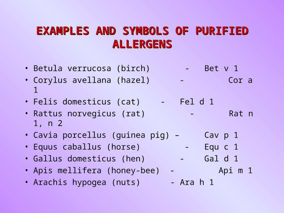
EXAMPLES AND SYMBOLS OF PURIFIED EXAMPLES AND SYMBOLS OF PURIFIED ALLERGENSALLERGENS
• Betula verrucosa (birch) - Bet v 1
• Corylus avellana (hazel) - Cor a 1
• Felis domesticus (cat) - Fel d 1
• Rattus norvegicus (rat) - Rat n 1, n 2
• Cavia porcellus (guinea pig) – Cav p 1
• Equus caballus (horse) - Equ c 1
• Gallus domesticus (hen) - Gal d 1
• Apis mellifera (honey-bee) - Api m 1
• Arachis hypogea (nuts) - Ara h 1
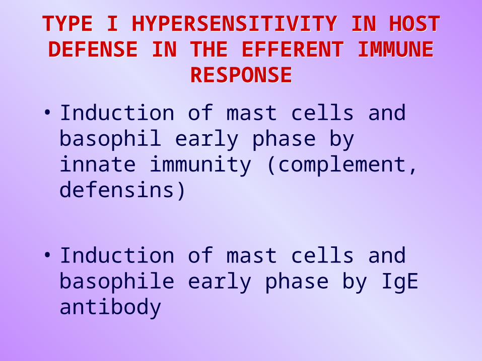
TYPE I HYPERSENSITIVITY IN HOST DEFENSE IN THE EFFERENT IMMUNE
RESPONSE
TYPE I HYPERSENSITIVITY IN HOST DEFENSE IN THE EFFERENT IMMUNE
RESPONSE
• Induction of mast cells and basophil early phase by innate immunity (complement, defensins)
• Induction of mast cells and basophile early phase by IgE antibody
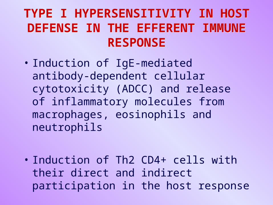
TYPE I HYPERSENSITIVITY IN HOST DEFENSE IN THE EFFERENT IMMUNE
RESPONSE
TYPE I HYPERSENSITIVITY IN HOST DEFENSE IN THE EFFERENT IMMUNE
RESPONSE
• Induction of IgE-mediated antibody-dependent cellular cytotoxicity (ADCC) and release of inflammatory molecules from macrophages, eosinophils and neutrophils
• Induction of Th2 CD4+ cells with their direct and indirect participation in the host response
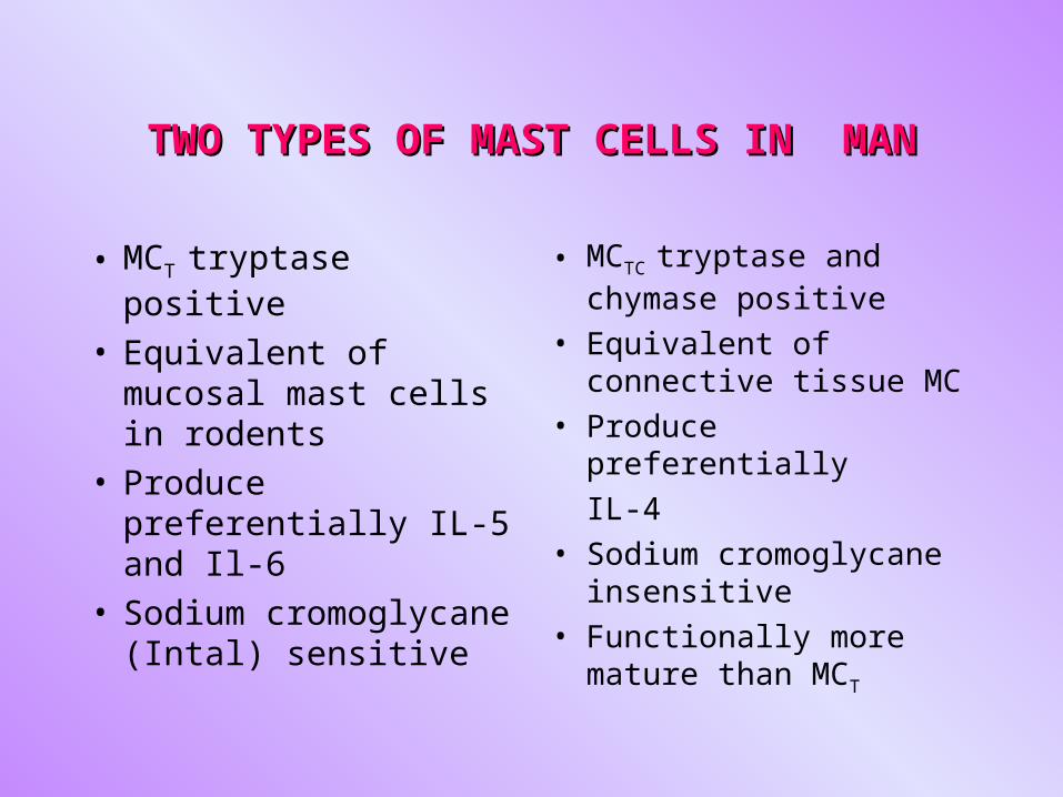
TWO TYPES OF MAST CELLS IN MANTWO TYPES OF MAST CELLS IN MAN
• MCT tryptase positive
• Equivalent of mucosal mast cells in rodents
• Produce preferentially IL-5 and Il-6
• Sodium cromoglycane (Intal) sensitive
• MCTC tryptase and chymase positive
• Equivalent of connective tissue MC
• Produce preferentially
IL-4
• Sodium cromoglycane insensitive
• Functionally more mature than MCT
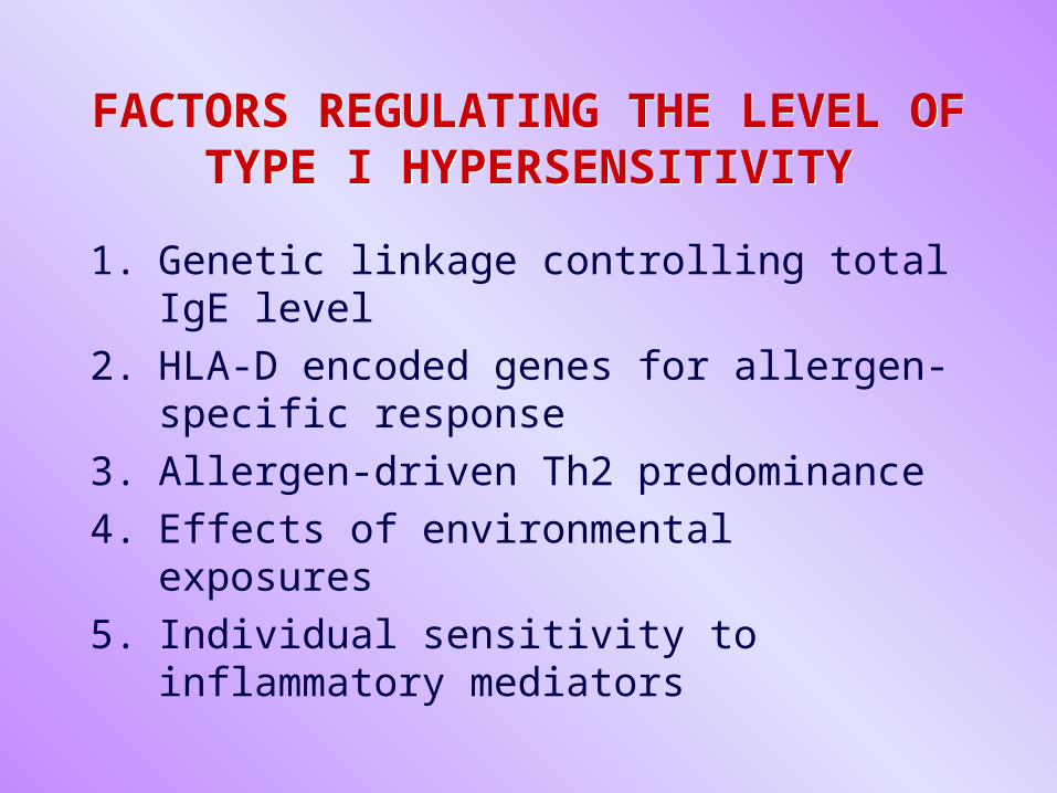
FACTORS REGULATING THE LEVEL OF TYPE I HYPERSENSITIVITY
FACTORS REGULATING THE LEVEL OF TYPE I HYPERSENSITIVITY
1. Genetic linkage controlling total IgE level
2. HLA-D encoded genes for allergen-specific response
3. Allergen-driven Th2 predominance
4. Effects of environmental exposures
5. Individual sensitivity to inflammatory mediators
ASTHMA PATHOGENESIS ASTHMA PATHOGENESIS
• It is a chronic inflammatory disorder of the airways: mucosal inflammation, reversible airway obstruction, bronchial hyperresponsiveness
• Bronchial epithelium, mast cells, T-helper cells and eosinophils are known to drive this process
ASTHMA PATHOGENESIS ASTHMA PATHOGENESIS
• It is consequence of dysregulation of selective cytokine networks in the airways due to Th2 cells
• Chronic mucosal inflammation, at least in part, leads to „airway remodeling”.
ROLE OF MAST TRYPTASE IN ASTHMA
ROLE OF MAST TRYPTASE IN ASTHMA
Mast tryptase induces: 1. Bronchoconstriction and
hyperresponsiveness
2. Stimulation of proliferation of fibroblasts, smooth muscle and epithelial cells
3. Generation of kinins
4. Stimulation of IL-8 release
5. Eosinophil chemotaxis
TYPE II HYPERSENSTIVITY REACTIONS
TYPE II HYPERSENSTIVITY REACTIONS
• Are caused by IgG and IgM antibodies directed against cell surface, extracellular matrix and intracellular antigens. The latter are usually non-pathogenic but diagnostically useful
• Transfusion reactions to erythrocytes are due to antibodies to blood group antigens
• The antibodies damage cells and tissues by activating complement and by binding and activating Fc receptor + effector cells
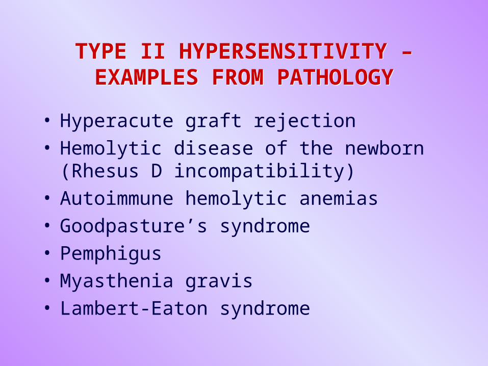
TYPE II HYPERSENSITIVITY – EXAMPLES FROM PATHOLOGY
TYPE II HYPERSENSITIVITY – EXAMPLES FROM PATHOLOGY
• Hyperacute graft rejection• Hemolytic disease of the newborn (Rhesus D
incompatibility)• Autoimmune hemolytic anemias• Goodpasture’s syndrome• Pemphigus • Myasthenia gravis• Lambert-Eaton syndrome
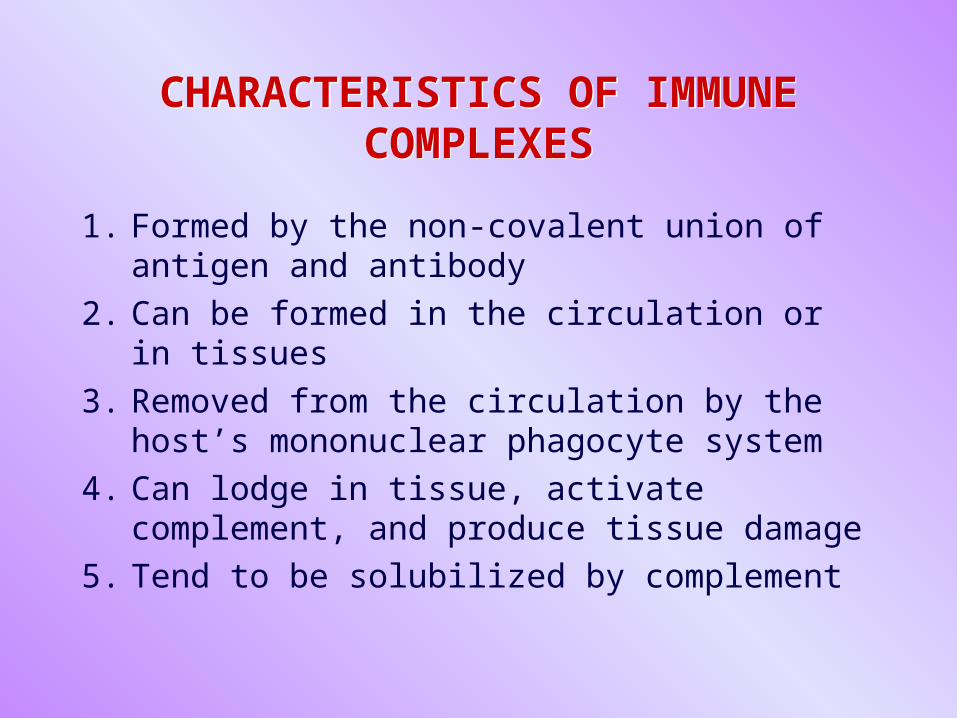
CHARACTERISTICS OF IMMUNE COMPLEXES
CHARACTERISTICS OF IMMUNE COMPLEXES
1. Formed by the non-covalent union of antigen and antibody
2. Can be formed in the circulation or in tissues
3. Removed from the circulation by the host’s mononuclear phagocyte system
4. Can lodge in tissue, activate complement, and produce tissue damage
5. Tend to be solubilized by complement
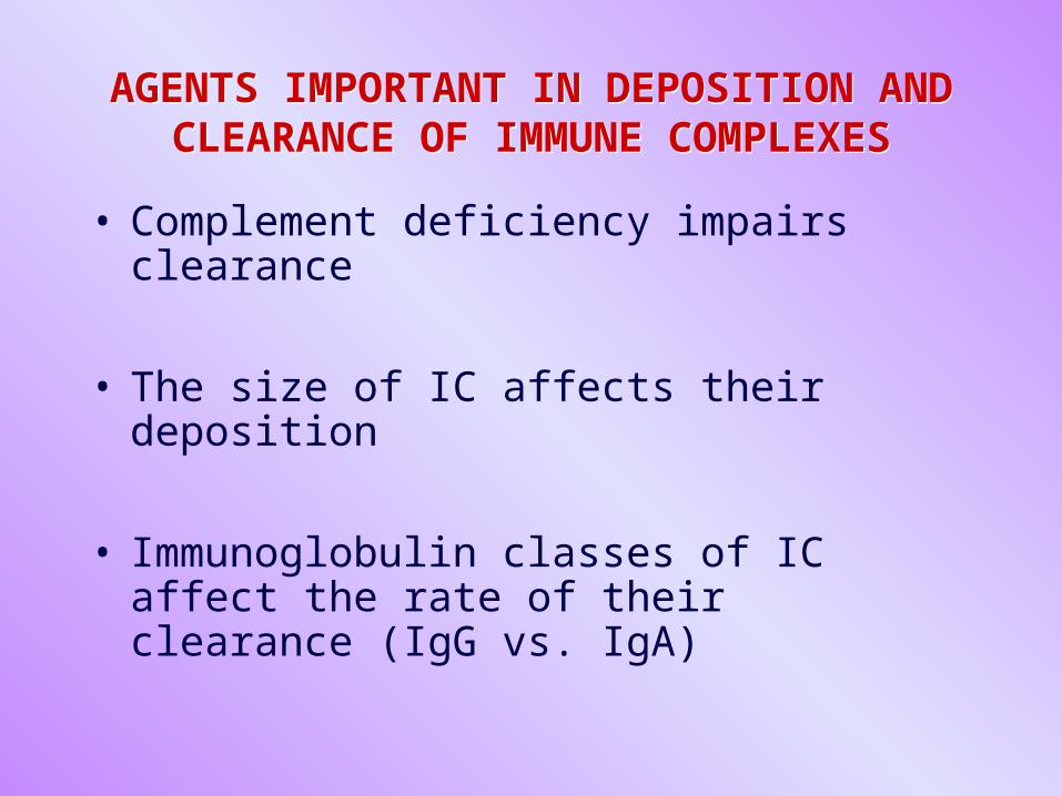
AGENTS IMPORTANT IN DEPOSITION AND CLEARANCE OF IMMUNE COMPLEXES
AGENTS IMPORTANT IN DEPOSITION AND CLEARANCE OF IMMUNE COMPLEXES
• Complement deficiency impairs clearance
• The size of IC affects their deposition
• Immunoglobulin classes of IC affect the rate of their clearance (IgG vs. IgA)
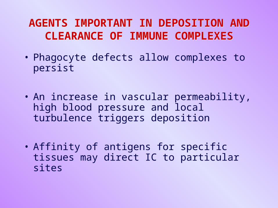
AGENTS IMPORTANT IN DEPOSITION AND CLEARANCE OF IMMUNE COMPLEXES
AGENTS IMPORTANT IN DEPOSITION AND CLEARANCE OF IMMUNE COMPLEXES
• Phagocyte defects allow complexes to persist
• An increase in vascular permeability, high blood pressure and local turbulence triggers deposition
• Affinity of antigens for specific tissues may direct IC to particular sites
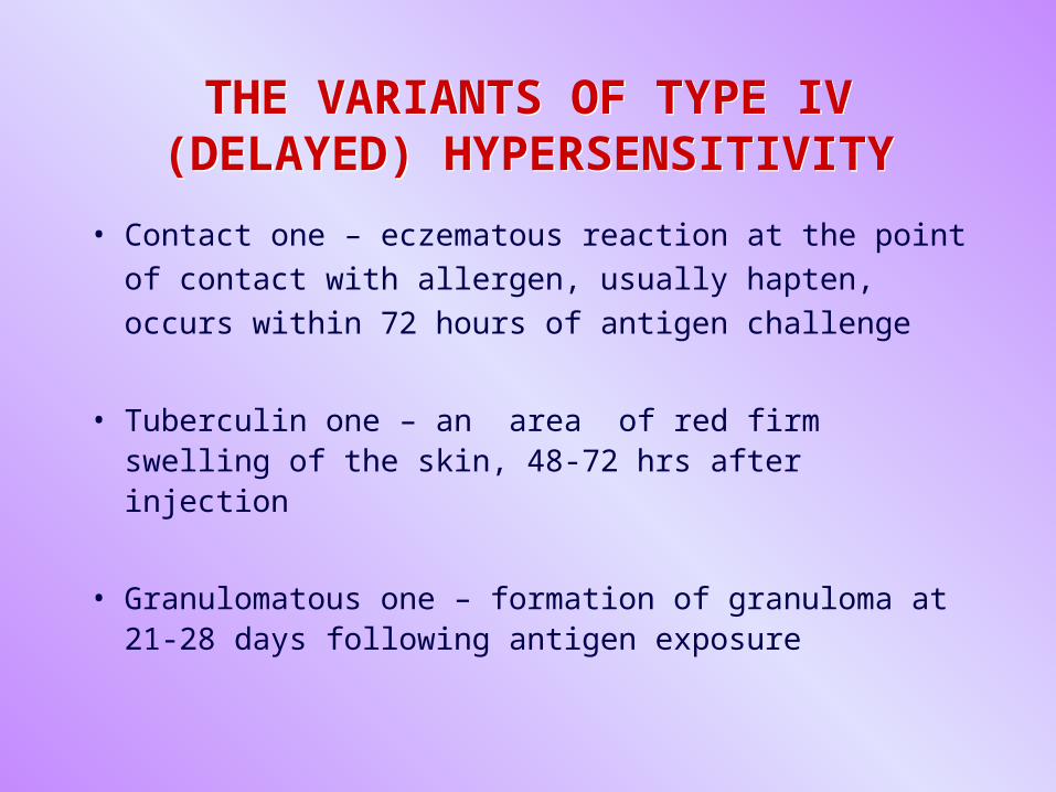
THE VARIANTS OF TYPE IV (DELAYED) HYPERSENSITIVITY
THE VARIANTS OF TYPE IV (DELAYED) HYPERSENSITIVITY
• Contact one – eczematous reaction at the point of
contact with allergen, usually hapten, occurs within
72 hours of antigen challenge
• Tuberculin one – an area of red firm swelling of the skin, 48-72 hrs after injection
• Granulomatous one – formation of granuloma at 21-28 days following antigen exposure
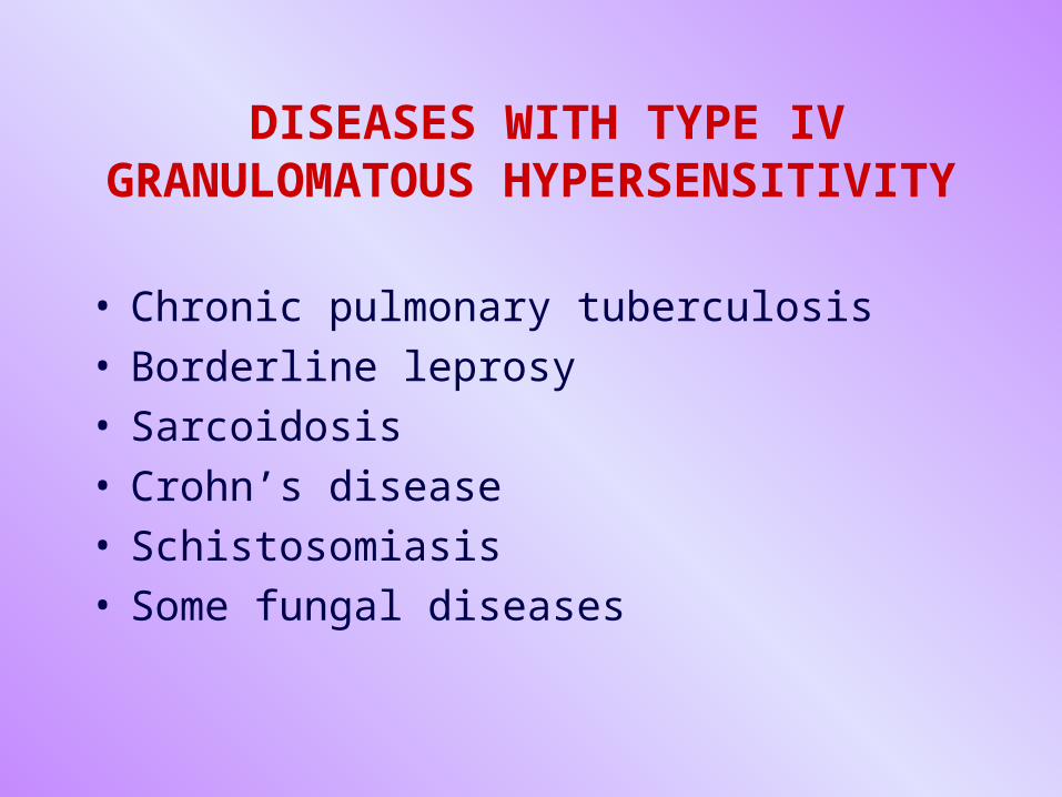
DISEASES WITH TYPE IV GRANULOMATOUS HYPERSENSITIVITY
• Chronic pulmonary tuberculosis• Borderline leprosy• Sarcoidosis• Crohn’s disease• Schistosomiasis• Some fungal diseases

THANK YOU!THANK YOU!


![PPT jan 2013 [Lecture seule]](https://img.pdfslide.us/doc/110x75/62602cecffb3104ab66000f3/ppt-jan-2013-lecture-seule.jpg)