Embed Size (px)
Citation preview
Lecture 7:Roles for MAGUKS in
Activity-dependent Synaptogenesis
MCP 9.013 ‘04
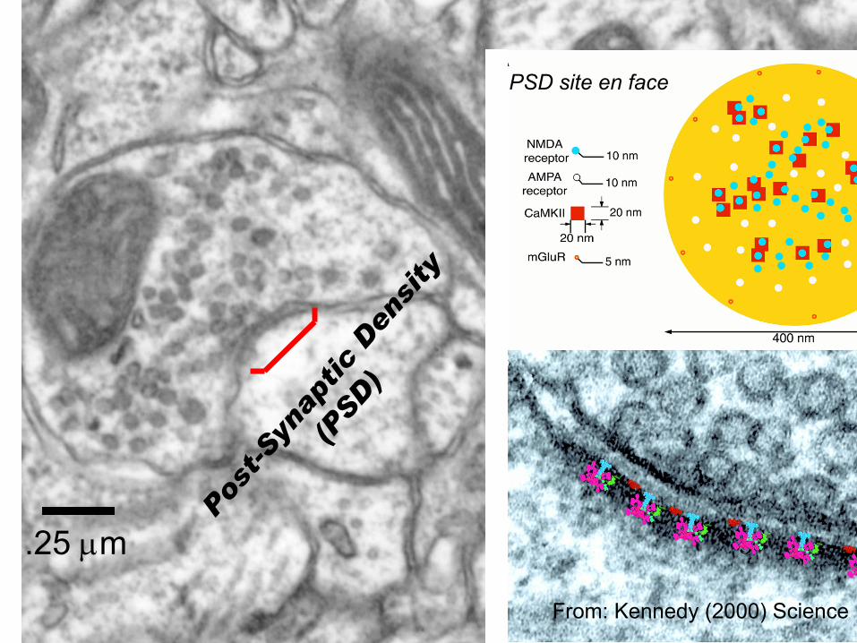
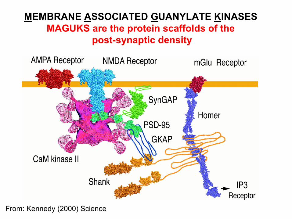
From: Kennedy (2000) Science
.25 µm
PSD site en face
Post
-Syn
aptic
Den
sity
(PSD
)
From: Kennedy (2000) Science
MEMBRANE ASSOCIATED GUANYLATE KINASES MAGUKS are the protein scaffolds of the
post-synaptic density
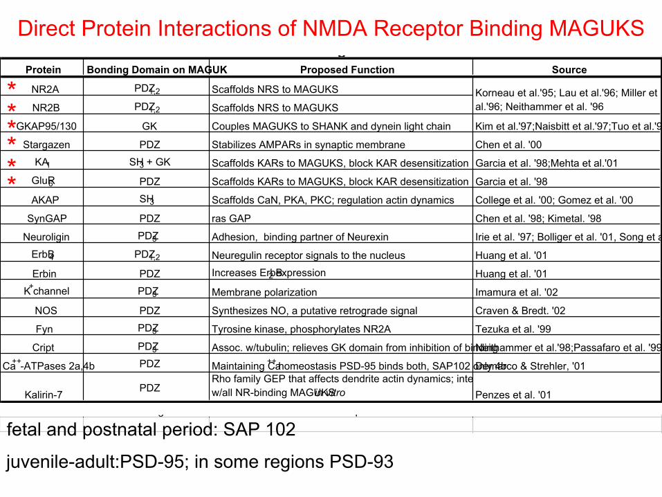
Protein Bonding Domain on MAGUK Proposed Function Source
NR2A PDZ1,2 Scaffolds NRS to MAGUKS
NR2B PDZ1,2 Scaffolds NRS to MAGUKS
GKAP95/130 GK Couples MAGUKS to SHANK and dynein light chain Kim et al.'97;Naisbitt et al.'97;Tuo et al.'9
Stargazen PDZ Stabilizes AMPARs in synaptic membrane Chen et al. '00
KA1 SH3 + GK Scaffolds KARs to MAGUKS, block KAR desensitization Garcia et al. '98;Mehta et al.'01
GluR6 PDZ Scaffolds KARs to MAGUKS, block KAR desensitization Garcia et al. '98
AKAP SH3 Scaffolds CaN, PKA, PKC; regulation actin dynamics College et al. '00; Gomez et al. '00
SynGAP PDZ ras GAP Chen et al. '98; Kimetal. '98
Neuroligin PDZ3 Adhesion, binding partner of Neurexin Irie et al. '97; Bolliger et al. '01, Song et a
ErbB4 PDZ1,2 Neuregulin receptor signals to the nucleus Huang et al. '01
Erbin PDZ Increases ErbB2 expression Huang et al. '01
K+channel PDZ3 Membrane polarization Imamura et al. '02
NOS PDZ Synthesizes NO, a putative retrograde signal Craven & Bredt. '02
Fyn PDZ3 Tyrosine kinase, phosphorylates NR2A Tezuka et al. '99
Cript PDZ3 Assoc. w/tubulin; relieves GK domain from inhibition of bindingNeithammer et al.'98;Passafaro et al. '99
Ca++-ATPases 2a,4b PDZ Maintaining Ca++ homeostasis PSD-95 binds both, SAP102 only 4bDemarco & Strehler, '01
Kalirin-7 PDZRho family GEP that affects dendrite actin dynamics; intew/all NR-binding MAGUKS in vitro Penzes et al. '01
TABLE I: Proteins that Bind Domains of NR-binding MAGUKS*
* not known whether all NR binding MAGUKS will interact with most of these proteins
Korneau et al.'95; Lau et al.'96; Miller et al.'96; Neithammer et al. '96
Direct Protein Interactions of NMDA Receptor Binding MAGUKS
fetal and postnatal period: SAP 102
juvenile-adult:PSD-95; in some regions PSD-93
******
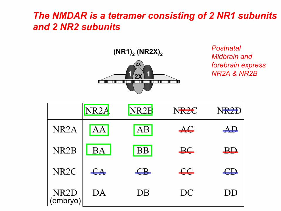
NR2A NR2B NR2C NR2D
AA AB AC AD
BA BB BC BD
CA CB CC CD
DA DB DC DD
NR2A
NR2B
NR2C
NR2D
(NR1)2 (NR2X)2
2X 112X
The NMDAR is a tetramer consisting of 2 NR1 subunitsand 2 NR2 subunits
PostnatalMidbrain andforebrain expressNR2A & NR2B
(embryo)
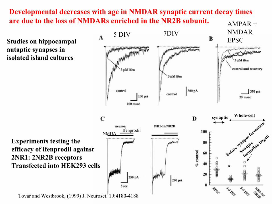
Tovar and Westbrook, (1999) J. Neurosci. 19:4180-4188
Developmental decreases with age in NMDAR synaptic current decay times are due to the loss of NMDARs enriched in the NR2B subunit.
Studies on hippocampalautaptic synapses inisolated island cultures
Experiments testing theefficacy of ifenprodil against2NR1: 2NR2B receptorsTransfected into HEK293 cells
5 DIV 7DIVAMPAR + NMDAR EPSC
NMDAIfenprodil
Before
synapse
formatio
n
Synapse
formatio
n begun
Whole-cellsynaptic
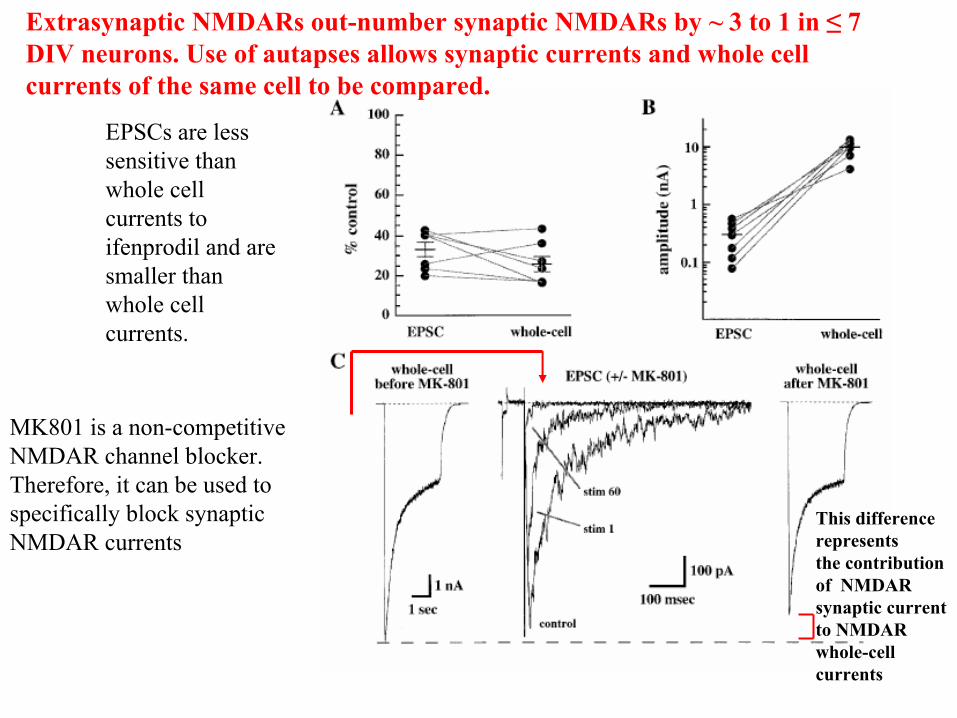
Extrasynaptic NMDARs out-number synaptic NMDARs by ~ 3 to 1 in ≤ 7 DIV neurons. Use of autapses allows synaptic currents and whole cell currents of the same cell to be compared.
EPSCs are less sensitive than whole cell currents to ifenprodil and are smaller than whole cell currents.
MK801 is a non-competitive NMDAR channel blocker. Therefore, it can be used to specifically block synaptic NMDAR currents
This differencerepresents the contributionof NMDARsynaptic currentto NMDAR whole-cell currents
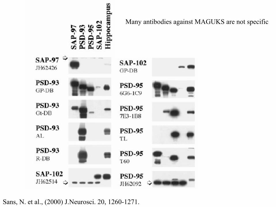
Many antibodies against MAGUKS are not specific
Sans, N. et al., (2000) J.Neurosci. 20, 1260-1271.
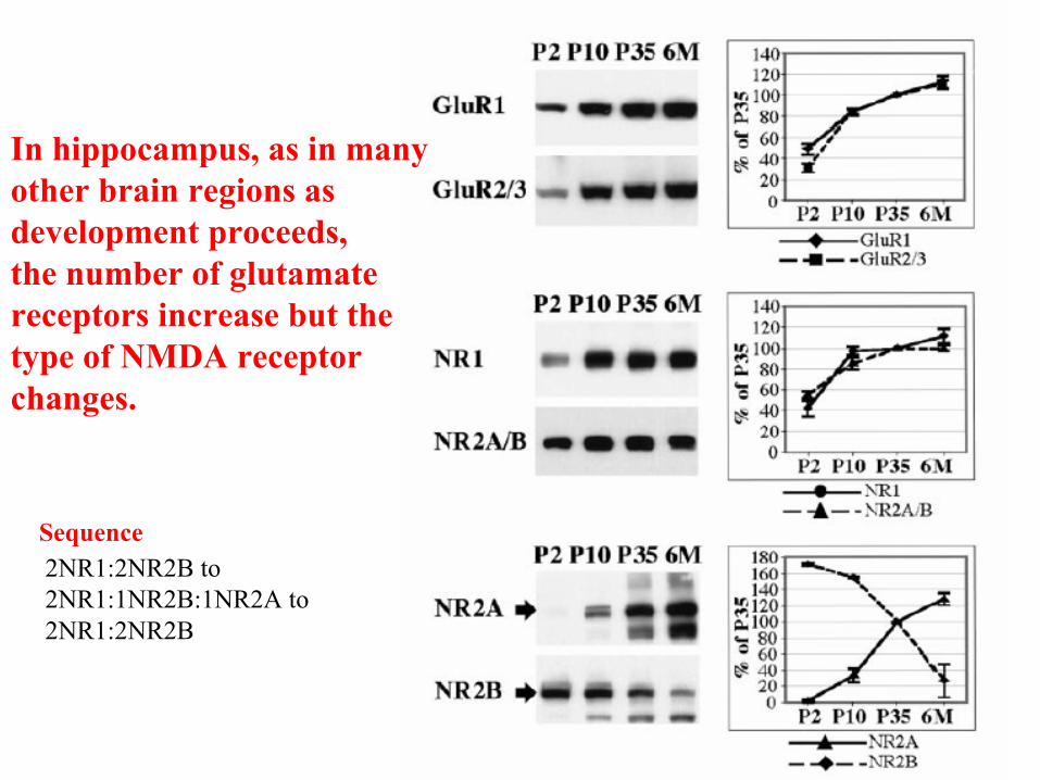
In hippocampus, as in manyother brain regions as development proceeds,the number of glutamatereceptors increase but thetype of NMDA receptorchanges.
2NR1:2NR2B to 2NR1:1NR2B:1NR2A to 2NR1:2NR2B
Sequence
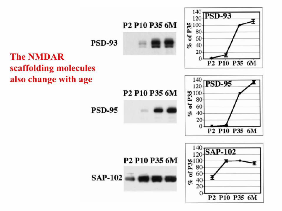
The NMDAR scaffolding molecules also change with age
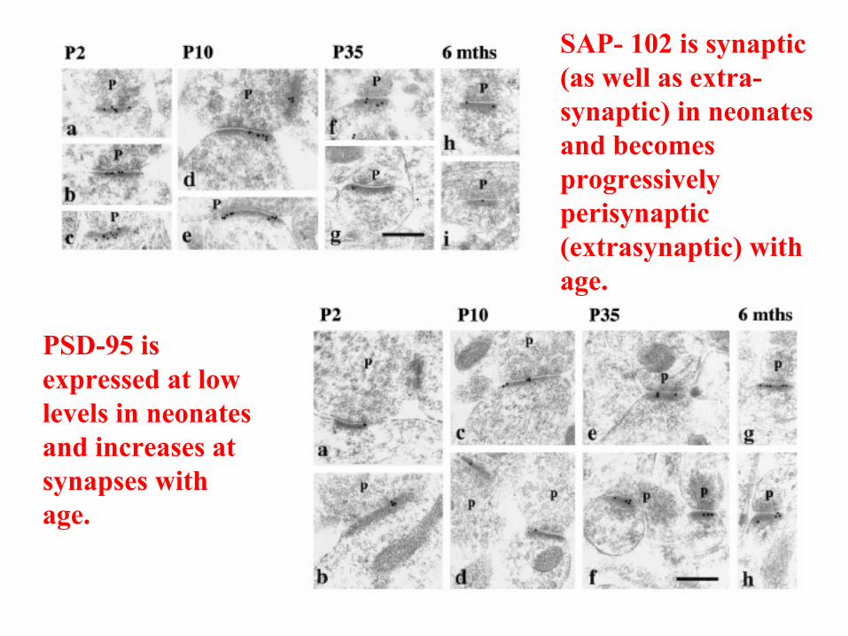
SAP- 102 is synaptic (as well as extra-synaptic) in neonates and becomes progressively perisynaptic (extrasynaptic) with age.
PSD-95 is expressed at low levels in neonates and increases at synapses withage.
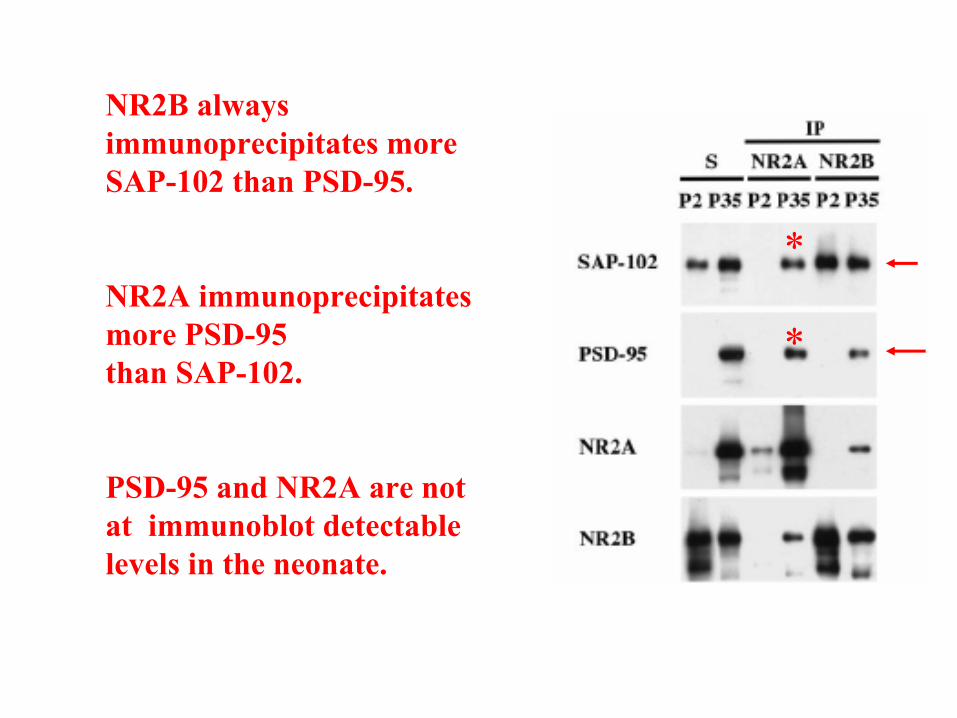
NR2B always immunoprecipitates moreSAP-102 than PSD-95.
NR2A immunoprecipitates more PSD-95than SAP-102.
PSD-95 and NR2A are notat immunoblot detectable levels in the neonate.
*
*
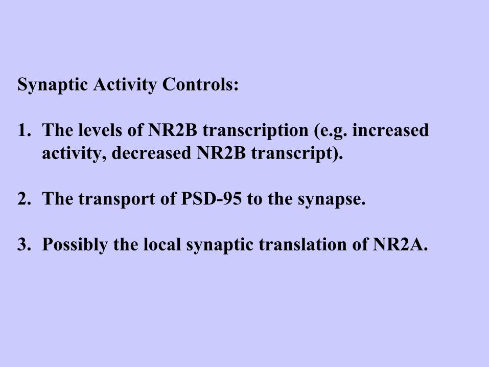
Synaptic Activity Controls:
1. The levels of NR2B transcription (e.g. increased activity, decreased NR2B transcript).
2. The transport of PSD-95 to the synapse.
3. Possibly the local synaptic translation of NR2A.
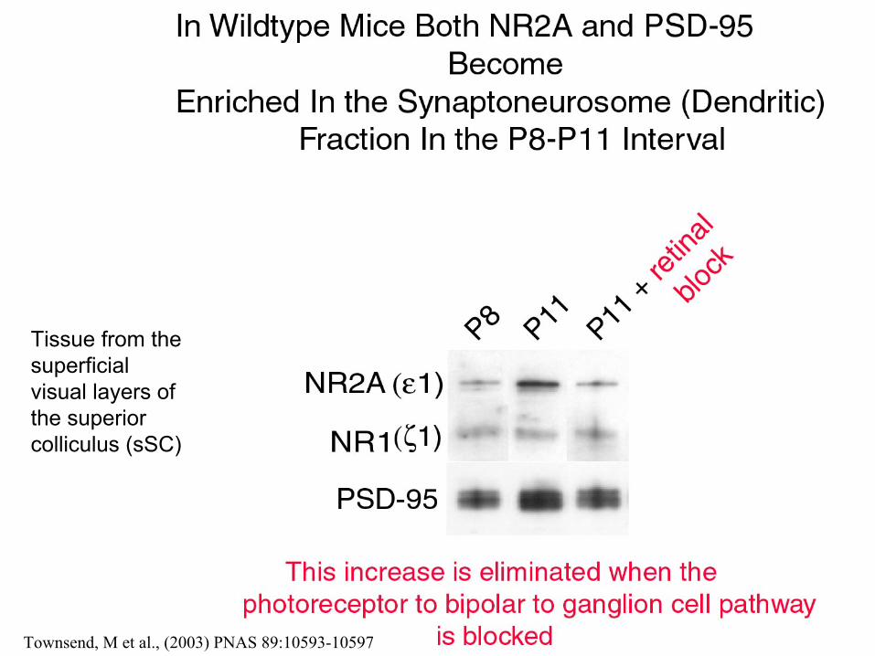
Tissue from the superficial visual layers ofthe superior colliculus (sSC)
Townsend, M et al., (2003) PNAS 89:10593-10597
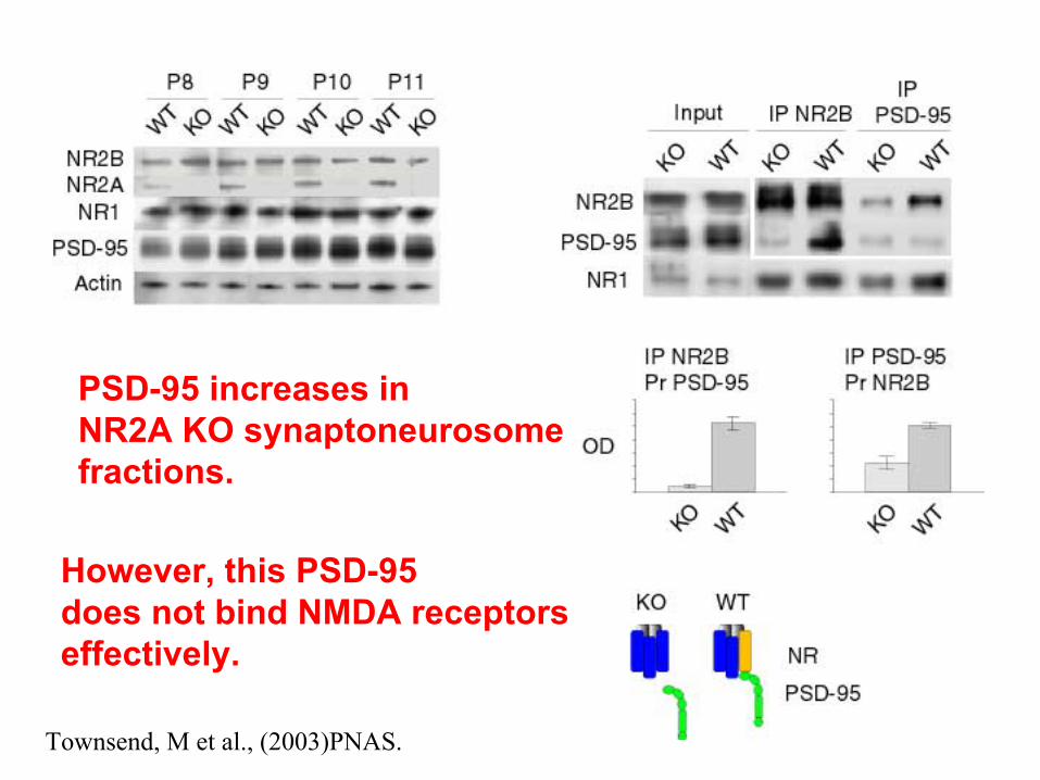
PSD-95 increases inNR2A KO synaptoneurosomefractions.
However, this PSD-95does not bind NMDA receptorseffectively.
Townsend, M et al., (2003)PNAS.
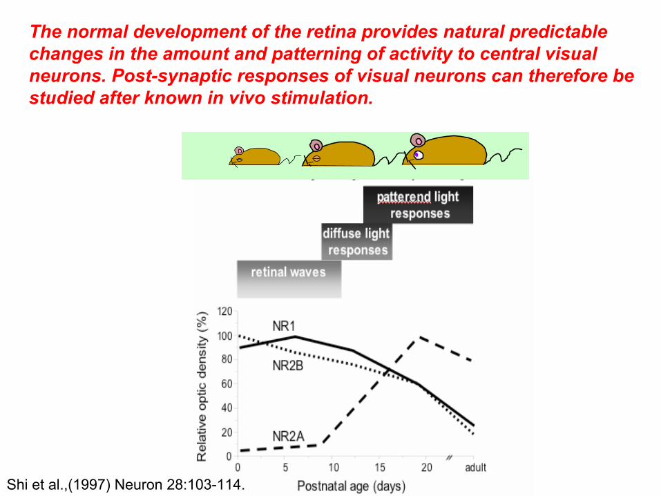
The normal development of the retina provides natural predictable changes in the amount and patterning of activity to central visual neurons. Post-synaptic responses of visual neurons can therefore be studied after known in vivo stimulation.
Shi et al.,(1997) Neuron 28:103-114.
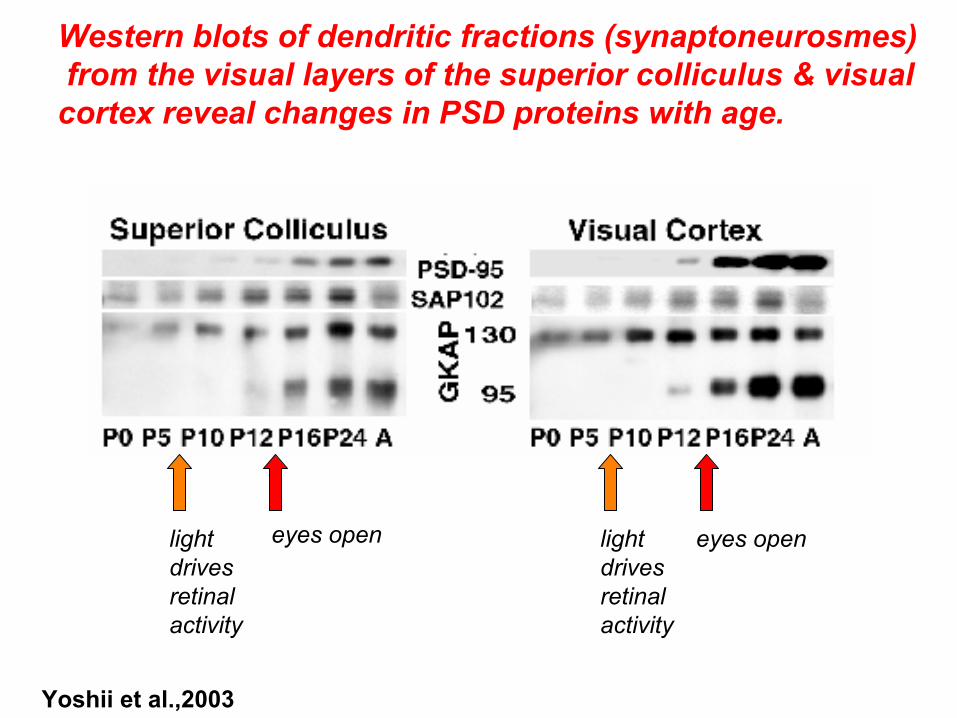
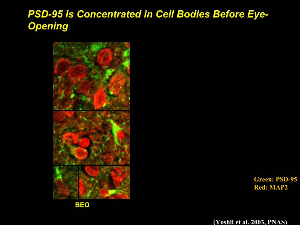
Western blots of dendritic fractions (synaptoneurosmes)from the visual layers of the superior colliculus & visualcortex reveal changes in PSD proteins with age.
eyes open eyes openlight drivesretinalactivity
light drivesretinalactivity
Yoshii et al.,2003
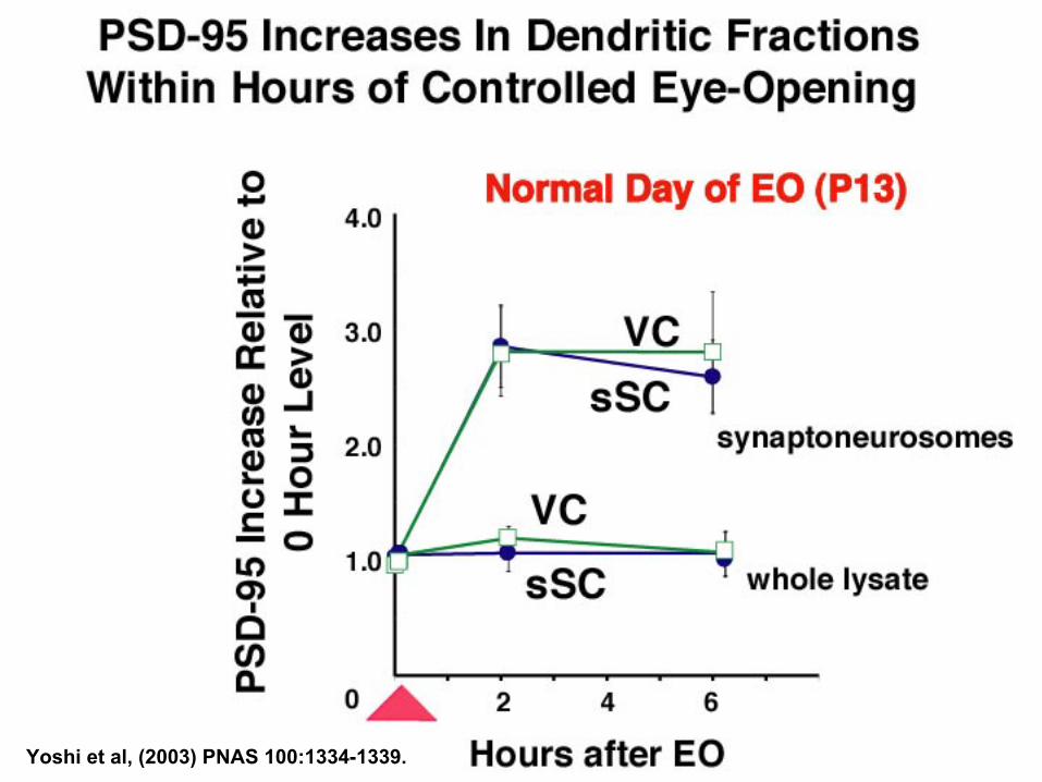
Yoshi et al, (2003) PNAS 100:1334-1339.
BEOAEO
(Yoshii et al. 2003, PNAS)
Green: PSD-95Red: MAP2
PSD-95 Is Concentrated in Cell Bodies Before Eye-Opening
BEO
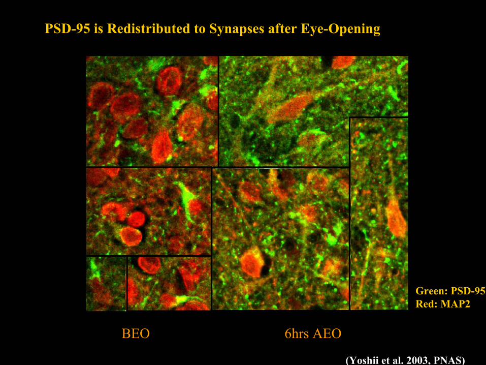
BEOBEO 6hrs AEO
PSD-95 is Redistributed to Synapses after Eye-Opening
(Yoshii et al. 2003, PNAS)
Green: PSD-95Red: MAP2
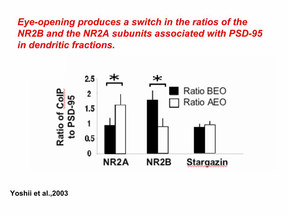
Eye-opening produces a switch in the ratios of the NR2B and the NR2A subunits associated with PSD-95in dendritic fractions.
Yoshii et al.,2003
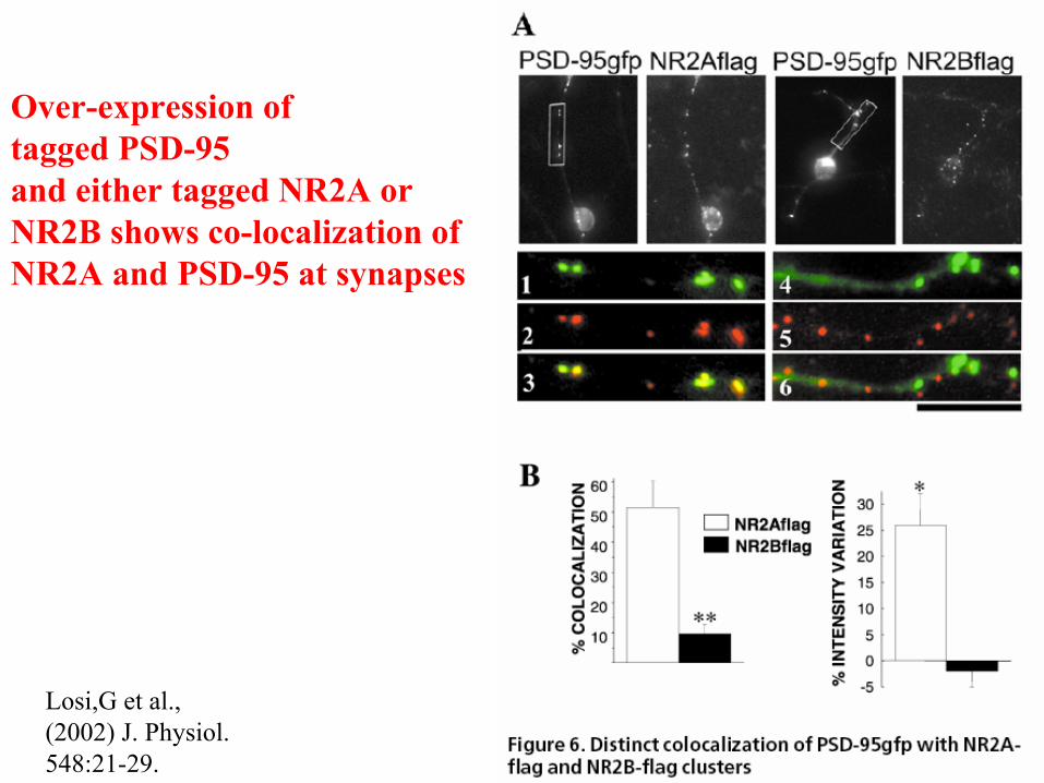
Over-expression of tagged PSD-95and either tagged NR2A orNR2B shows co-localization of NR2A and PSD-95 at synapses
Losi,G et al., (2002) J. Physiol. 548:21-29.
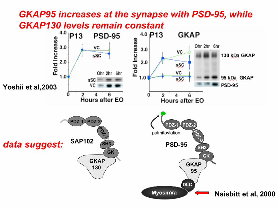
GKAP95 increases at the synapse with PSD-95, while GKAP130 levels remain constant
SAP102
GKAP130
SH3
GK
PDZ-1 PDZ-2
PDZ-3 palmitoylation
GKAP95
PSD-95 SH3
GK
PDZ-1 PDZ-2
PDZ-3
DLC
MyosinVa
data suggest:
Naisbitt et al, 2000
Yoshii et al,2003
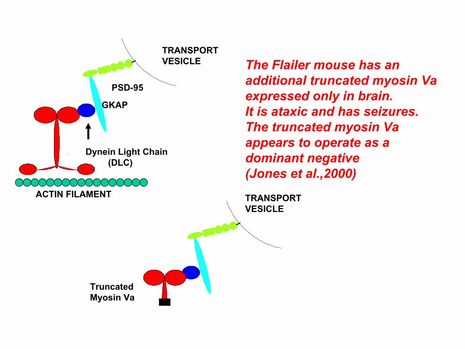
Dynein Light Chain(DLC)
PSD-95
GKAP
TRANSPORT VESICLE
ACTIN FILAMENT
Truncated Myosin Va
TRANSPORT VESICLE
The Flailer mouse has anadditional truncated myosin Vaexpressed only in brain.It is ataxic and has seizures.The truncated myosin Vaappears to operate as adominant negative (Jones et al.,2000)
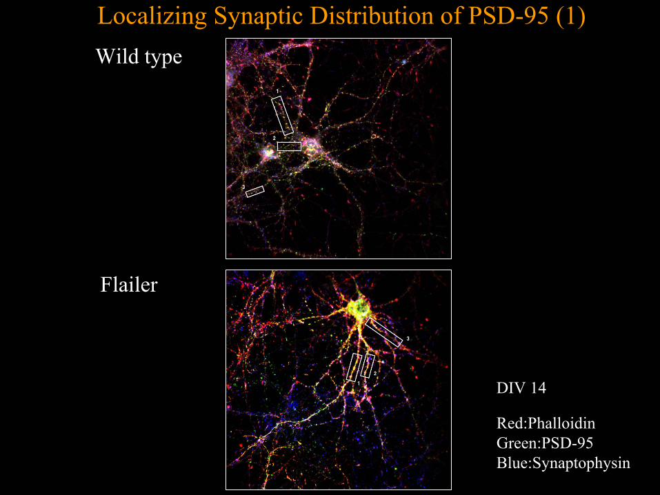
Wild type
Flailer
Localizing Synaptic Distribution of PSD-95 (1)
DIV 14
Red:PhalloidinGreen:PSD-95Blue:Synaptophysin
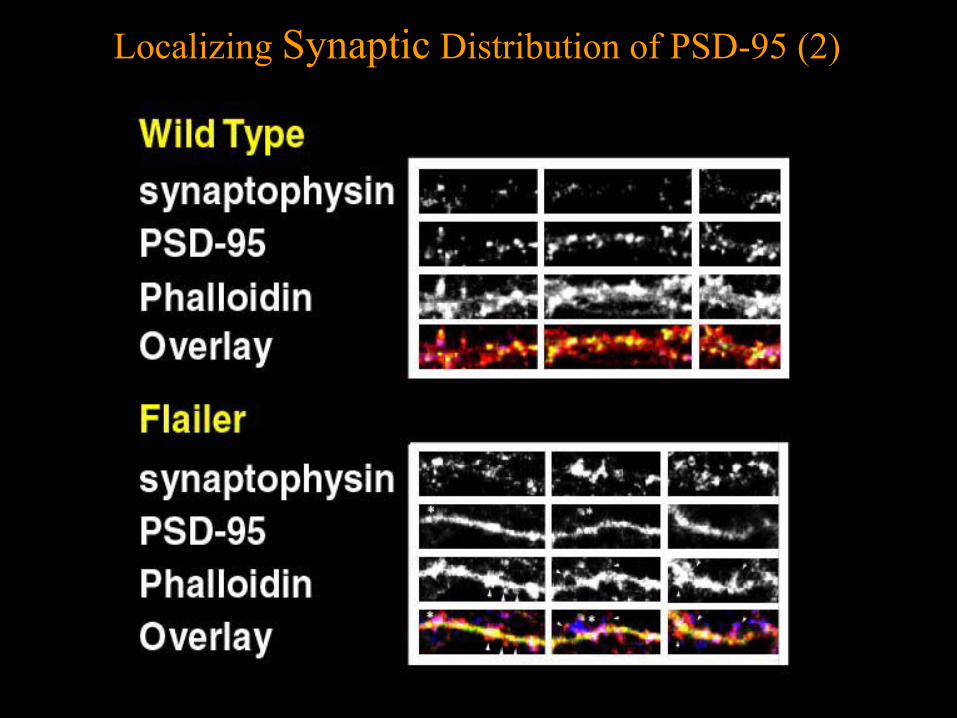
Localizing Synaptic Distribution of PSD-95 (2)
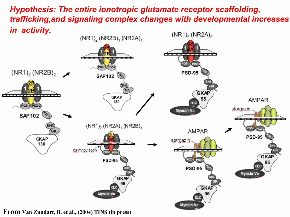
Hypothesis: The entire ionotropic glutamate receptor scaffolding, trafficking,and signaling complex changes with developmental increasesin activity.
From Van Zundart, B. et al., (2004) TINS (in press)
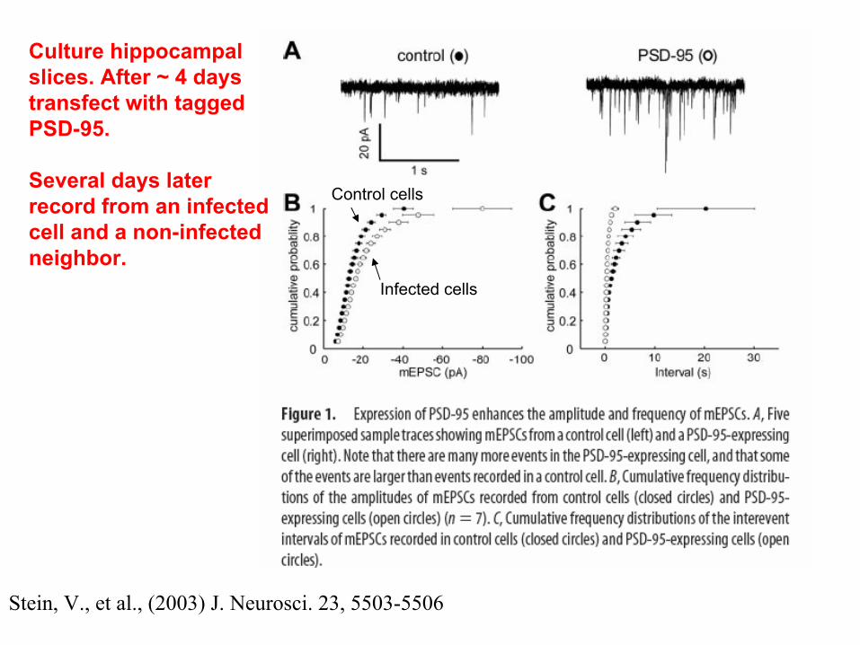
Stein, V., et al., (2003) J. Neurosci. 23, 5503-5506
Culture hippocampalslices. After ~ 4 daystransfect with taggedPSD-95.
Several days laterrecord from an infectedcell and a non-infectedneighbor.
Control cells
Infected cells
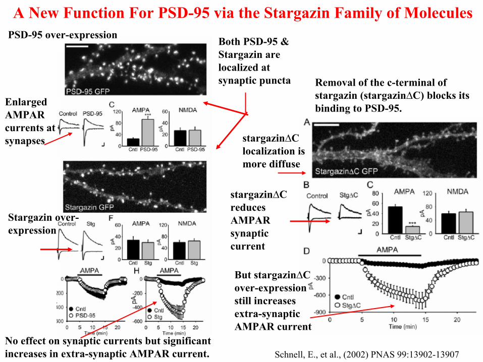
A New Function For PSD-95 via the Stargazin Family of Molecules
Both PSD-95 & Stargazin are localized at synaptic puncta
Enlarged AMPAR currents at synapses
PSD-95 over-expression
Stargazin over-expression
No effect on synaptic currents but significant increases in extra-synaptic AMPAR current.
Removal of the c-terminal ofstargazin (stargazin∆C) blocks its binding to PSD-95.
stargazin∆C localization is more diffuse
stargazin∆C reduces AMPAR synaptic current
But stargazin∆C over-expression still increases extra-synaptic AMPAR current
Schnell, E., et al., (2002) PNAS 99:13902-13907
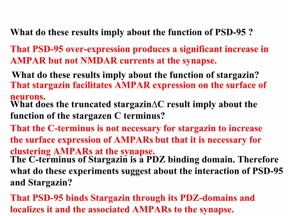
What do these results imply about the function of stargazin?That stargazin facilitates AMPAR expression on the surface of neurons.
What do these results imply about the function of PSD-95 ?
That PSD-95 over-expression produces a significant increase in AMPAR but not NMDAR currents at the synapse.
What does the truncated stargazin∆C result imply about the function of the stargazen C terminus?That the C-terminus is not necessary for stargazin to increase the surface expression of AMPARs but that it is necessary for clustering AMPARs at the synapse. The C-terminus of Stargazin is a PDZ binding domain. Therefore what do these experiments suggest about the interaction of PSD-95 and Stargazin?That PSD-95 binds Stargazin through its PDZ-domains and localizes it and the associated AMPARs to the synapse.
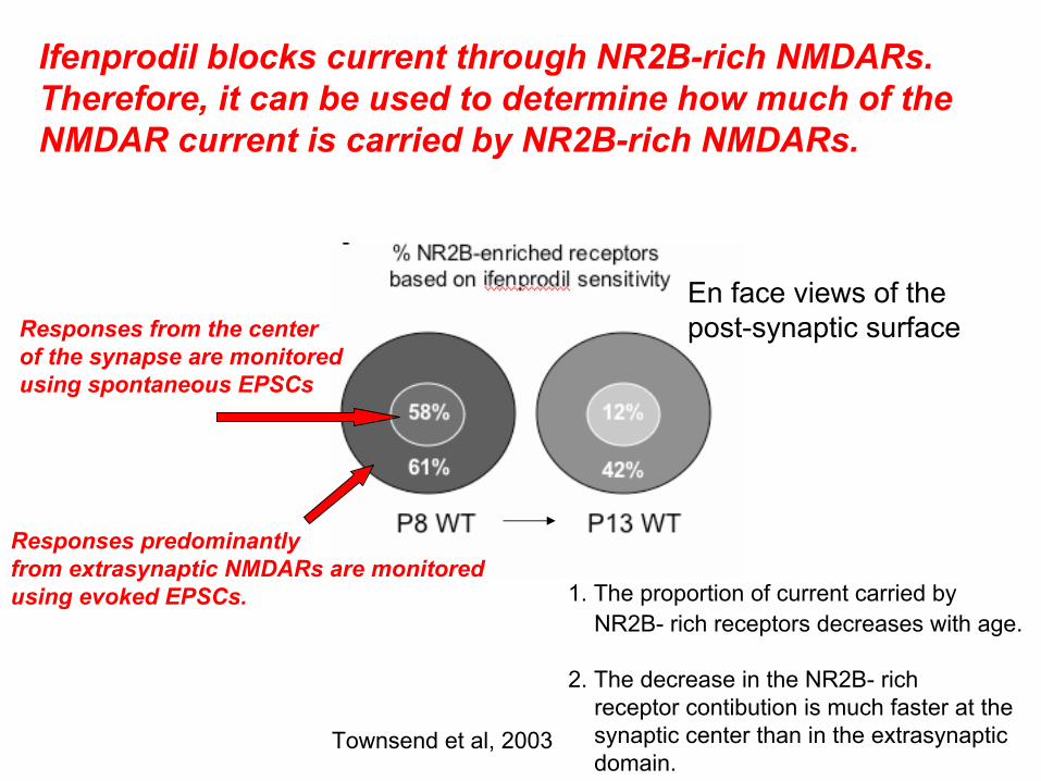
Ifenprodil blocks current through NR2B-rich NMDARs. Therefore, it can be used to determine how much of the NMDAR current is carried by NR2B-rich NMDARs.
Responses from the centerof the synapse are monitoredusing spontaneous EPSCs
Responses predominantlyfrom extrasynaptic NMDARs are monitoredusing evoked EPSCs.
En face views of thepost-synaptic surface
1. The proportion of current carried byNR2B- rich receptors decreases with age.
2. The decrease in the NR2B- rich receptor contibution is much faster at thesynaptic center than in the extrasynapticdomain.
Townsend et al, 2003
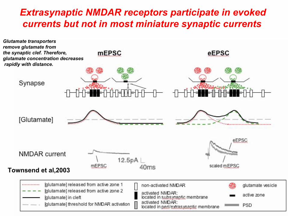
Extrasynaptic NMDAR receptors participate in evokedcurrents but not in most miniature synaptic currents
Glutamate transportersremove glutamate fromthe synaptic clef. Therefore,glutamate concentration decreasesrapidly with distance.
Townsend et al,2003
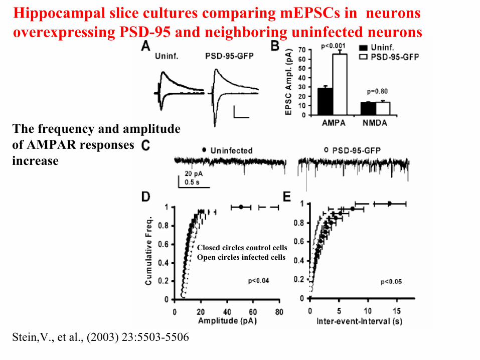
Hippocampal slice cultures comparing mEPSCs in neurons overexpressing PSD-95 and neighboring uninfected neurons
Closed circles control cells Open circles infected cells
The frequency and amplitudeof AMPAR responses increase
Stein,V., et al., (2003) 23:5503-5506