Embed Size (px)
Citation preview
Lecture 56: Development of Heart II
Learning Objectives
• By the end of this session, the student should be able to:– Describe septum formation in the common atrium &
ventricles.– Describe septum formation in the atrioventricular
canal.– Describe septum formation in the truncus arteriosus
and conus cordis.– Describe formation of conducting system of the heart.– Correlate this knowledge to clinical conditions.
• Reference: Langman's Medical Embryology: T.W. Sadler, 12th ed., CH. 13, P. 171 –185.
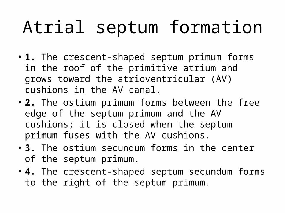
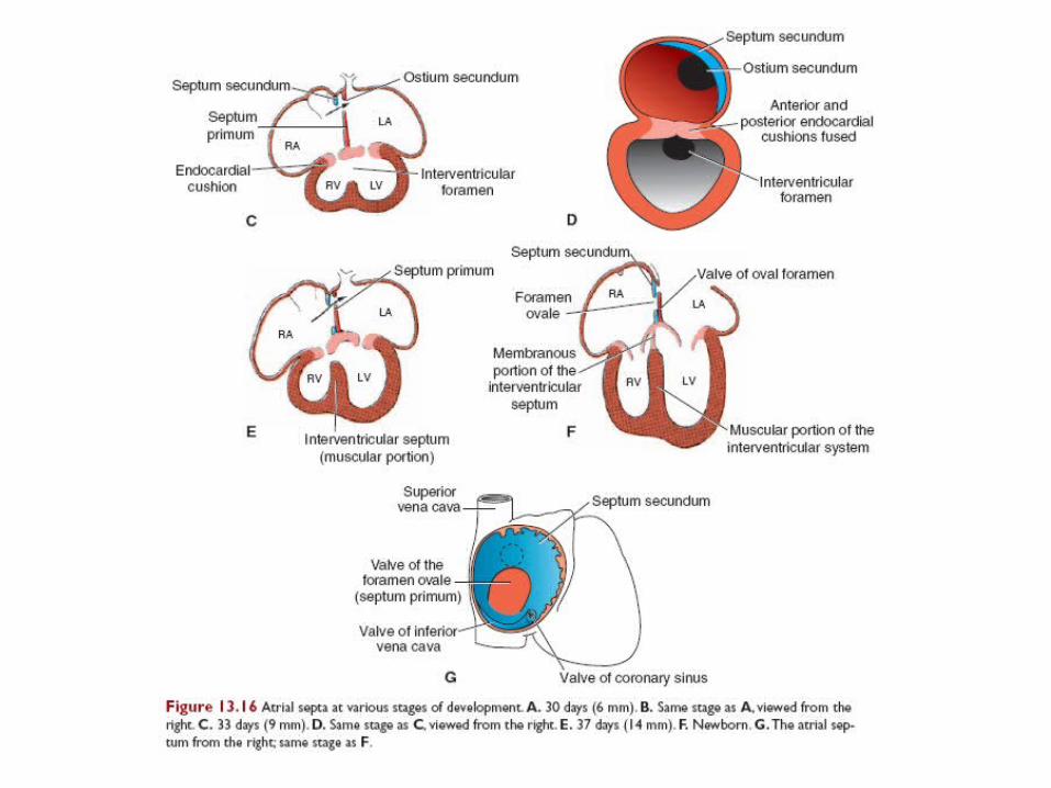
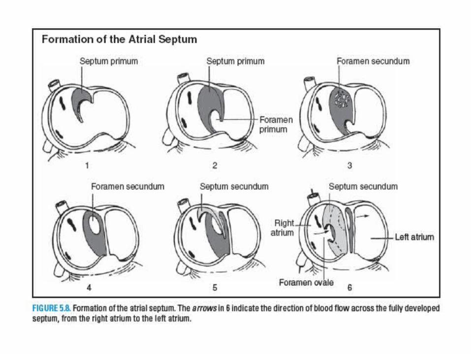
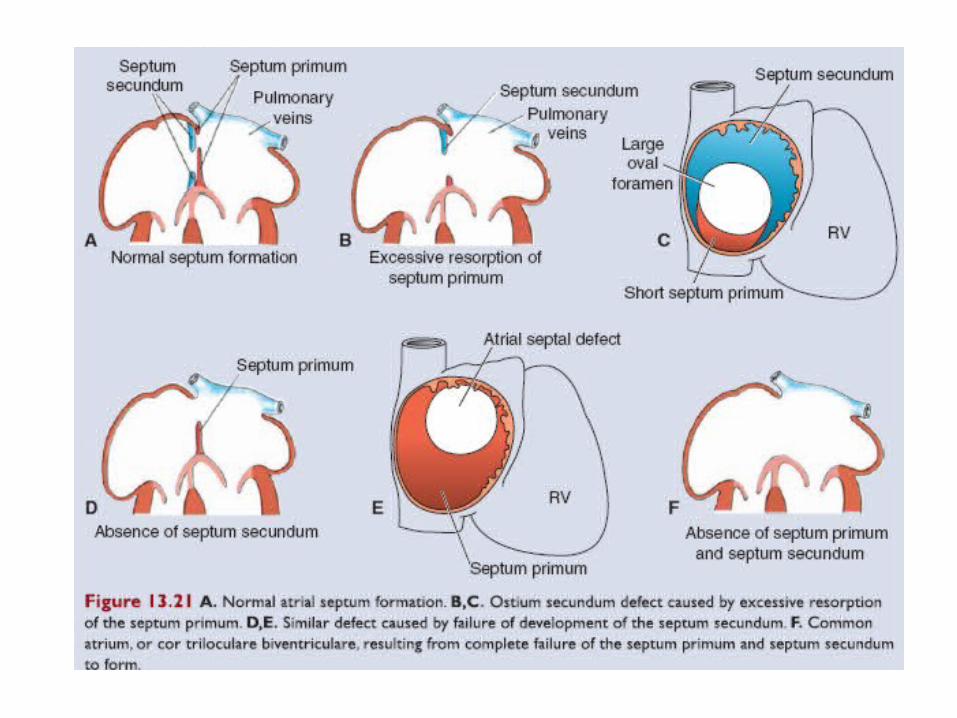
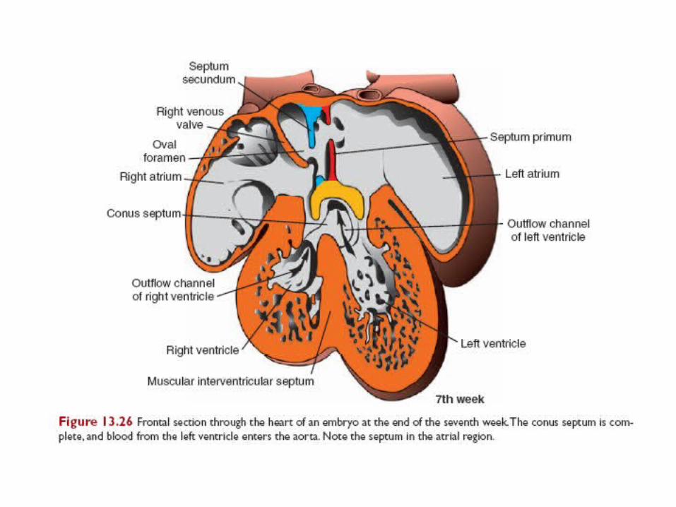
Atrial septum formation
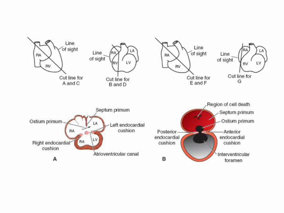
• 1. The crescent-shaped septum primum forms in the roof of the primitive atrium and grows toward the atrioventricular (AV) cushions in the AV canal.
• 2. The ostium primum forms between the free edge of the septum primum and the AV cushions; it is closed when the septum primum fuses with the AV cushions.
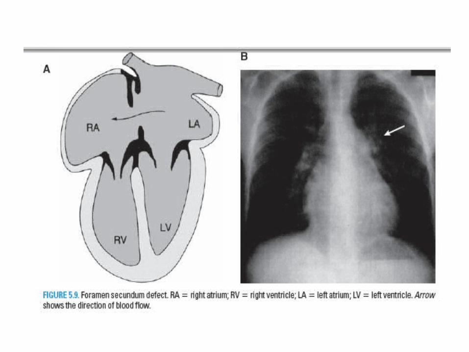
• 3. The ostium secundum forms in the center of the septum primum.
• 4. The crescent-shaped septum secundum forms to the right of the septum primum.
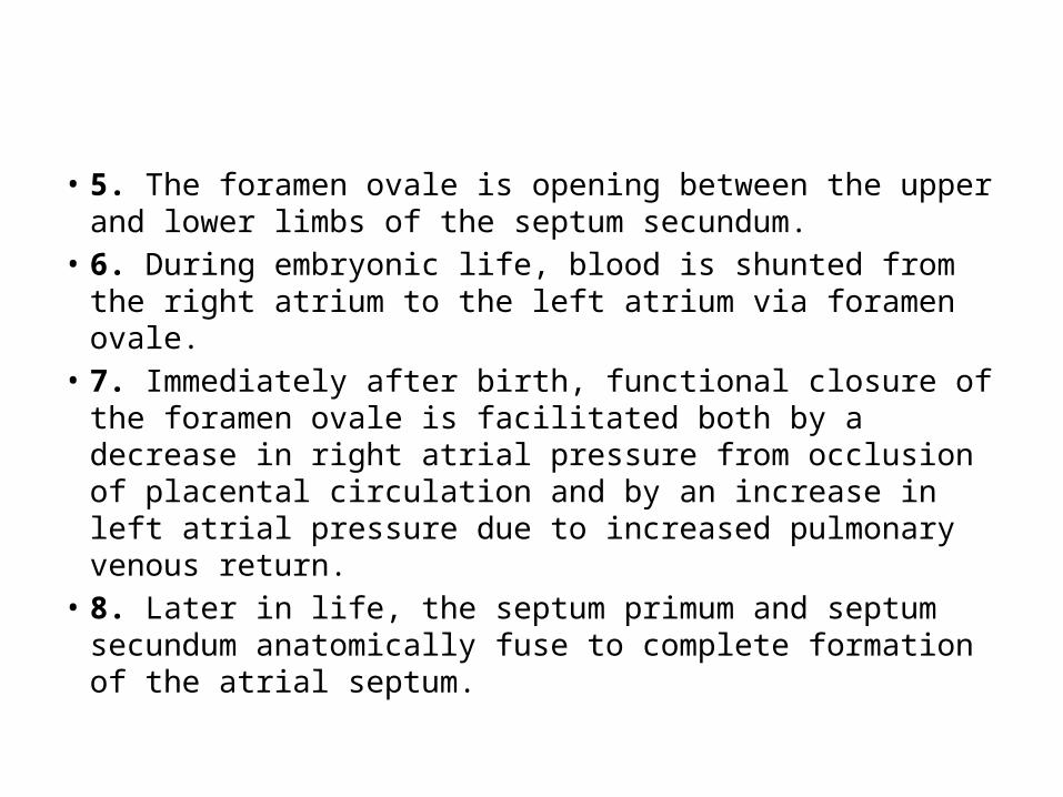
• 5. The foramen ovale is opening between the upper and lower limbs of the septum secundum.
• 6. During embryonic life, blood is shunted from the right atrium to the left atrium via foramen ovale.
• 7. Immediately after birth, functional closure of the foramen ovale is facilitated both by a decrease in right atrial pressure from occlusion of placental circulation and by an increase in left atrial pressure due to increased pulmonary venous return.
• 8. Later in life, the septum primum and septum secundum anatomically fuse to complete formation of the atrial septum.
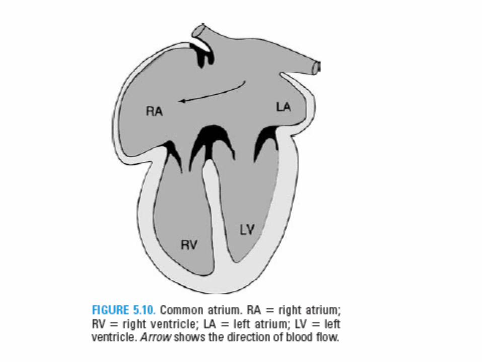
• Atrial septal defect• Premature closure of foramen ovale• Foramen secundum defect• Common atrium
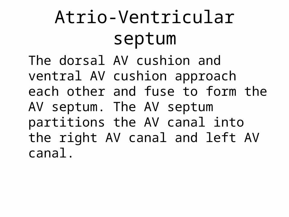
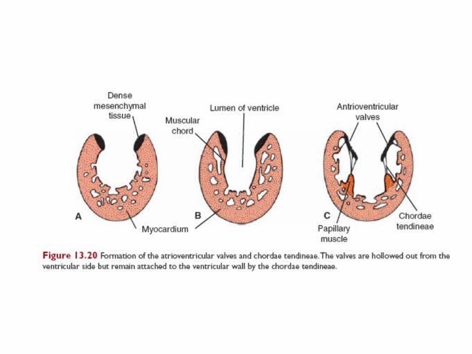
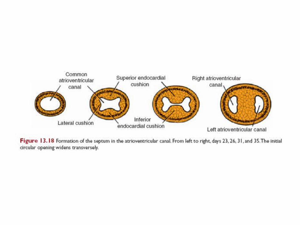
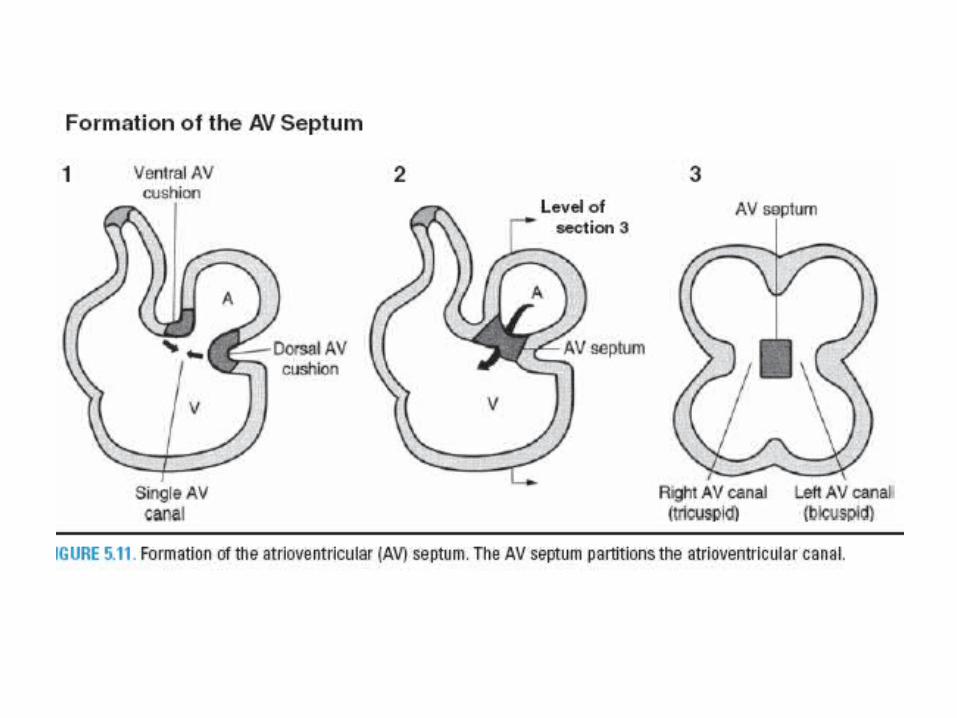
Atrio-Ventricular septum
The dorsal AV cushion and ventral AV cushion approach each other and fuse to form the AV septum. The AV septum partitions the AV canal into the right AV canal and left AV canal.

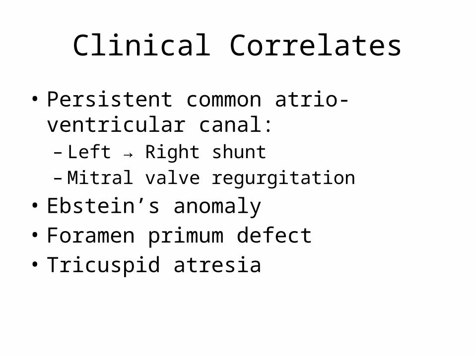
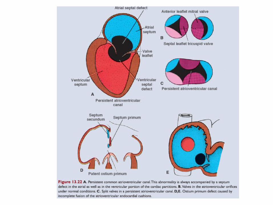
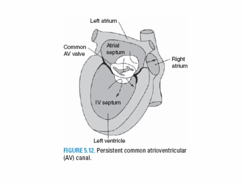
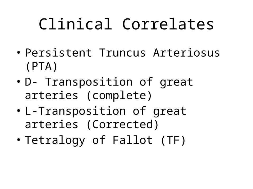
Clinical Correlates
• Persistent common atrio-ventricular canal:– Left → Right shunt– Mitral valve regurgitation
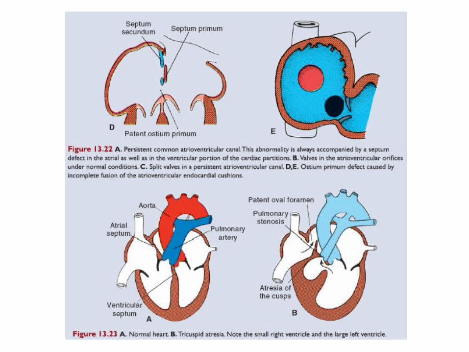
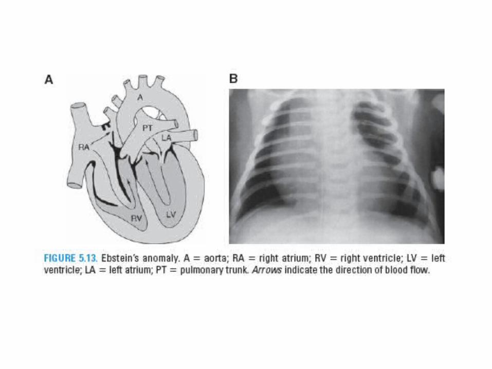
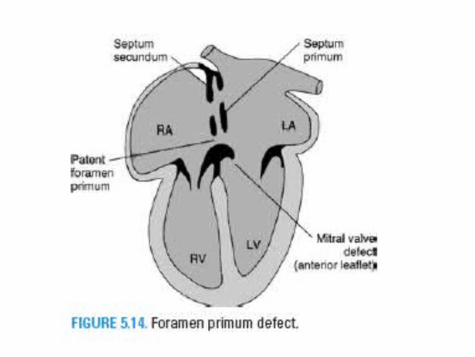
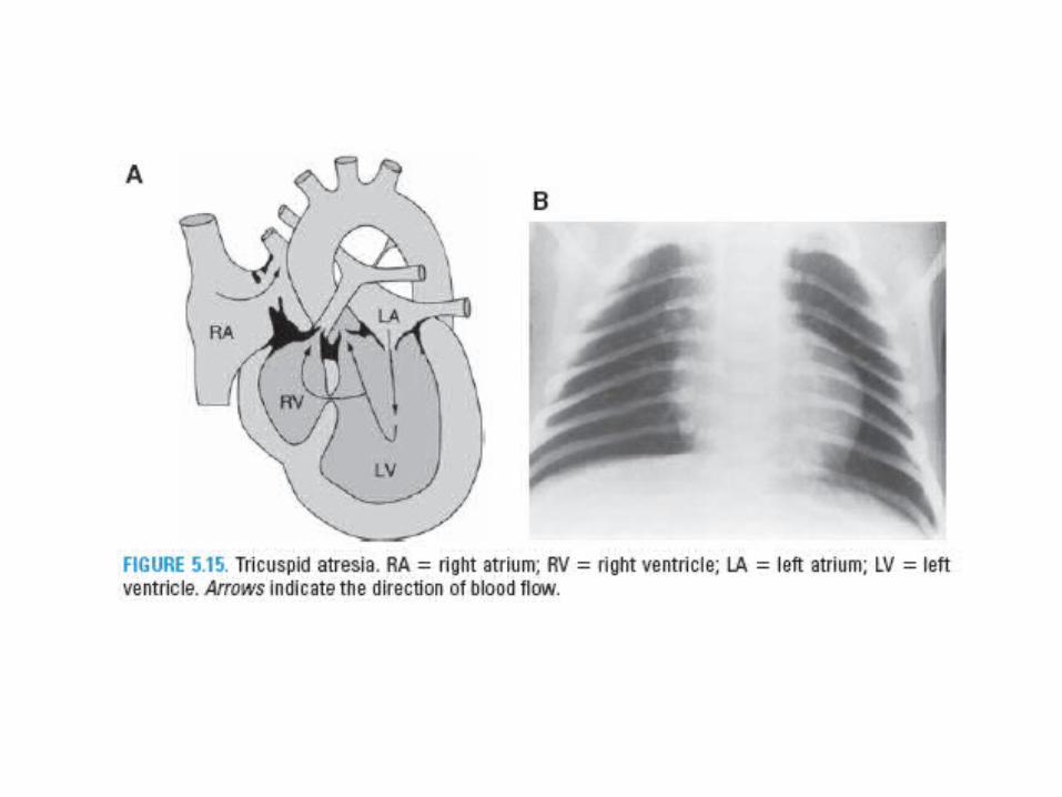
• Ebstein’s anomaly• Foramen primum defect• Tricuspid atresia
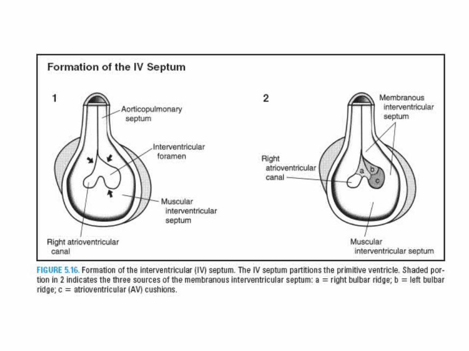
Inter-ventricular septum
• 1. The muscular IV septum develops in the midline on the floor of the primitive ventricle and grows toward the fused AV cushions.
• 2. The IV foramen is located between the free edge of the muscular IV septum and the fused AV cushions.
• 3. The IV foramen is closed by the membranous IV septum.
• 4. The membranous IV septum forms by the proliferation and fusion of tissue from three sources: the right bulbar ridge, left bulbar ridge, and AV cushions.
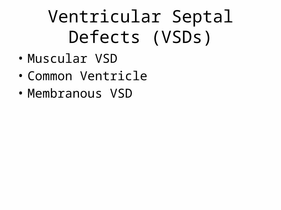
Ventricular Septal Defects (VSDs)
• Muscular VSD• Common Ventricle• Membranous VSD

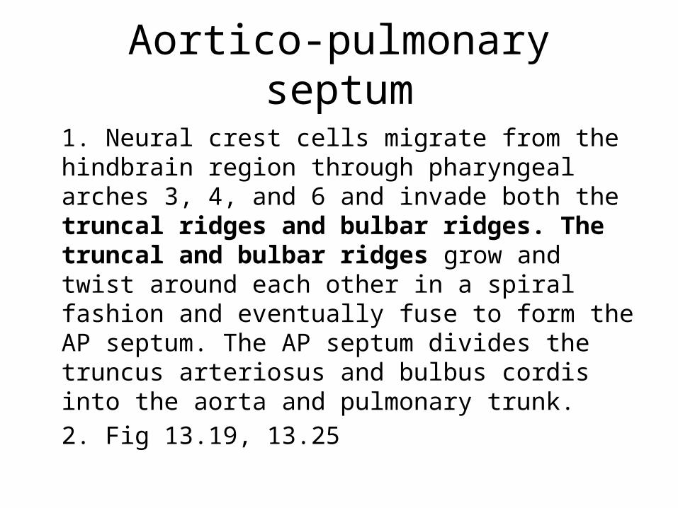
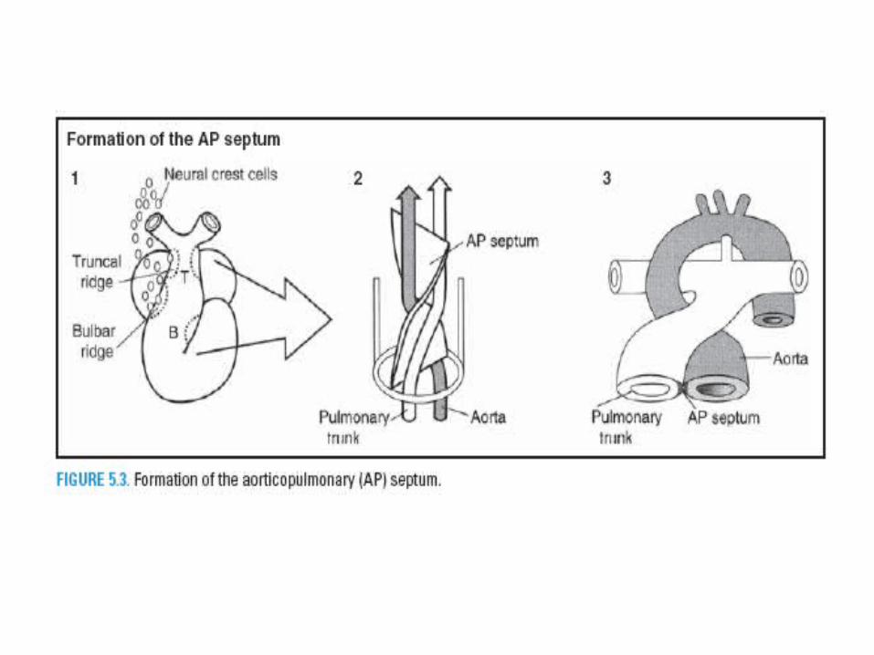
Aortico-pulmonary septum
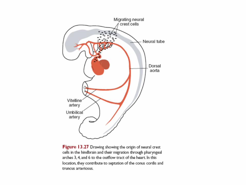
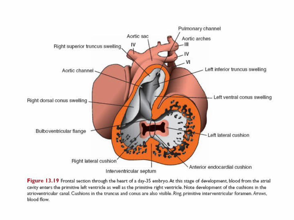
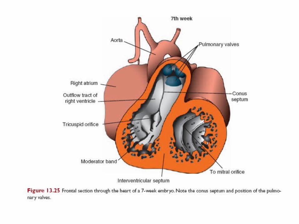
1. Neural crest cells migrate from the hindbrain region through pharyngeal arches 3, 4, and 6 and invade both the truncal ridges and bulbar ridges. The truncal and bulbar ridges grow and twist around each other in a spiral fashion and eventually fuse to form the AP septum. The AP septum divides the truncus arteriosus and bulbus cordis into the aorta and pulmonary trunk.2. Fig 13.19, 13.25
Clinical Correlates
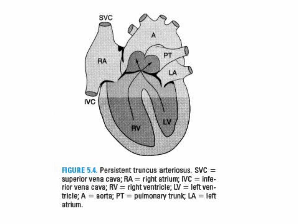
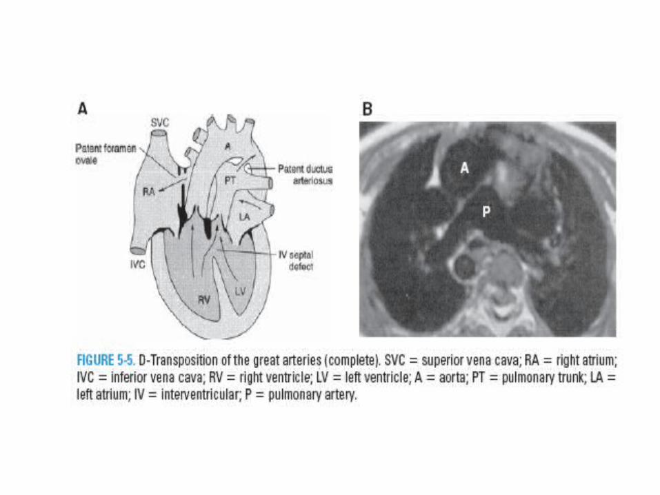
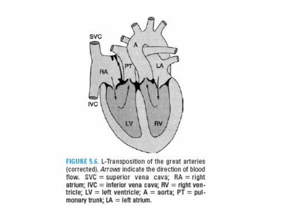
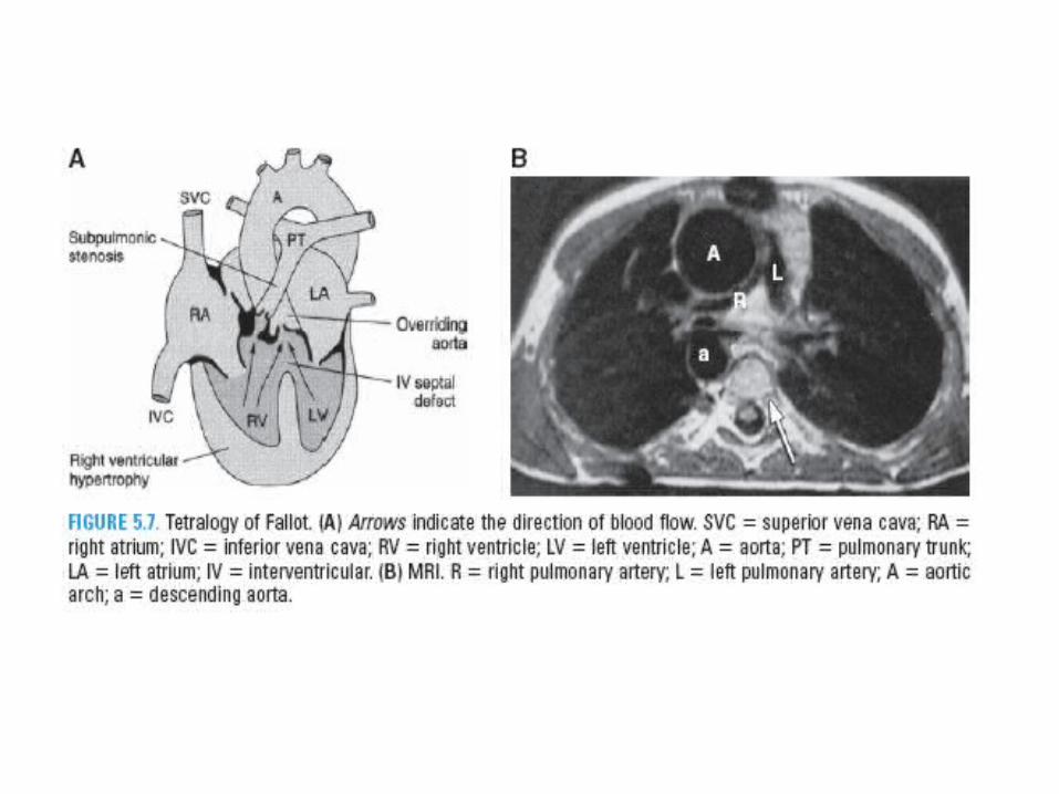
• Persistent Truncus Arteriosus (PTA)• D- Transposition of great arteries (complete)• L-Transposition of great arteries (Corrected)• Tetralogy of Fallot (TF)
Conducting system of the heart• A. At week 5, cardiac myocytes in sinus venosus region of
primitive heart tube begin to undergo spontaneous electrical depolarizations at a faster rate than cardiac myocytes in other regions.
• B. As dextral looping occurs, sinus venous becomes incorporated into right atrium, and these fast-rate depolarizing cardiac myocytes become sinoatrial (SA) node and atrioventricular (AV) node.
• C. In the adult, the cardiac myocytes of the SA and AV nodes remain committed to a fast rate of electrical depolarizations instead of developing contractile properties.
• D. As the atria and ventricles become electrically isolated by the formation of fibrous skeleton of heart, the AV node provides only pathway for depolarizations to flow from atria to ventricles.
• E. The AV bundle or bundle of His develops from a ringlike cluster of cells found at the AV junction that specifically expresses the homeobox gene, msx-2.
• F. The intramural network of Purkinje myocytes have a distinct embryological origin (versus the bundle of His), in that Purkinje myocytes develop from already contractile cardiac myocytes within the myocardium and can therefore be considered as modified cardiac myocytes.