Embed Size (px)
Citation preview
1
Protein Translocation & Nucleocytoplasmic Transport
Chapter 12 MBoC (5th Edition) Alberts et al. Reference paper: Tran and Wente, Cell 125, 1041-1053, 2006
2/8/2012
Lecture 4

2 Page 713 Molecular Biology of the Cell (© Garland Science 2008)
3
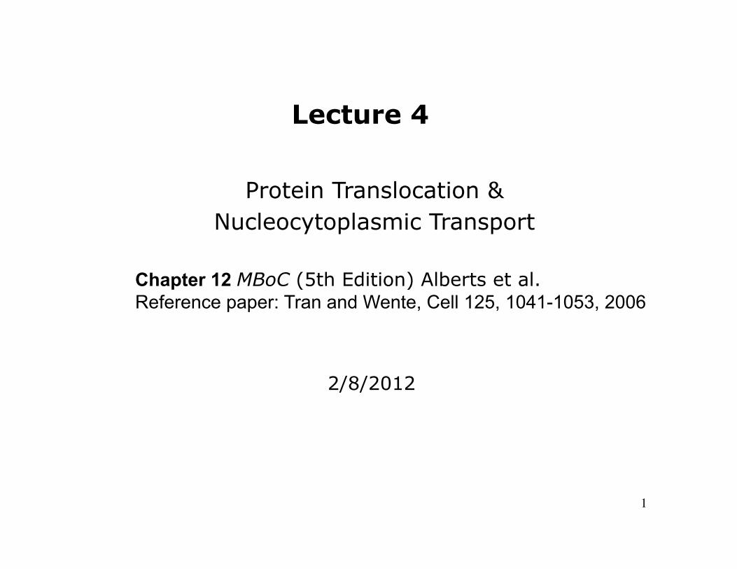
Figure 14-53
The Production of Mitochondrial and Chloroplast proteins by Two Separate Genetic Systems
Most of the proteins in these organelles are encoded by the nucleus and must be imported from the cytosol.

4
The Organization of the Human Mitochondrial genome
The genome contains 2rRNA genes, 22 tRNA genes, and 13 protein-coding genes. The DNAs of many other animal mitochondrial genomes have also been completely sequenced. Most of these animal mitochondrial DNAs encode precisely the same genes as humans, with the gene order being identical for animals that range from fish to mammals.
Figure 14-60
5
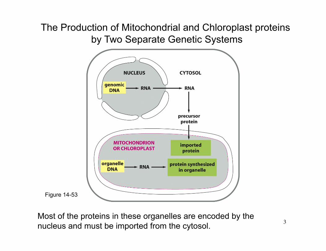
Protein Transport into Mitochondria Matrix
TOM: translocator in the outer membrane of mitochondria TIM: translocator in the inner membrane of mitochondria
Figure 12-25
Mitochondria proteins have a signal peptide. Most proteins are too large to fit through pore and are unfolded by hsp70. Mitochondria proteins has specialized membrane proteins to assist the translocation. The signal peptide binds receptor on outer membrane. The receptor/protein complex diffuses until it reaches a contact site. When a mitochondria matrix protein is translocated into matrix by passing through two
membranes, the signal peptide is cleaved.
To get to the inner membrane or the intermembrane space, a protein is translocated out of matrix.

6
Figure 12-23
The Protein Translocators in the Mitochondrial Membranes
TOM: required for the import of all nucleus-encoded mitochondrial proteins SAM: helps mitochondrian proteins to fold properly in the outer membrane. TIM23: transports some soluble proteins into the matrix space and helps to insert transmembrane proteins into the inner membrane. TIM22: mediates the insertion of a subclass of inner membrane proteins, including the transporter that moves ADP, ATP, and phosphate in and out of mitochondria.
OXA: mediates the insertion of those inner membrane proteins that are synthesized within mitochondria and some imported inner membrane proteins.
7
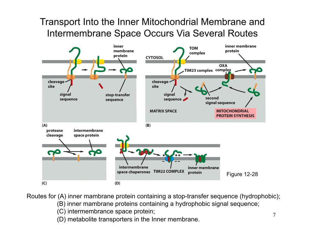
Routes for (A) inner mambrane protein containing a stop-transfer sequence (hydrophobic); (B) inner mambrane proteins containing a hydrophobic signal sequence; (C) intermembrance space protein; (D) metabolite transporters in the Inner membrane.
Transport Into the Inner Mitochondrial Membrane and Intermembrane Space Occurs Via Several Routes
Figure 12-28
8
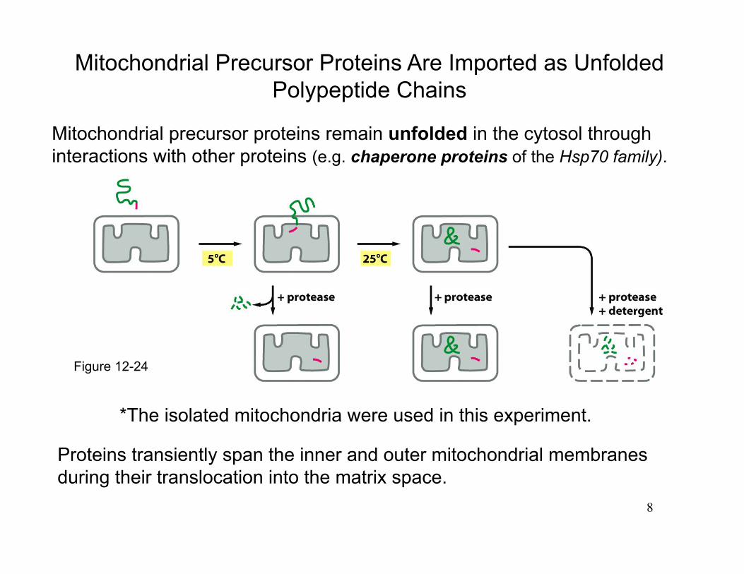
Mitochondrial Precursor Proteins Are Imported as Unfolded Polypeptide Chains
Proteins transiently span the inner and outer mitochondrial membranes during their translocation into the matrix space.
Figure 12-24
Mitochondrial precursor proteins remain unfolded in the cytosol through interactions with other proteins (e.g. chaperone proteins of the Hsp70 family).
*The isolated mitochondria were used in this experiment.
9
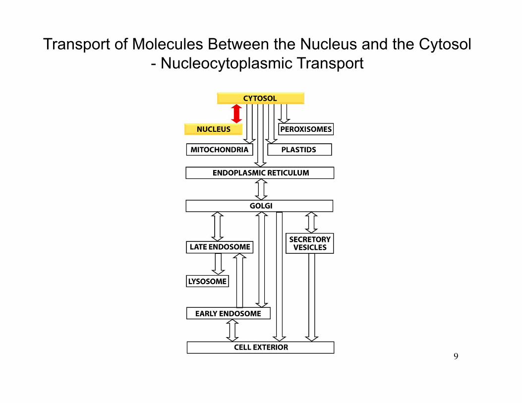
Transport of Molecules Between the Nucleus and the Cytosol - Nucleocytoplasmic Transport
10
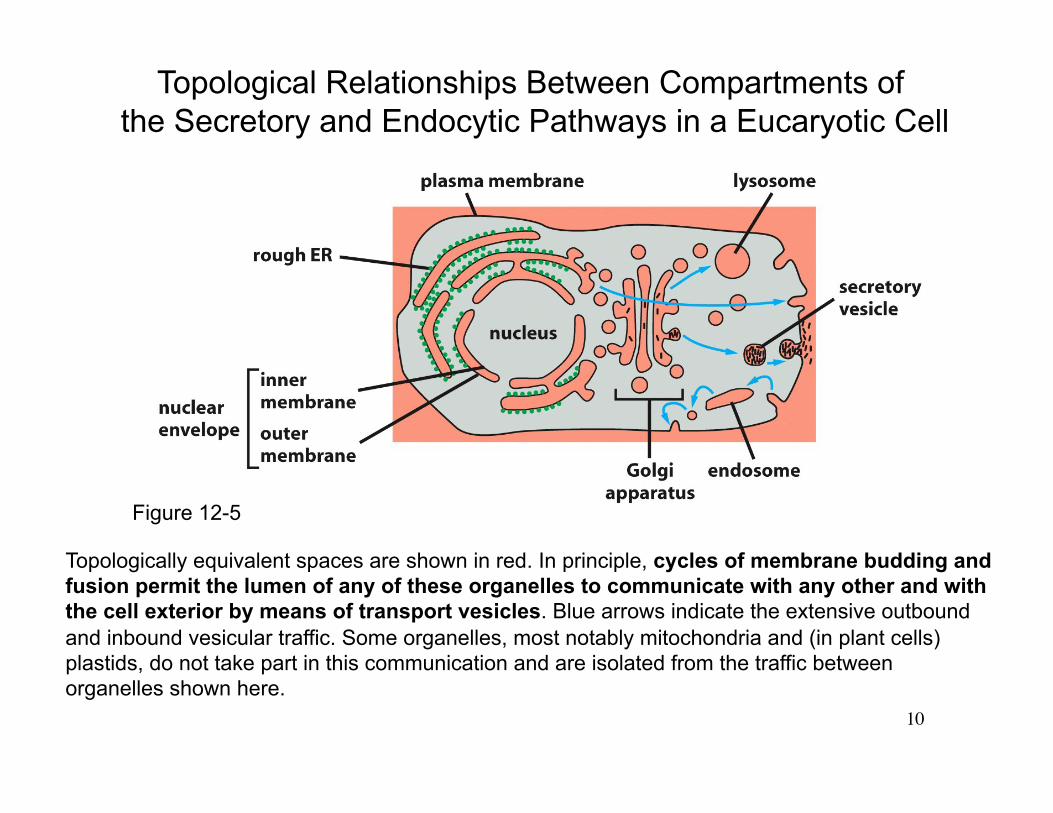
Topologically equivalent spaces are shown in red. In principle, cycles of membrane budding and fusion permit the lumen of any of these organelles to communicate with any other and with the cell exterior by means of transport vesicles. Blue arrows indicate the extensive outbound and inbound vesicular traffic. Some organelles, most notably mitochondria and (in plant cells) plastids, do not take part in this communication and are isolated from the traffic between organelles shown here.
Topological Relationships Between Compartments of the Secretory and Endocytic Pathways in a Eucaryotic Cell
Figure 12-5
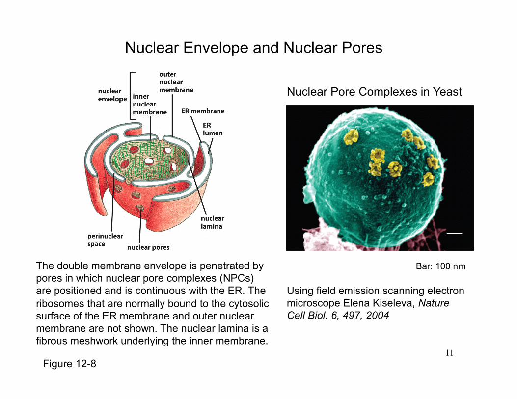
11
The double membrane envelope is penetrated by pores in which nuclear pore complexes (NPCs) are positioned and is continuous with the ER. The ribosomes that are normally bound to the cytosolic surface of the ER membrane and outer nuclear membrane are not shown. The nuclear lamina is a fibrous meshwork underlying the inner membrane.
Bar: 100 nm
Using field emission scanning electron microscope Elena Kiseleva, Nature Cell Biol. 6, 497, 2004
Nuclear Pore Complexes in Yeast
Nuclear Envelope and Nuclear Pores
Figure 12-8

12
(A) A small region of the nuclear envelope (cross section). (B) A scanning electron micrograph of the nuclear side of the nuclear envelope of an oocyte. (C) An electron micrograph showing a side view of two NPCs (brackets). The inner and outer nuclear membranes are continuous at the edges of the pore. (D) An electron micrograph showing face-on views of negatively stained NPCs.
The Arrangement of NPCs in the Nuclear Envelope
Figure 12-9

13
The Nuclear Pore Complex
(a) Cut-away view of the structure of the NPC from Dictyostelium determined by cryo-ET (electron tomogram). The central plug or transporter is transparently rendered. (b) Schematic diagram of nucleoporin localization within the metazoan NPC. Biochemically characterized subcomplexes are boxed. Note that this two-dimensional representation is simplified and not exhaustive.
Schwartz t. Current Opinion in Structural Biology 2005, 15:221–226
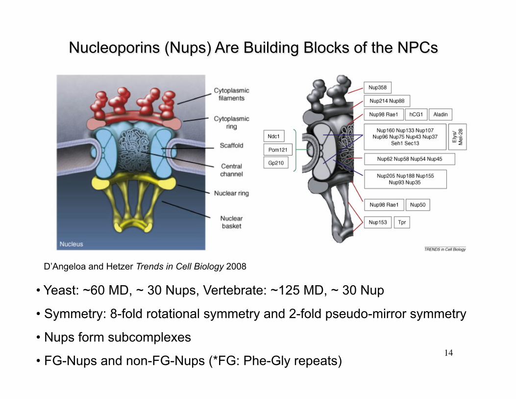
14
• Yeast: ~60 MD, ~ 30 Nups, Vertebrate: ~125 MD, ~ 30 Nup
• Symmetry: 8-fold rotational symmetry and 2-fold pseudo-mirror symmetry
• Nups form subcomplexes
• FG-Nups and non-FG-Nups (*FG: Phe-Gly repeats)
D’Angeloa and Hetzer Trends in Cell Biology 2008

15 Tran and Wente, Cell 125, 1041-1053, 2006
Nuclear Pore Complex (NPC) Assembly and the Dynamics of Nucleoporins Are Correlated with their Proposed Functions
within the NPC
A cross-section of an NPC is shown, with the central region magnified in the right three panels. The order of Nup assembly (late to early) into NPCs following mitosis and the relative Nup NPC residence time or shuttling activity (stable to transient) are illustrated. The predicted function (structural to transport) for each Nup or subcomplex is also shown. Common colors between the three figures in each structure indicate correlations in assembly, dynamics, and function. Those that are early in assembly and stable in residence time are likely structural in function. In contrast, those that are late in assembly and transient in residence time are likely directly involved in transport.
16
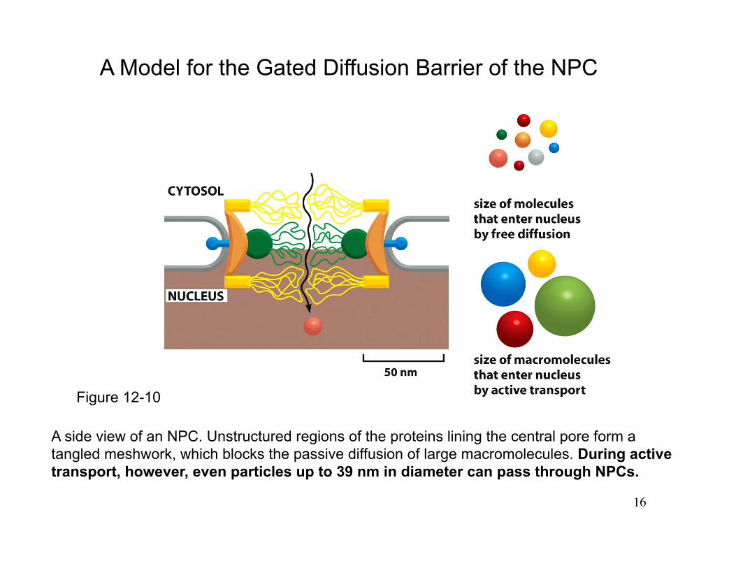
A side view of an NPC. Unstructured regions of the proteins lining the central pore form a tangled meshwork, which blocks the passive diffusion of large macromolecules. During active transport, however, even particles up to 39 nm in diameter can pass through NPCs.
A Model for the Gated Diffusion Barrier of the NPC
Figure 12-10
17
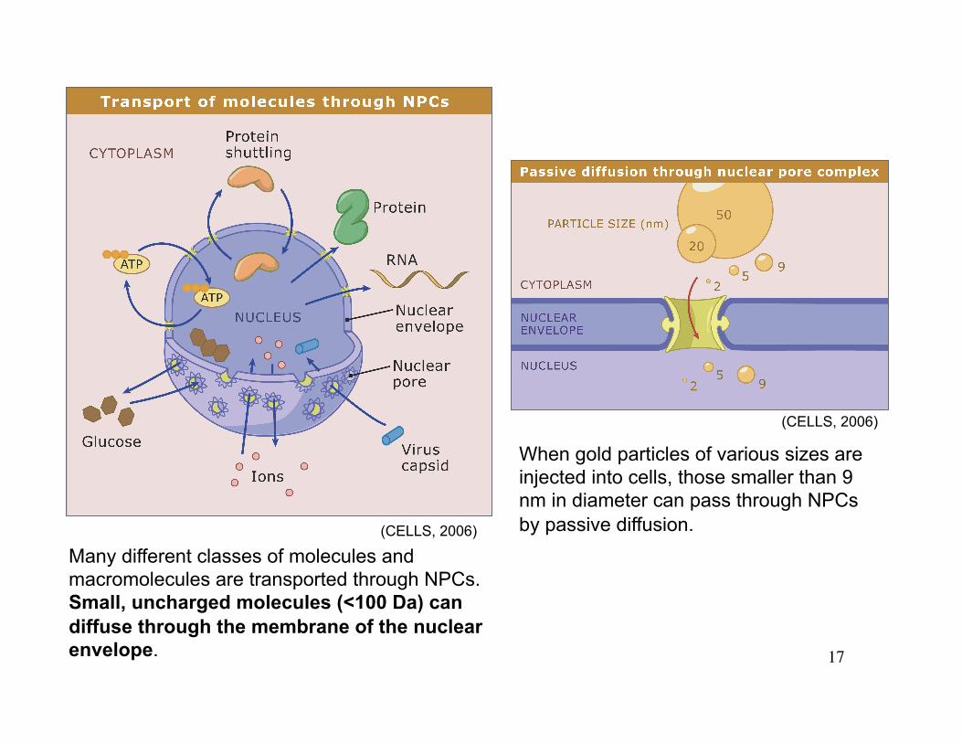
Many different classes of molecules and macromolecules are transported through NPCs. Small, uncharged molecules (<100 Da) can diffuse through the membrane of the nuclear envelope.
When gold particles of various sizes are injected into cells, those smaller than 9 nm in diameter can pass through NPCs by passive diffusion. (CELLS, 2006)
(CELLS, 2006)

18
(Samuel Dales, Blobel Lab)
Bidirectional Nucleocytoplasmic Transport
19
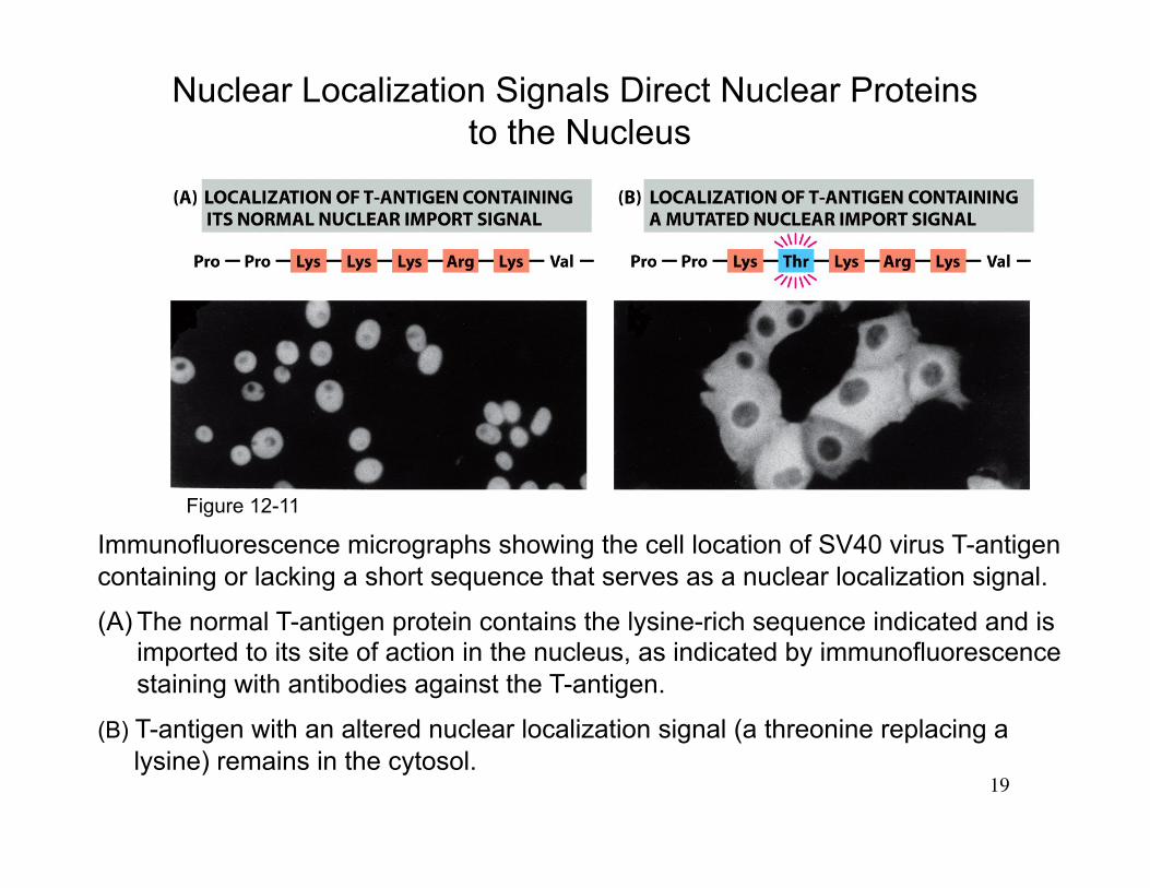
Immunofluorescence micrographs showing the cell location of SV40 virus T-antigen containing or lacking a short sequence that serves as a nuclear localization signal.
(A) The normal T-antigen protein contains the lysine-rich sequence indicated and is imported to its site of action in the nucleus, as indicated by immunofluorescence staining with antibodies against the T-antigen.
(B) T-antigen with an altered nuclear localization signal (a threonine replacing a lysine) remains in the cytosol.
Nuclear Localization Signals Direct Nuclear Proteins to the Nucleus
Figure 12-11
20
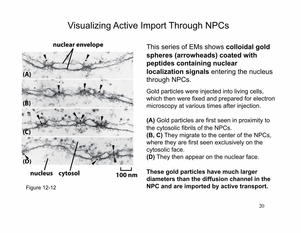
This series of EMs shows colloidal gold spheres (arrowheads) coated with peptides containing nuclear localization signals entering the nucleus through NPCs.
Gold particles were injected into living cells, which then were fixed and prepared for electron microscopy at various times after injection.
(A) Gold particles are first seen in proximity to the cytosolic fibrils of the NPCs. (B, C) They migrate to the center of the NPCs, where they are first seen exclusively on the cytosolic face. (D) They then appear on the nuclear face.
These gold particles have much larger diameters than the diffusion channel in the NPC and are imported by active transport.
Visualizing Active Import Through NPCs
Figure 12-12

21 Ref: Au and Pante, Journal of Structural Biology, 2012, 177 (1): 90-8.
Nuclear transport of baculovirus: Revealing the nuclear pore complex passage
EM of NPC cross-sections from Xenopus oocytes microinjected with baculovirus AcMNPV capsid and incubated at room temperature for 3.5 h. Capsids of 250–300 nm in length are seen traversing the NPCs. Capsids appear fully intact in its native conformation while crossing the NPC. Note the capsid in the middle panel appear shorter due to the variability in the length of these capsids. Bar, 100 nm. n, nucleus; c, cytoplasm.
EM of Xenopus oocytes microinjected with WGA-gold into either the cytoplasm (A) or nucleus (B), incubated at room temperature for 2 h, followed by cytoplasmic injection of baculovirus AcMNPV capsids and further incubated for 8 h. When NPCs were inhibited by WGA-gold particles, capsids remained interacting with the NPC cytoplasmic filaments 8 h post micro-injection. No capsids were found inside the nucleus when NPCs were inhibited with WGA. Bar, 200 nm. n, nucleus; c, cytoplasm. Black arrows point to capsids and white arrowheads point to WGA-gold particles at NPCs.
22
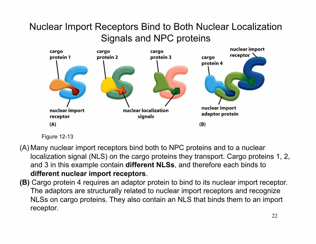
(A) Many nuclear import receptors bind both to NPC proteins and to a nuclear localization signal (NLS) on the cargo proteins they transport. Cargo proteins 1, 2, and 3 in this example contain different NLSs, and therefore each binds to different nuclear import receptors.
(B) Cargo protein 4 requires an adaptor protein to bind to its nuclear import receptor. The adaptors are structurally related to nuclear import receptors and recognize NLSs on cargo proteins. They also contain an NLS that binds them to an import receptor.
Nuclear Import Receptors Bind to Both Nuclear Localization Signals and NPC proteins
Figure 12-13

23
Proteins in Kapβ Receptor Family
Chook and Blobel, Curr Opin Struct Biol. 2001.11(6):703-15.

24
The compartmentalization of Ran-GDP and Ran-GTP. Localization of Ran-GDP in the cytosol and Ran-GTP in the nucleus results from the localization of two Ran regulatory proteins: Ran GTPase-activating protein (Ran-GAP) is located in the cytosol and Ran guanine nucleotide exchange factor (Ran-GEF) binds to chromatin and is therefore located in the nucleus.
* Ran-REF is also call RCC1 (regulator of chromosome condensation 1).
The Ran GTPase Imposes Directionality on Transport Through NPCs
Figure 12-14

25 Figure 12-15
Movement through the NPC of loaded nuclear transport receptors occurs along the FG-repeats displayed by certain NPC proteins. The differential localization of Ran-GTP in the nucleus and Ran-GDP in the cytosol provides directionality (red arrows) to both nuclear import (left) and nuclear export (right).
Ran-GAP stimulates the hydrolysis of GTP to produce Ran-GDP on the cytosolic side of the NPC.
Ran-GDP is imported into the nucleus by its own import receptor, which is specific for the GDP-bound conformation of Ran. The Ran-GDP receptor is structurally unrelated to the main family of nuclear transport receptors. However, it also binds to FG-repeats in NPC proteins and hops across the NPC.
A Model Explaining How GTP Hydrolysis by Ran in the Cytosol Provides Directionality to Nuclear Transport

26 Xylourgidis and Fornerod, Dev. Cell 2009
27
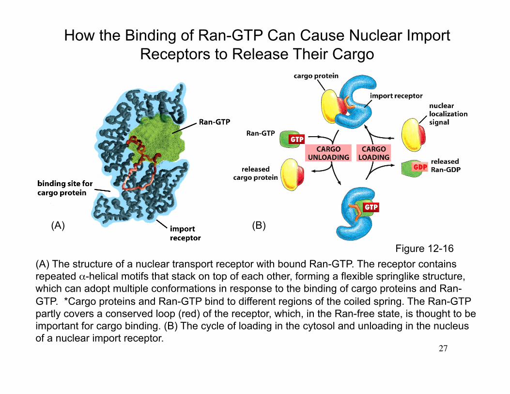
(A) The structure of a nuclear transport receptor with bound Ran-GTP. The receptor contains repeated α-helical motifs that stack on top of each other, forming a flexible springlike structure, which can adopt multiple conformations in response to the binding of cargo proteins and Ran-GTP. *Cargo proteins and Ran-GTP bind to different regions of the coiled spring. The Ran-GTP partly covers a conserved loop (red) of the receptor, which, in the Ran-free state, is thought to be important for cargo binding. (B) The cycle of loading in the cytosol and unloading in the nucleus of a nuclear import receptor.
How the Binding of Ran-GTP Can Cause Nuclear Import Receptors to Release Their Cargo
(A) (B)
Figure 12-16

28
Figure 12-18
The nuclear factor of activated T cells (NF-AT) is a gene regulatory protein that, in the resting T cell, is found in the cytosol in a phosphorylated state. When T cells are activated by foreign antigen, the intracellular Ca2+ concentration increases. In high Ca2+, the protein phosphatase calcineurin binds to NF-AT and dephosphorylates it. The dephosphorylation exposes nuclear import signals and blocks a nuclear export signal. The complex of NF-AT and calcineurin is then imported into the nucleus, where NF-AT activates the transcription of numerous genes required for T cell activation. The response shuts off when Ca2+ levels decrease, releasing NF-AT from calcineurin. Rephosphorylation of NF-AT inactivates the nuclear import signals and re-exposes the nuclear export signal, causing NF-AT to relocate to the cytosol.
The Control of Nuclear Import During T Cell Activation
Some of the most potent immunosuppressive drugs, including cyclosporin A and FK506, inhibit the ability of calcineurin to dephosphorylate NF-AT and thereby block the nuclear accumulation of NF-AT and T cell activation.
29
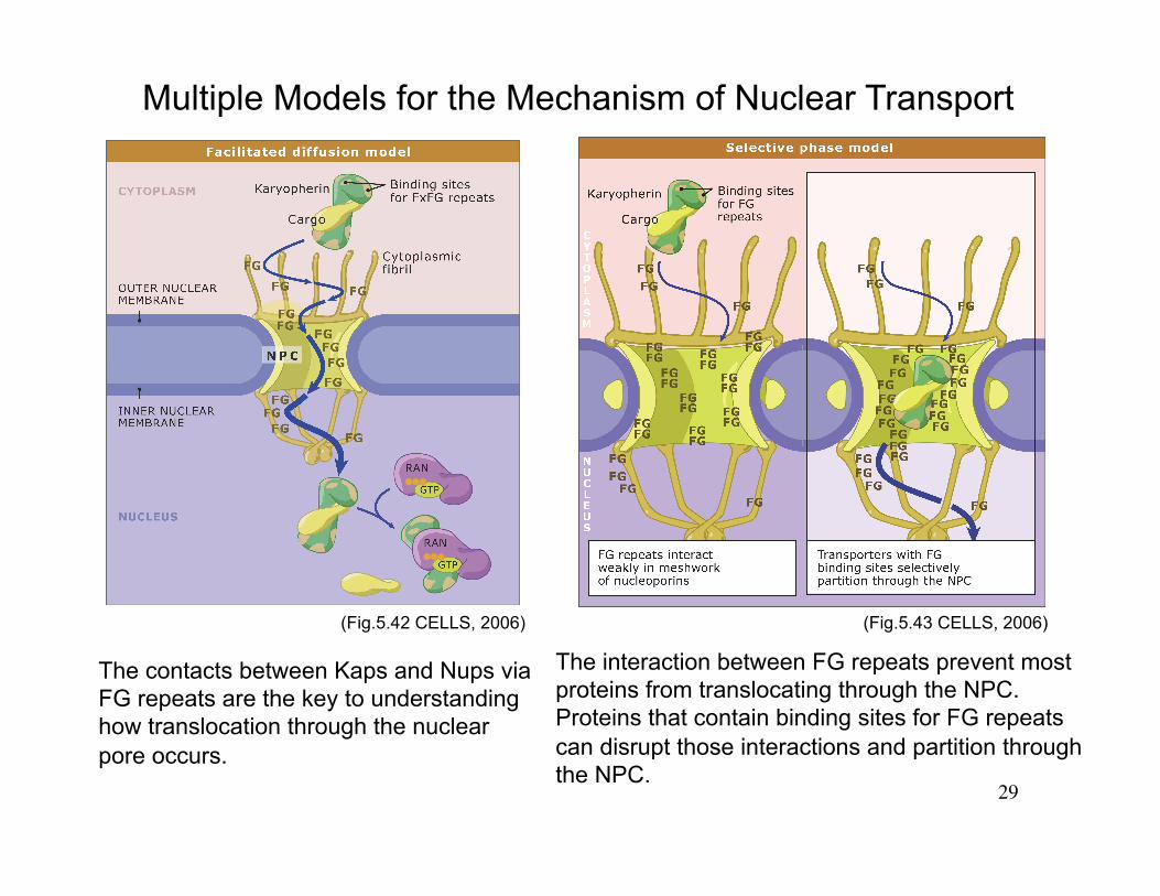
Multiple Models for the Mechanism of Nuclear Transport
The contacts between Kaps and Nups via FG repeats are the key to understanding how translocation through the nuclear pore occurs.
The interaction between FG repeats prevent most proteins from translocating through the NPC. Proteins that contain binding sites for FG repeats can disrupt those interactions and partition through the NPC.
(Fig.5.42 CELLS, 2006) (Fig.5.43 CELLS, 2006)


![Molecular biology of the cell : [MBOC]Molecular Biology Beganwith aSpotlight on£ coli 22 Summary 22 GENETIC INFORMATION IN EUKARYOTES 23 Eukaryotic Cells MayHaveOriginated asPredators](https://img.pdfslide.us/doc/110x75/5f0dcac57e708231d43c1bd0/molecular-biology-of-the-cell-mboc-molecular-biology-beganwith-aspotlight-on.jpg)