Embed Size (px)
Citation preview
Lecture 2
Molecular Structure of DNA and RNA part 2
Chapter 9, pages 237 - 250
9-31Copyright ©The McGraw-Hill Companies, Inc. Permission required for reproduction or display
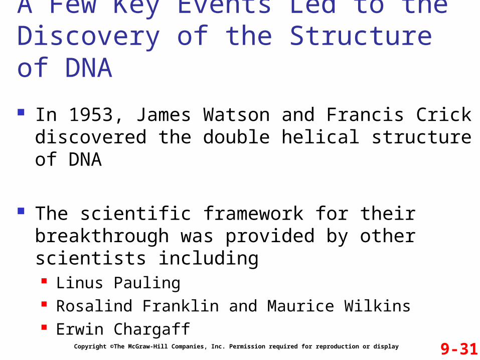
In 1953, James Watson and Francis Crick discovered the double helical structure of DNA
The scientific framework for their breakthrough was provided by other scientists including Linus Pauling Rosalind Franklin and Maurice Wilkins Erwin Chargaff
A Few Key Events Led to the Discovery of the Structure of DNA
9-32Copyright ©The McGraw-Hill Companies, Inc. Permission required for reproduction or display
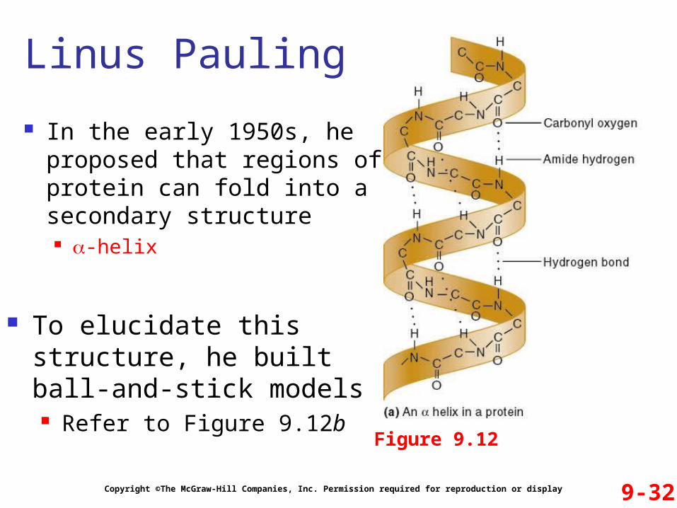
In the early 1950s, he proposed that regions of protein can fold into a secondary structure -helix
Linus Pauling
Figure 9.12
To elucidate this structure, he built ball-and-stick models Refer to Figure 9.12b
9-33Copyright ©The McGraw-Hill Companies, Inc. Permission required for reproduction or display
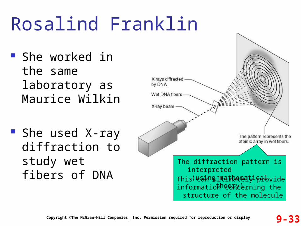
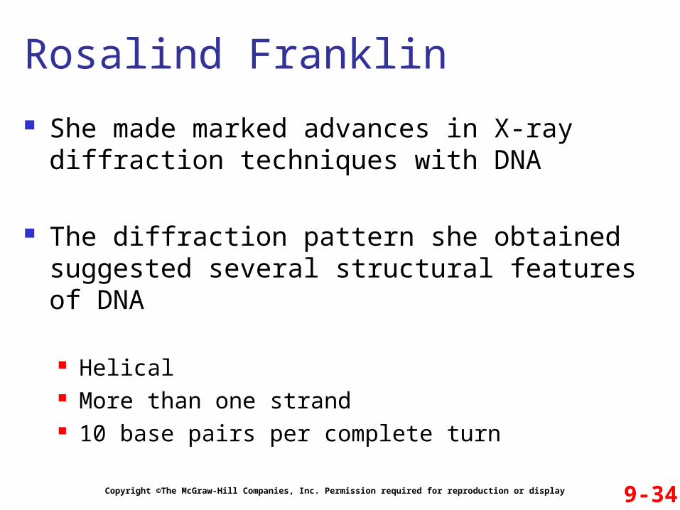
She worked in the same laboratory as Maurice Wilkins
She used X-ray diffraction to study wet fibers of DNA
Rosalind Franklin
The diffraction pattern is interpreted (using mathematical theory)
This can ultimately provide information concerning the
structure of the molecule
9-34Copyright ©The McGraw-Hill Companies, Inc. Permission required for reproduction or display
She made marked advances in X-ray diffraction techniques with DNA
The diffraction pattern she obtained suggested several structural features of DNA
Helical More than one strand 10 base pairs per complete turn
Rosalind Franklin
9-35Copyright ©The McGraw-Hill Companies, Inc. Permission required for reproduction or display
Chargaff pioneered many of the biochemical techniques for the isolation, purification and measurement of nucleic acids from living cells
It was already known then that DNA contained the four bases: A, G, C and T
Erwin Chargaff’s Experiment
The Hypothesis An analysis of the base composition of DNA in
different species may reveal important features about the structure of DNA
9-36Copyright ©The McGraw-Hill Companies, Inc. Permission required for reproduction or display
Testing the Hypothesis
Refer to Figure 9.14
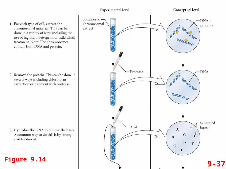
9-37Figure 9.14
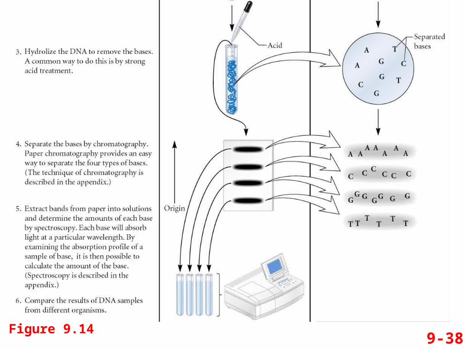
9-38Figure 9.14
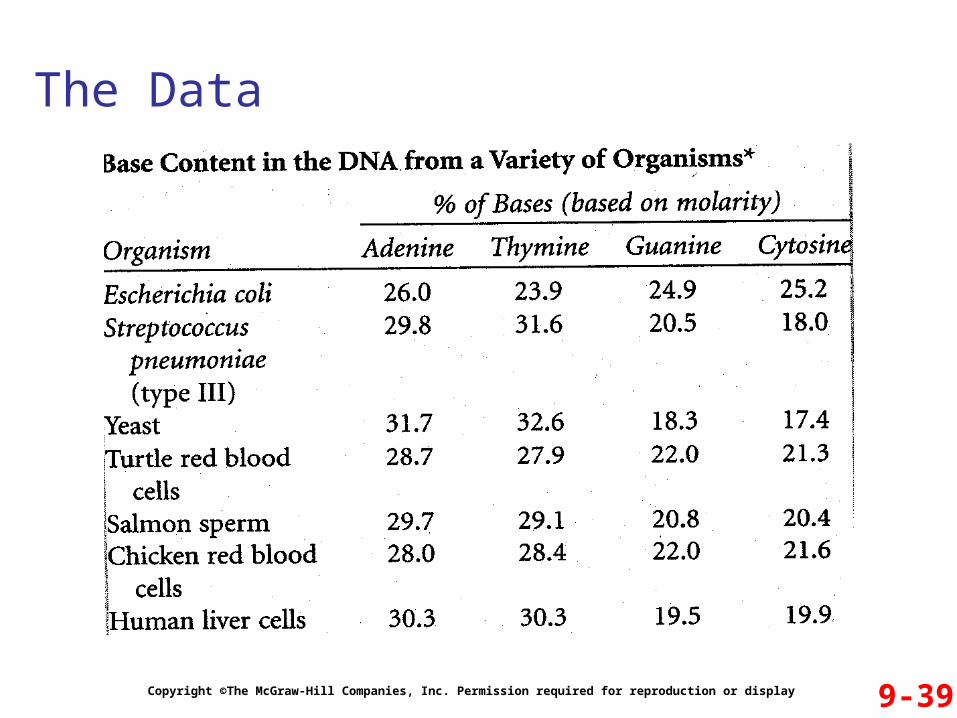
The Data
9-39Copyright ©The McGraw-Hill Companies, Inc. Permission required for reproduction or display
Interpreting the Data
9-40Copyright ©The McGraw-Hill Companies, Inc. Permission required for reproduction or display
The data shown in Figure 9.14 are only a small sampling of Chargaff’s results
The compelling observation was that Percent of adenine = percent of thymine Percent of cytosine = percent of guanine
This observation became known as Chargaff’s rule It was crucial evidence that Watson and Crick used to
elucidate the structure of DNA
9-41Copyright ©The McGraw-Hill Companies, Inc. Permission required for reproduction or display
Familiar with all of these key observations, Watson and Crick set out to solve the structure of DNA They tried to build ball-and-stick models that incorporated
all known experimental observations
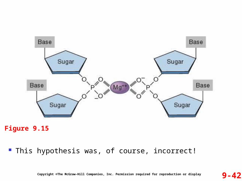
A critical question was how the two (or more strands) would interact An early hypothesis proposed that the strands interact
through phosphate-Mg++ crosslinks Refer to Figure 9.15
Watson and Crick
9-42Copyright ©The McGraw-Hill Companies, Inc. Permission required for reproduction or display
Figure 9.15
This hypothesis was, of course, incorrect!
9-43Copyright ©The McGraw-Hill Companies, Inc. Permission required for reproduction or display
They went back to the ball-and-stick units They then built models with the
Sugar-phosphate backbone on the outside Bases projecting toward each other
They first considered a structure in which bases form H bonds with identical bases in the opposite strand ie., A to A, T to T, C to C, and G to G
Model building revealed that this also was incorrect
Watson and Crick
9-44Copyright ©The McGraw-Hill Companies, Inc. Permission required for reproduction or display
They then realized that the hydrogen bonding of A and T resembled that between C and G So they built ball-and-stick models with AT and CG
interactions These were consistent with all known data about DNA structure
Refer to Figure 9.16
Watson, Crick and Maurice Wilkins were awarded the Nobel Prize in 1962 Rosalind Franklin died in 1958, and Nobel prizes are not
awarded posthumously
Watson and Crick
9-45Copyright ©The McGraw-Hill Companies, Inc. Permission required for reproduction or display
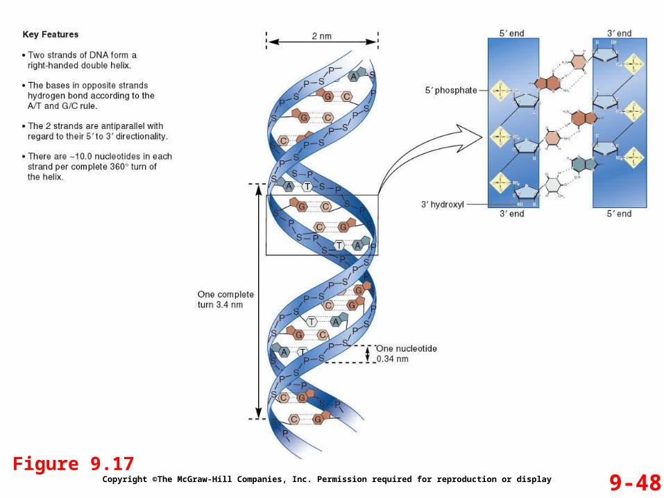
General structural features (Figures 9.17 & 9.18)
The DNA Double Helix
Two strands are twisted together around a common axis
There are 10 bases per complete twist The two strands are antiparallel
One runs in the 5’ to 3’ direction and the other 3’ to 5’ The helix is right-handed
As it spirals away from you, the helix turns in a clockwise direction
9-46Copyright ©The McGraw-Hill Companies, Inc. Permission required for reproduction or display
General structural features (Figures 9.17 & 9.18)
The DNA Double Helix
The double-bonded structure is stabilized by
1. Hydrogen bonding between complementary bases A bonded to T by two hydrogen bonds C bonded to G by three hydrogen bonds
2. Base stacking Within the DNA, the bases are oriented so that the flattened
regions are facing each other
9-47Copyright ©The McGraw-Hill Companies, Inc. Permission required for reproduction or display
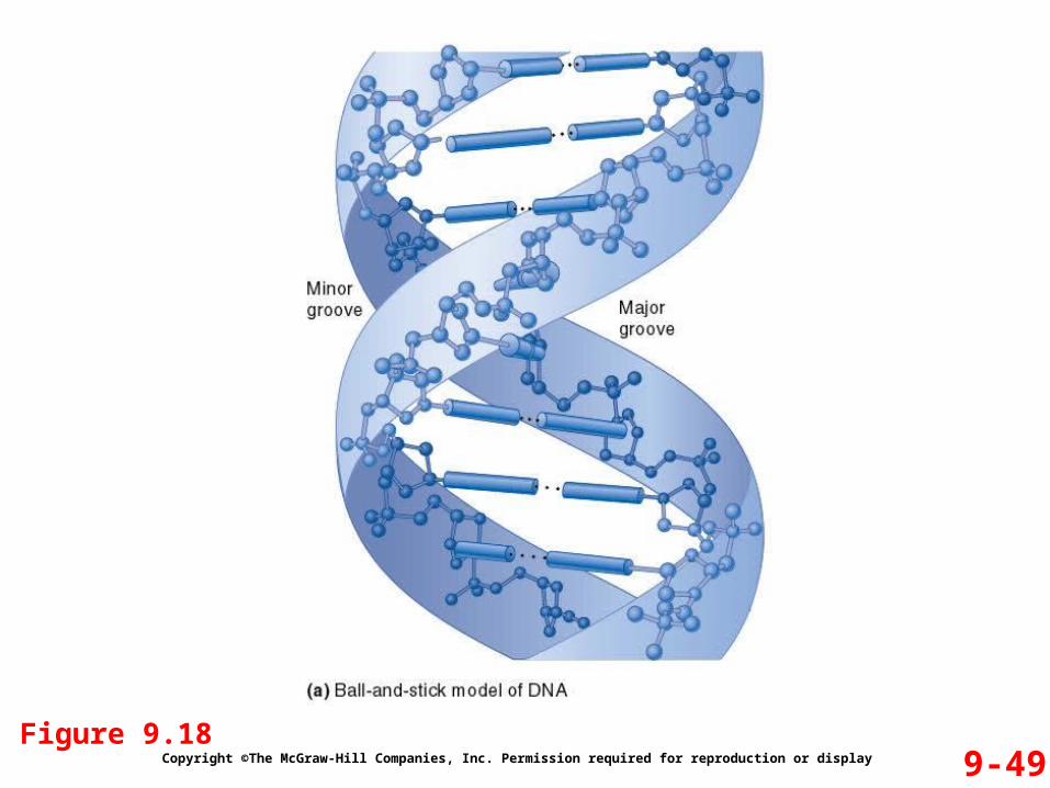
General structural features (Figures 9.17 & 9.18)
The DNA Double Helix
There are two asymmetrical grooves on the outside of the helix
1. Major groove
2. Minor groove
Certain proteins can bind within these grooves They can thus interact with a particular sequence of bases
9-48Copyright ©The McGraw-Hill Companies, Inc. Permission required for reproduction or display
Figure 9.17
9-49Copyright ©The McGraw-Hill Companies, Inc. Permission required for reproduction or display
Figure 9.18
9-50Copyright ©The McGraw-Hill Companies, Inc. Permission required for reproduction or display
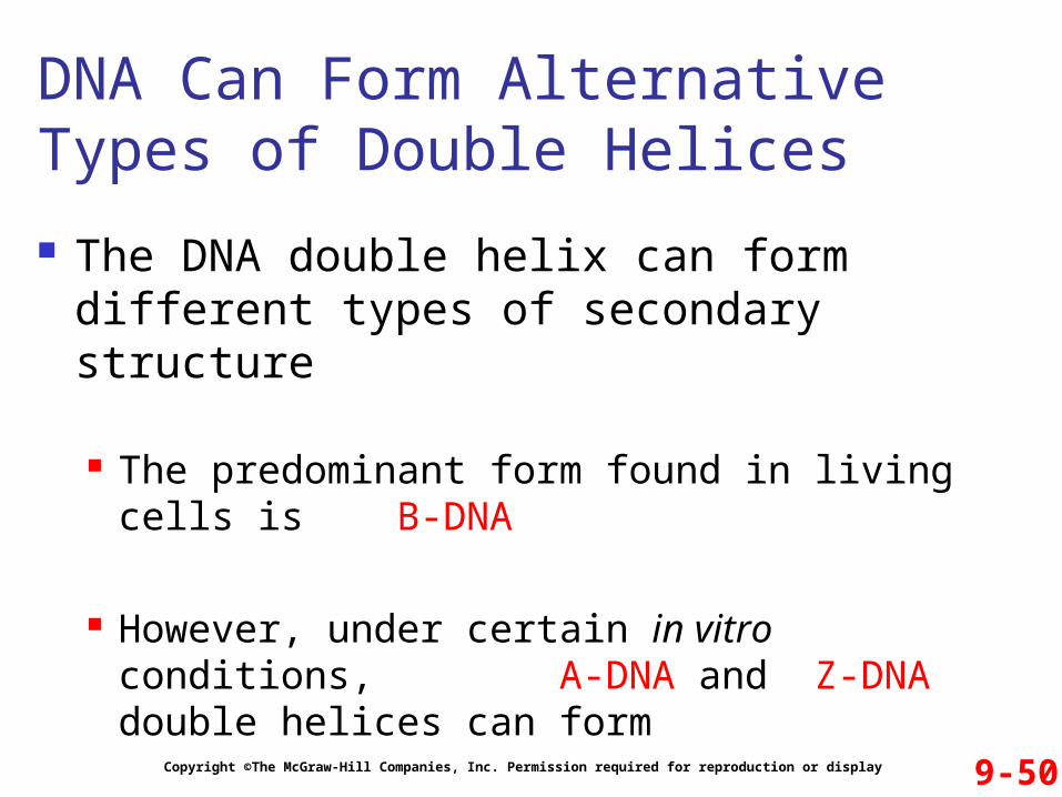
The DNA double helix can form different types of secondary structure
The predominant form found in living cells is B-DNA
However, under certain in vitro conditions, A-DNA and Z-DNA double helices can form
DNA Can Form Alternative Types of Double Helices
9-51Copyright ©The McGraw-Hill Companies, Inc. Permission required for reproduction or display
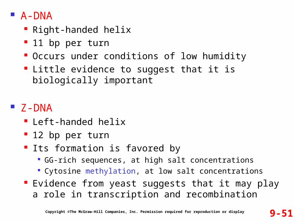
A-DNA Right-handed helix 11 bp per turn Occurs under conditions of low humidity Little evidence to suggest that it is biologically important
Z-DNA Left-handed helix 12 bp per turn Its formation is favored by
GG-rich sequences, at high salt concentrations Cytosine methylation, at low salt concentrations
Evidence from yeast suggests that it may play a role in transcription and recombination
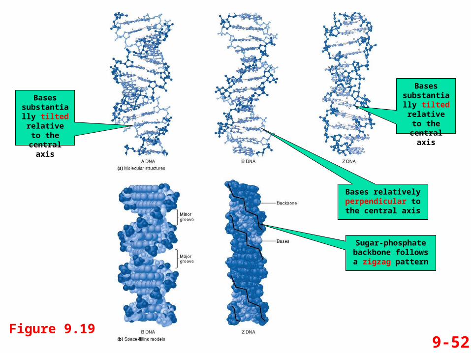
9-52Figure 9.19
Bases substantially tilted relative to the central
axis
Bases substantially tilted relative to the central
axis
Sugar-phosphate backbone follows a
zigzag pattern
Bases relatively perpendicular to the
central axis
9-53Copyright ©The McGraw-Hill Companies, Inc. Permission required for reproduction or display
In the late 1950s, Alexander Rich et al discovered triplex DNA It was formed in vitro using DNA pieces that were made
synthetically
In the 1980s, it was discovered that natural double- stranded DNA can join with a synthetic strand of DNA to form triplex DNA The synthetic strand binds to the major groove of the
naturally-occurring double-stranded DNA Refer to Figure 9.20
DNA Can Form a Triple Helix
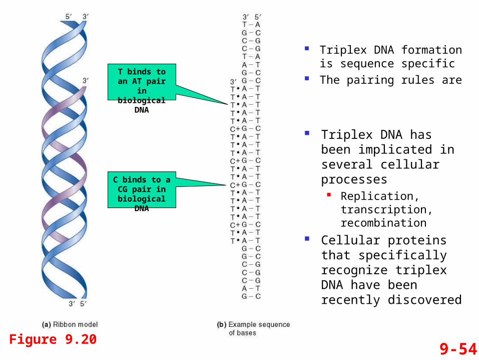
9-54Figure 9.20
Triplex DNA formation is sequence specific
The pairing rules are
Triplex DNA has been implicated in several cellular processes
Replication, transcription, recombination
Cellular proteins that specifically recognize triplex DNA have been recently discovered
T binds to an AT pair in
biological DNA
C binds to a CG pair in
biological DNA
9-55Copyright ©The McGraw-Hill Companies, Inc. Permission required for reproduction or display
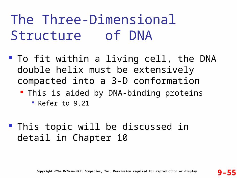
To fit within a living cell, the DNA double helix must be extensively compacted into a 3-D conformation This is aided by DNA-binding proteins
Refer to 9.21
This topic will be discussed in detail in Chapter 10
The Three-Dimensional Structure of DNA
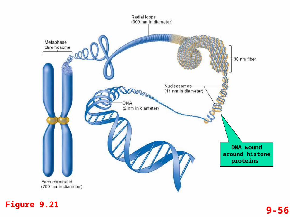
9-56Figure 9.21
DNA wound around histone
proteins
9-57Copyright ©The McGraw-Hill Companies, Inc. Permission required for reproduction or display
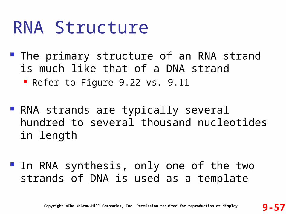
The primary structure of an RNA strand is much like that of a DNA strand Refer to Figure 9.22 vs. 9.11
RNA strands are typically several hundred to several thousand nucleotides in length
In RNA synthesis, only one of the two strands of DNA is used as a template
RNA Structure
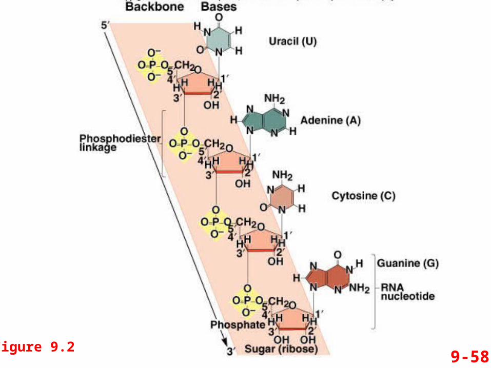
9-58Figure 9.22
9-59Copyright ©The McGraw-Hill Companies, Inc. Permission required for reproduction or display
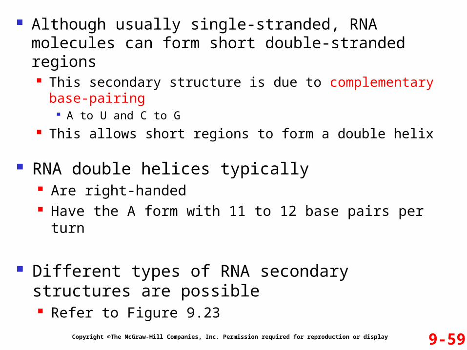
Although usually single-stranded, RNA molecules can form short double-stranded regions This secondary structure is due to complementary base-
pairing A to U and C to G
This allows short regions to form a double helix
RNA double helices typically Are right-handed Have the A form with 11 to 12 base pairs per turn
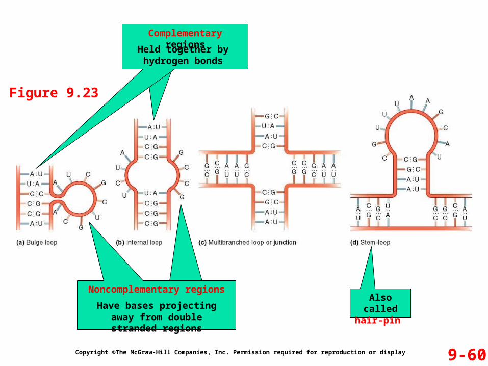
Different types of RNA secondary structures are possible Refer to Figure 9.23
9-60Copyright ©The McGraw-Hill Companies, Inc. Permission required for reproduction or display
Figure 9.23
Also called hair-pin
Complementary regions
Noncomplementary regions
Held together by hydrogen bonds
Have bases projecting away from double stranded regions
9-61
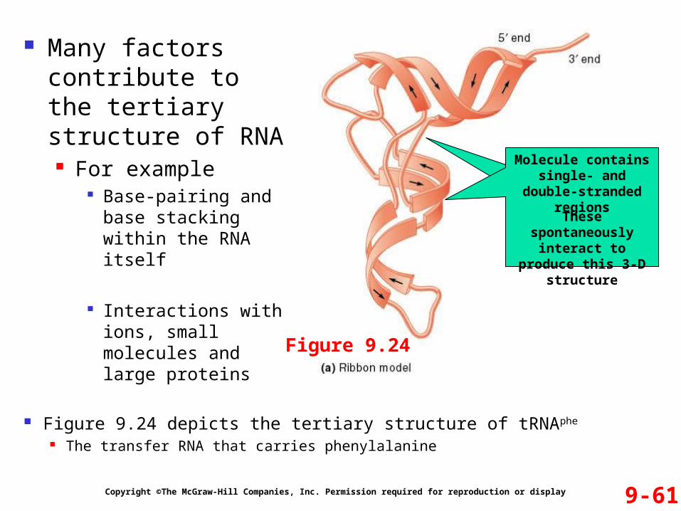
Many factors contribute to the tertiary structure of RNA For example
Base-pairing and base stacking within the RNA itself
Interactions with ions, small molecules and large proteins
Copyright ©The McGraw-Hill Companies, Inc. Permission required for reproduction or display
Figure 9.24 depicts the tertiary structure of tRNAphe
The transfer RNA that carries phenylalanine
Molecule contains single- and double-stranded regions
These spontaneously interact to produce this 3-D structure
Figure 9.24