Embed Size (px)
Citation preview
Lecture 17
– Exams in Chemistry office with M’Lis. Please show your ID to her to pick up your exam.
– Quiz on Friday– Enzyme mechanisms
Terms to review for enzymes
• Cofactor
• Coenzyme
• Prosthetic group
• Holoenzyme
• Apoenzyme
• Lock and Key
• Transition analog model
• Induced fit
• Active site, binding site, recognition site, catalytic site

Catalytic Mechanisms
• Acid-base catalysis
• Covalent catalysis
• Metal ion catalysis
• Proximity and orientation effects (ex. anhydride)
• Preferential binding of the transition state complex
General Acid-Base Catalysis
• Large number of possible amino acids
• Requires that they can accept and donate a proton
• Glu, Asp
• Lys, His, Arg
• Cys, Ser, Thr
• Also can include metal cofactors (metal ion catalysis)
• Example can be observed in RNAse
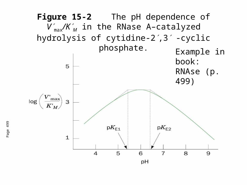
Figure 15-2 The pH dependence of Vmax/KM in the RNase A–catalyzed hydrolysis of cytidine-2,3 -cyclic
phosphate.
Pag
e 49
9
Example in book: RNAse (p. 499)
Pag
e 49
9
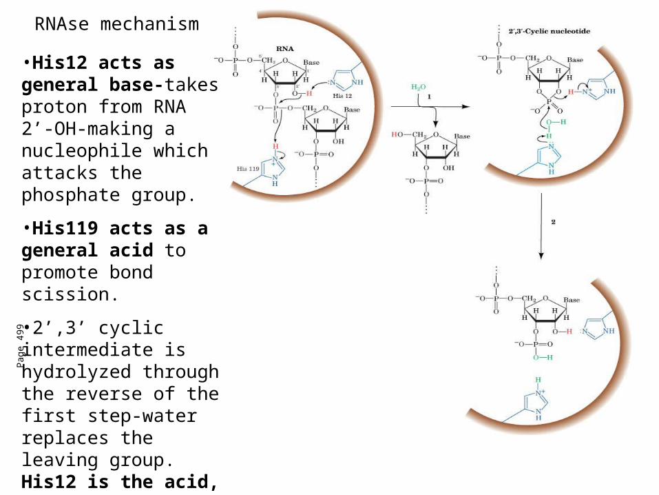
•His12 acts as general base-takes proton from RNA 2’-OH-making a nucleophile which attacks the phosphate group.
•His119 acts as a general acid to promote bond scission.
•2’,3’ cyclic intermediate is hydrolyzed through the reverse of the first step-water replaces the leaving group. His12 is the acid, His119 acts as the base
RNAse mechanism

Covalent catalysis
• Rate acceleration through the transient formation of a catalyst-substrate covalent bond.
• Example-decarboxylation of acetoacetate by primary amines
• Amine nucleophilically attacks carbonyl group of acetoacetate to form a Schiff base (imine bound)
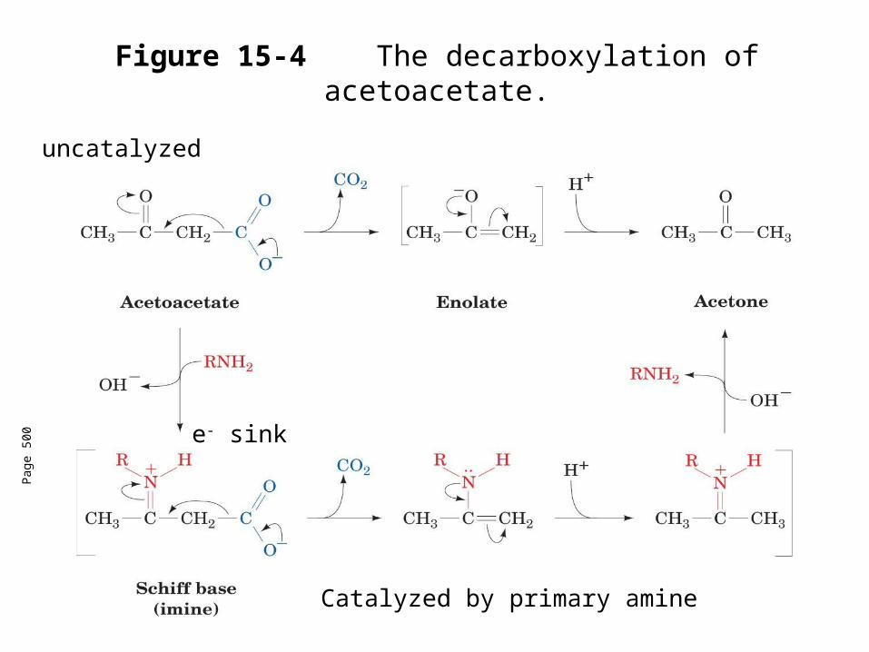
Figure 15-4 The decarboxylation of acetoacetate.
Pag
e 50
0 e- sink
uncatalyzed
Catalyzed by primary amine

Covalent catalysis
• Made up of three stages
1. The nucleophilic reaction between the catalyst and the substrate to form a covalent bond.
2. The withdrawal of electrons from the reaction center by the now electrophilic catalyst
3. The elimination of the catalyst (reverse of 1.)
• Nucleophilic catalysis - covalent bond formation is limiting.
• Electrophilic catalysis-withdrawal of electrons is rate limiting
Covalent catalysis
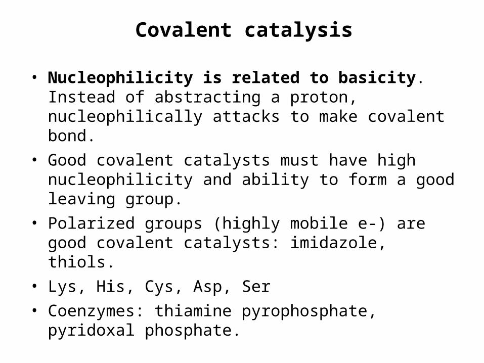
• Nucleophilicity is related to basicity. Instead of abstracting a proton, nucleophilically attacks to make covalent bond.
• Good covalent catalysts must have high nucleophilicity and ability to form a good leaving group.
• Polarized groups (highly mobile e-) are good covalent catalysts: imidazole, thiols.
• Lys, His, Cys, Asp, Ser
• Coenzymes: thiamine pyrophosphate, pyridoxal phosphate.
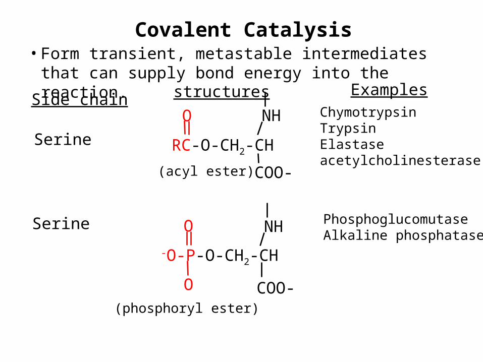
Covalent Catalysis• Form transient, metastable intermediates that can supply
bond energy into the reaction.
Serine
Side chainNH
RC-O-CH2-CH
O
COO-(acyl ester)
ChymotrypsinTrypsinElastaseacetylcholinesterase
structures Examples
Serine
-O-P-O-CH2-CH
O
(phosphoryl ester)
O
NH
COO-
PhosphoglucomutaseAlkaline phosphatase
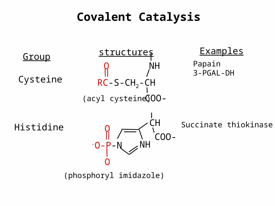
Covalent Catalysis
Cysteine
GroupNH
RC-S-CH2-CH
O
COO-(acyl cysteine)
Papain3-PGAL-DH
structures Examples
Histidine
-O-P-N
O
(phosphoryl imidazole)
O
NHCOO-
Succinate thiokinase CH
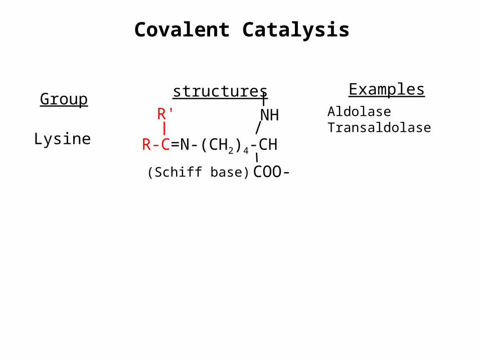
Covalent Catalysis
Lysine
GroupNH
R-C=N-(CH2)4-CH
R'
COO-(Schiff base)
AldolaseTransaldolase
structures Examples

Metal ion catalysis
• Almost 1/3 of all enzymes use metal ions for catalytic activity. 2 main types:
1. Metalloenzymes-have tightly bound metal ions, mmost commonly transition metal ions such as Fe2+, Fe3+, Cu2+, Zn2+, Mn2+, or Co3+
2. Metal-activated enzymes-loosely bind metal ions form solution-usually alkali or alkaline earth metals-Na+, K+, Ca2+
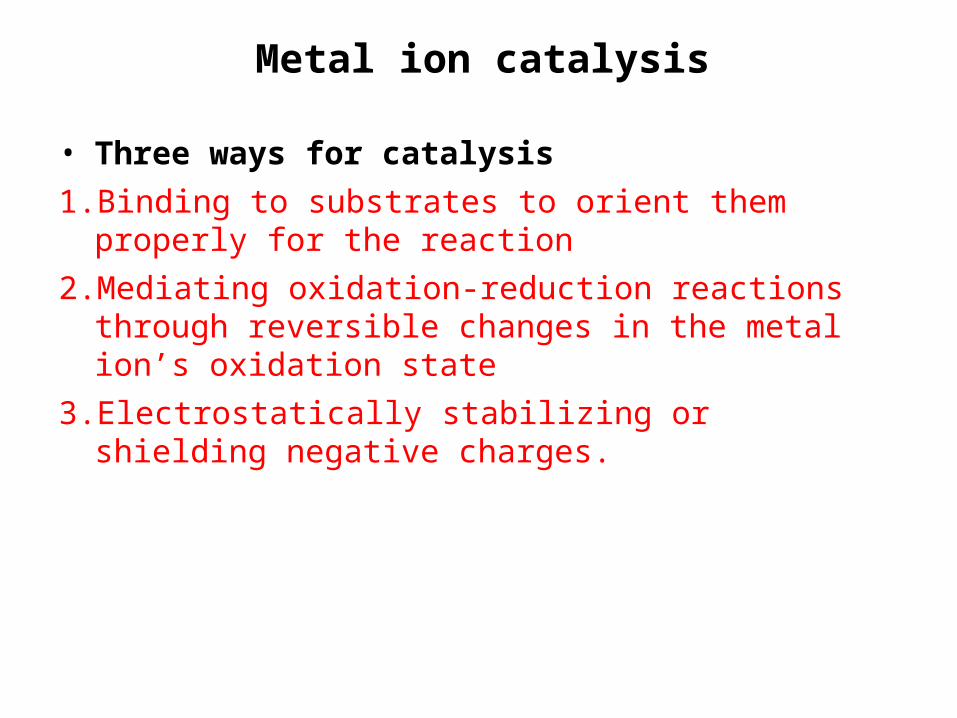
Metal ion catalysis
• Three ways for catalysis
1. Binding to substrates to orient them properly for the reaction
2. Mediating oxidation-reduction reactions through reversible changes in the metal ion’s oxidation state
3. Electrostatically stabilizing or shielding negative charges.
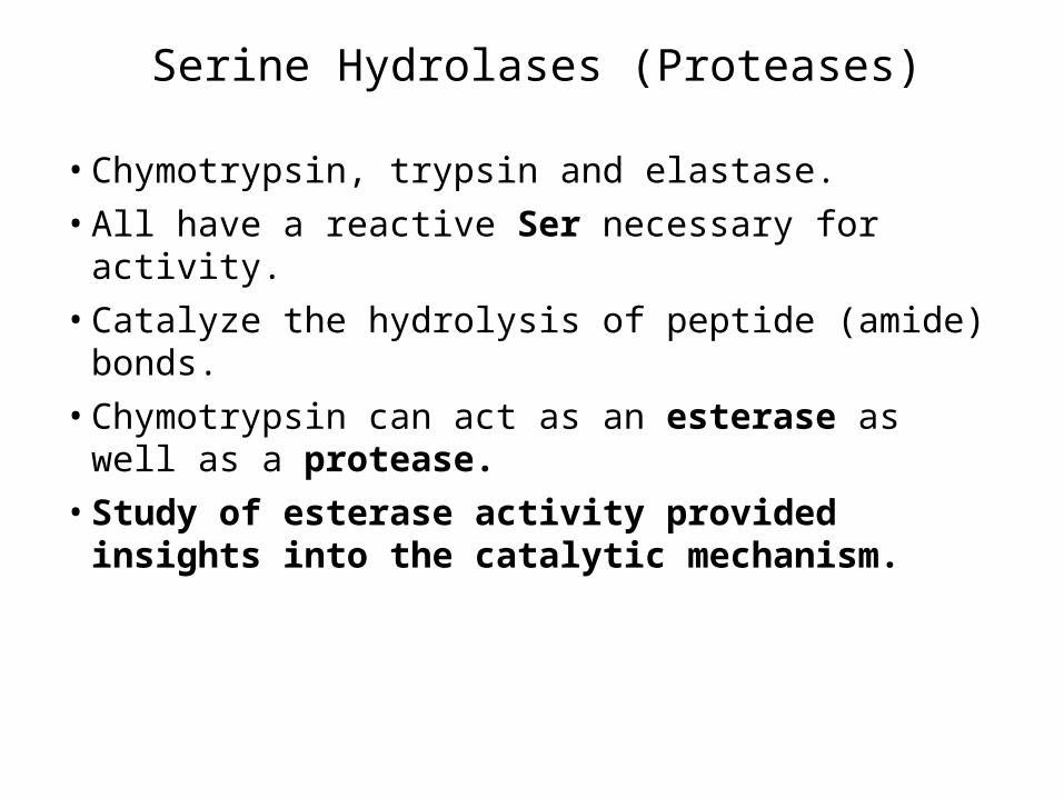
Serine Hydrolases (Proteases)
• Chymotrypsin, trypsin and elastase.
• All have a reactive Ser necessary for activity.
• Catalyze the hydrolysis of peptide (amide) bonds.
• Chymotrypsin can act as an esterase as well as a protease.
• Study of esterase activity provided insights into the catalytic mechanism.
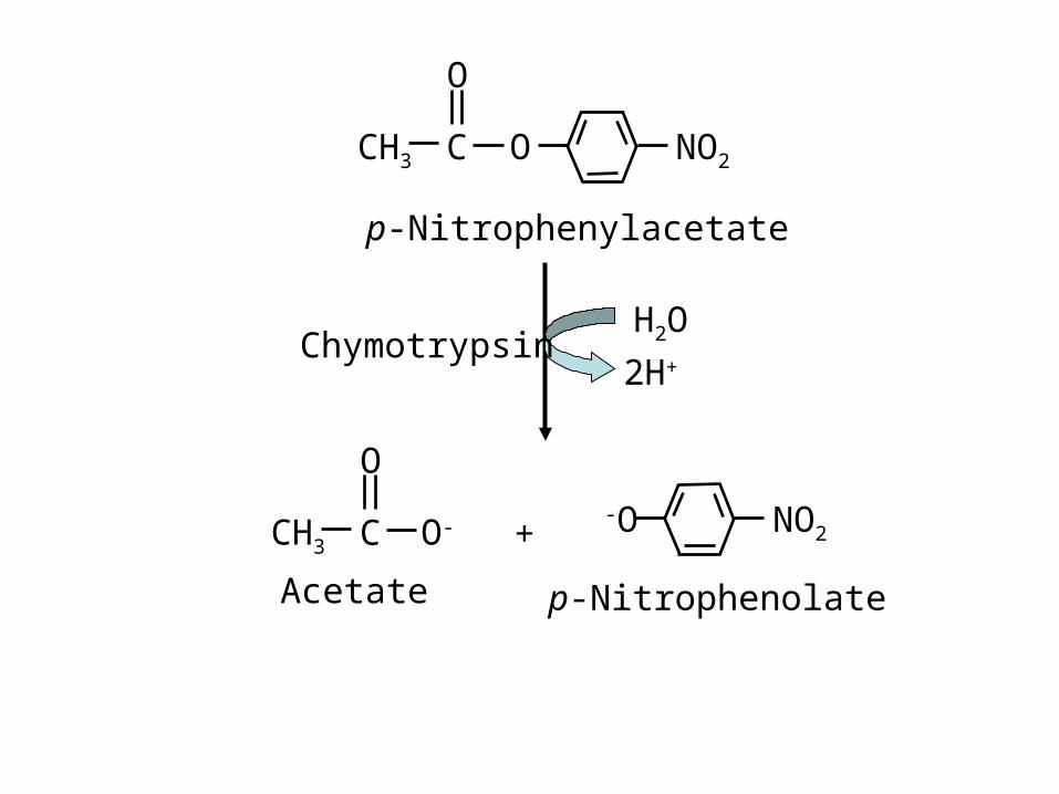
NO2
p-Nitrophenylacetate
CH3 O
O
C
CH3 O-
O
C NO2-O
p-NitrophenolateAcetate
ChymotrypsinH2O
2H+
+

Serine Hydrolases (Proteases)
• Reaction takes place in 2 phases
1. The “burst phase”-fast generation of p-nitrophenolate in stoichiometric amounts with enzyme added
2. The “steady-state phase”-p-nitrophenolate generated at reduced but constant rate; independent of substrate concentration.
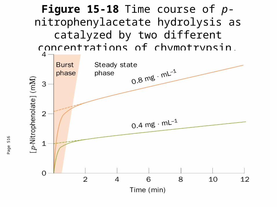
Figure 15-18 Time course of p-nitrophenylacetate hydrolysis as catalyzed by two different concentrations of chymotrypsin.
Pag
e 51
6
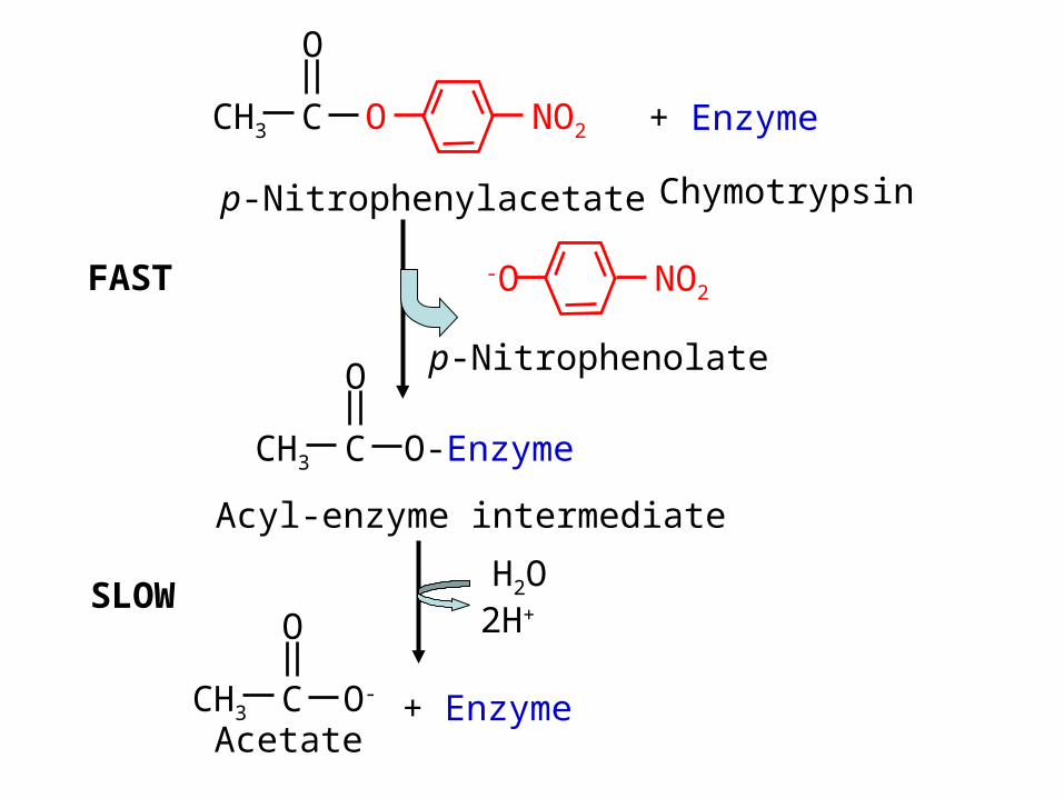
NO2
p-Nitrophenylacetate
CH3 O
O
C
CH3 O-Enzyme
O
C
NO2-O
p-Nitrophenolate
Acyl-enzyme intermediate
Chymotrypsin
H2O2H+
+ Enzyme
CH3 O-
O
CAcetate
+ Enzyme
SLOW
FAST
Chymotrypsin
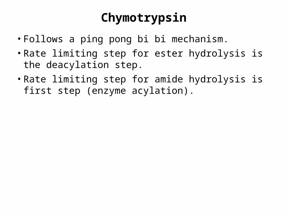
• Follows a ping pong bi bi mechanism.
• Rate limiting step for ester hydrolysis is the deacylation step.
• Rate limiting step for amide hydrolysis is first step (enzyme acylation).
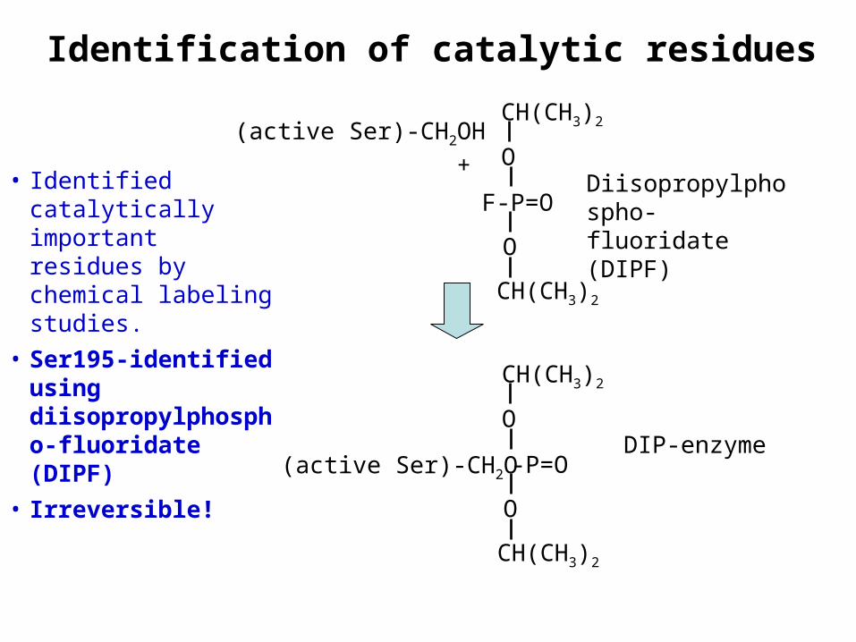
Identification of catalytic residues
• Identified catalytically important residues by chemical labeling studies.
• Ser195-identified using diisopropylphospho-fluoridate (DIPF)
• Irreversible!
(active Ser)-CH2OH
F-P=O
O
Diisopropylphospho-fluoridate (DIPF)
O+
CH(CH3)2
CH(CH3)2
(active Ser)-CH2O -P=O
O
O
CH(CH3)2
CH(CH3)2
DIP-enzyme

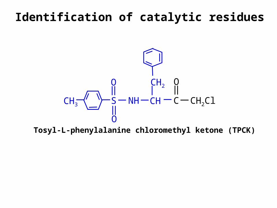
Identification of catalytic residues
• His57 was identified through affinity labeling
• Substrate analog with a reactive group that specifically binds to the active site of the enzyme forms a stable covalent bond with a nearby susceptible group.
• Reactive substrate analogs are sometimes called “Trojan horses” of biochemistry.
• Affinity labeled groups can be identified by peptide mapping.
• For chymotrypsin, they used an analog to Phe.
CH2ClCH3 C
O
NHS
OO
CH
CH2
Identification of catalytic residues
Tosyl-L-phenylalanine chloromethyl ketone (TPCK)

Figure 15-19Reaction of TPCK with chymotrypsin to alkylate His 57.
Pag
e 51
7
Homology among enzymes
• Bovine chymotrypsin, bovine trypsin and porcine elastase are highly homologous
• ~40% identical over ~240 residues.
• All enzymes have active Ser and catalytically essential His
• X-ray structures closely related.
• Asp102 buried in a solvent inaccessible pocket (third enzyme in the “catalytic triad”)
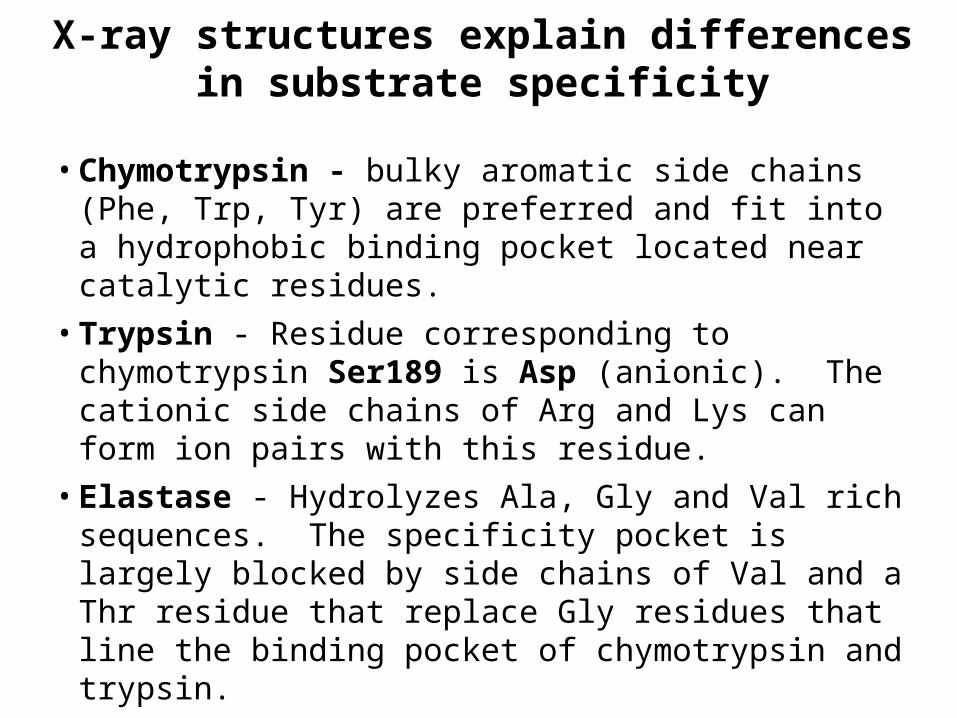
X-ray structures explain differences in substrate specificity
• Chymotrypsin - bulky aromatic side chains (Phe, Trp, Tyr) are preferred and fit into a hydrophobic binding pocket located near catalytic residues.
• Trypsin - Residue corresponding to chymotrypsin Ser189 is Asp (anionic). The cationic side chains of Arg and Lys can form ion pairs with this residue.
• Elastase - Hydrolyzes Ala, Gly and Val rich sequences. The specificity pocket is largely blocked by side chains of Val and a Thr residue that replace Gly residues that line the binding pocket of chymotrypsin and trypsin.
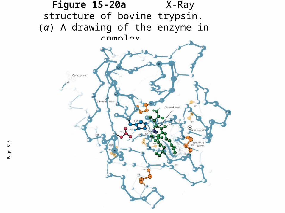
Figure 15-20a X-Ray structure of bovine trypsin.
(a) A drawing of the enzyme in complex.
Pag
e 51
8

Figure 15-20b X-Ray structure of bovine trypsin. (b) A ribbon diagram of trypsin.
Pag
e 51
9
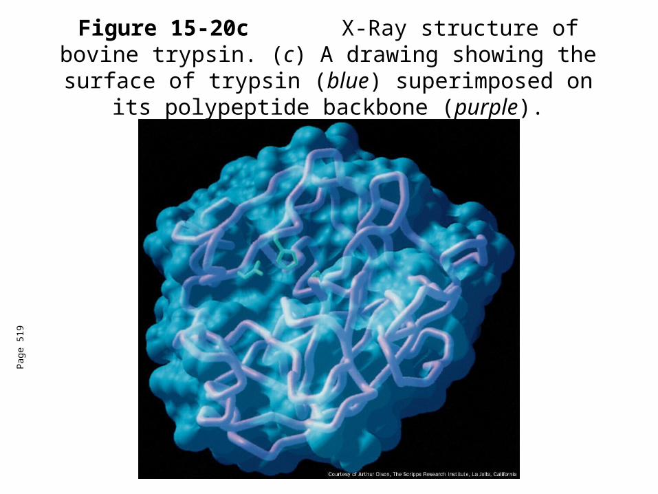
Figure 15-20c X-Ray structure of bovine trypsin. (c) A drawing showing the surface of trypsin
(blue) superimposed on its polypeptide backbone (purple).
Pag
e 51
9
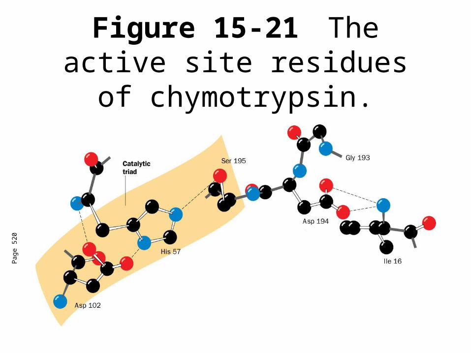
Figure 15-21 The active site residues of chymotrypsin.
Pag
e 52
0
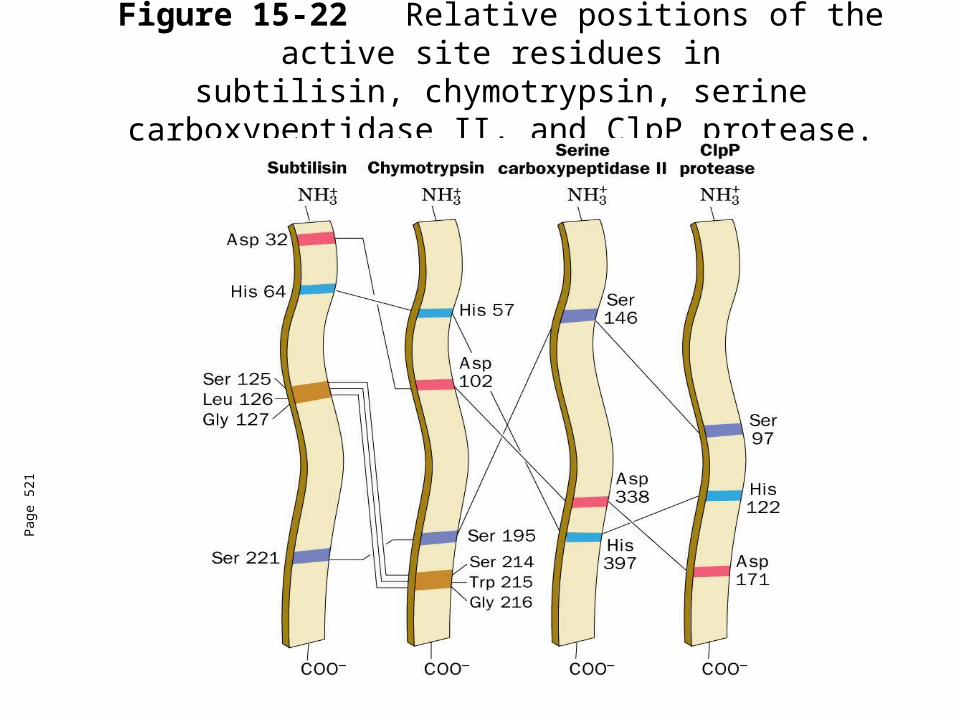
Figure 15-22Relative positions of the active site residues insubtilisin, chymotrypsin, serine carboxypeptidase II, and
ClpP protease.
Pag
e 52
1

Figure 15-23Catalytic
mechanism of the serine proteases.
Pag
e 52
2