Embed Size (px)
Citation preview
Massachusetts Institute of TechnologyHarvard Medical School
Brigham and Women’s HospitalVA Boston Healthcare System
2.79J/3.96J/20.441/HST522J
CHRONIC RESPONSE TO IMPLANTS:a-Smooth Muscle Actin and Lubricin
M. Spector, Ph.D.
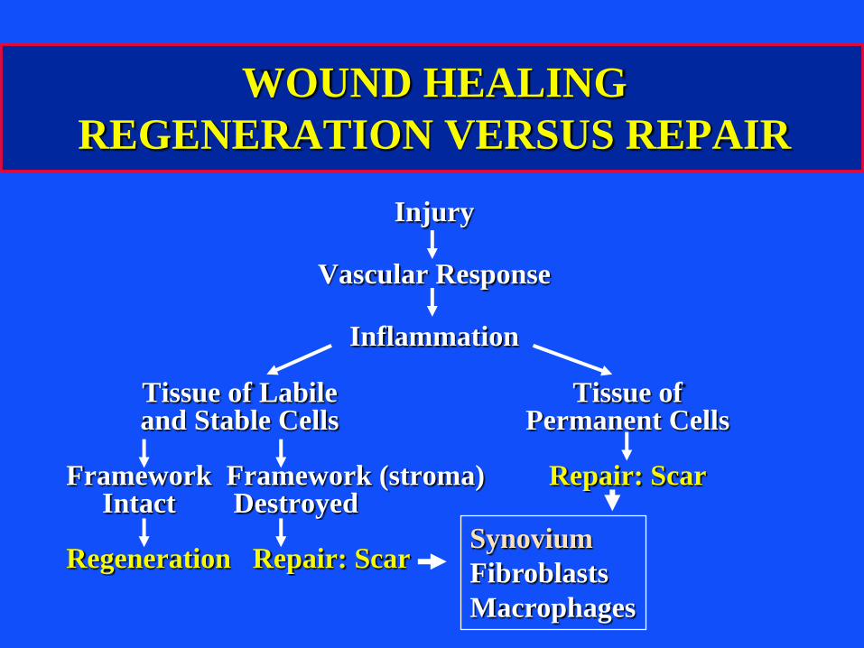
Injury
Vascular Response
Inflammation
Tissue of Labile Tissue ofand Stable Cells Permanent Cells
Framework Framework (stroma) Repair: ScarIntact Destroyed
Regeneration Repair: Scar
WOUND HEALING
REGENERATION VERSUS REPAIR
Synovium
Fibroblasts
Macrophages
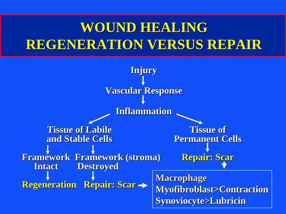
Injury
Vascular Response
Inflammation
Tissue of Labile Tissue ofand Stable Cells Permanent Cells
Framework Framework (stroma) Repair: ScarIntact Destroyed
Regeneration Repair: Scar
WOUND HEALING
REGENERATION VERSUS REPAIR
Macrophage
Myofibroblast>Contraction
Synoviocyte>Lubricin
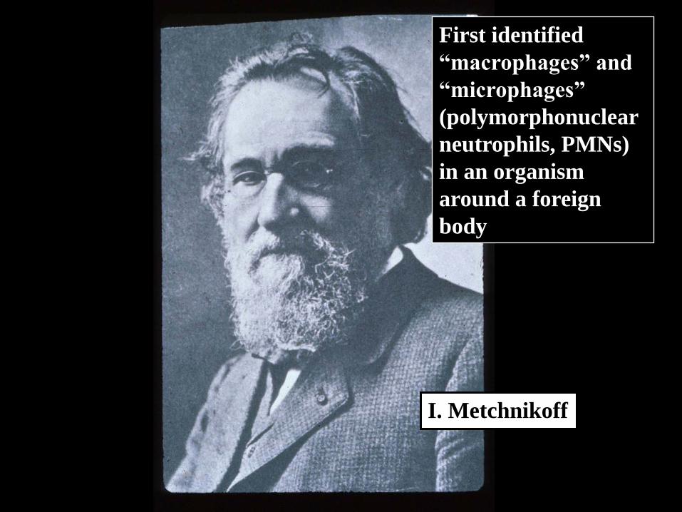
I. Metchnikoff
First identified
“macrophages” and
“microphages”
(polymorphonuclear
neutrophils, PMNs)
in an organism
around a foreign
body
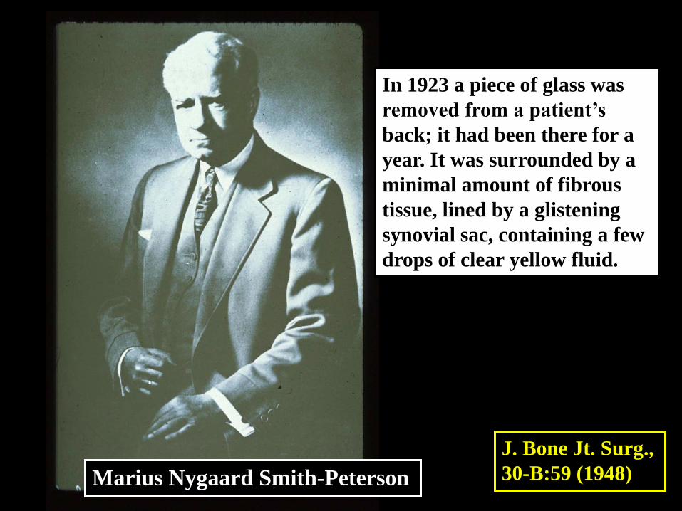
Marius Nygaard Smith-Peterson
In 1923 a piece of glass was
removed from a patient’s
back; it had been there for a
year. It was surrounded by a
minimal amount of fibrous
tissue, lined by a glistening
synovial sac, containing a few
drops of clear yellow fluid.
J. Bone Jt. Surg.,
30-B:59 (1948)
Rabbit Ear Chamber Wound Healing and
Wound Infection (1980)
IA Silver in, TK Hunt,
DR Knighton, et al., Surg 90:62 (1981)
Diagrams removed due to copyright restrictions.
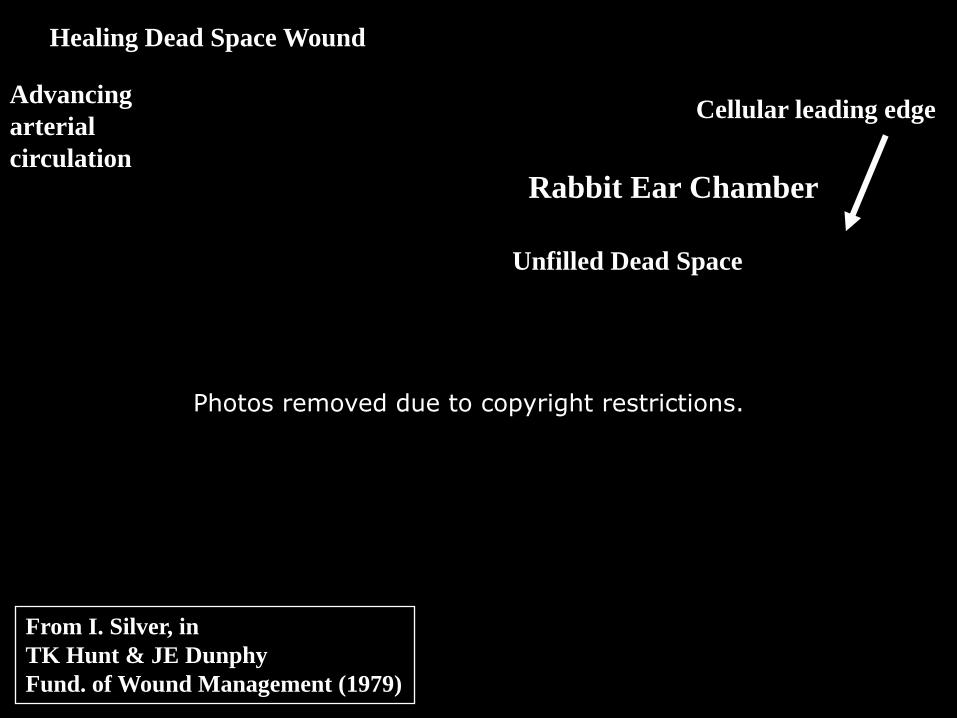
From I. Silver, in
Hunt & JE Dunphy
nd. of Wound Management (1979)
TK
Fu
Healing Dead Space Wound
vancing
terial
culation
Rabbit Ear Chamber
Unfilled Dead Space
Cellular leading edg
Photos removed due to copyright restrictions.
Ad
ar
cir
e
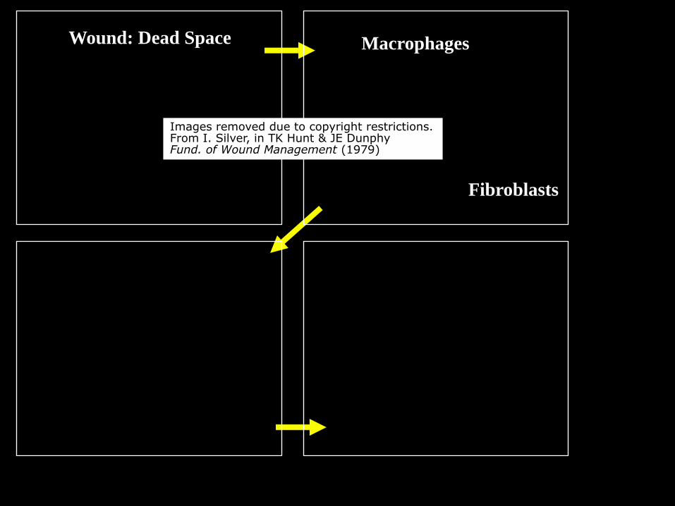
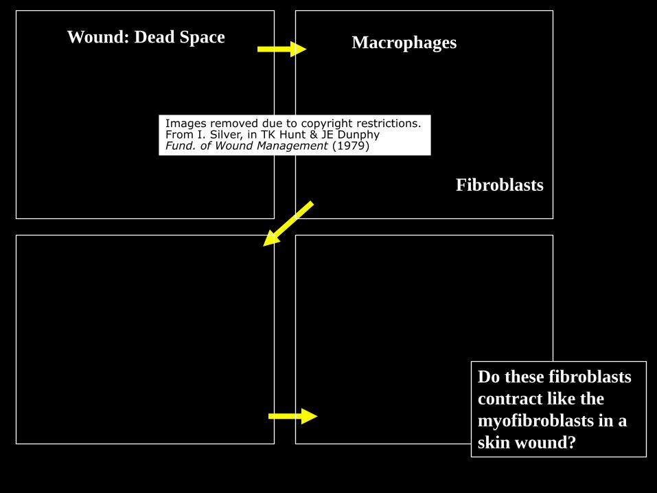
Wound: Dead Space Macrophages
Fibroblasts
Images removed due to copyright restrictions.From I. Silver, in TK Hunt & JE DunphyFund. of Wound Management (1979)
Wound: Dead Space Macrophages
Fibroblasts
Images removed due to copyright restrictions.From I. Silver, in TK Hunt & JE DunphyFund. of Wound Management (1979)
Do these fibroblasts
contract like the
myofibroblasts in a
skin wound?
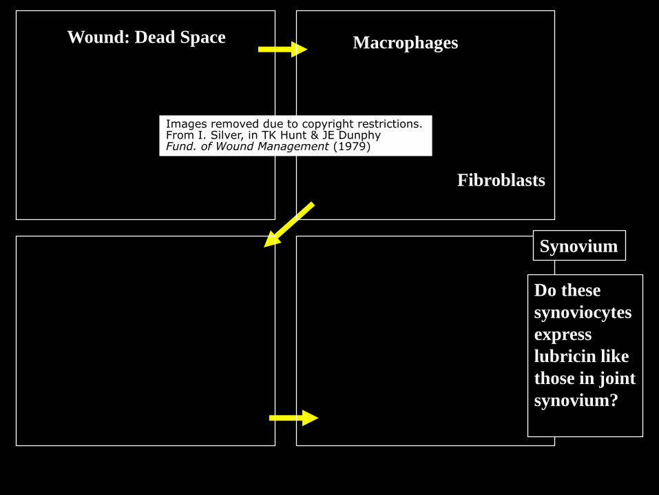
Wound: Dead Space Macrophages
Fibroblasts
Images removed due to copyright restrictions.From I. Silver, in TK Hunt & JE DunphyFund. of Wound Management (1979)
Synovium
Do these
synoviocytes
express
lubricin like
those in joint
synovium?
IA Silver in, TK Hunt,
Wound Healing and
Wound Infection (1980)
Image removed due to copyright restrictions.Graph of regional oxygen tension vs. distance around the foreign body.
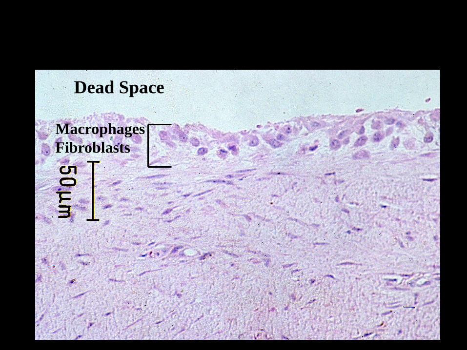
Macrophages
Fibroblasts
Dead Space
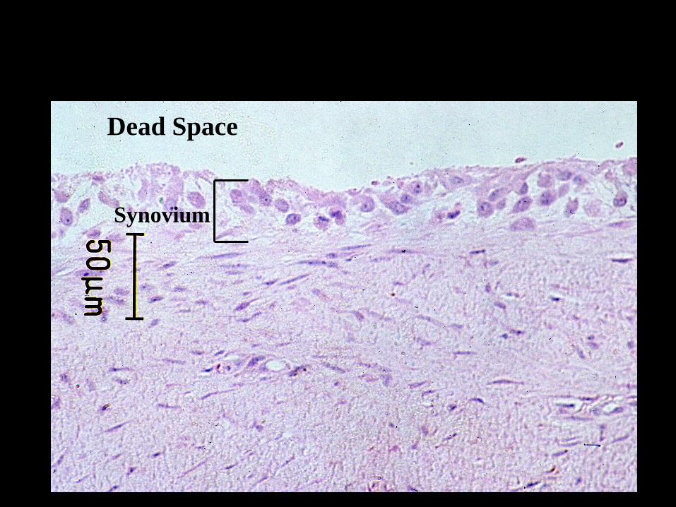
Synovium
Dead Space
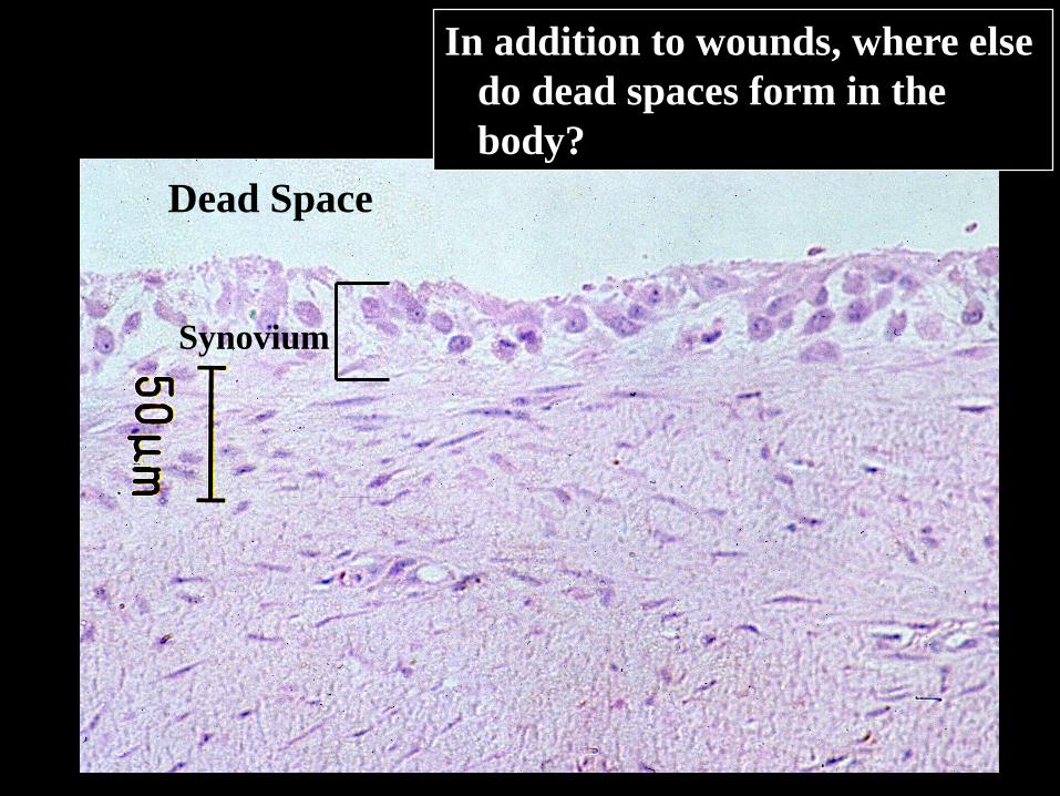
Synovium
Dead Space
In addition to wounds, where else
do dead spaces form in the
body?
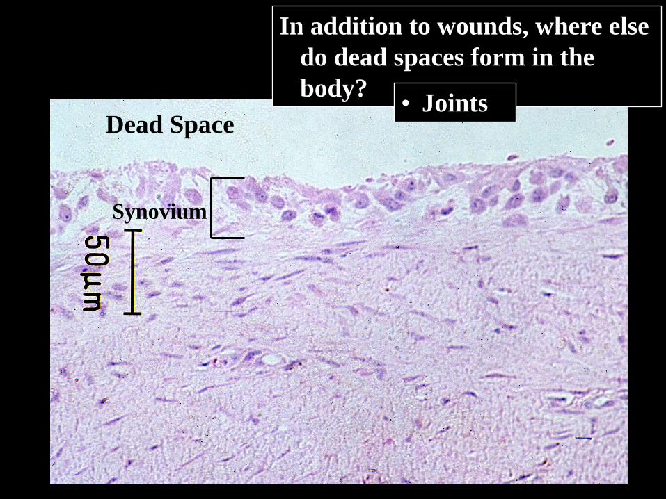
Synovium
Dead Space
In addition to wounds, where else
do dead spaces form in the
body?• Joints
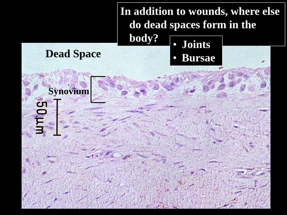
Synovium
Dead Space
In addition to wounds, where else
do dead spaces form in the
body?• Joints
• Bursae
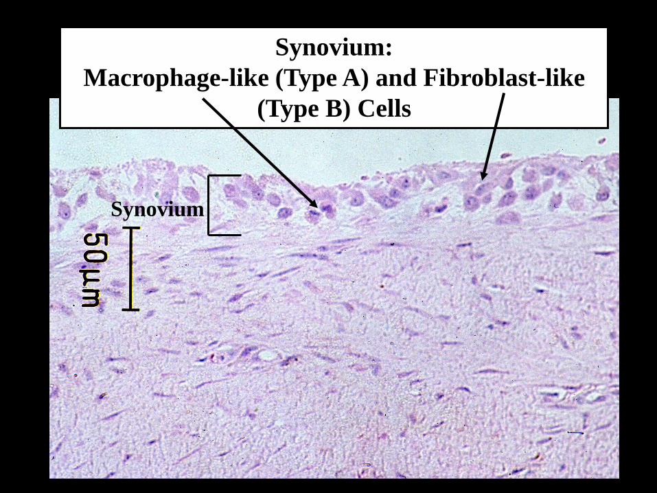
Synovium:
Macrophage-like (Type A) and Fibroblast-like
(Type B) Cells
Synovium

Injury
Vascular Response
Inflammation
Tissue of Labile Tissue ofand Stable Cells Permanent Cells
Framework Framework (stroma) Repair: ScarIntact Destroyed
Regeneration Repair: Scar
WOUND HEALING
REGENERATION VERSUS REPAIR
Macrophage
Myofibroblast>Contraction
Synoviocyte>Lubricin
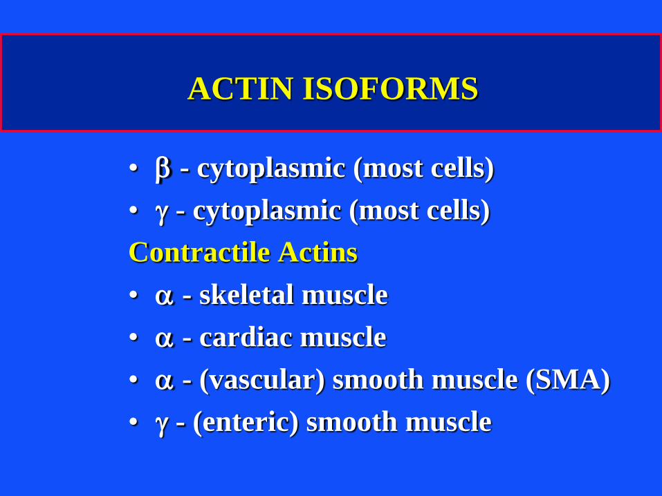
ACTIN ISOFORMS
• b - cytoplasmic (most cells)
• g - cytoplasmic (most cells)
Contractile Actins
• a - skeletal muscle
• a - cardiac muscle
• a - (vascular) smooth muscle (SMA)
• g - (enteric) smooth muscle
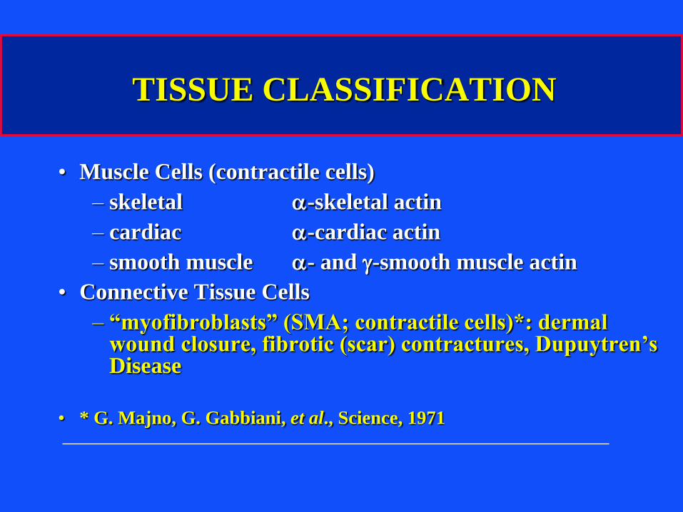
TISSUE CLASSIFICATION
• Muscle Cells (contractile cells)
– skeletal a-skeletal actin
– cardiac a-cardiac actin
– smooth muscle a- and g-smooth muscle actin
• Connective Tissue Cells
– “myofibroblasts” (SMA; contractile cells)*: dermal wound closure, fibrotic (scar) contractures, Dupuytren’s Disease
• * G. Majno, G. Gabbiani, et al., Science, 1971
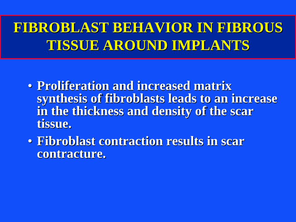
FIBROBLAST BEHAVIOR IN FIBROUS
TISSUE AROUND IMPLANTS
• Proliferation and increased matrix synthesis of fibroblasts leads to an increase in the thickness and density of the scar tissue.
• Fibroblast contraction results in scar contracture.
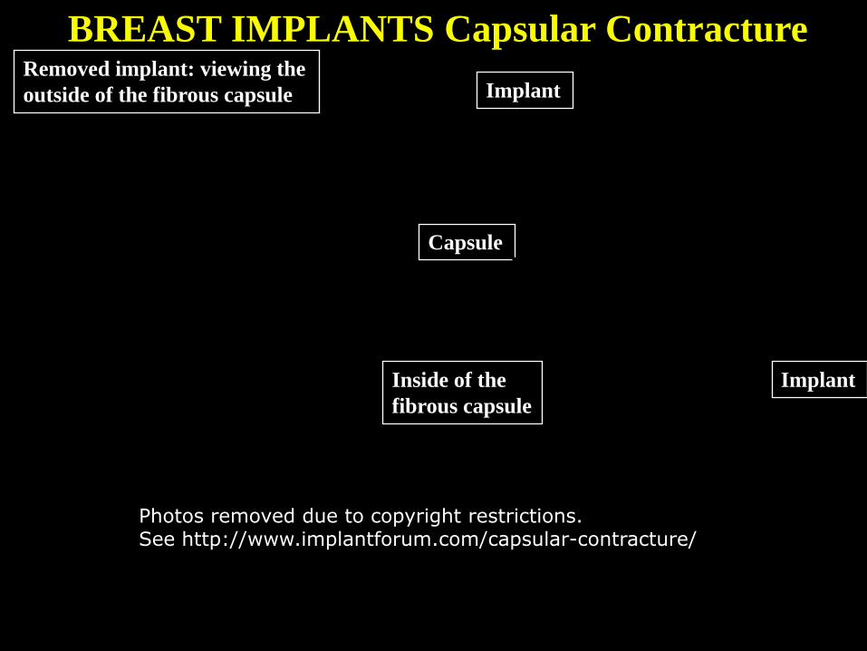
BREAST IMPLANTS Capsular ContractureRemoved implant: viewing the
outside of the fibrous capsule
Capsule
Implant
Inside of the
fibrous capsule
Implant
Photos removed due to copyright restrictions.See http://www.implantforum.com/capsular-contracture/
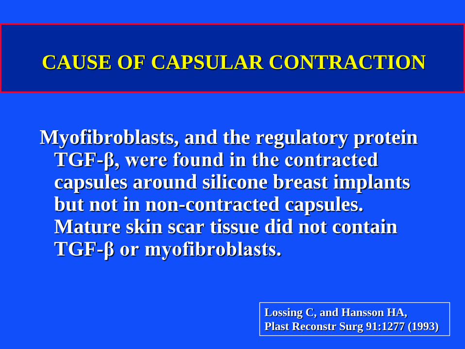
CAUSE OF CAPSULAR CONTRACTION
Myofibroblasts, and the regulatory protein TGF-β, were found in the contracted capsules around silicone breast implants but not in non-contracted capsules. Mature skin scar tissue did not contain TGF-β or myofibroblasts.
Lossing C, and Hansson HA,
Plast Reconstr Surg 91:1277 (1993)
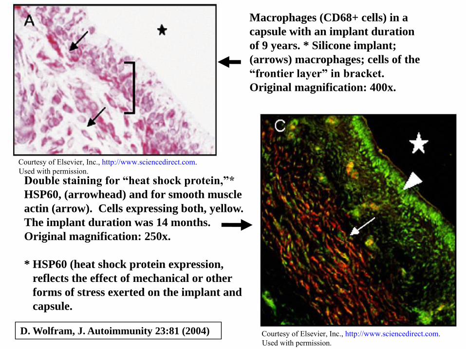
Macrophages (CD68+ cells) in a
capsule with an implant duration
of 9 years. * Silicone implant;
(arrows) macrophages; cells of the
“frontier layer” in bracket.
Original magnification: 400x.
D. Wolfram, J. Autoimmunity 23:81 (2004)
Double staining for “heat shock protein,”*
HSP60, (arrowhead) and for smooth muscle
actin (arrow). Cells expressing both, yellow.
The implant duration was 14 months.
Original magnification: 250x.
* HSP60 (heat shock protein expression,
reflects the effect of mechanical or other
forms of stress exerted on the implant and
capsule.
Courtesy of Elsevier, Inc., http://www.sciencedirect.com. Used with permission.
Courtesy of Elsevier, Inc., http://www.sciencedirect.com. Used with permission.
D. Wolfram, J. Autoimmunity 23:81 (2004)
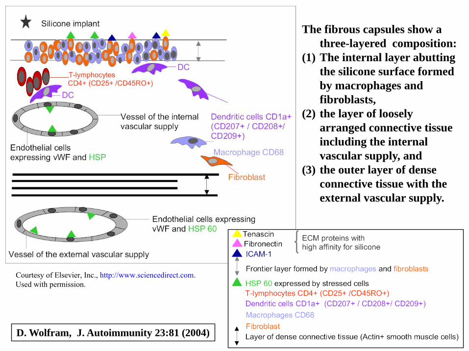
The fibrous capsules show a
three-layered composition:
(1) The internal layer abutting
the silicone surface formed
by macrophages and
fibroblasts,
(2) the layer of loosely
arranged connective tissue
including the internal
vascular supply, and
(3) the outer layer of dense
connective tissue with the
external vascular supply.
Courtesy of Elsevier, Inc., http://www.sciencedirect.com. Used with permission.
J Bone Joint Surg 2005;87A:1284
White arrows:
Radiolucencies due to
osteolysis associated with
particulate wear debris
and movement of the
prosthesis (loosening)
• What is the make-up of the
periprosthetic tissue?
• Why is it so persistent?
Photos removed due to copyright restrictions.
Fig. 1 Kaplan-Meier survival curves with clinical and radiographic failure as the end points.
Martin S. D. et.al. J Bone Joint Surg 2005:87:1284-1292
Graph removed due to copyright restrictions.
Fig. 3 Prevalence of radiolucent lines around the glenoid components.
Martin S. D. et.al. J Bone Joint Surg 2005:87:1284-1292
Graph removed due to copyright restrictions.
J. Biomed. Mater. Res. (in press)
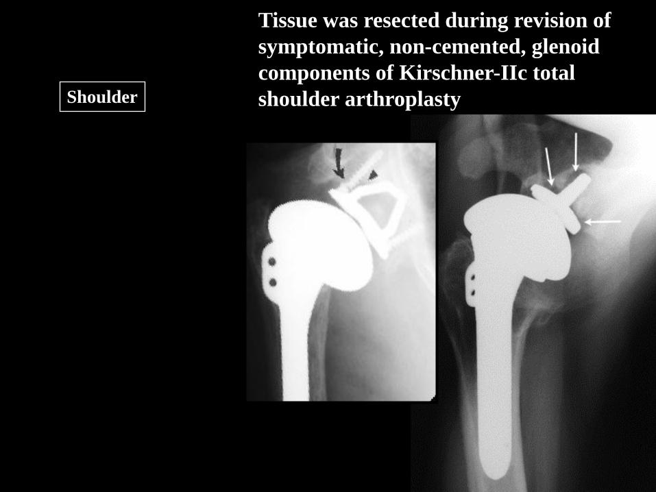
Tissue was resected during revision of
symptomatic, non-cemented, glenoid
components of Kirschner-IIc total
shoulder arthroplasty Shoulder
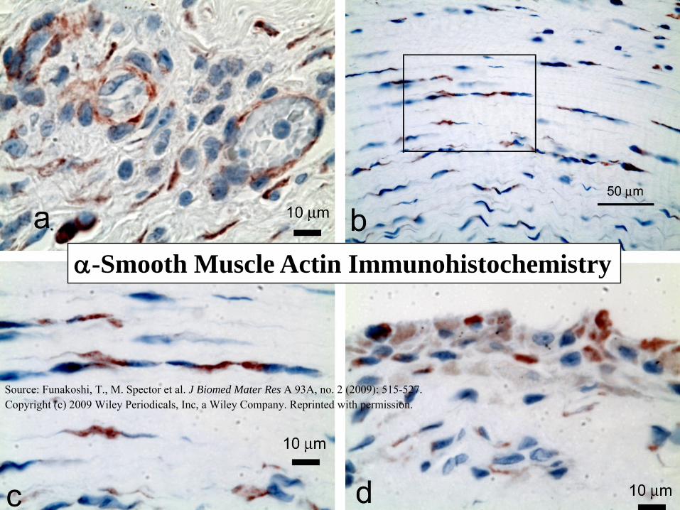
a-Smooth Muscle Actin Immunohistochemistry
Source: Funakoshi, T., M. Spector et al. J Biomed Mater Res A 93A, no. 2 (2009): 515-527. Copyright (c) 2009 Wiley Periodicals, Inc, a Wiley Company. Reprinted with permission.
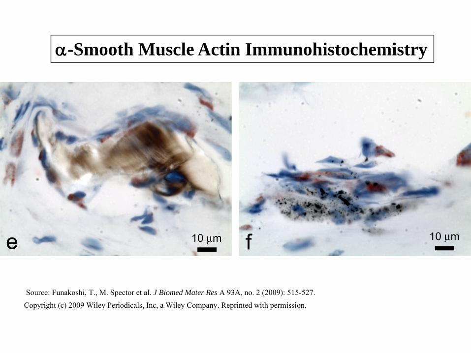
a-Smooth Muscle Actin Immunohistochemistry
Source: Funakoshi, T., M. Spector et al. J Biomed Mater Res A 93A, no. 2 (2009): 515-527. Copyright (c) 2009 Wiley Periodicals, Inc, a Wiley Company. Reprinted with permission.
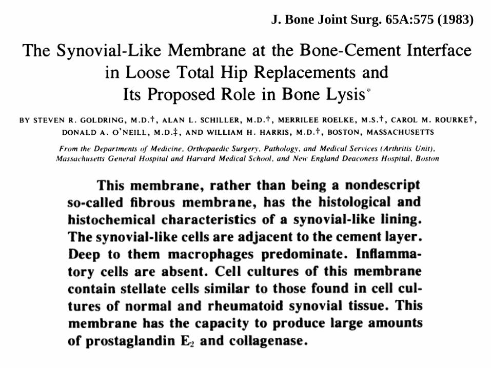
J. Bone Joint Surg. 65A:575 (1983)

S. Goldring, et al.,
J. Bone Joint Surg. 65A:575 (1983)
Four photos removed due to copyright restrictions.
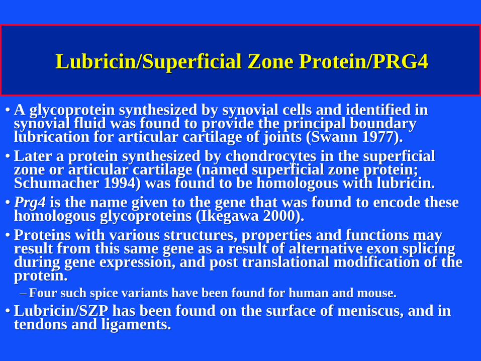
Lubricin/Superficial Zone Protein/PRG4
• A glycoprotein synthesized by synovial cells and identified in synovial fluid was found to provide the principal boundary lubrication for articular cartilage of joints (Swann 1977).
• Later a protein synthesized by chondrocytes in the superficial zone or articular cartilage (named superficial zone protein; Schumacher 1994) was found to be homologous with lubricin.
• Prg4 is the name given to the gene that was found to encode these homologous glycoproteins (Ikegawa 2000).
• Proteins with various structures, properties and functions may result from this same gene as a result of alternative exon splicing during gene expression, and post translational modification of the protein.– Four such spice variants have been found for human and mouse.
• Lubricin/SZP has been found on the surface of meniscus, and in tendons and ligaments.
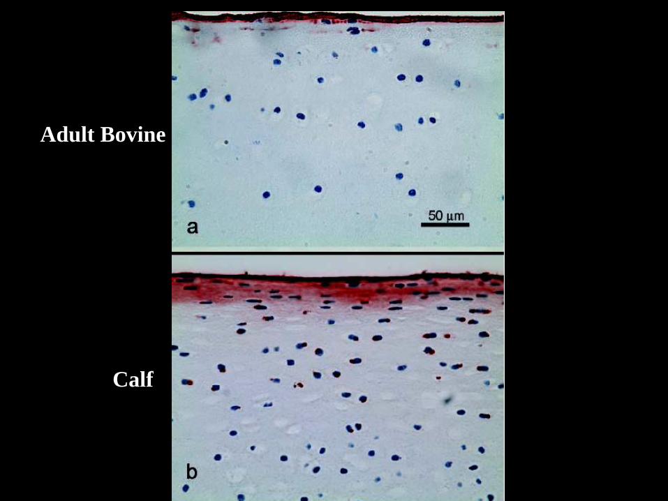
Adult Bovine
Calf
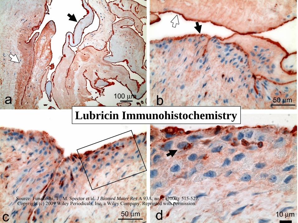
Lubricin Immunohistochemistry
Source: Funakoshi, T., M. Spector et al. J Biomed Mater Res A 93A, no. 2 (2009): 515-527. Copyright (c) 2009 Wiley Periodicals, Inc, a Wiley Company. Reprinted with permission.
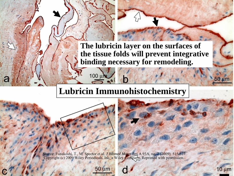
Lubricin Immunohistochemistry
The lubricin layer on the surfaces of the tissue folds will prevent integrative binding necessary for remodeling.
Source: Funakoshi, T., M. Spector et al. J Biomed Mater Res A 93A, no. 2 (2009): 515-527. Copyright (c) 2009 Wiley Periodicals, Inc, a Wiley Company. Reprinted with permission.
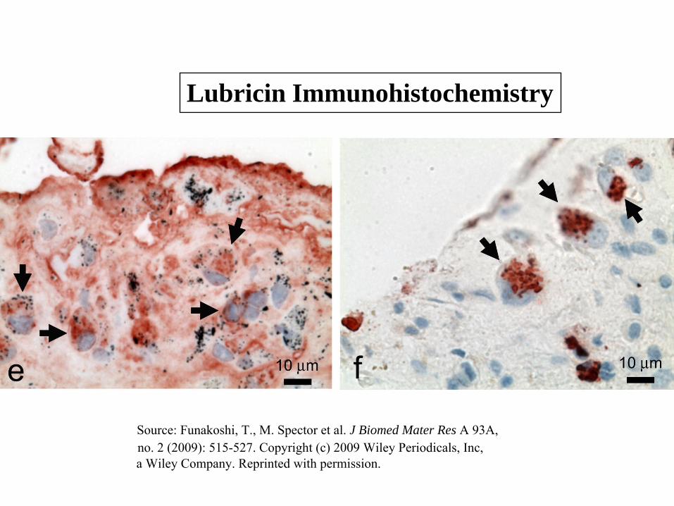
Lubricin Immunohistochemistry
Source: Funakoshi, T., M. Spector et al. J Biomed Mater Res A 93A, no. 2 (2009): 515-527. Copyright (c) 2009 Wiley Periodicals, Inc,a Wiley Company. Reprinted with permission.
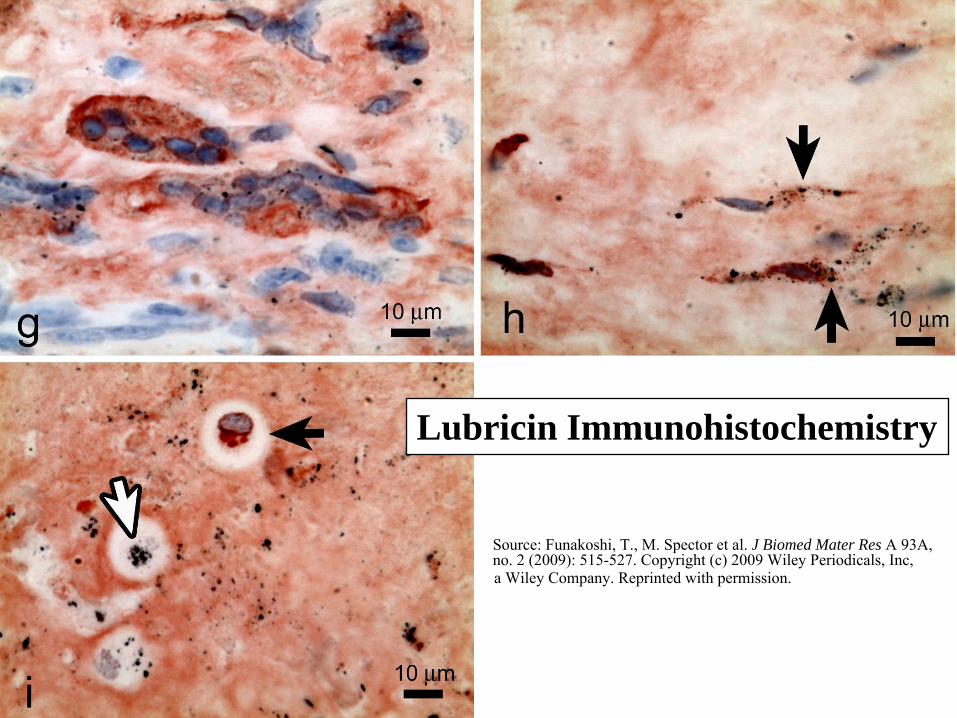
Lubricin Immunohistochemistry
Source: Funakoshi, T., M. Spector et al. J Biomed Mater Res A 93A, no. 2 (2009): 515-527. Copyright (c) 2009 Wiley Periodicals, Inc, a Wiley Company. Reprinted with permission.
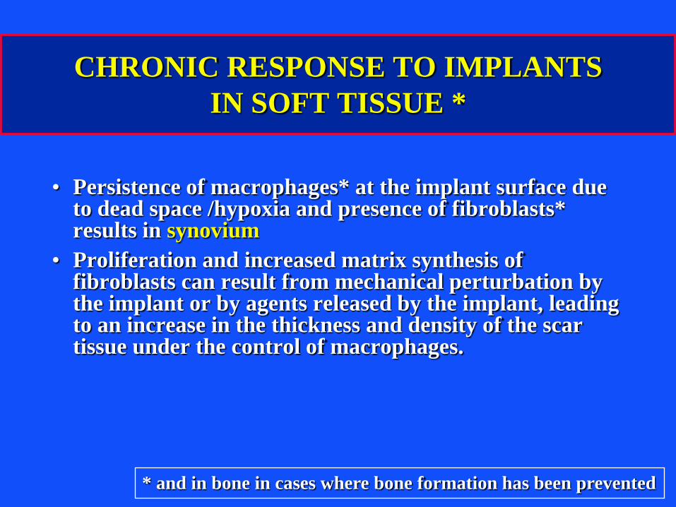
CHRONIC RESPONSE TO IMPLANTS
IN SOFT TISSUE *
• Persistence of macrophages* at the implant surface due to dead space /hypoxia and presence of fibroblasts* results in synovium
• Proliferation and increased matrix synthesis of fibroblasts can result from mechanical perturbation by the implant or by agents released by the implant, leading to an increase in the thickness and density of the scar tissue under the control of macrophages.
* and in bone in cases where bone formation has been prevented
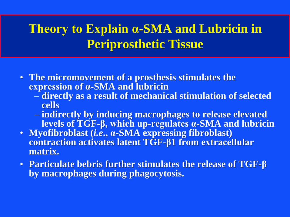
Theory to Explain α-SMA and Lubricin in
Periprosthetic Tissue
• The micromovement of a prosthesis stimulates the expression of α-SMA and lubricin– directly as a result of mechanical stimulation of selected
cells– indirectly by inducing macrophages to release elevated
levels of TGF-β, which up-regulates α-SMA and lubricin• Myofibroblast (i.e., α-SMA expressing fibroblast)
contraction activates latent TGF-β1 from extracellular matrix.
• Particulate bebris further stimulates the release of TGF-β by macrophages during phagocytosis.
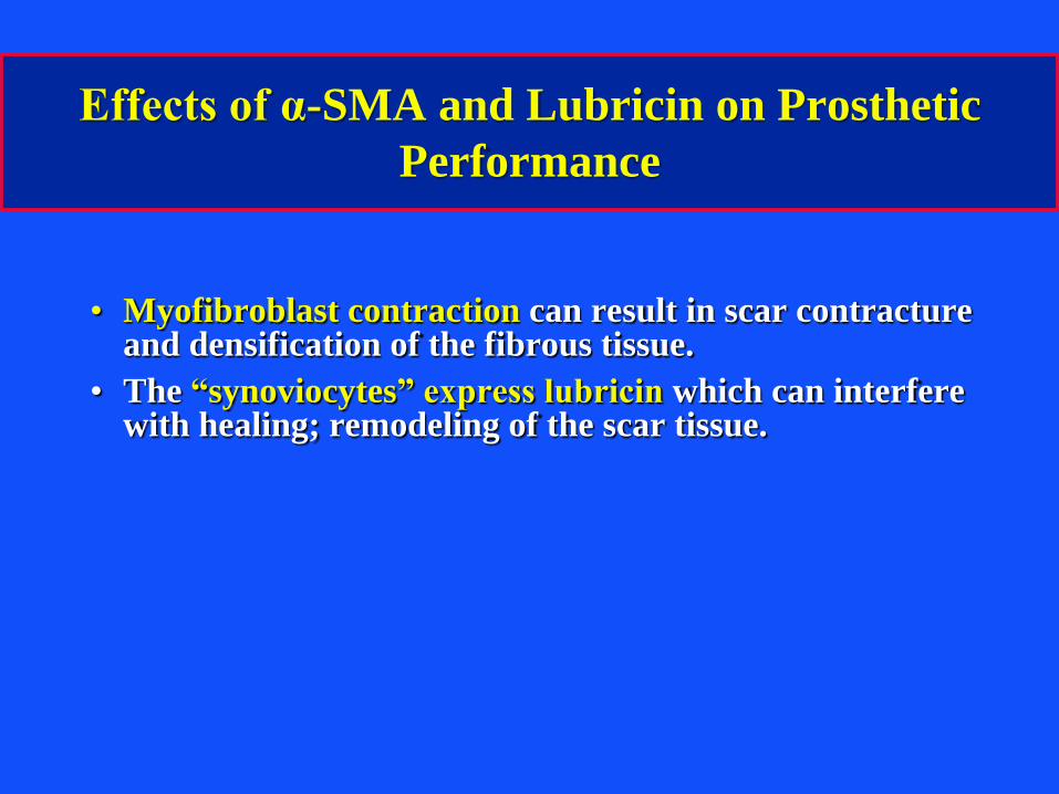
Effects of α-SMA and Lubricin on Prosthetic
Performance
• Myofibroblast contraction can result in scar contracture and densification of the fibrous tissue.
• The “synoviocytes” express lubricin which can interfere with healing; remodeling of the scar tissue.
MIT OpenCourseWarehttp://ocw.mit.edu
20.441J / 2.79J / 3.96J / HST.522J Biomaterials-Tissue InteractionsFall 2009 For information about citing these materials or our Terms of Use, visit: http://ocw.mit.edu/terms.