Embed Size (px)
Citation preview

Lecture 11b.
Neurophysiology
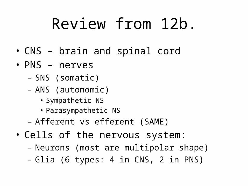
Review from 12b.
• CNS – brain and spinal cord• PNS – nerves
– SNS (somatic)– ANS (autonomic)
• Sympathetic NS• Parasympathetic NS
– Afferent vs efferent (SAME)
• Cells of the nervous system:– Neurons (most are multipolar shape)– Glia (6 types: 4 in CNS, 2 in PNS)
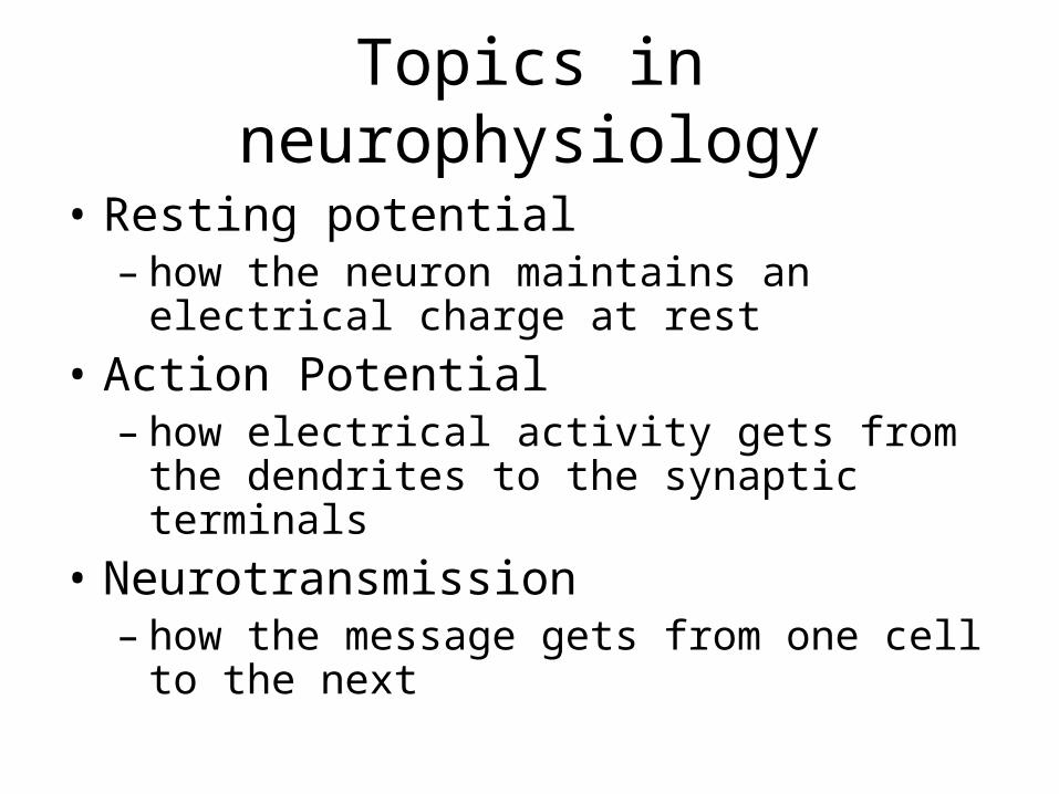
Topics in neurophysiology
• Resting potential– how the neuron maintains an electrical charge
at rest
• Action Potential– how electrical activity gets from the dendrites
to the synaptic terminals
• Neurotransmission– how the message gets from one cell to the
next
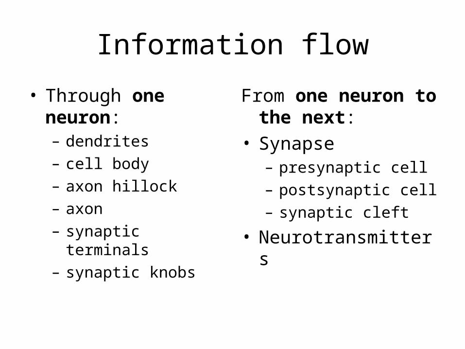
Information flow
• Through one neuron: – dendrites– cell body– axon hillock– axon– synaptic terminals– synaptic knobs
From one neuron to the next:
• Synapse– presynaptic cell– postsynaptic cell– synaptic cleft
• Neurotransmitters
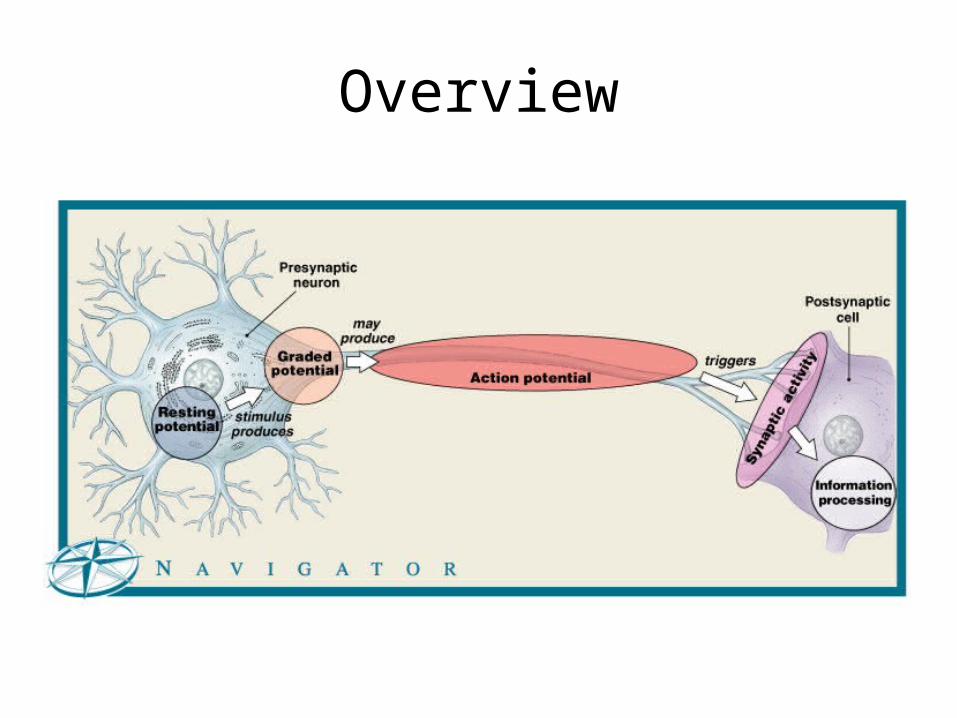
Figure 12–7 (Navigator)
Overview
Big Picture
• Don’ t lose sight of the larger significance of what we are talking about:– the ability to move, think, and feel, everything
that makes us human, everything that makes all animals live and breathe…
All of these things are caused by the movement of a ions into and out of the membranes of neural cells.
If neurons are excitable cells…
• What does this mean?
• Have we talked about a model in another cell for propagation of an impulse?
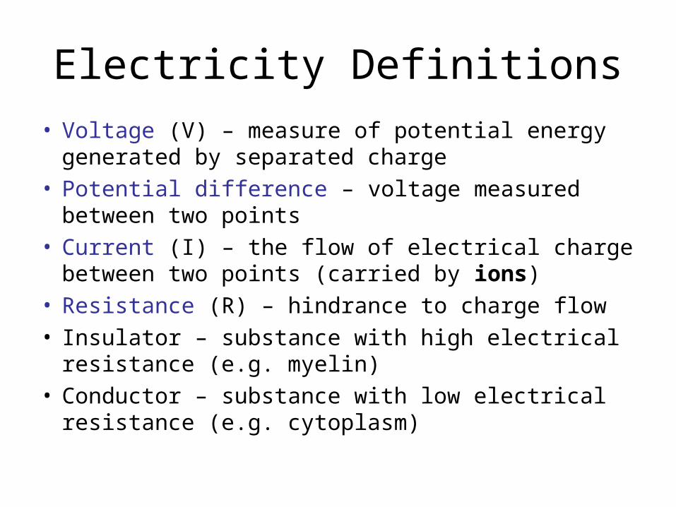
Electricity Definitions
• Voltage (V) – measure of potential energy generated by separated charge
• Potential difference – voltage measured between two points
• Current (I) – the flow of electrical charge between two points (carried by ions)
• Resistance (R) – hindrance to charge flow• Insulator – substance with high electrical resistance
(e.g. myelin)• Conductor – substance with low electrical resistance
(e.g. cytoplasm)
Transmembrane Potential
• All cells have an excess of negative and charges inside versus outside (in the extracellular fluid)
• This transmembrane potential is particularly important to neurons because changes in the membrane potential can be used for signaling or transmitting information
• In neurons, it is abround -70mV (millivolts)
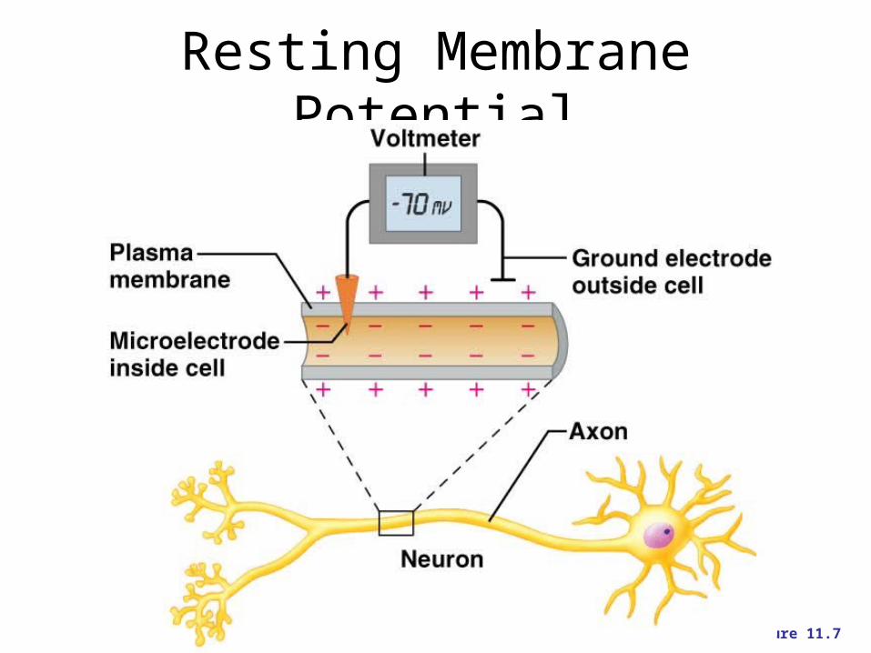
Resting Membrane Potential
Figure 11.7
Ion Movements and Electrical Signals
• Ion movements, or changes in the distribution of ions on either side of a cell membrane cause changes in the membrane potential which propagate (spread) along cells = action potentials
• To understand this, we first must look at the resting conditions, when no changes are occuring
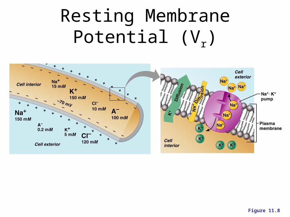
Resting Membrane Potential (Vr)
Figure 11.8
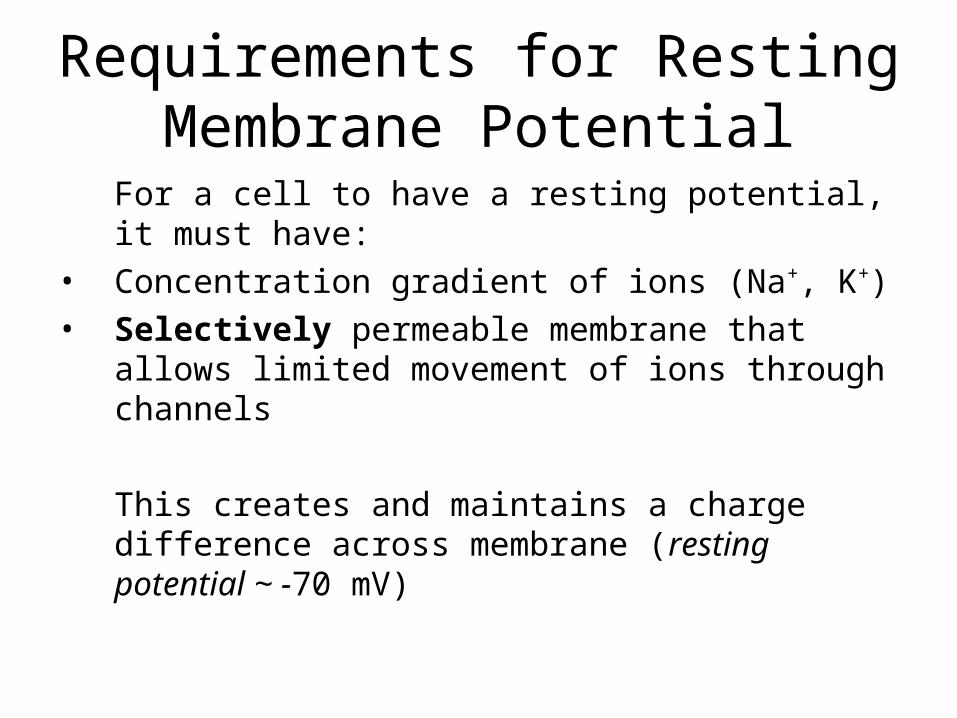
Requirements for Resting Membrane Potential
For a cell to have a resting potential, it must have:
• Concentration gradient of ions (Na+, K+) • Selectively permeable membrane that allows
limited movement of ions through channels
This creates and maintains a charge difference across membrane (resting potential ~ -70 mV)
Potential Energy
• Energy can be stored in a form that can later be released = turned into active energy, energy that does something
• This stored energy = potential energy
• Examples: waterfall, raised book
The semipermeable cell membrane is key.
• Remember that the membrane allows for the separation of ions because it is selectively permeable.
• Which ions are in greater concentration inside the cell? Outside the cell?
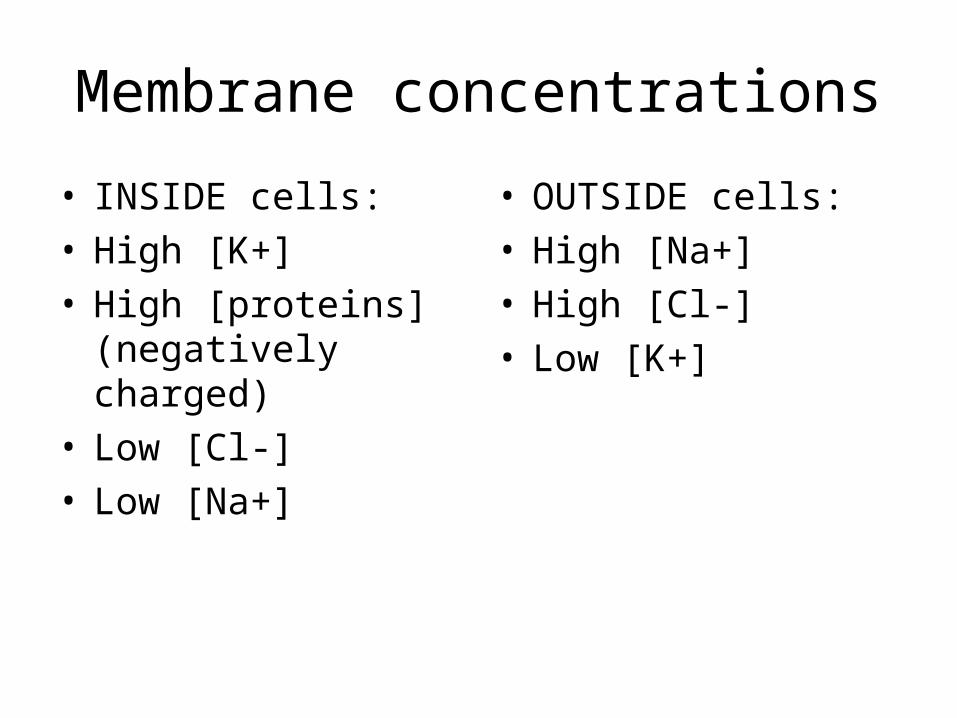
Membrane concentrations
• INSIDE cells:• High [K+]• High [proteins]
(negatively charged)• Low [Cl-]• Low [Na+]
• OUTSIDE cells:• High [Na+]• High [Cl-]• Low [K+]
So far we know the following:
• Intracellular fluid contains a high concentration of K+ and negatively charged proteins)
• Extracellular fluid contains a high concentration of Na+ and Cl- ions
• What would happen if the cell membrane were freely permeable? Gradients? Potential?
Key to resting potential
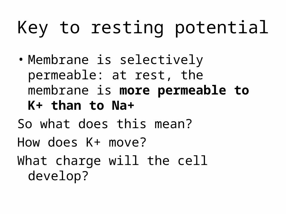
• Membrane is selectively permeable: at rest, the membrane is more permeable to K+ than to Na+
So what does this mean?
How does K+ move?
What charge will the cell develop?
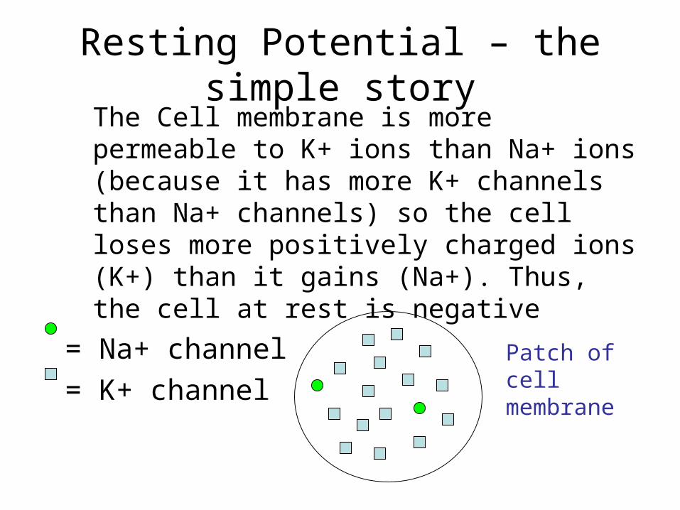
Resting Potential – the simple storyThe Cell membrane is more permeable to K+ ions than Na+ ions (because it has more K+ channels than Na+ channels) so the cell loses more positively charged ions (K+) than it gains (Na+). Thus, the cell at rest is negative
= Na+ channel
= K+ channelPatch of cell membrane

Resting Membrane Potential (Vr)
Figure 11.8
Resting potential overview• At rest, the cell is almost exclusively permeable to
K+• So K+ leaves the cell continuously, but will it
reach concentration equilibrium (same conc. on both sides)?
• NO, because as + ions leave, they leave behind an excess of negative charge and an electrical potential develops (due to separation of + and – charges) which is EQUAL and OPPOSITE to the concentration force.
• This balance point is called the equilibrium potential.
Two types of gradients
1. Chemical (concentration) gradient: caused by different concentrations of a single ion on either side of the plasma membrane– e.g. high potassium concentration inside the
cell tends to make K+ ions want to leave the cell
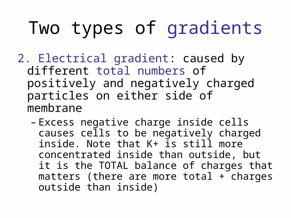
Two types of gradients
2. Electrical gradient: caused by different total numbers of positively and negatively charged particles on either side of membrane– Excess negative charge inside cells causes
cells to be negatively charged inside. Note that K+ is still more concentrated inside than outside, but it is the TOTAL balance of charges that matters (there are more total + charges outside than inside)
Cell model
• Imagine a cell with high K+ inside and high Na+ outside.
• At time = 0, the membrane is impermeable
• Say that we now put K+ channels in the cell, making it permeable to only K+. What happens to K+?
• Will it continue to move until it is the same on both sides (in and out)?
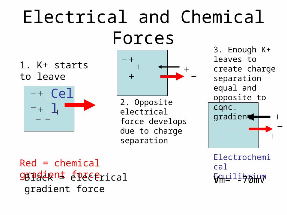
Electrical and Chemical Forces
Red = chemical gradient force
Black = electrical gradient force
Electrochemical Equilibrium
Vm= -70mV
1. K+ starts to leave
Cell2. Opposite electrical force develops due to charge separation
3. Enough K+ leaves to create charge separation equal and opposite to conc. gradient
Equilibrium = balance
• Chemical equilibrium: The point at which diffusion causes equal amounts of a particular ion on either side of the membrane
• Electrical equilibrium: The point at which the TOTAL number of + and – ions is equal on both sides of the membrane (potential = 0)
The electrochemical gradient
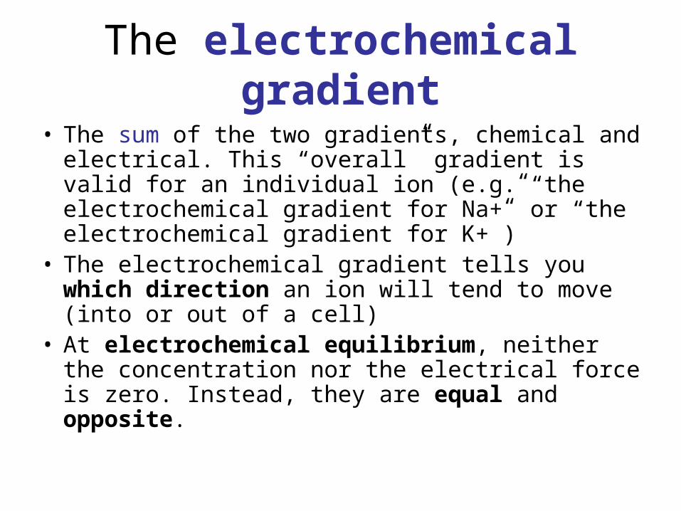
• The sum of the two gradients, chemical and electrical. This “overall” gradient is valid for an individual ion (e.g. “the electrochemical gradient for Na+” or “the electrochemical gradient for K+”)
• The electrochemical gradient tells you which direction an ion will tend to move (into or out of a cell)
• At electrochemical equilibrium, neither the concentration nor the electrical force is zero. Instead, they are equal and opposite.
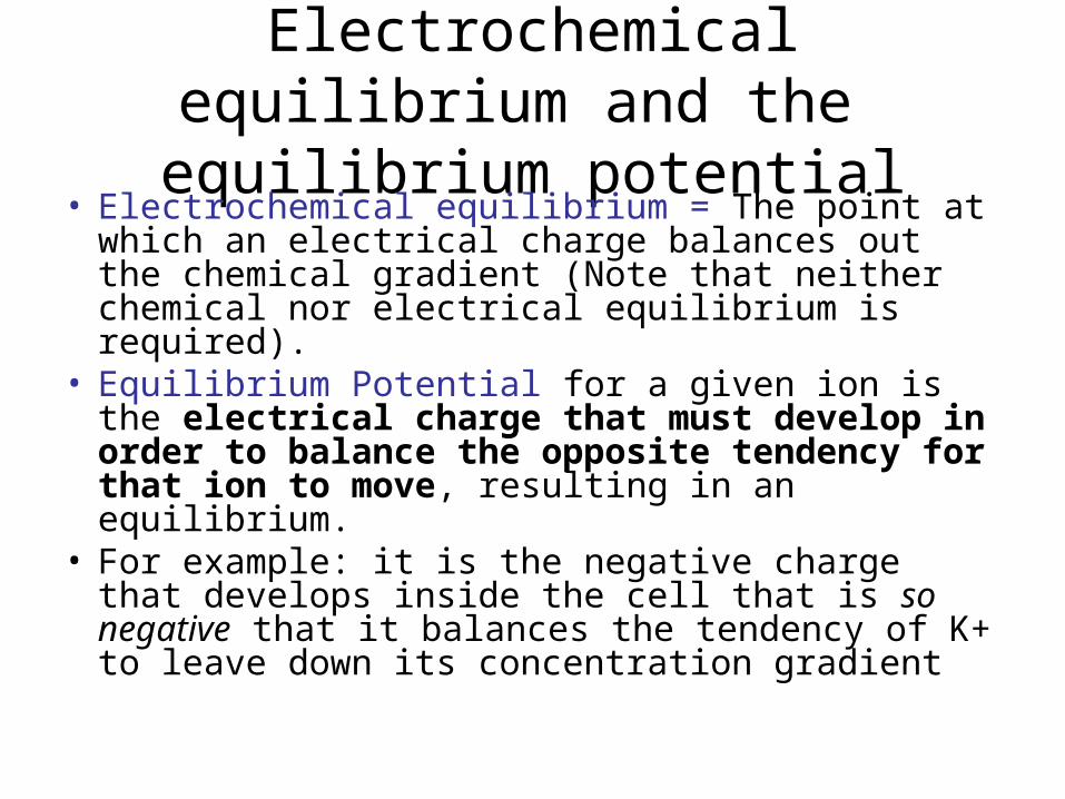
Electrochemical equilibrium and the equilibrium potential
• Electrochemical equilibrium = The point at which an electrical charge balances out the chemical gradient (Note that neither chemical nor electrical equilibrium is required).
• Equilibrium Potential for a given ion is the electrical charge that must develop in order to balance the opposite tendency for that ion to move, resulting in an equilibrium.
• For example: it is the negative charge that develops inside the cell that is so negative that it balances the tendency of K+ to leave down its concentration gradient
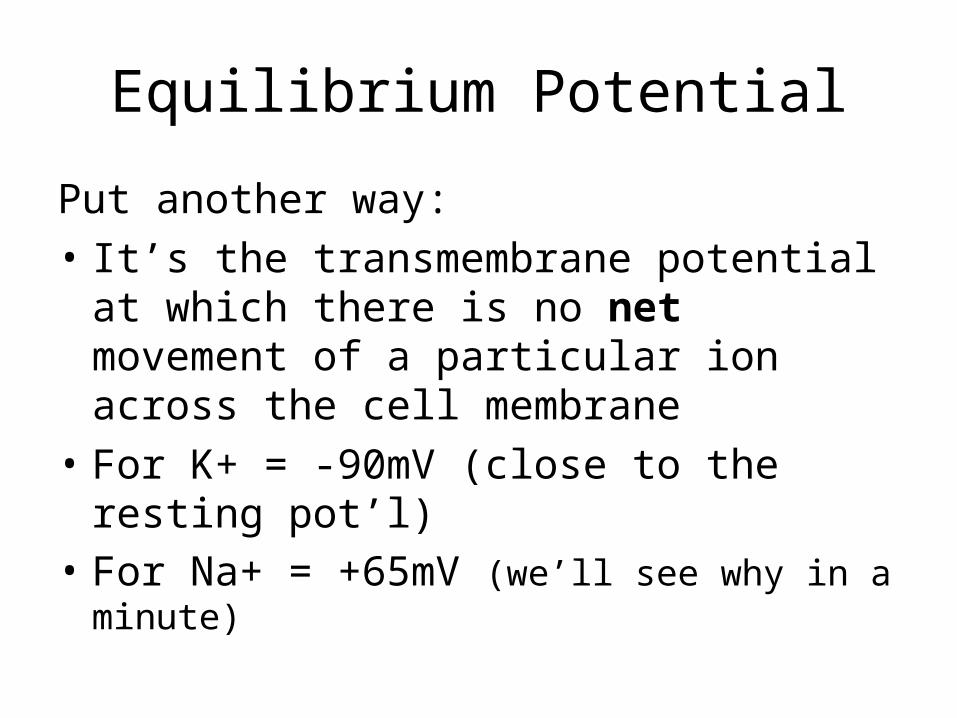
Equilibrium Potential
Put another way:
• It’s the transmembrane potential at which there is no net movement of a particular ion across the cell membrane
• For K+ = -90mV (close to the resting pot’l)• For Na+ = +65mV (we’ll see why in a minute)
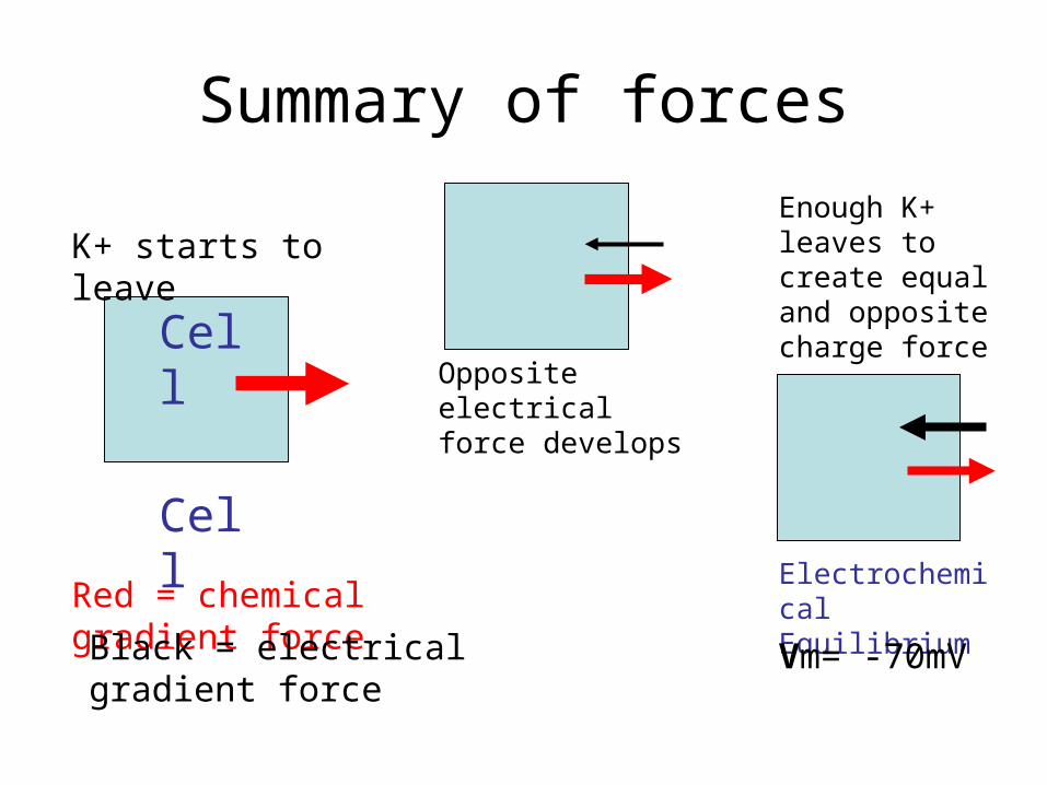
Summary of forces
Red = chemical gradient force
Black = electrical gradient force
Electrochemical Equilibrium
Vm= -70mV
K+ starts to leave
CellOpposite electrical force develops
Enough K+ leaves to create equal and opposite charge force
Cell
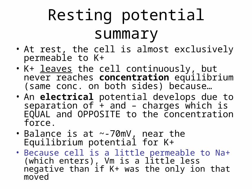
Resting potential summary
• At rest, the cell is almost exclusively permeable to K+
• K+ leaves the cell continuously, but never reaches concentration equilibrium (same conc. on both sides) because…
• An electrical potential develops due to separation of + and – charges which is EQUAL and OPPOSITE to the concentration force.
• Balance is at ~-70mV, near the Equilibrium potential for K+
• Because cell is a little permeable to Na+ (which enters), Vm is a little less negative than if K+ was the only ion that moved
Interesting note
• The amount of potassium ions that must leave the cell in order to leave behind a negative charge is negligible.
• Hard to believe, but true
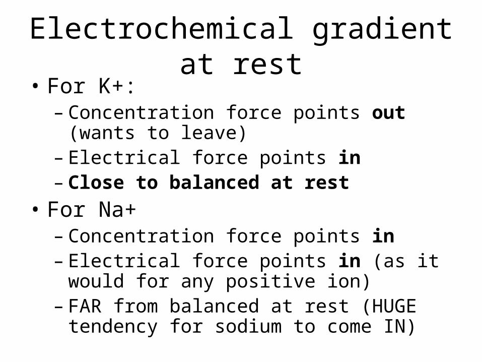
Electrochemical gradient at rest• For K+:
– Concentration force points out (wants to leave)
– Electrical force points in – Close to balanced at rest
• For Na+– Concentration force points in– Electrical force points in (as it would for any
positive ion)– FAR from balanced at rest (HUGE tendency
for sodium to come IN)
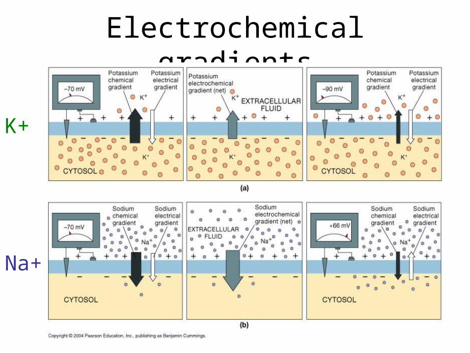
Electrochemical gradients
Na+
K+
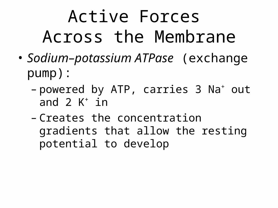
Active Forces Across the Membrane
• Sodium–potassium ATPase (exchange pump): – powered by ATP, carries 3 Na+ out and 2 K+ in– Creates the concentration gradients that allow
the resting potential to develop
Ion movements in neurons
• Ions can only move through the membrane using channels
• There are different kinds of channels – Passive or non-gated channels– Active or gated channels
Passive channels
• Always open
• The reason that neurons are more permeable to K+ than to Na+ at rest is there are far more passive, non-gated K+ channels in the membrane than there are passive Na+ channels
Gated channels
Types:
• Chemically regulated (receptors)
• Voltage regulated
• Mechanically regulated (rare)
Have we seen examples of these channels in other tissue?
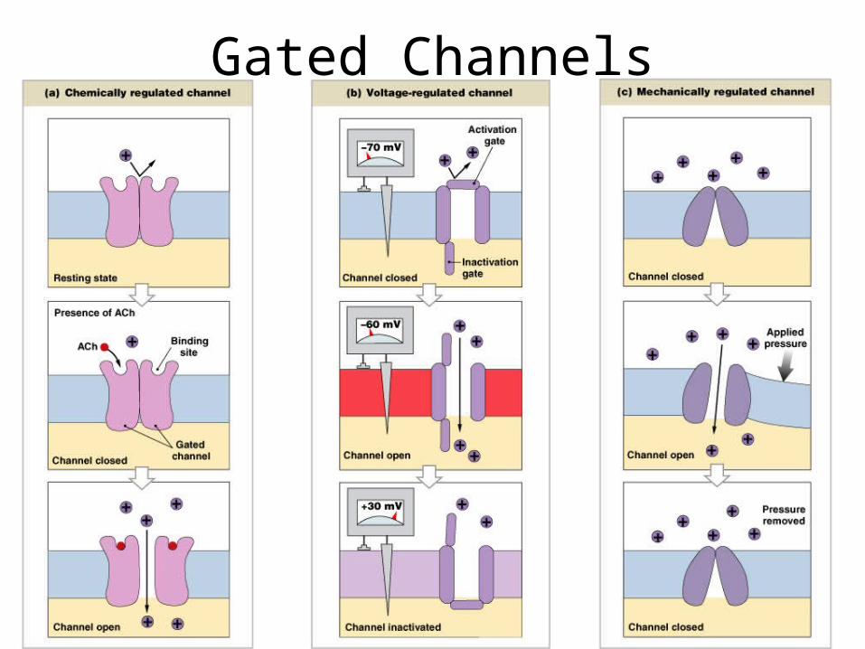
Gated Channels
Figure 12–10
If the gated channels are opened..
• What happens to the membrane potential?
• At a synapse, what type of channels are likely to be activated first, chemically regulated or voltage dependent?
Changes in Membrane Potential
• Transmembrane potential rises or falls:– in response to temporary changes in
membrane permeability resulting from opening or closing gated membrane channels
– Remember it is ion movement that causes electrical signals
• Types of signals – graded potentials and action potentials
Graded Potentials Action Potentials
• Graded Potentials, caused by chemically gated channels, often start off the activity in a neuron, which then gets passed on down the cell via electrically gated channels
• Graded potentials occur at a synapse caused by neurotransmitters, then lead to action potentials
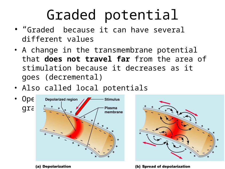
Graded potential• “Graded” because it can have several different values
• A change in the transmembrane potential that does not travel far from the area of stimulation because it decreases as it goes (decremental)
• Also called local potentials• Opening gated sodium channels produces graded
potentials
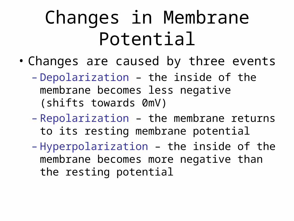
Changes in Membrane Potential
• Changes are caused by three events– Depolarization – the inside of the membrane
becomes less negative (shifts towards 0mV)– Repolarization – the membrane returns to its
resting membrane potential– Hyperpolarization – the inside of the
membrane becomes more negative than the resting potential
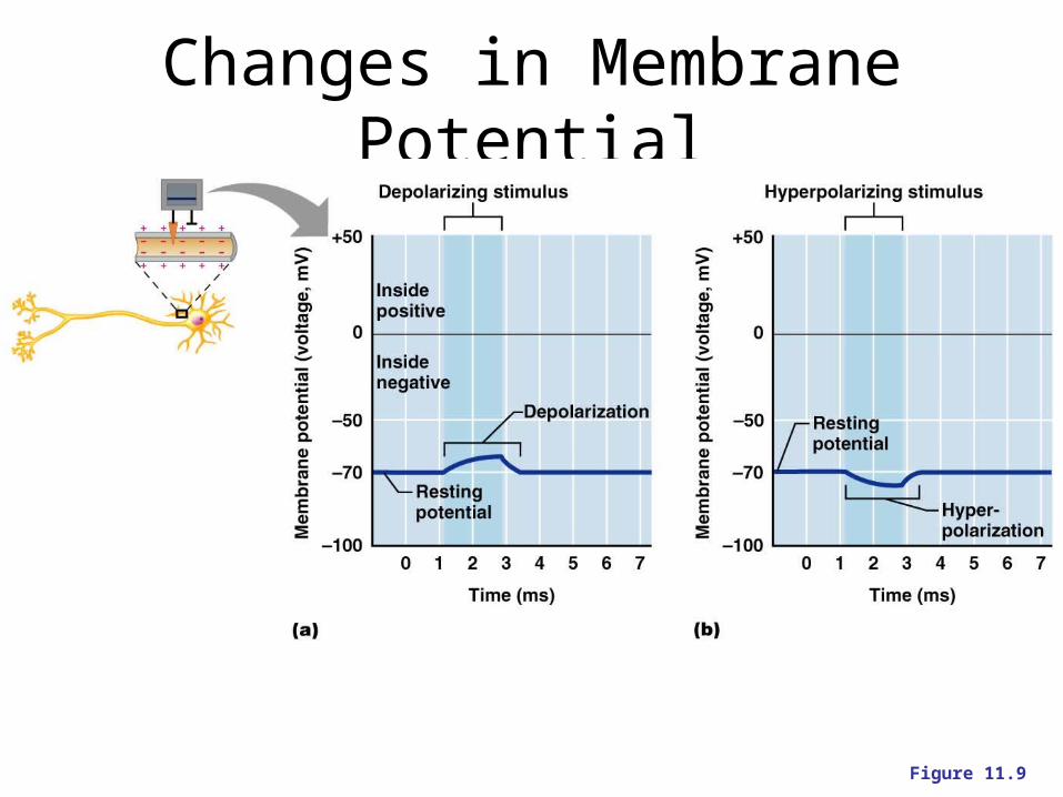
Changes in Membrane Potential
Figure 11.9
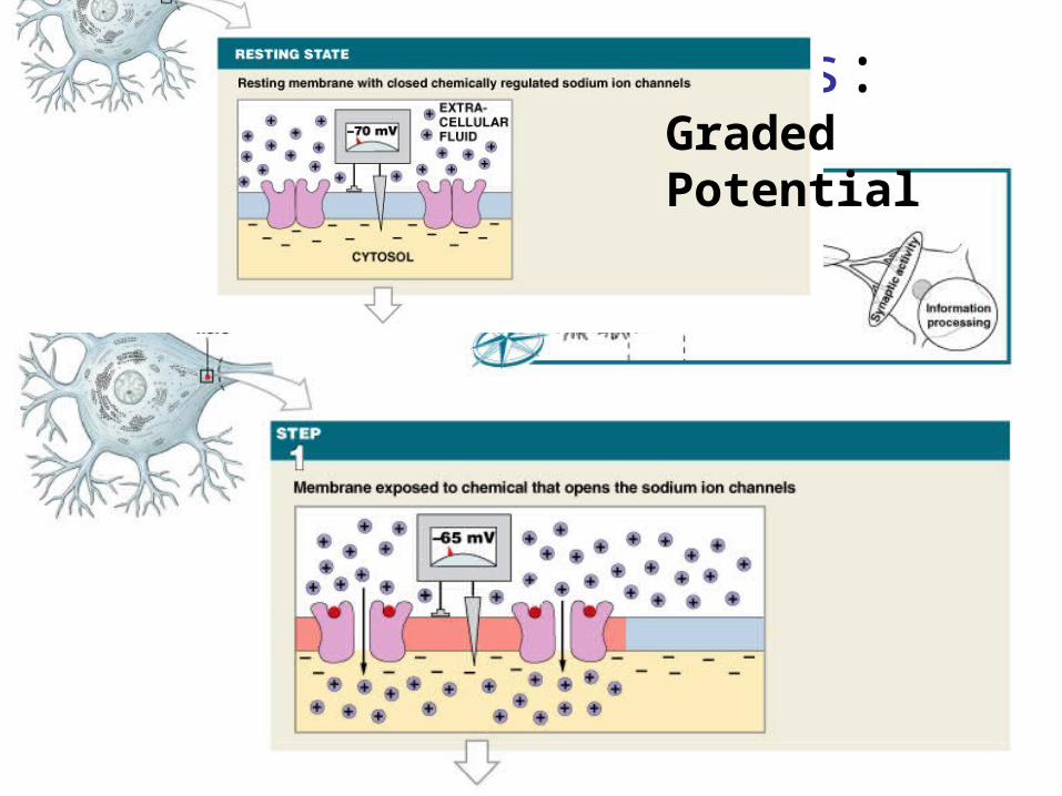
Graded Potentials:
Figure 12–11 (Step 1)
Graded Potential
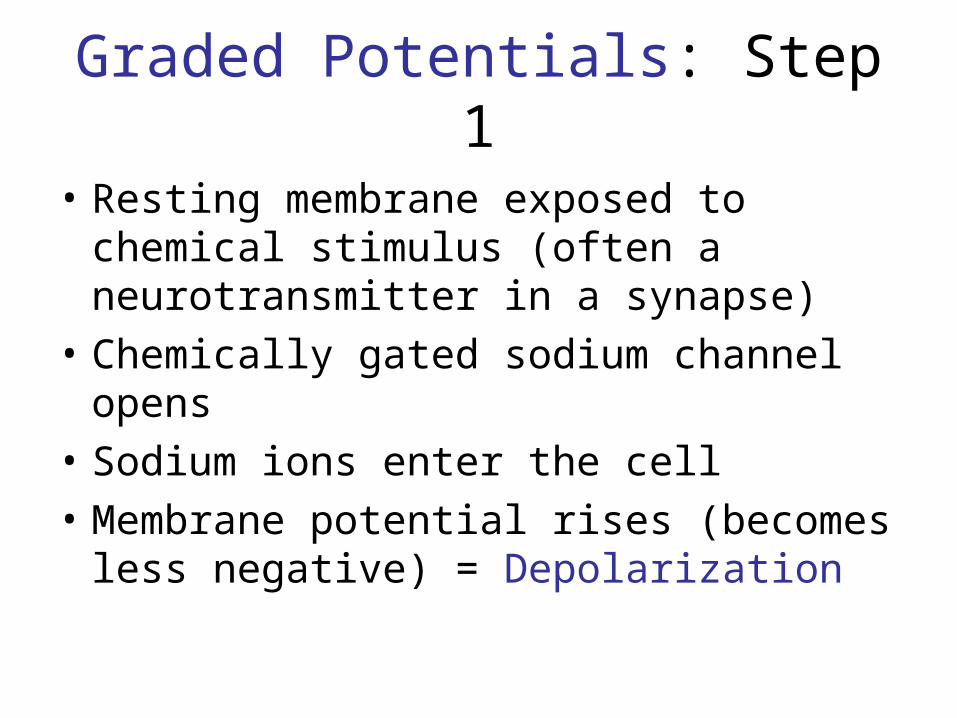
Graded Potentials: Step 1
• Resting membrane exposed to chemical stimulus (often a neurotransmitter in a synapse)
• Chemically gated sodium channel opens
• Sodium ions enter the cell
• Membrane potential rises (becomes less negative) = Depolarization
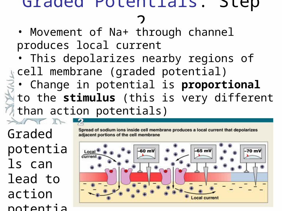
Graded Potentials: Step 2
Figure 12–11 (Step 2)
• Movement of Na+ through channel produces local current • This depolarizes nearby regions of cell membrane (graded potential)• Change in potential is proportional to the stimulus (this is very different than action potentials)
Graded potentials can lead to action potentials
Action Potentials
• Propagated changes in the transmembrane potential that spread through a neuron from the axon hillock to the synaptic terminals and result in release of neurotransmitters
• Key Features:– Change in membrane potential– Travels length of axon in one direction– Always the same regardless of stimulus– Function: Link activity at cell body and dendrites with
synaptic activity at terminals
Initiating Action Potential
• Initial stimulus: – a graded depolarization must reach the axon
hillock and be large enough (10 to 15 mV) to change membrane potential from -70 mV (resting) to threshold (about -60 to -55 mV)
– Threshold = voltage that, if it is attained at the axon hillock, will always* cause an action potential
• It is the point at which voltage-gated sodium channels open
• Hillock is the critical decision point because it has the highest concentration of voltage gated sodium channels
All-or-None Principle
• If a stimulus exceeds threshold :– the action potential is the same no matter how
large the stimulus
• It is decidedly NOT proportional to the stimulus; as long as you get there, it’s all the same
• Action potential is either triggered, or not
• Gun analogy
Keys to the AP
• Electrochemical gradient for Na+ is really big pointing in (both concentration and electrical forces are pulling Na+ in)
• When threshold is reached, voltage-gated Na+ channels open, allowing Na+ to “rush” in
Keys to the AP
• In a positive feedback mechanism, Na+ coming in through chemically gated channels (graded potential) depolarize the membrane causing voltage gated Na+ channels to open.
• Voltage-gated Na+ channels allow more Na+ in, this depolarizes the membrane more, opens more voltage-gated Na+ channels, etc.
• Na+ indepolarizationopens Na+ channels
Keys to the AP
• At about +30 mV, Na+ channels close, ending the depolarization
• Now voltage gated K+ channels open (in addition to the passive K+ channels that are always open) to help return the voltage to its resting value of -70mV
• These cause the potential to overshoot -70 and go a little more negative.
• When they close, Vm returns to -70mV
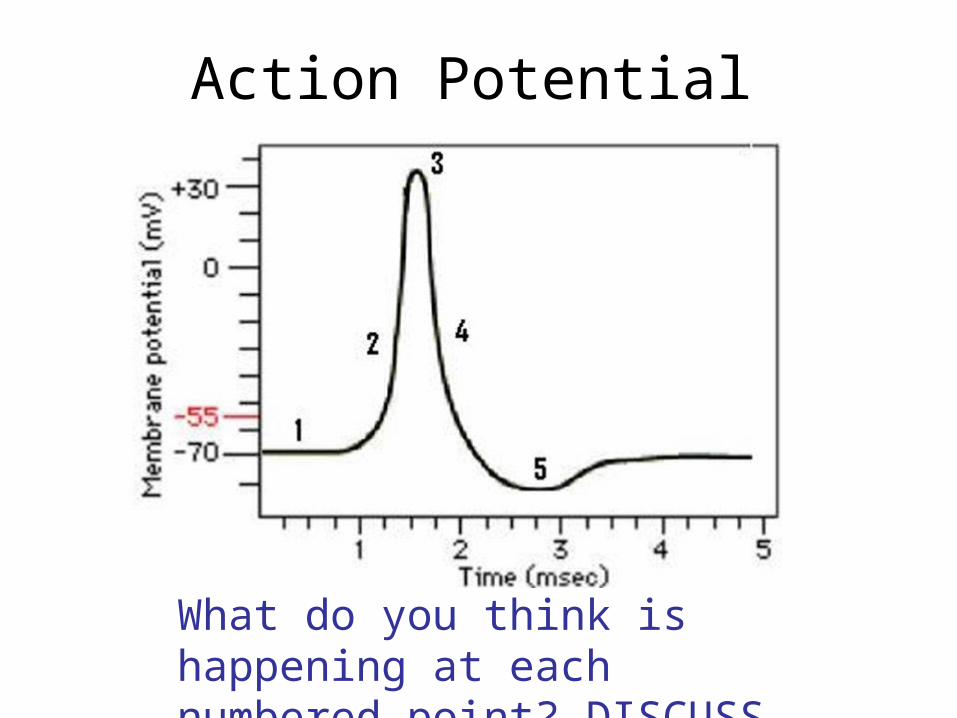
Action Potential
What do you think is happening at each numbered point? DISCUSS

Movie
• Action potential
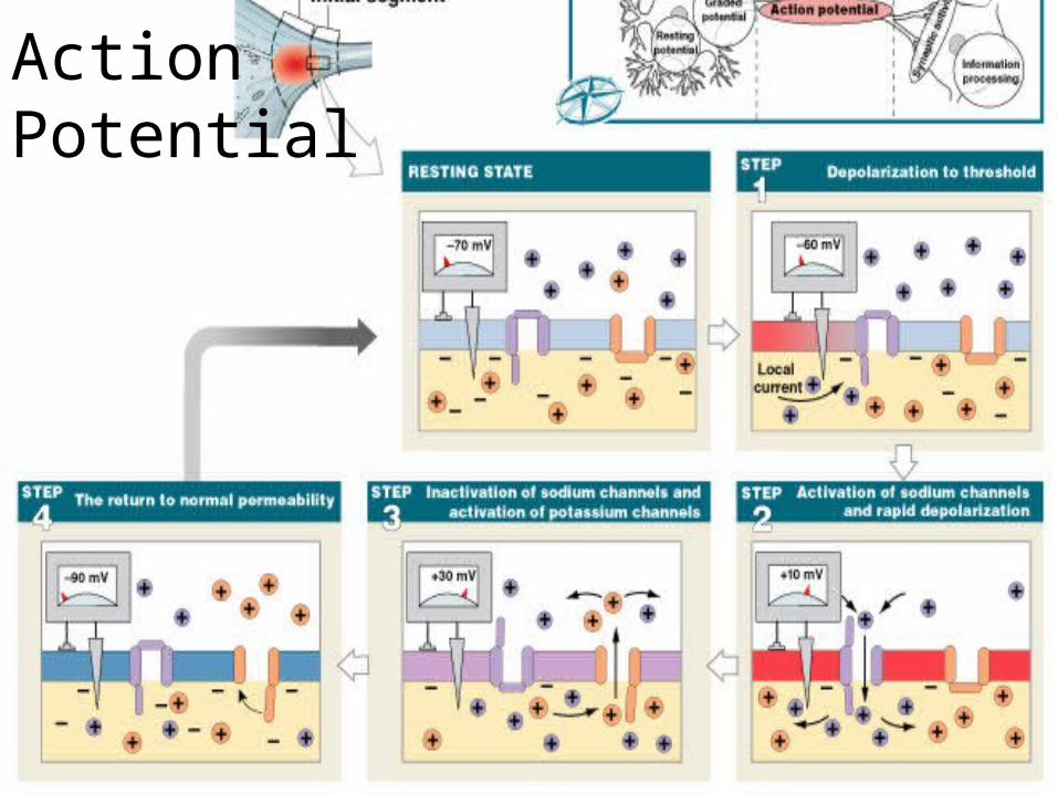
Figure 12–13 (Navigator)
Action Potential
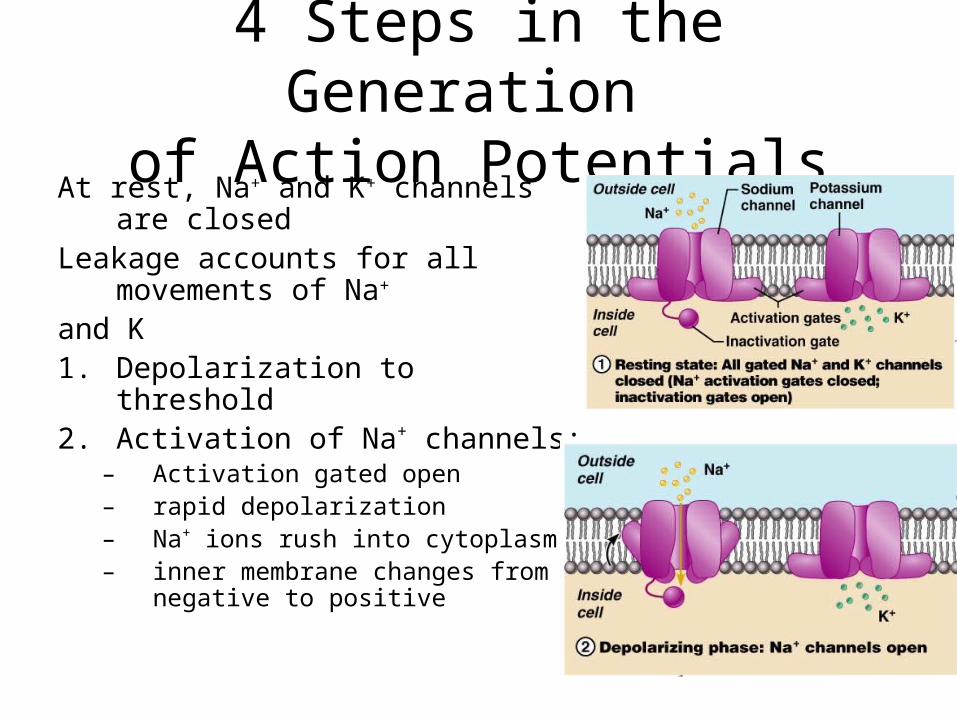
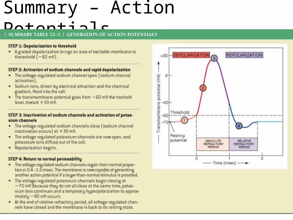
4 Steps in the Generation of Action Potentials
At rest, Na+ and K+ channels are closed
Leakage accounts for all movements of Na+
and K1. Depolarization to threshold2. Activation of Na+ channels:
– Activation gated open– rapid depolarization– Na+ ions rush into cytoplasm– inner membrane changes from
negative to positive
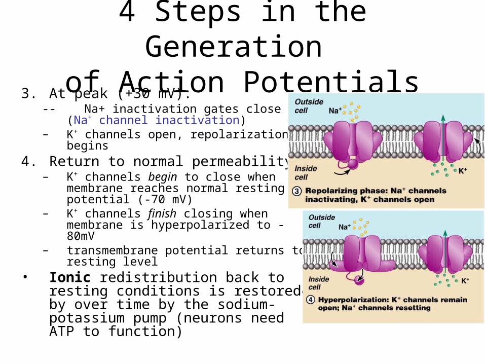
4 Steps in the Generation of Action Potentials
3. At peak (+30 mV):-- Na+ inactivation gates close (Na+
channel inactivation)– K+ channels open, repolarization
begins
4. Return to normal permeability:– K+ channels begin to close when
membrane reaches normal resting potential (-70 mV)
– K+ channels finish closing when membrane is hyperpolarized to -80mV
– transmembrane potential returns to resting level
• Ionic redistribution back to resting conditions is restored by over time by the sodium-potassium pump (neurons need ATP to function)
Summary – Action Potentials
Table 12-3
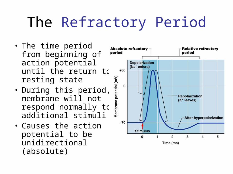
The Refractory Period
• The time period from beginning of action potential until the return to resting state
• During this period, membrane will not respond normally to additional stimuli
• Causes the action potential to be unidirectional (absolute)
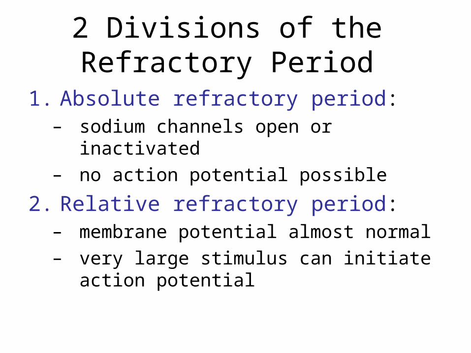
2 Divisions of the Refractory Period
1. Absolute refractory period:– sodium channels open or inactivated– no action potential possible
2. Relative refractory period:– membrane potential almost normal– very large stimulus can initiate action
potential
Propagation (Conduction) of Action Potentials
• Propagation:– moves action potentials generated in axon hillock
along entire length of axon– This occurs in a series of repeated actions, not by
passive flow
• Rate of impulse propagation is determined by:– Axon diameter – the larger the diameter, the faster
the impulse (less resistance in large axons)– Presence of a myelin sheath – myelination
dramatically increases impulse speed
2 Methods of Propagating Action Potentials
1. Continuous propagation:– unmyelinated axons
2. Saltatory propagation:– myelinated axons
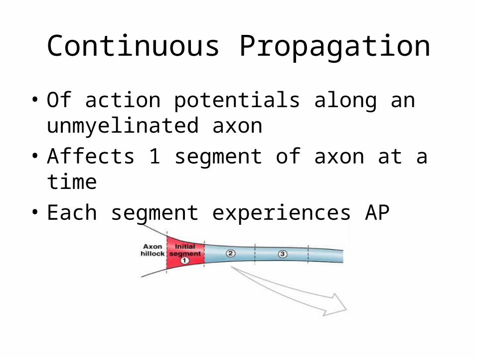
Figure 12–14
Continuous Propagation
• Of action potentials along an unmyelinated axon
• Affects 1 segment of axon at a time
• Each segment experiences AP
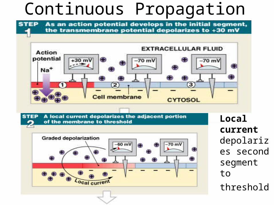
Continuous Propagation
Figure 12–14 (Step 1)
Local current depolarizes second segment to
threshold
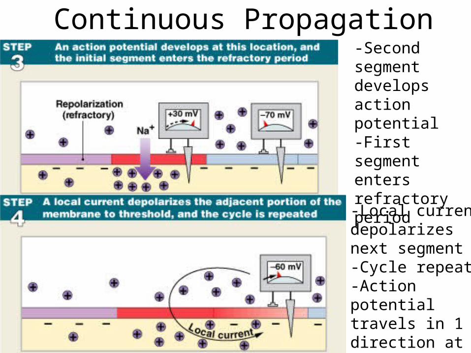
Continuous Propagation
Figure 12–14 (Step 2)
-Second segment develops action potential-First segment enters refractory period
-Local current depolarizes next segment-Cycle repeats-Action potential travels in 1 direction at 1 m/sec
Why can APs only travel in one direction?
• What if you started one right in the middle of the axon (say, by injecting a bunch of Na+)?
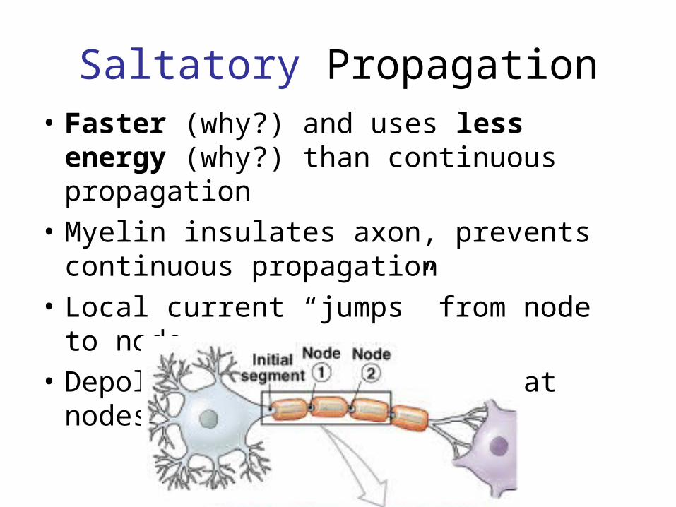
Saltatory Propagation• Faster (why?) and uses less energy
(why?) than continuous propagation
• Myelin insulates axon, prevents continuous propagation
• Local current “jumps” from node to node
• Depolarization occurs only at nodes
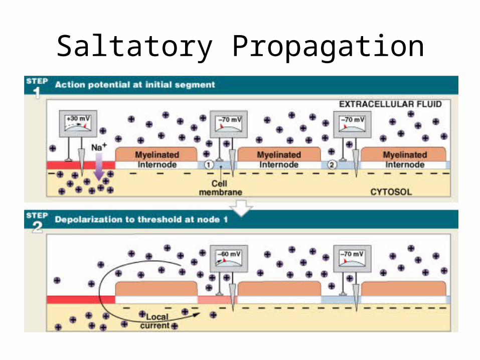
Saltatory Propagation
Figure 12–15 (Steps 1, 2)
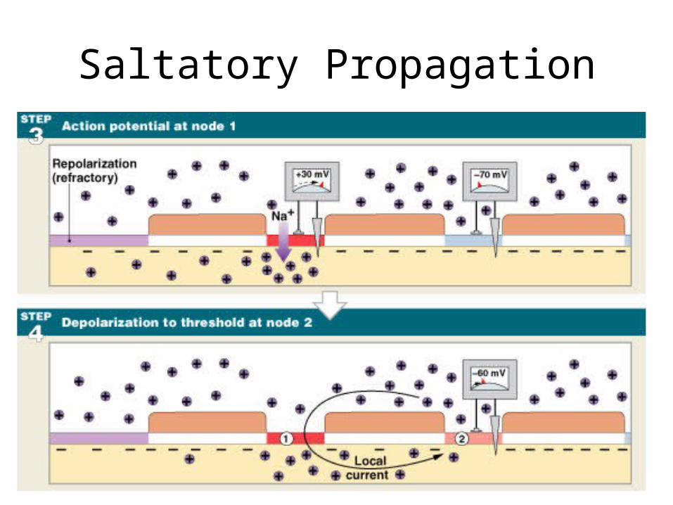
Saltatory Propagation
Figure 12–15 (Steps 3, 4)
Multiple Sclerosis (MS)• An autoimmune disease that mainly affects
young adults• Symptoms: visual disturbances, weakness,
loss of muscular control, and urinary incontinence
• Nerve fibers are severed and myelin sheaths in the CNS become nonfunctional scleroses
• Shunting and short-circuiting of nerve impulses occurs
KEY CONCEPT
• “Information” travels within the nervous system as propagated electrical signals (action potentials)
• The most important information (vision, balance, motor commands) is carried by large-diameter myelinated axons
Movie
• Synapse activity
• Action potentials:– are transmitted from presynaptic neuron to
postsynaptic neuron (or other postsynaptic cell) across a synapse
2 Types of Synapses
1. Electrical synapses:– direct physical contact between cells
2. Chemical synapses:– signal transmitted across a gap by chemical
neurotransmitters
Synapse types
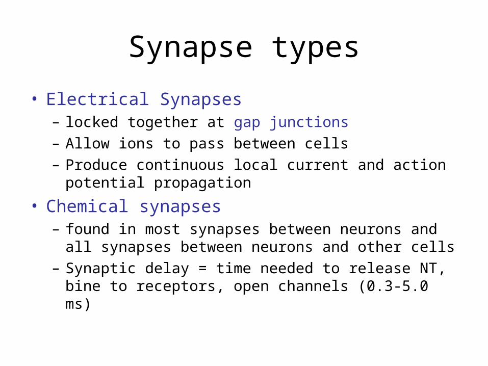
• Electrical Synapses – locked together at gap junctions– Allow ions to pass between cells– Produce continuous local current and action potential
propagation
• Chemical synapses– found in most synapses between neurons and all
synapses between neurons and other cells– Synaptic delay = time needed to release NT, bine to
receptors, open channels (0.3-5.0 ms)
Chemical Synapses
• The most common type of synapse (think about why this might be)
• Response of the postsynaptic cell is dependent on the neurotransmitter AND the type of receptor found in the cell membrane of the postsynaptic cell
An example
• ACh causes a depolarization in the membrane of skeletal muscles and in the heart, ACh causes a transient hyperpolarization
• The response to ACh depends on the receptor in the membrane of the post-synaptic cell
Acetylcholine
• Synapses that release ACh are called cholinergic synapses
• Released at – All neuromuscular junctions involving skeletal
muscles– Synapses in the CNS– Neuron-neuron synapses in the PNS– All neuromuscular and neuroglandular junctions in the
Parasympathetic Nervous System (division of the autonomic NS)
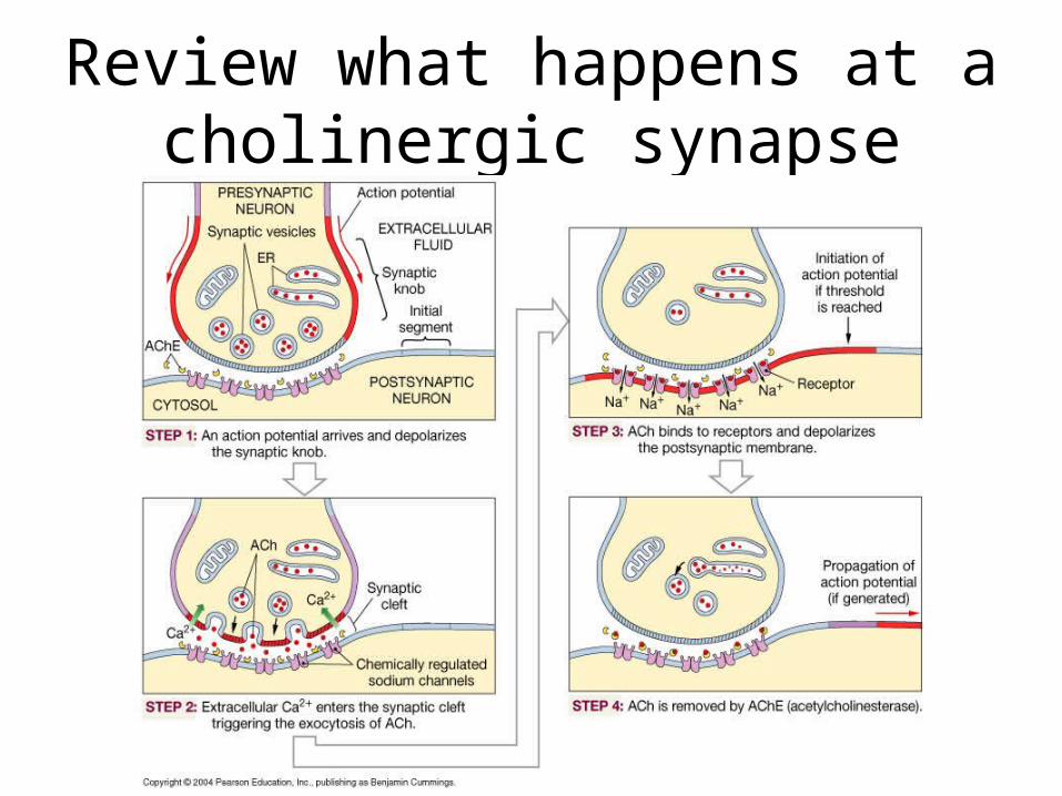
Review what happens at a cholinergic synapse
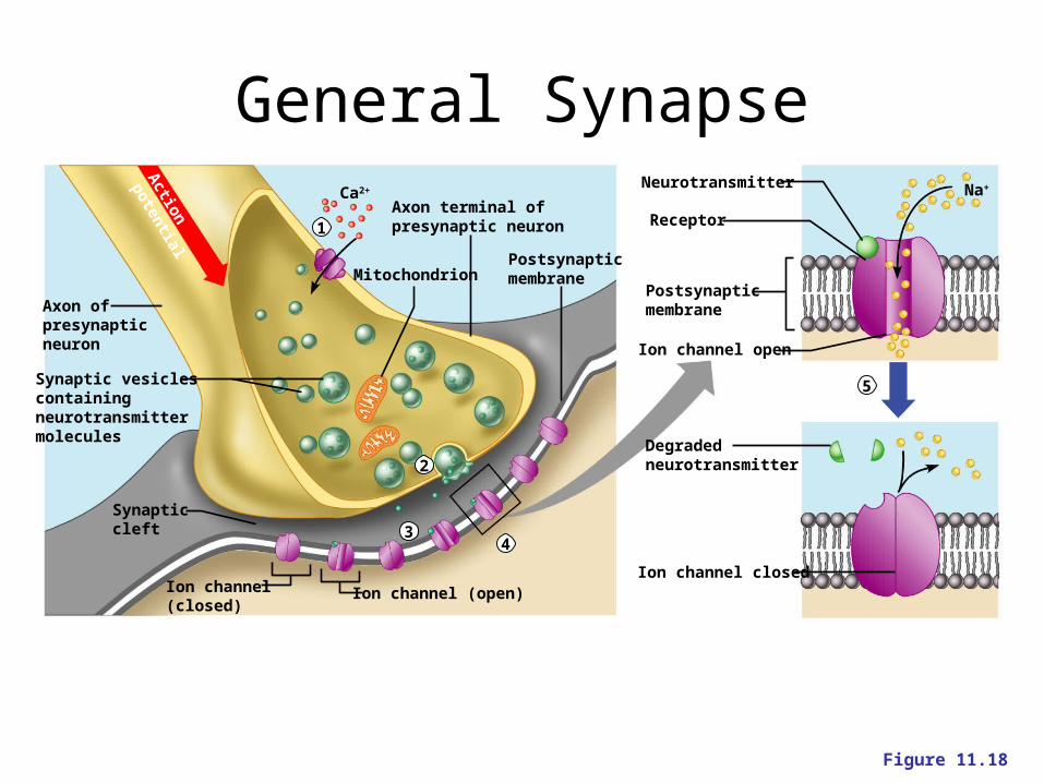
Synaptic vesiclescontaining neurotransmitter molecules
Axon of presynapticneuron
Synapticcleft
Ion channel(closed)
Ion channel (open)
Axon terminal of presynaptic neuron
PostsynapticmembraneMitochondrion
Ion channel closed
Ion channel open
Neurotransmitter
Receptor
Postsynapticmembrane
Degradedneurotransmitter
Na+Ca2+
1
2
34
5
Action
potential
Figure 11.18
General Synapse
Stopping the Message
• Removal of neurotransmitters occurs when they:– Are degraded by enzymes (like AChE)– Are reabsorbed by astrocytes or the
presynaptic terminals (reuptake)– Diffuse from the synaptic cleft
Acetylcholine (ACh)
• Two types of receptors that bind Ach– Nicotinic (the ones in muscles)
• Fast acting
– Muscarinic• Slower, modulatory
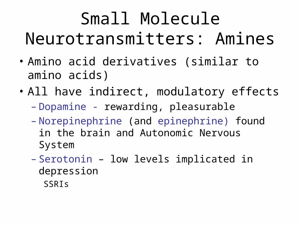
Small Molecule Neurotransmitters: Amines
• Amino acid derivatives (similar to amino acids)
• All have indirect, modulatory effects– Dopamine - rewarding, pleasurable– Norepinephrine (and epinephrine) found in the
brain and Autonomic Nervous System– Serotonin – low levels implicated in
depression SSRIs
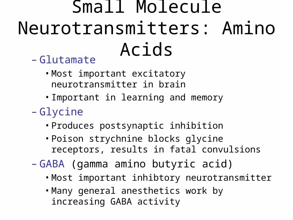
Small Molecule Neurotransmitters: Amino Acids
– Glutamate• Most important excitatory neurotransmitter in brain• Important in learning and memory
– Glycine• Produces postsynaptic inhibition• Poison strychnine blocks glycine receptors, results
in fatal convulsions
– GABA (gamma amino butyric acid)• Most important inhibtory neurotransmitter• Many general anesthetics work by increasing
GABA activity
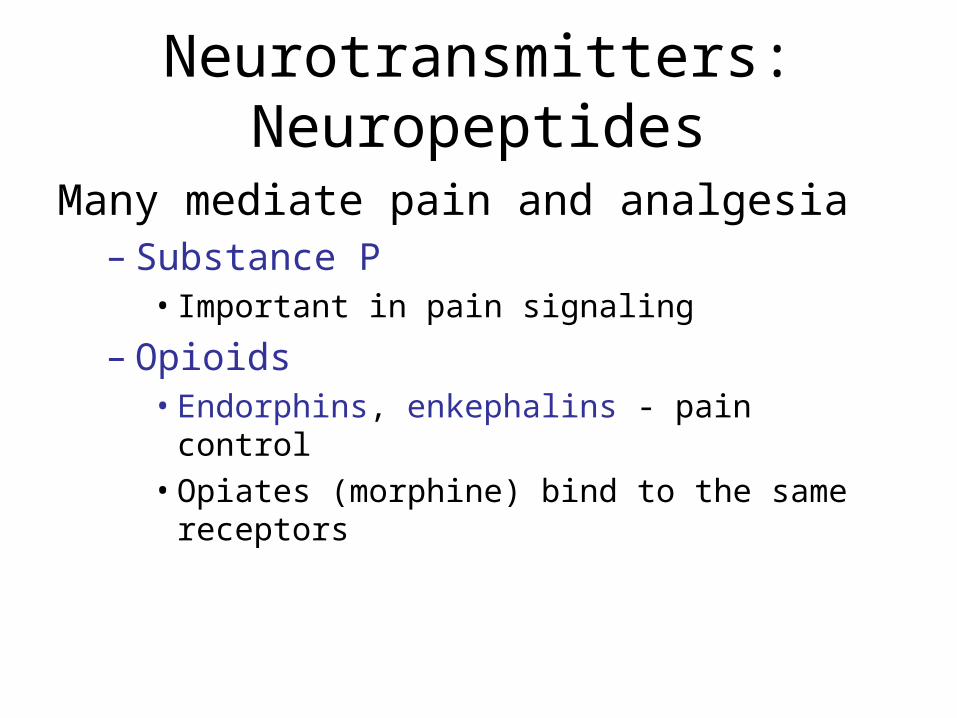
Neurotransmitters: Neuropeptides
Many mediate pain and analgesia– Substance P
• Important in pain signaling
– Opioids• Endorphins, enkephalins - pain control• Opiates (morphine) bind to the same receptors
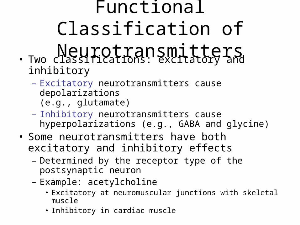
Functional Classification of Neurotransmitters
• Two classifications: excitatory and inhibitory– Excitatory neurotransmitters cause depolarizations
(e.g., glutamate)– Inhibitory neurotransmitters cause hyperpolarizations
(e.g., GABA and glycine)
• Some neurotransmitters have both excitatory and inhibitory effects – Determined by the receptor type of the postsynaptic
neuron – Example: acetylcholine
• Excitatory at neuromuscular junctions with skeletal muscle• Inhibitory in cardiac muscle
Neurotransmitters activate other cells by three main mechanisms • Direct effects (e.g. ACh)
• Indirect via G proteins (e.g. serotonin)
• Indirect via intracellular enzymes
Remember: several neurotransmitters can have wither direct or indirect effects, depending on what receptors are present at a synapse
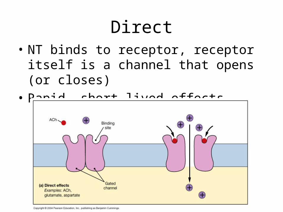
Direct• NT binds to receptor, receptor itself is a
channel that opens (or closes)
• Rapid, short-lived effects
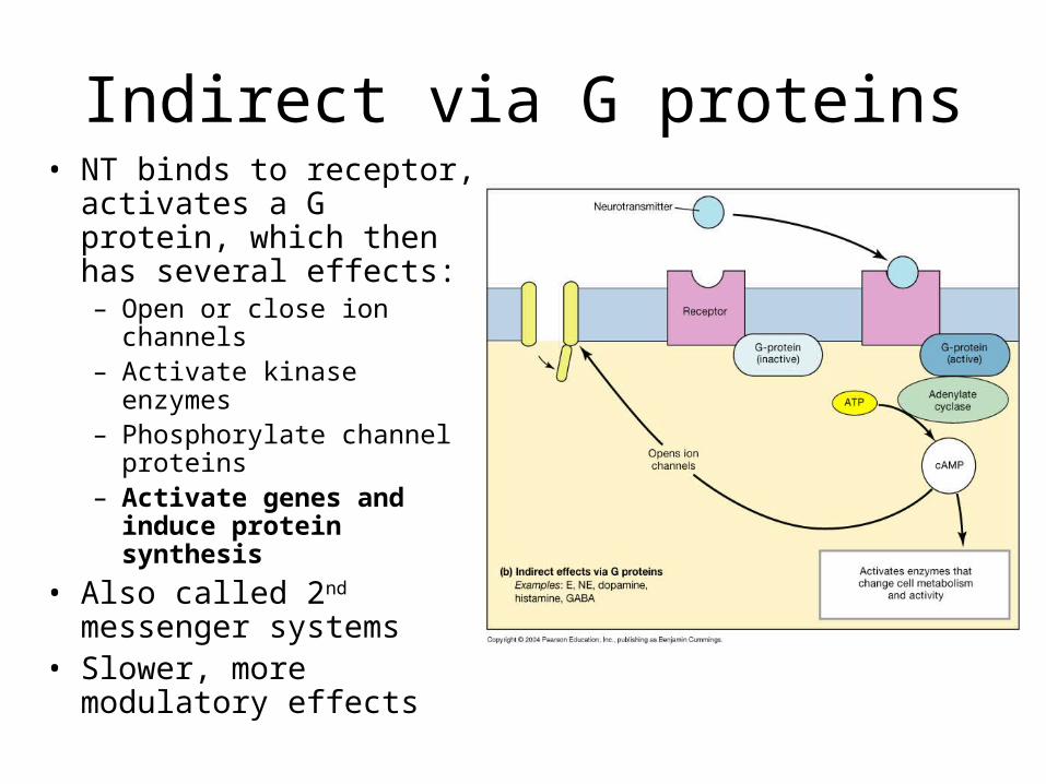
Indirect via G proteins• NT binds to receptor,
activates a G protein, which then has several effects: – Open or close ion
channels– Activate kinase enzymes– Phosphorylate channel
proteins – Activate genes and
induce protein synthesis
• Also called 2nd messenger systems
• Slower, more modulatory effects
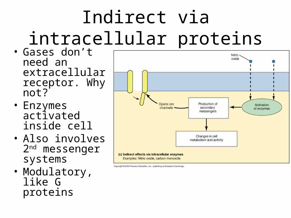
Indirect via intracellular proteins• Gases don’t
need an extracellular receptor. Why not?
• Enzymes activated inside cell
• Also involves 2nd messenger systems
• Modulatory, like G proteins
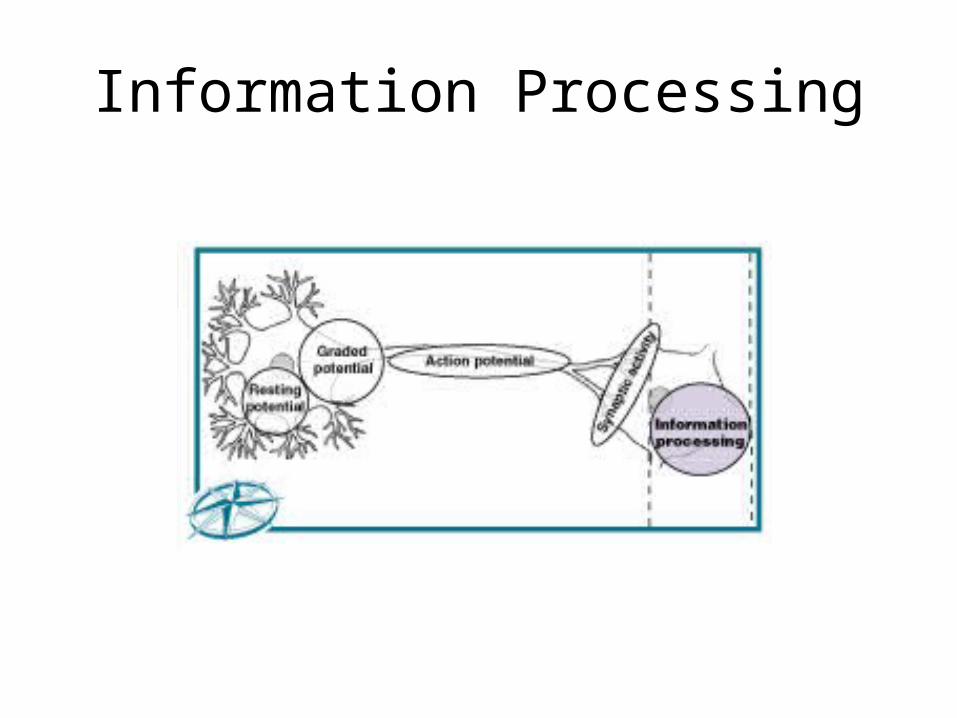
Information Processing
Figure 12–18 (Navigator)
Synapses
• WTF?
• Why do neurons go right up next to each other and then stop, leaving a little space?
• Wouldn’t it just be better if they were all connected by gap junctions? Why is a system with many discrete cells better than cells being just little parts of a huge aggregation?
Information Processing
• At the simplest level (individual neurons):– many dendrites receive neurotransmitter
messages simultaneously– some excitatory, some inhibitory– net effect on axon hillock determines if action
potential is produced
2 Types of Postsynaptic Potentials
1. Excitatory postsynaptic potential (EPSP):– graded depolarization of postsynaptic
membrane (postsynaptic membranes do not generate action potentials – why not?)
2. Inhibitory postsynaptic potential (IPSP):– graded hyperpolarization of postsynaptic
membrane
Inhibition
• A neuron that receives many IPSPs:– is inhibited from producing an action potential– because the stimulation needed to reach
threshold is increased
Summation
• To trigger an action potential:– 1 EPSP is not enough – EPSPs (and IPSPs) combine through
summation:• temporal summation• spatial summation
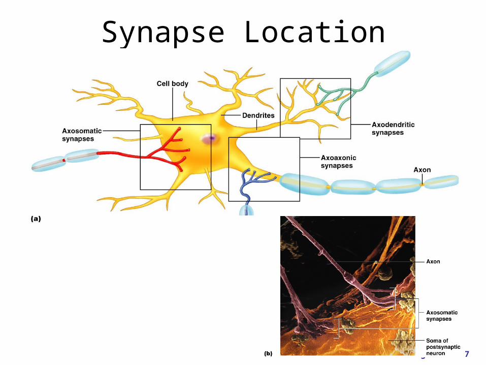
Synapse Location
Figure 11.17
Coding for Stimulus Intensity
• All action potentials are alike and are independent of stimulus intensity
• Frequency of action potentials depends on degree of depolarization above threshold – Strong stimuli can generate an action
potential more often than weaker stimuli
• The CNS determines stimulus intensity by the frequency of impulse transmission
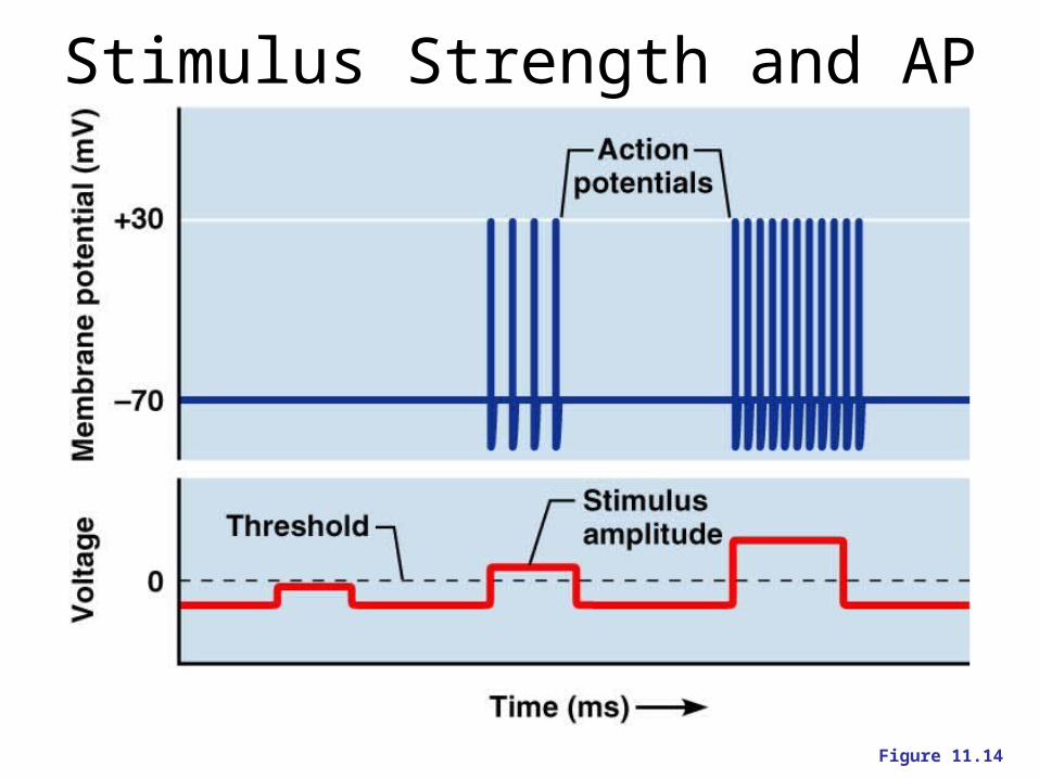
Stimulus Strength and AP Frequency
Figure 11.14
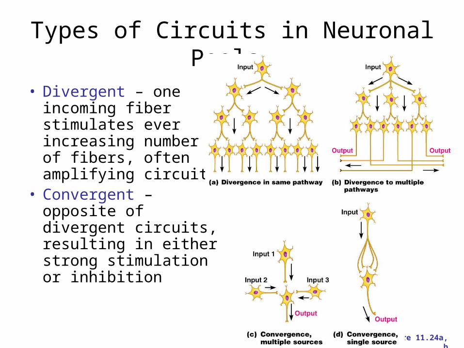
Types of Circuits in Neuronal Pools
• Divergent – one incoming fiber stimulates ever increasing number of fibers, often amplifying circuits
• Convergent – opposite of divergent circuits, resulting in either strong stimulation or inhibition
Figure 11.24a, b
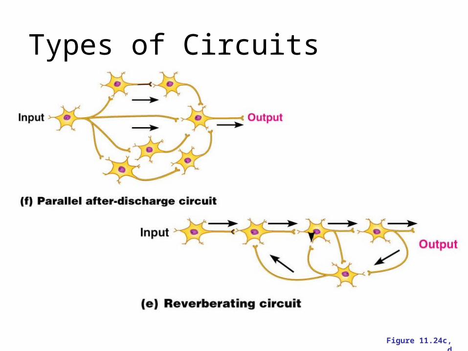
Types of Circuits
Figure 11.24c, d
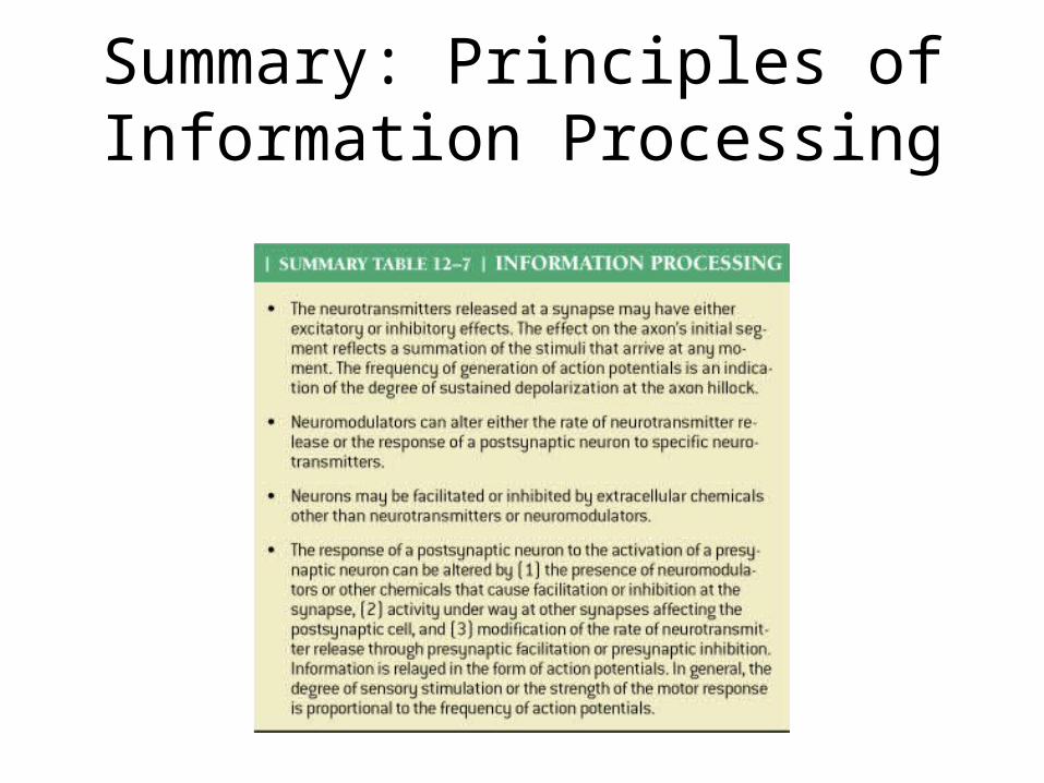
Summary: Principles of Information Processing
Table 12-7