Embed Size (px)
Citation preview
7/28/2019 Lecture 10 - Hemolytic Anemias - Extracorpuscular Defects1
http://slidepdf.com/reader/full/lecture-10-hemolytic-anemias-extracorpuscular-defects1 1/28
What is destroying this RBC?
7/28/2019 Lecture 10 - Hemolytic Anemias - Extracorpuscular Defects1
http://slidepdf.com/reader/full/lecture-10-hemolytic-anemias-extracorpuscular-defects1 2/28
Extracorpuscular defects
•
Immune hemolytic anemias – These anemias result from a shortened RBC
survival mediated by the immune response,specifically humoral antibodies.
– There are three broad categories:
• Alloimmune in which the patient producesalloantibodies to foreign RBC antigens introduced
through transfusion or pregnancy –Transfusion reaction
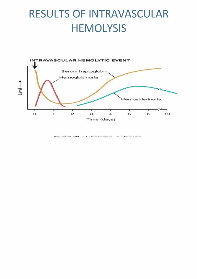
» An immediate transfusion reaction is characterized byacute intravascular hemolysis, mostly associated with
ABO IgM isoantibodies. The patient’s antibodiesdestroy the donor’s cells.
7/28/2019 Lecture 10 - Hemolytic Anemias - Extracorpuscular Defects1
http://slidepdf.com/reader/full/lecture-10-hemolytic-anemias-extracorpuscular-defects1 3/28
RESULTS OF INTRAVASCULAR
HEMOLYSIS
7/28/2019 Lecture 10 - Hemolytic Anemias - Extracorpuscular Defects1
http://slidepdf.com/reader/full/lecture-10-hemolytic-anemias-extracorpuscular-defects1 4/28
Extracorpuscular defects»A delayed transfusion reaction occurs 2-14
days after transfusion and usually is the result
of an anamnestic response in which IgG
antibodies are made in an individual who has
been previously sensitized. Extravascularhemolysis of IgG coated antibodies occurs in
the spleen.
–Hemolytic disease of the newborn
»RBCs of the fetus are destroyed by maternal
IgG antibodies that cross the placenta.
»The baby is often born jaundiced, anemic and
with hepatosplenomegaly.
7/28/2019 Lecture 10 - Hemolytic Anemias - Extracorpuscular Defects1
http://slidepdf.com/reader/full/lecture-10-hemolytic-anemias-extracorpuscular-defects1 5/28
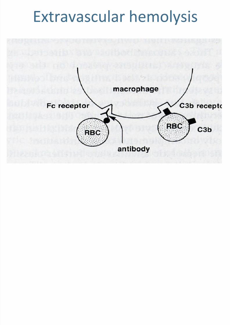
Extravascular hemolysis
7/28/2019 Lecture 10 - Hemolytic Anemias - Extracorpuscular Defects1
http://slidepdf.com/reader/full/lecture-10-hemolytic-anemias-extracorpuscular-defects1 6/28
Extracorpuscular defects•
Autoimmune hemolytic anemia – This represents anabnormality in which the immune system’s ability for
self-recognition is lost and antibodies are made to the
RBC antigens (autoantibodies). They bind to the RBCs
and initiate hemolysis.
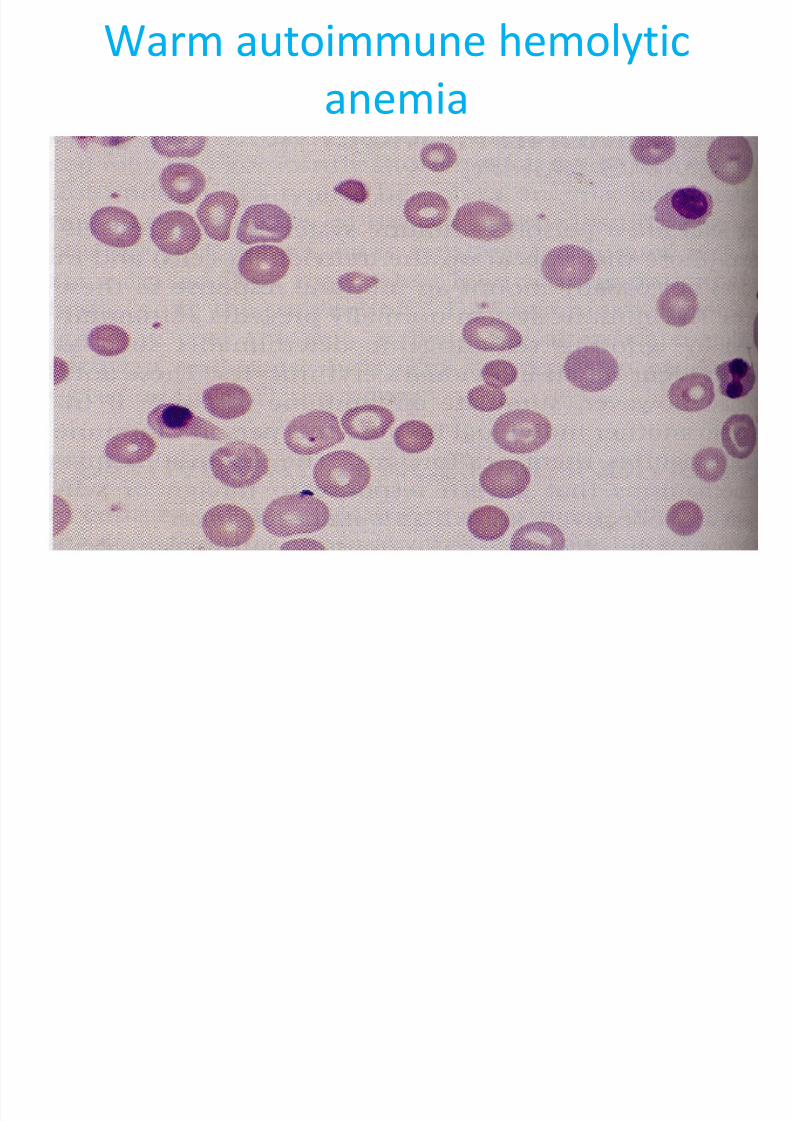
– Warm autoimmune hemolytic anemia - In this type
of immune hemolytic anemia the serologic
reactivity of the IgG antibody involved is optimal at
370 C.
» Primary, idiopathic - severe, but self-limiting anemias that
may last several weeks to years.
» Secondary – associated with some underlying disease
(lymphoproliferative, neoplastic, SLE, RA, viral or bacterial
infection, chronic inflammatory disease)
7/28/2019 Lecture 10 - Hemolytic Anemias - Extracorpuscular Defects1
http://slidepdf.com/reader/full/lecture-10-hemolytic-anemias-extracorpuscular-defects1 7/28
Extracorpuscular defects
» In both the primary and secondary form of the
disease, most hemolysis is extravascular and
complement is not necessary for cell destruction,
though it may be involved (Ag-Ab complexes may bepitted from the cell membrane in the spleen or the
cell itself may be ingested by phagocytic cells).
»The anemia is moderate to severe, the RBCs are
normochromic, normocytic with polychromasia(increased reticulocytes).
»Spherocytes, schistocytes, etc. may be seen and are
indicative of the hemolytic process.
7/28/2019 Lecture 10 - Hemolytic Anemias - Extracorpuscular Defects1
http://slidepdf.com/reader/full/lecture-10-hemolytic-anemias-extracorpuscular-defects1 8/28
Extracorpuscular defects
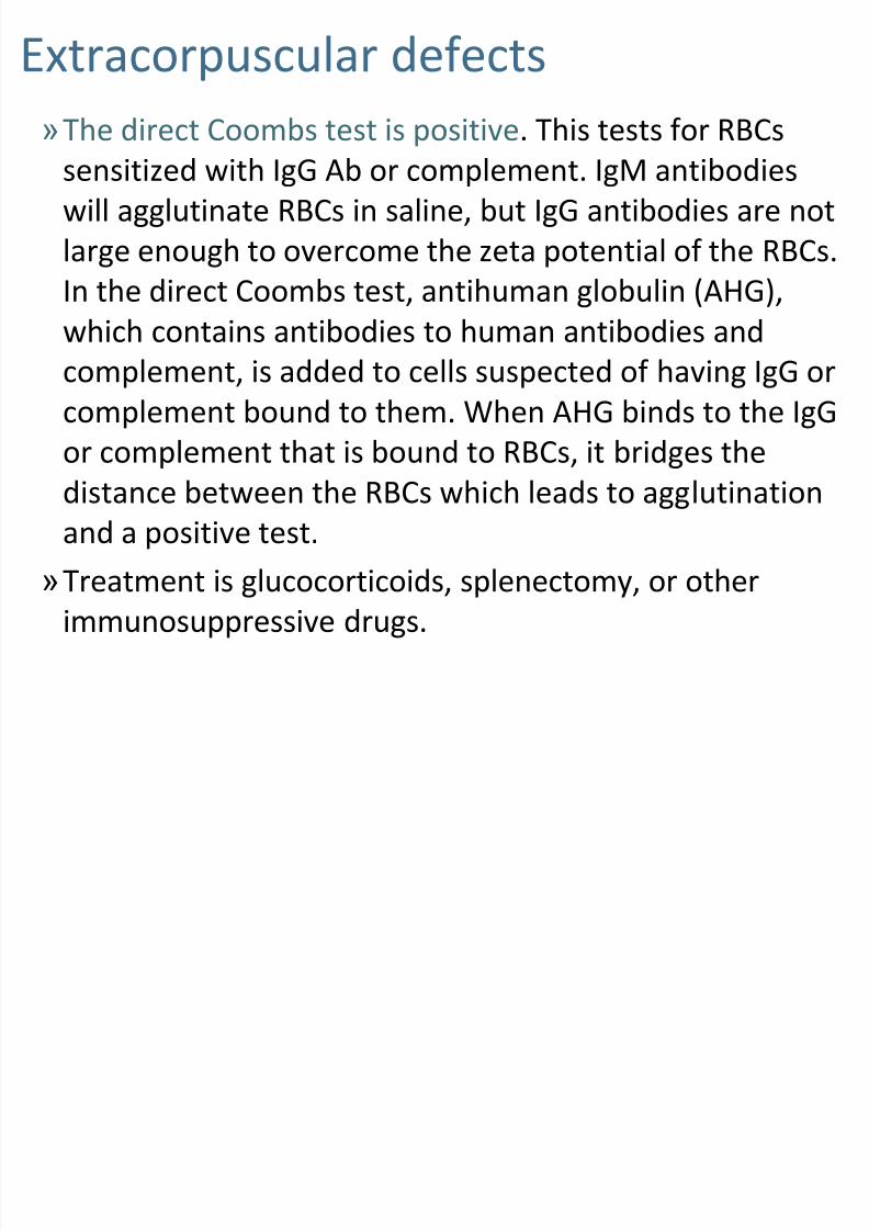
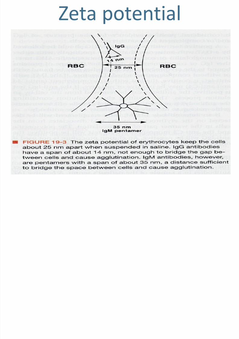
»The direct Coombs test is positive. This tests for RBCs
sensitized with IgG Ab or complement. IgM antibodies
will agglutinate RBCs in saline, but IgG antibodies are not
large enough to overcome the zeta potential of the RBCs.
In the direct Coombs test, antihuman globulin (AHG),which contains antibodies to human antibodies and
complement, is added to cells suspected of having IgG or
complement bound to them. When AHG binds to the IgG
or complement that is bound to RBCs, it bridges thedistance between the RBCs which leads to agglutination
and a positive test.
»Treatment is glucocorticoids, splenectomy, or other
immunosuppressive drugs.
7/28/2019 Lecture 10 - Hemolytic Anemias - Extracorpuscular Defects1
http://slidepdf.com/reader/full/lecture-10-hemolytic-anemias-extracorpuscular-defects1 9/28
Zeta potential
7/28/2019 Lecture 10 - Hemolytic Anemias - Extracorpuscular Defects1
http://slidepdf.com/reader/full/lecture-10-hemolytic-anemias-extracorpuscular-defects1 10/28
Warm autoimmune hemolytic
anemia
7/28/2019 Lecture 10 - Hemolytic Anemias - Extracorpuscular Defects1
http://slidepdf.com/reader/full/lecture-10-hemolytic-anemias-extracorpuscular-defects1 11/28
Extracorpuscular defects
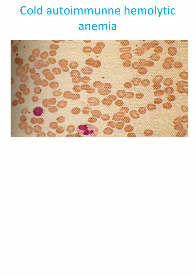
–Cold autoimmune hemolytic anemia – pathologic coldautoantibodies are usually IgM antibodies that fixcomplement and are optimally reactive below 370 C.It is normal to have benign cold autoantibodies, buttheir thermal amplitude and concentration are not
high enough to cause problems. The pathologic formscan be divided into three types:
»Cold agglutinin syndrome – This is idiopathic, chronic,usually in individuals older than 50, and usually due to an
IgM monoclonal antibody.» Secondary, cold autoimmune hemolytic anemia – due to
polyclonal IgM antibodies that develop with Mycoplasma
pneumonia infections, infectious mononucleosis, orlymphoproliferative disease. Is usually transient.
7/28/2019 Lecture 10 - Hemolytic Anemias - Extracorpuscular Defects1
http://slidepdf.com/reader/full/lecture-10-hemolytic-anemias-extracorpuscular-defects1 12/28
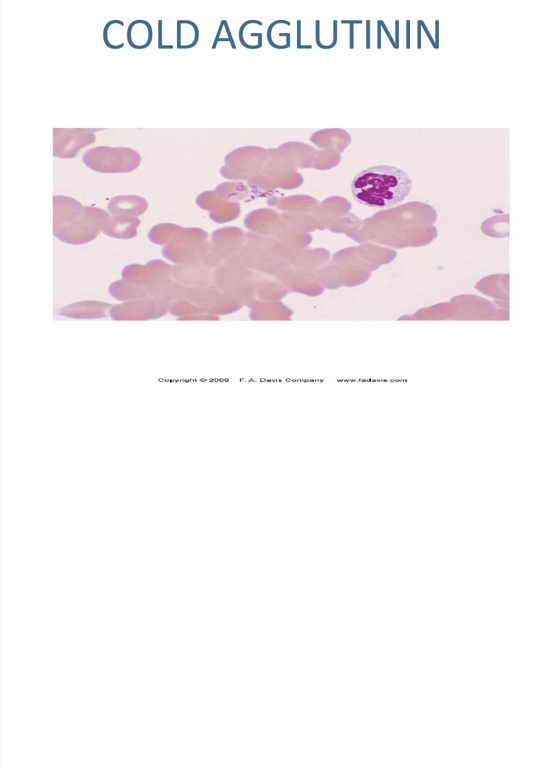
COLD AGGLUTININ
7/28/2019 Lecture 10 - Hemolytic Anemias - Extracorpuscular Defects1
http://slidepdf.com/reader/full/lecture-10-hemolytic-anemias-extracorpuscular-defects1 13/28
Extracorpuscular defects
»
In both a and b the extent of the disease is related to thethermal amplitude of the antibody – if it reacts at 30-320 C,it can cause problems when the peripheral circulation coolsto that temperature:
» IgM binds and fixes complement, upon warming the
antibody dissociates, but complement remains boundleading to either intravascular or extravascular hemolysis.
» The patient may experience acrocyanosis of hands, feet,ears, and nose (with agglutination blood flow slows down,the skin turns white and then blue; upon warming, the skin
turns red).
» Blood counts are difficult to perform unless the blood iswarmed.
» The Coombs test with anti-complement antibody is positive.
7/28/2019 Lecture 10 - Hemolytic Anemias - Extracorpuscular Defects1
http://slidepdf.com/reader/full/lecture-10-hemolytic-anemias-extracorpuscular-defects1 14/28
Extracorpuscular defects
» The cold agglutinin test is positive at 0-200 C and usually upto 300 C. The titer is usually 1:1000 or greater.
» Paroxysmal cold hemoglobinuria (the third type of coldautoimmune hemolytic anemia) – is found in associationwith viral disorders and syphilis and may be chronic.
» This is characterized by massive, intermittent, acuteintravascular hemolysis and hemoglobinuria upon exposureto cold.
» It is caused by a biphasic IgG antibody that binds at low
temperature and fixes complement.»Upon warming, to body temperature, the intravascular
hemolysis occurs and is accompanied by fever, shaking chills,and abdominal and back cramps.
7/28/2019 Lecture 10 - Hemolytic Anemias - Extracorpuscular Defects1
http://slidepdf.com/reader/full/lecture-10-hemolytic-anemias-extracorpuscular-defects1 15/28
Cold autoimmunne hemolytic
anemia
7/28/2019 Lecture 10 - Hemolytic Anemias - Extracorpuscular Defects1
http://slidepdf.com/reader/full/lecture-10-hemolytic-anemias-extracorpuscular-defects1 16/28
Extracorpuscular defects
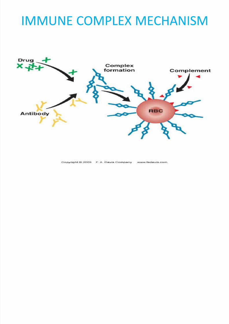
•Drug induced immune hemolytic anemia – many different drugs can cause this and3-4 different mechanisms may be
involved. – Immune complex mechanism
• The drug binds to plasma proteins andantibodies are made against the drug.
• The antibodies bind to the drug to form animmune complex which adsorbs nonspecificallyto the patients RBCs, complement is fixed, andacute intravascular hemolysis occurs.
7/28/2019 Lecture 10 - Hemolytic Anemias - Extracorpuscular Defects1
http://slidepdf.com/reader/full/lecture-10-hemolytic-anemias-extracorpuscular-defects1 17/28
IMMUNE COMPLEX MECHANISM
7/28/2019 Lecture 10 - Hemolytic Anemias - Extracorpuscular Defects1
http://slidepdf.com/reader/full/lecture-10-hemolytic-anemias-extracorpuscular-defects1 18/28
Extracorpuscular defects
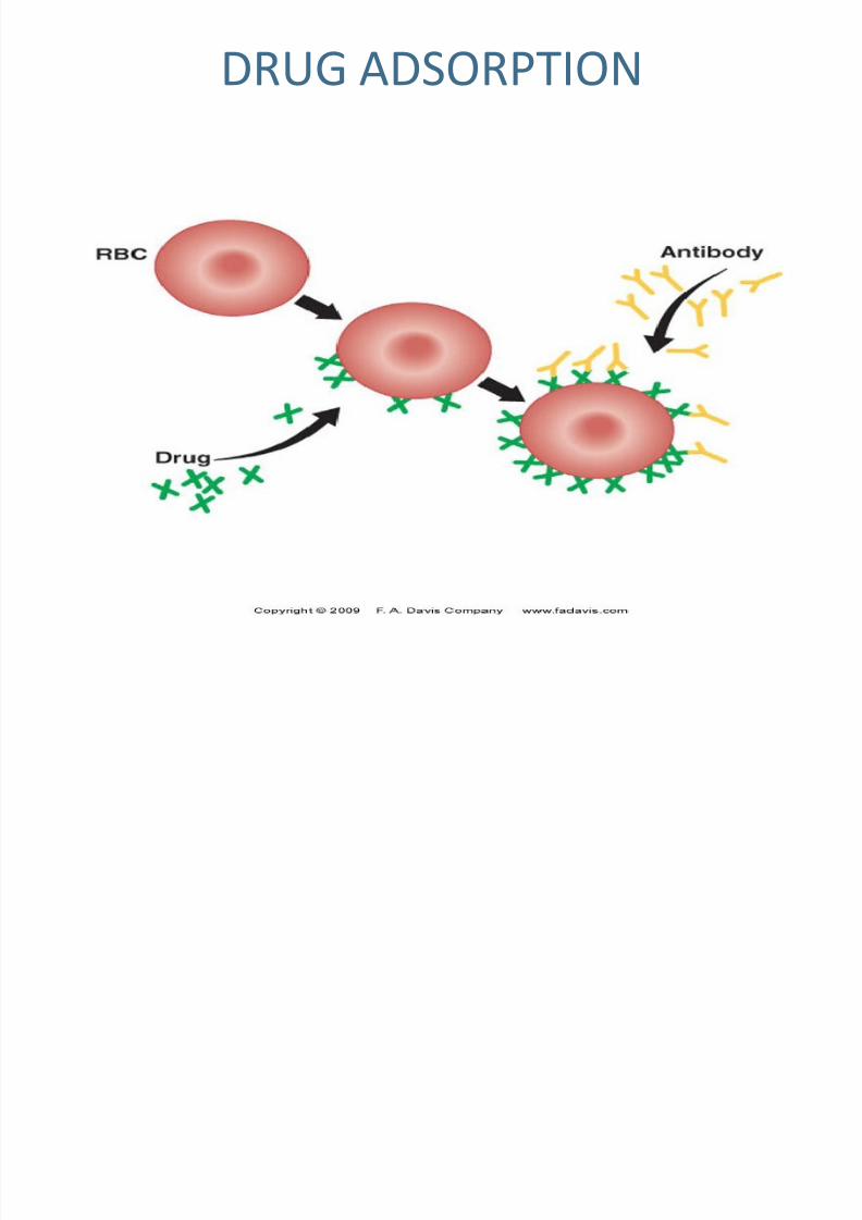
– Drug adsorption (hapten) mechanism
• The drug binds nonspecifically to proteins on the RBCmembrane, antibodies are made (usually IgG), theybind to the drug and extravascular hemolysis occurs.
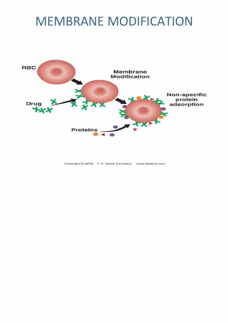
– Membrane modification mechanism
•
The drug modifies the RBC membrane so that normalplasma proteins bind nonimmunologically.
• In rare instances a cross reacting antibody causes ahemolytic anemia.
–
Methyldopa induced mechanism• The drug induces formation of autoantibodies causing
extravascular destruction.
• It may change autoproteins so that they are no longerrecognized as self, or it may cause a direct loss of T
suppressor cells.
7/28/2019 Lecture 10 - Hemolytic Anemias - Extracorpuscular Defects1
http://slidepdf.com/reader/full/lecture-10-hemolytic-anemias-extracorpuscular-defects1 19/28
DRUG ADSORPTION
7/28/2019 Lecture 10 - Hemolytic Anemias - Extracorpuscular Defects1
http://slidepdf.com/reader/full/lecture-10-hemolytic-anemias-extracorpuscular-defects1 20/28
MEMBRANE MODIFICATION
7/28/2019 Lecture 10 - Hemolytic Anemias - Extracorpuscular Defects1
http://slidepdf.com/reader/full/lecture-10-hemolytic-anemias-extracorpuscular-defects1 21/28
Extracorpuscular defects
• Nonimmune hemolytic anemias
– Caused by antagonists in blood or abnormalities inplasma lipids.
• Chemicals and drugs
– Include drugs that cause oxidative injury
– Inhalation of arsine gas
– Lead intoxication (in addition to interfering withheme synthesis, lead can cause membrane damageby interfering with energy production)
– Injection of large volumes of water.
• Animal venoms – bees, wasps, spiders, scorpions insusceptible individuals, rarely snakebites
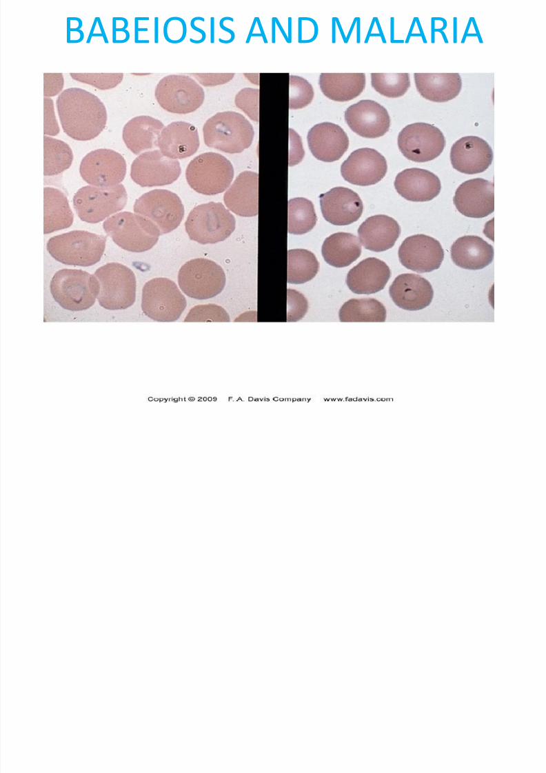
• Infectious agents – malarial parasites, Babeiosis,
Clostridium perfringens, Bartonella bacilliformis
7/28/2019 Lecture 10 - Hemolytic Anemias - Extracorpuscular Defects1
http://slidepdf.com/reader/full/lecture-10-hemolytic-anemias-extracorpuscular-defects1 22/28
BABEIOSIS AND MALARIA
7/28/2019 Lecture 10 - Hemolytic Anemias - Extracorpuscular Defects1
http://slidepdf.com/reader/full/lecture-10-hemolytic-anemias-extracorpuscular-defects1 23/28
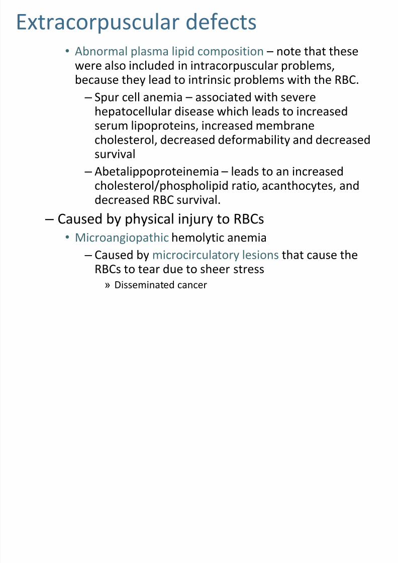
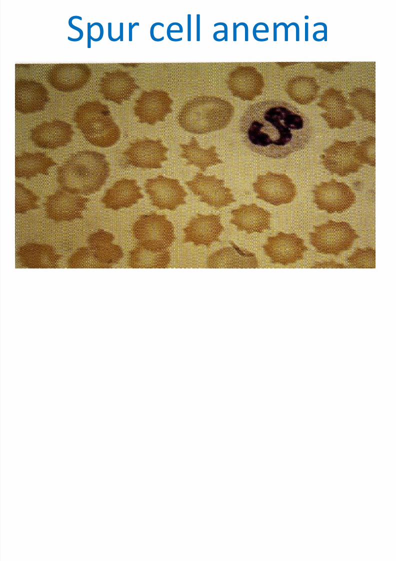
Extracorpuscular defects• Abnormal plasma lipid composition – note that these
were also included in intracorpuscular problems,because they lead to intrinsic problems with the RBC.
– Spur cell anemia – associated with severehepatocellular disease which leads to increasedserum lipoproteins, increased membrane
cholesterol, decreased deformability and decreasedsurvival
– Abetalippoproteinemia – leads to an increasedcholesterol/phospholipid ratio, acanthocytes, anddecreased RBC survival.
– Caused by physical injury to RBCs
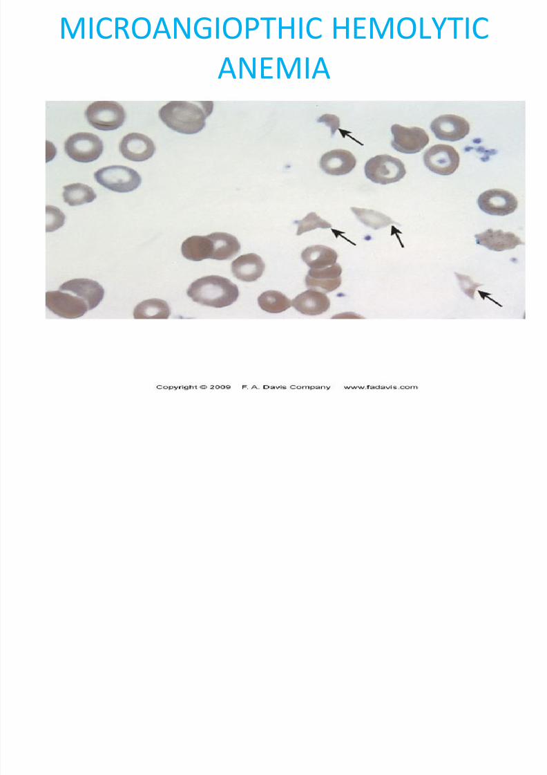
• Microangiopathic hemolytic anemia
– Caused by microcirculatory lesions that cause theRBCs to tear due to sheer stress
» Disseminated cancer
7/28/2019 Lecture 10 - Hemolytic Anemias - Extracorpuscular Defects1
http://slidepdf.com/reader/full/lecture-10-hemolytic-anemias-extracorpuscular-defects1 24/28
Spur cell anemia
7/28/2019 Lecture 10 - Hemolytic Anemias - Extracorpuscular Defects1
http://slidepdf.com/reader/full/lecture-10-hemolytic-anemias-extracorpuscular-defects1 25/28
7/28/2019 Lecture 10 - Hemolytic Anemias - Extracorpuscular Defects1
http://slidepdf.com/reader/full/lecture-10-hemolytic-anemias-extracorpuscular-defects1 26/28
MICROANGIOPTHIC HEMOLYTIC
ANEMIA
7/28/2019 Lecture 10 - Hemolytic Anemias - Extracorpuscular Defects1
http://slidepdf.com/reader/full/lecture-10-hemolytic-anemias-extracorpuscular-defects1 27/28
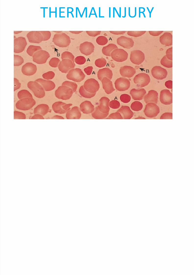
THERMAL INJURY
7/28/2019 Lecture 10 - Hemolytic Anemias - Extracorpuscular Defects1
http://slidepdf.com/reader/full/lecture-10-hemolytic-anemias-extracorpuscular-defects1 28/28
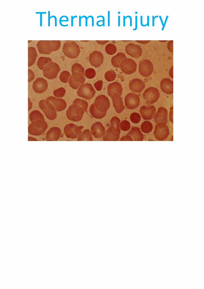
Thermal injury


![Clinical Presentation and Management of Hemolytic Anemias...Clinical Presentation and Management of Hemolytic Anemias Review Article [1] | September 03, 2002 By Kalust Ucar, MD, FACP](https://img.pdfslide.us/doc/110x75/5ed3b0b669d8e83fb45ede5c/clinical-presentation-and-management-of-hemolytic-anemias-clinical-presentation.jpg)