Embed Size (px)
Citation preview
Lecture 1. Arterial Hypertension
V. Babadzhan, D.M. Professor of Medicine,
Kharkov State Medical University Department of Internal Medicine 2,
Clinical Immunology and Allergology
Internal Medicine
Arterial Hypertension
Arterial hypertension defines as an elevation of
systolic and/or diastolic blood pressures to
above 140/90 mm Hg.
In a suburban population like that in the
Framingham Study, nearly one-fifth of individuals
have blood pressures greater than 160/95, while
almost one-half have pressures greater than
140/90.
Patients with arterial hypertension and no definable cause
are said to have primary, essential hypertension.
Individuals in whom a specific structural organ or gene
defect is responsible for hypertension are defined as having
a secondary form of hypertension. The prevalence of
various forms of secondary hypertension in the general
population, although in middle-aged males it has been
reported to be 6 percent. In contrast, individuals in whom
generalized or functional abnormalities may be the cause of
hypertension, even if the abnormalities are discrete, are
defined as having essential hypertension.
CLASSIFICATION OF ARTERIAL HYPERTENSION Systolic hypertension with wide pulse pressure
I. Decreased compliance of aorta (arteriosclerosis) II. Increased stroke volume: A. Aortic regurgitation; B. Thyrotoxicosis; C. Hyperkinetic heart syndrome; D. Fever; E. Arteriovenous fistula; F. Patent ductus arteriosus
Systolic and diastolic hypertension (increased peripheral vascular resistance)
I. Renal A.Chronic pyelonephritis; B.Acute and chronic glomerulonephritis; C.Polycystic renal disease; D.Renovascular stenosis or renal infarction; E.Most other severe renal diseases (arteriolar nephrosclerosis, diabetic nephropathy);
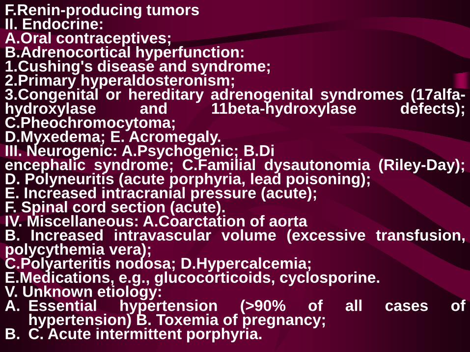
F.Renin-producing tumors II. Endocrine: A.Oral contraceptives; B.Adrenocortical hyperfunction: 1.Cushing's disease and syndrome; 2.Primary hyperaldosteronism; 3.Congenital or hereditary adrenogenital syndromes (17alfa-hydroxylase and 11beta-hydroxylase defects); C.Pheochromocytoma; D.Myxedema; E. Acromegaly. III. Neurogenic: A.Psychogenic; B.Di encephalic syndrome; C.Familial dysautonomia (Riley-Day); D. Polyneuritis (acute porphyria, lead poisoning); E. Increased intracranial pressure (acute); F. Spinal cord section (acute). IV. Miscellaneous: A.Coarctation of aorta B. Increased intravascular volume (excessive transfusion, polycythemia vera); C.Polyarteritis nodosa; D.Hypercalcemia; E.Medications, e.g., glucocorticoids, cyclosporine. V. Unknown etiology: A. Essential hypertension (>90% of all cases of
hypertension) B. Toxemia of pregnancy; B. C. Acute intermittent porphyria.
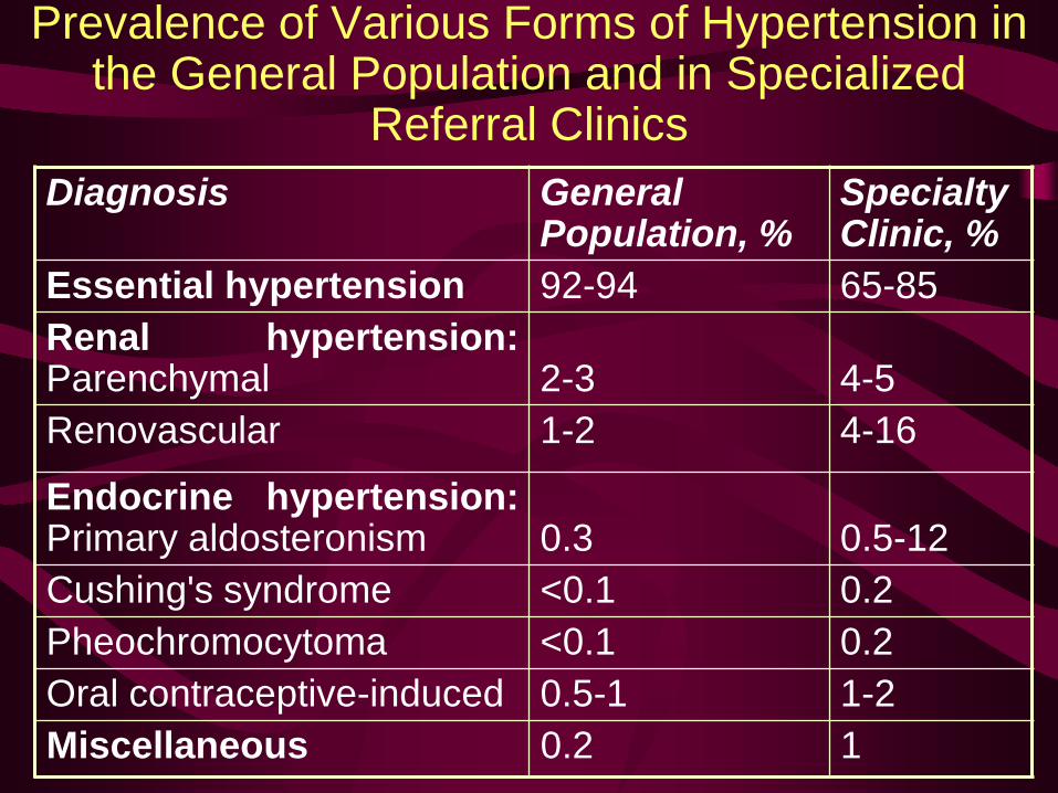
Prevalence of Various Forms of Hypertension in the General Population and in Specialized
Referral Clinics Diagnosis General
Population, % Specialty Clinic, %
Essential hypertension 92-94 65-85 Renal hypertension: Parenchymal
2-3
4-5
Renovascular 1-2 4-16
Endocrine hypertension: Primary aldosteronism
0.3
0.5-12
Cushing's syndrome <0.1 0.2 Pheochromocytoma <0.1 0.2 Oral contraceptive-induced 0.5-1 1-2 Miscellaneous 0.2 1
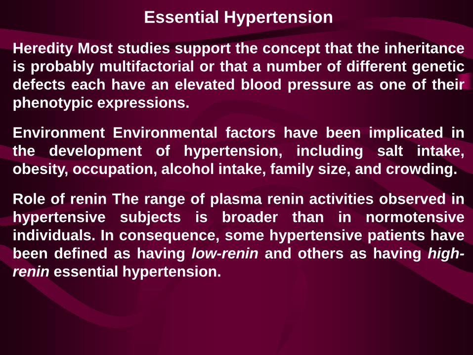
Essential Hypertension
Heredity Most studies support the concept that the inheritance is probably multifactorial or that a number of different genetic defects each have an elevated blood pressure as one of their phenotypic expressions.
Environment Environmental factors have been implicated in the development of hypertension, including salt intake, obesity, occupation, alcohol intake, family size, and crowding.
Role of renin The range of plasma renin activities observed in hypertensive subjects is broader than in normotensive individuals. In consequence, some hypertensive patients have been defined as having low-renin and others as having high-renin essential hypertension.
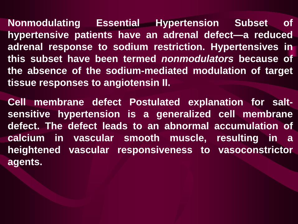
Nonmodulating Essential Hypertension Subset of hypertensive patients have an adrenal defect—a reduced adrenal response to sodium restriction. Hypertensives in this subset have been termed nonmodulators because of the absence of the sodium-mediated modulation of target tissue responses to angiotensin II.
Cell membrane defect Postulated explanation for salt-sensitive hypertension is a generalized cell membrane defect. The defect leads to an abnormal accumulation of calcium in vascular smooth muscle, resulting in a heightened vascular responsiveness to vasoconstrictor agents.
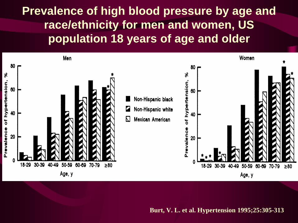
Prevalence of high blood pressure by age and race/ethnicity for men and women, US population 18 years of age and older
Burt, V. L. et al. Hypertension 1995;25:305-313
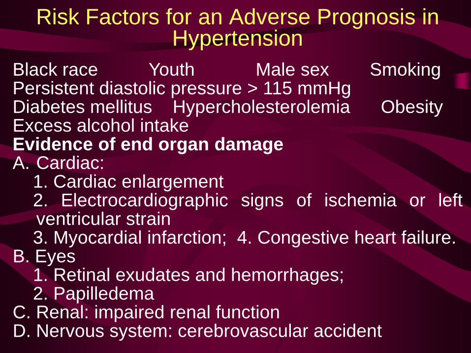
Risk Factors for an Adverse Prognosis in Hypertension
Black race Youth Male sex Smoking Persistent diastolic pressure > 115 mmHg Diabetes mellitus Hypercholesterolemia Obesity Excess alcohol intake Evidence of end organ damage A. Cardiac: 1. Cardiac enlargement 2. Electrocardiographic signs of ischemia or left
ventricular strain 3. Myocardial infarction; 4. Congestive heart failure. B. Eyes 1. Retinal exudates and hemorrhages; 2. Papilledema C. Renal: impaired renal function D. Nervous system: cerebrovascular accident
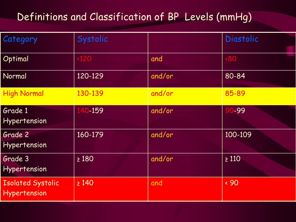
Category Systolic Diastolic
Optimal <120 and <80
Normal 120-129 and/or 80-84
High Normal 130-139 and/or 85-89
Grade 1 Hypertension
140-159 and/or 90-99
Grade 2 Hypertension
160-179 and/or 100-109
Grade 3 Hypertension
≥ 180 and/or ≥ 110
Isolated Systolic Hypertension
≥ 140 and < 90
Definitions and Classification of BP Levels (mmHg)
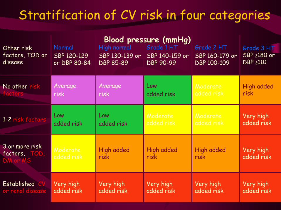
Stratification of CV risk in four categories
Blood pressure (mmHg) Other risk
factors, TOD or disease
Normal SBP 120-129 or DBP 80-84
High normal SBP 130-139 or DBP 85-89
Grade 1 HT SBP 140-159 or DBP 90-99
Grade 2 HT SBP 160-179 or DBP 100-109
Grade 3 HT SBP ≥180 or DBP ≥110
No other risk factors
Average risk
Average risk
Low added risk
Moderate added risk
High added risk
1-2 risk factors Low added risk
Low added risk
Moderate added risk
Moderate added risk
Very high added risk
3 or more risk factors, TOD, DM or MS
Moderate added risk
High added risk
High added risk
High added risk
Very high added risk
Established CV or renal disease
Very high added risk
Very high added risk
Very high added risk
Very high added risk
Very high added risk
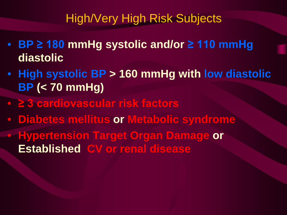
High/Very High Risk Subjects
• BP ≥ 180 mmHg systolic and/or ≥ 110 mmHg diastolic
• High systolic BP > 160 mmHg with low diastolic BP (< 70 mmHg)
• ≥ 3 cardiovascular risk factors • Diabetes mellitus or Metabolic syndrome • Hypertension Target Organ Damage or
Established CV or renal disease
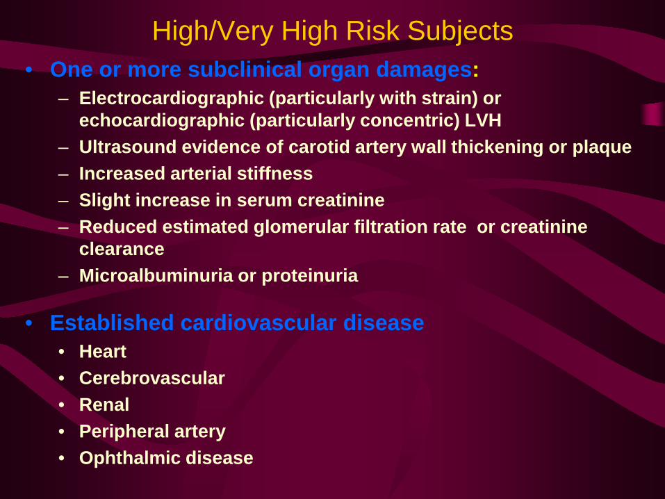
High/Very High Risk Subjects • One or more subclinical organ damages:
– Electrocardiographic (particularly with strain) or echocardiographic (particularly concentric) LVH
– Ultrasound evidence of carotid artery wall thickening or plaque – Increased arterial stiffness – Slight increase in serum creatinine – Reduced estimated glomerular filtration rate or creatinine
clearance – Microalbuminuria or proteinuria
• Established cardiovascular disease • Heart • Cerebrovascular • Renal • Peripheral artery • Ophthalmic disease
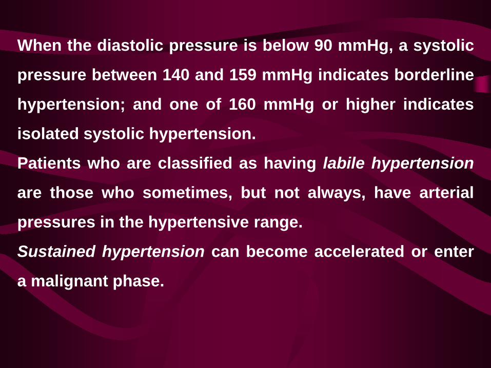
When the diastolic pressure is below 90 mmHg, a systolic
pressure between 140 and 159 mmHg indicates borderline
hypertension; and one of 160 mmHg or higher indicates
isolated systolic hypertension.
Patients who are classified as having labile hypertension
are those who sometimes, but not always, have arterial
pressures in the hypertensive range.
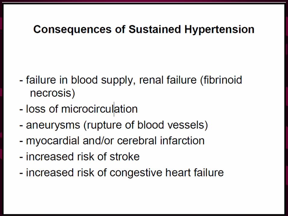
Sustained hypertension can become accelerated or enter
a malignant phase.
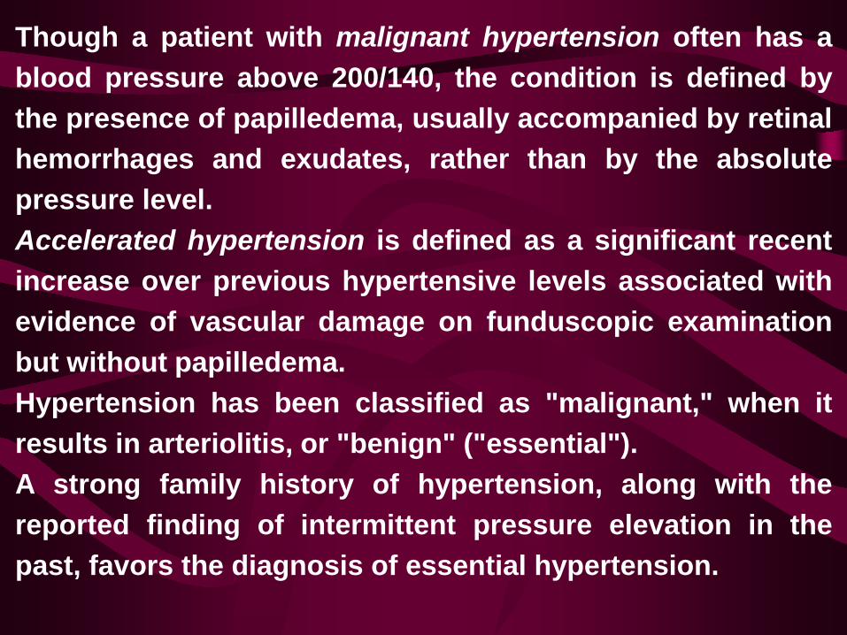
Though a patient with malignant hypertension often has a blood pressure above 200/140, the condition is defined by the presence of papilledema, usually accompanied by retinal hemorrhages and exudates, rather than by the absolute pressure level. Accelerated hypertension is defined as a significant recent increase over previous hypertensive levels associated with evidence of vascular damage on funduscopic examination but without papilledema. Hypertension has been classified as "malignant," when it results in arteriolitis, or "benign" ("essential"). A strong family history of hypertension, along with the reported finding of intermittent pressure elevation in the past, favors the diagnosis of essential hypertension.
Symptoms And Signs of arterial hypertension
Symptoms are related to the elevated blood pressure include
headache, dizziness, palpitations, easy fatigability, and
impotence. Headaches are localized to the occipital region and
are present when the patient awakens in the morning but subside
spontaneously after several hours.
Symptoms are related to the vascular disease include epistaxis,
blurring of vision owing to retinal changes, episodes of
weakness or dizziness due to transient cerebral ischemia, angina
pectoris, and dyspnea due to cardiac failure. Pain due to
dissection of the aorta or to a leaking aneurysm is an occasional
presenting symptom.
Symptoms related to the underlying disease in secondary hypertension are hematuria (glomerulonephritis), polyuria, polydipsia (diabetis melitus with nephrosclerosis), and muscle weakness secondary to hypokalemia (primary aldosteronism) or weight gain, and emotional lability (Cushing's syndrome), episodic headaches, palpitations, diaphoresis, and postural dizziness (a pheochromocytoma). Aspects of the history aid in determining whether vascular disease has progressed to a dangerous stage include angina pectoris and symptoms of cerebrovascular insufficiency, congestive heart failure, and/or peripheral vascular insufficiency. Risk factors: cigarette smoking, diabetes mellitus, lipid disorders, a family history of early cardiovascular deaths.
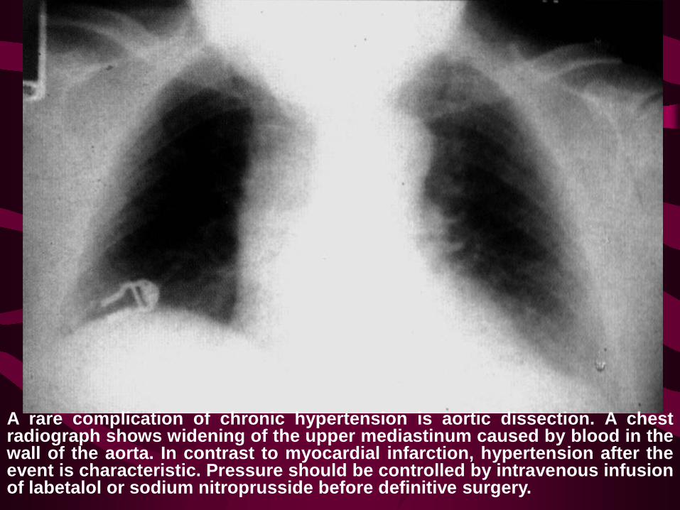
A rare complication of chronic hypertension is aortic dissection. A chest radiograph shows widening of the upper mediastinum caused by blood in the wall of the aorta. In contrast to myocardial infarction, hypertension after the event is characteristic. Pressure should be controlled by intravenous infusion of labetalol or sodium nitroprusside before definitive surgery.
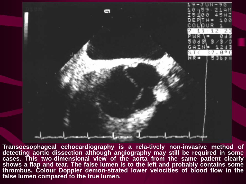
Transoesophageal echocardiography is a rela-tively non-invasive method of detecting aortic dissection although angiography may still be required in some cases. This two-dimensional view of the aorta from the same patient clearly shows a flap and tear. The false lumen is to the left and probably contains some thrombus. Colour Doppler demon-strated lower velocities of blood flow in the false lumen compared to the true lumen.
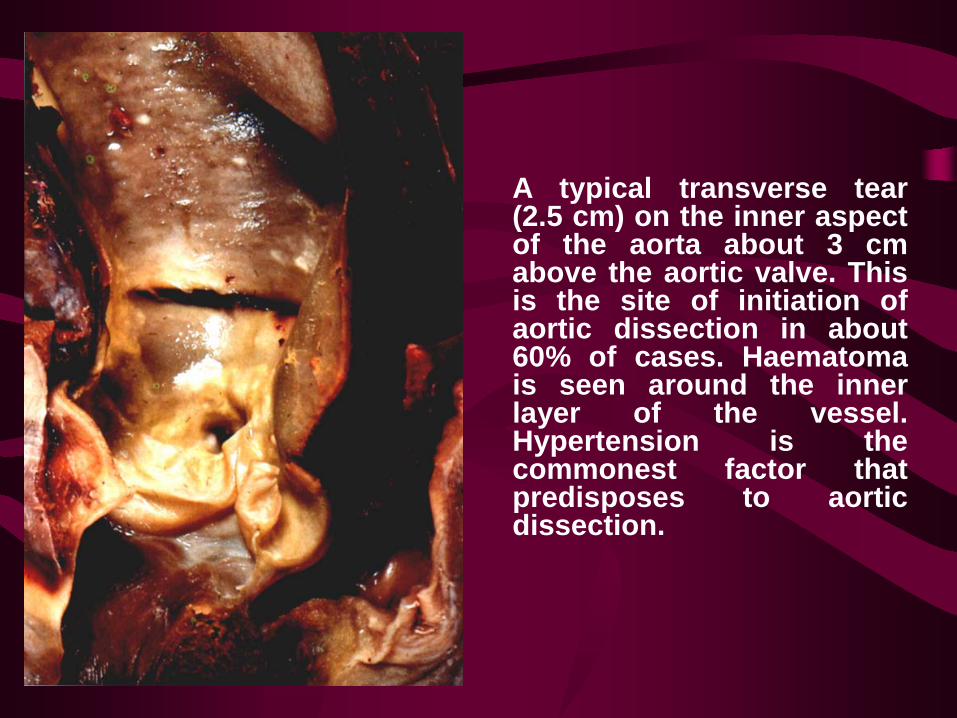
A typical transverse tear (2.5 cm) on the inner aspect of the aorta about 3 cm above the aortic valve. This is the site of initiation of aortic dissection in about 60% of cases. Haematoma is seen around the inner layer of the vessel. Hypertension is the commonest factor that predisposes to aortic dissection.
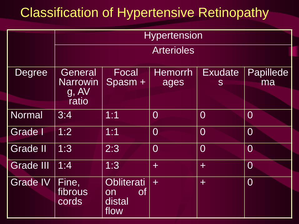
Classification of Hypertensive Retinopathy Hypertension
Arterioles
Degree General Narrowin
g, AV ratio
Focal Spasm +
Hemorrhages
Exudates
Papilledema
Normal 3:4 1:1 0 0 0
Grade I 1:2 1:1 0 0 0
Grade II 1:3 2:3 0 0 0
Grade III 1:4 1:3 + + 0
Grade IV Fine, fibrous cords
Obliteration of distal flow
+ + 0
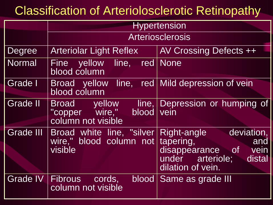
Classification of Arteriolosclerotic Retinopathy Hypertension
Arteriosclerosis Degree Arteriolar Light Reflex AV Crossing Defects ++ Normal Fine yellow line, red
blood column None
Grade I Broad yellow line, red blood column
Mild depression of vein
Grade II Broad yellow line, "copper wire," blood column not visible
Depression or humping of vein
Grade III Broad white line, "silver wire," blood column not visible
Right-angle deviation, tapering, and disappearance of vein under arteriole; distal dilation of vein.
Grade IV Fibrous cords, blood column not visible
Same as grade III

Health Consequences – Risk Factors ↓ Risk factors → ↑ life expectancy
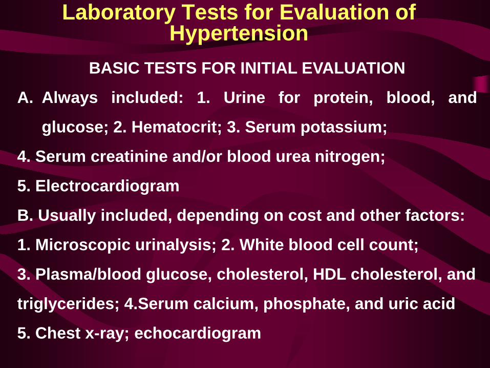
Laboratory Tests for Evaluation of Hypertension
BASIC TESTS FOR INITIAL EVALUATION
A. Always included: 1. Urine for protein, blood, and
glucose; 2. Hematocrit; 3. Serum potassium;
4. Serum creatinine and/or blood urea nitrogen;
5. Electrocardiogram
B. Usually included, depending on cost and other factors:
1. Microscopic urinalysis; 2. White blood cell count;
3. Plasma/blood glucose, cholesterol, HDL cholesterol, and
triglycerides; 4.Serum calcium, phosphate, and uric acid
5. Chest x-ray; echocardiogram
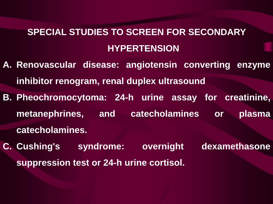
SPECIAL STUDIES TO SCREEN FOR SECONDARY
HYPERTENSION
A. Renovascular disease: angiotensin converting enzyme
inhibitor renogram, renal duplex ultrasound
B. Pheochromocytoma: 24-h urine assay for creatinine,
metanephrines, and catecholamines or plasma
catecholamines.
C. Cushing's syndrome: overnight dexamethasone
suppression test or 24-h urine cortisol.
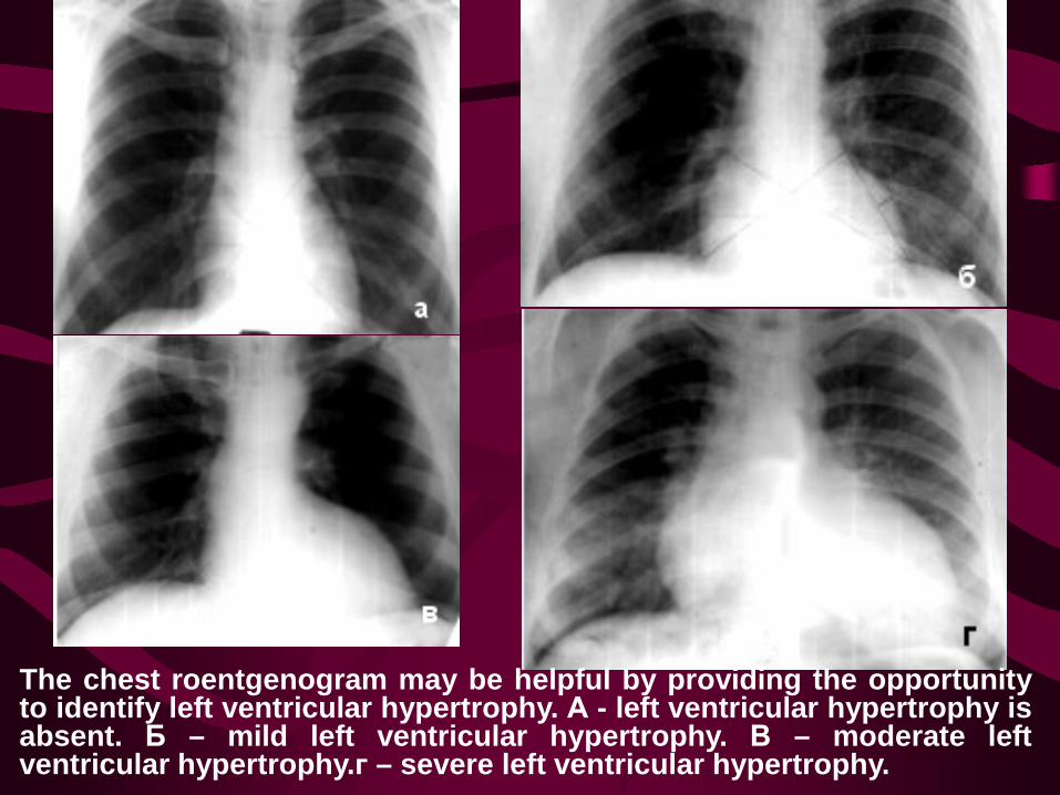
The chest roentgenogram may be helpful by providing the opportunity to identify left ventricular hypertrophy. А - left ventricular hypertrophy is absent. Б – mild left ventricular hypertrophy. В – moderate left ventricular hypertrophy.г – severe left ventricular hypertrophy.
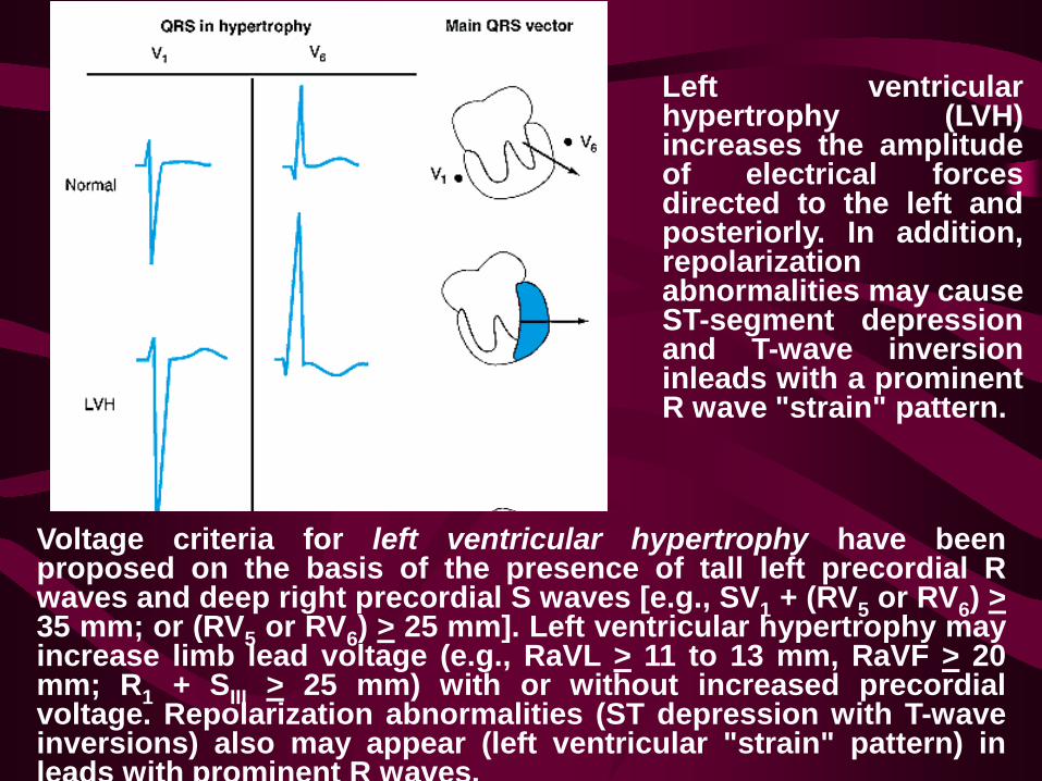
Left ventricular hypertrophy (LVH) increases the amplitude of electrical forces directed to the left and posteriorly. In addition, repolarization abnormalities may cause ST-segment depression and T-wave inversion inleads with a prominent R wave "strain" pattern.
Voltage criteria for left ventricular hypertrophy have been proposed on the basis of the presence of tall left precordial R waves and deep right precordial S waves [e.g., SV1 + (RV5 or RV6) > 35 mm; or (RV5 or RV6) > 25 mm]. Left ventricular hypertrophy may increase limb lead voltage (e.g., RaVL > 11 to 13 mm, RaVF > 20 mm; R1 + SIII > 25 mm) with or without increased precordial voltage. Repolarization abnormalities (ST depression with T-wave inversions) also may appear (left ventricular "strain" pattern) in leads with prominent R waves.
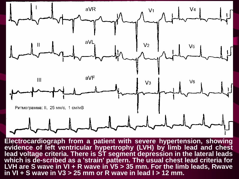
Electrocardiograph from a patient with severe hypertension, showing evidence of left ventricular hypertrophy (LVH) by limb lead and chest lead voltage criteria. There is ST segment depression in the lateral leads which is de-scribed as a 'strain' pattern. The usual chest lead criteria for LVH are S wave in VI + R wave in V5 > 35 mm. For the limb leads, Rwave in VI + S wave in V3 > 25 mm or R wave in lead I > 12 mm.
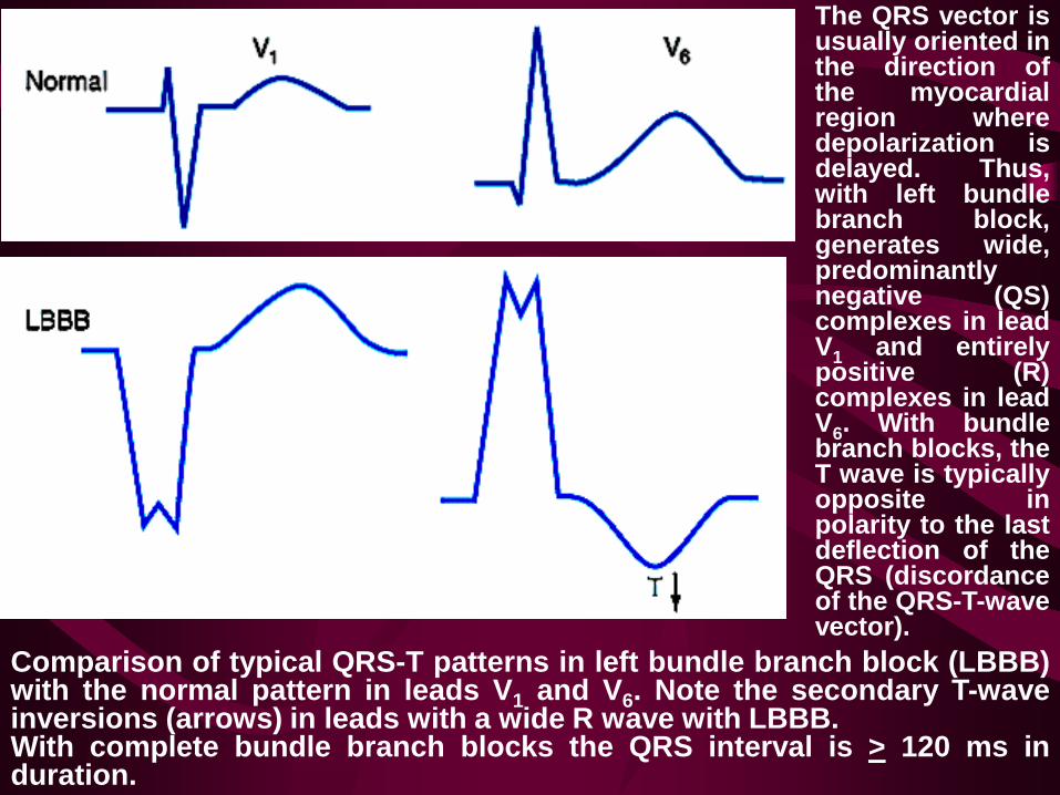
Comparison of typical QRS-T patterns in left bundle branch block (LBBB) with the normal pattern in leads V1 and V6. Note the secondary T-wave inversions (arrows) in leads with a wide R wave with LBBB. With complete bundle branch blocks the QRS interval is > 120 ms in duration.
The QRS vector is usually oriented in the direction of the myocardial region where depolarization is delayed. Thus, with left bundle branch block, generates wide, predominantly negative (QS) complexes in lead V1 and entirely positive (R) complexes in lead V6. With bundle branch blocks, the T wave is typically opposite in polarity to the last deflection of the QRS (discordance of the QRS-T-wave vector).
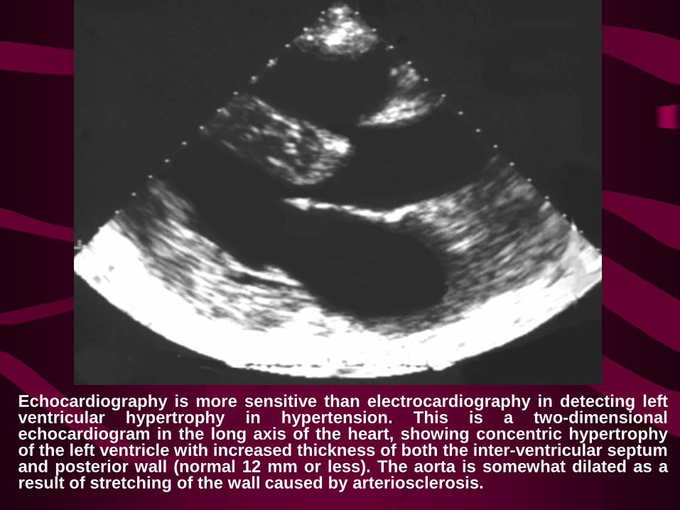
Echocardiography is more sensitive than electrocardiography in detecting left ventricular hypertrophy in hypertension. This is a two-dimensional echocardiogram in the long axis of the heart, showing concentric hypertrophy of the left ventricle with increased thickness of both the inter-ventricular septum and posterior wall (normal 12 mm or less). The aorta is somewhat dilated as a result of stretching of the wall caused by arteriosclerosis.
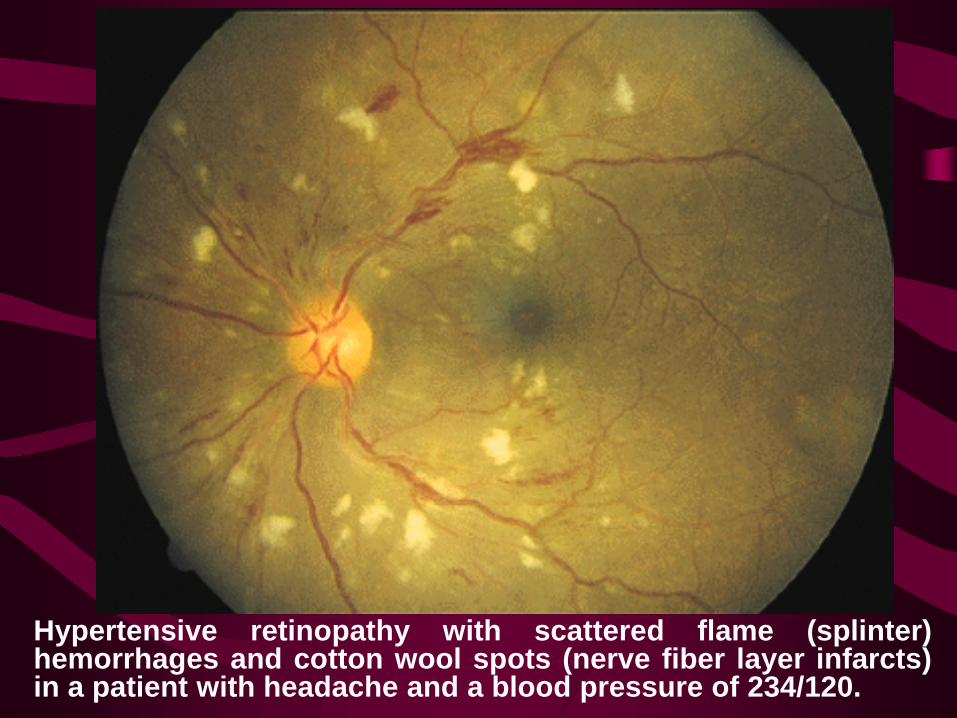
Hypertensive retinopathy with scattered flame (splinter) hemorrhages and cotton wool spots (nerve fiber layer infarcts) in a patient with headache and a blood pressure of 234/120.
Central Nervous System Dysfunction
In Patients With Hypertension Occipital headaches, most often occurring in the morning,
are the most prominent early symptoms of hypertension.
Dizziness, light-headedness, vertigo, tinnitus, and dimmed
vision or syncope may be observed.
Manifestations are due to vascular occlusion, hemorrhage,
or encephalopathy.
Cerebral infarction is secondary to the increased
atherosclerosis observed in hypertensive patients.
Cerebral hemorrhage is the result of both the elevated
arterial pressure and the development of cerebral vascular
microaneurysms (Charcot-Bouchard aneurysms).
Hypertensive encephalopathy consists of the following
symptom complex: severe hypertension, disordered
consciousness, increased intracranial pressure, retinopathy
with papilledema, and seizures. The pathogenesis is
uncertain but probably is not related to arteriolar spasm or
cerebral edema. Focal neurologic signs are infrequent and,
if present, suggest that infarction, hemorrhage, or transient
ischemic attacks are more likely diagnoses.
Coarctation Of The Aorta Narrowing or constriction of the lumen of the aorta may occur most common distal to the origin of the left subclavian artery near the insertion of the ligamentum arteriosum. Headache, epistaxis, cold extremities, and claudication with exercise may occur. Marked diminution, or delayed pulsations in the femoral arteries are detected on physical examination. Enlarged and pulsatile collateral vessels may be palpated in the intercostalspaces anteriorly, in the axillae, or posteriorly in the interscapular area. The upper extremities and thorax may be more developed than the lower extremities. A midsystolic murmur over the anterior part of the chest, back, and spinous processes may become continuous if the lumen is narrowed sufficiently. Roentgenograms may show a dilated ascending aorta. Indentation of the aorta at the site of coarctation and pre- and poststenotic dilatation (the "3" sign) along the left paramediastinal shadow. Notching of the ribs, an important radiographic sign, is due to erosion by dilated collateral vessels. Treatment is usually surgical.
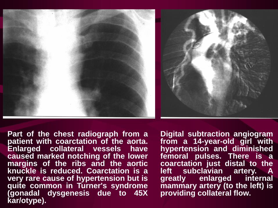
Part of the chest radiograph from a patient with coarctation of the aorta. Enlarged collateral vessels have caused marked notching of the lower margins of the ribs and the aortic knuckle is reduced. Coarctation is a very rare cause of hypertension but is quite common in Turner's syndrome (gonadal dysgenesis due to 45X kar/otype).
Digital subtraction angiogram from a 14-year-old girl with hypertension and diminished femoral pulses. There is a coarctation just distal to the left subclavian artery. A greatly enlarged internal mammary artery (to the left) is providing collateral flow.
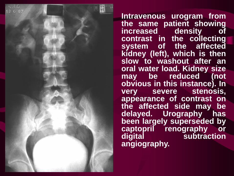
Intravenous urogram from the same patient showing increased density of contrast in the collecting system of the affected kidney (left), which is then slow to washout after an oral water load. Kidney size may be reduced (not obvious in this instance). In very severe stenosis, appearance of contrast on the affected side may be delayed. Urography has been largely superseded by captopril renography or digital subtraction angiography.
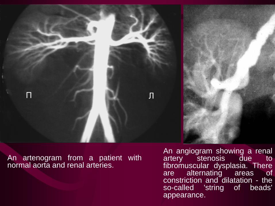
An artenogram from a patient with normal aorta and renal arteries.
An angiogram showing a renal artery stenosis due to fibromuscular dysplasia. There are alternating areas of constriction and dilatation - the so-called 'string of beads' appearance.
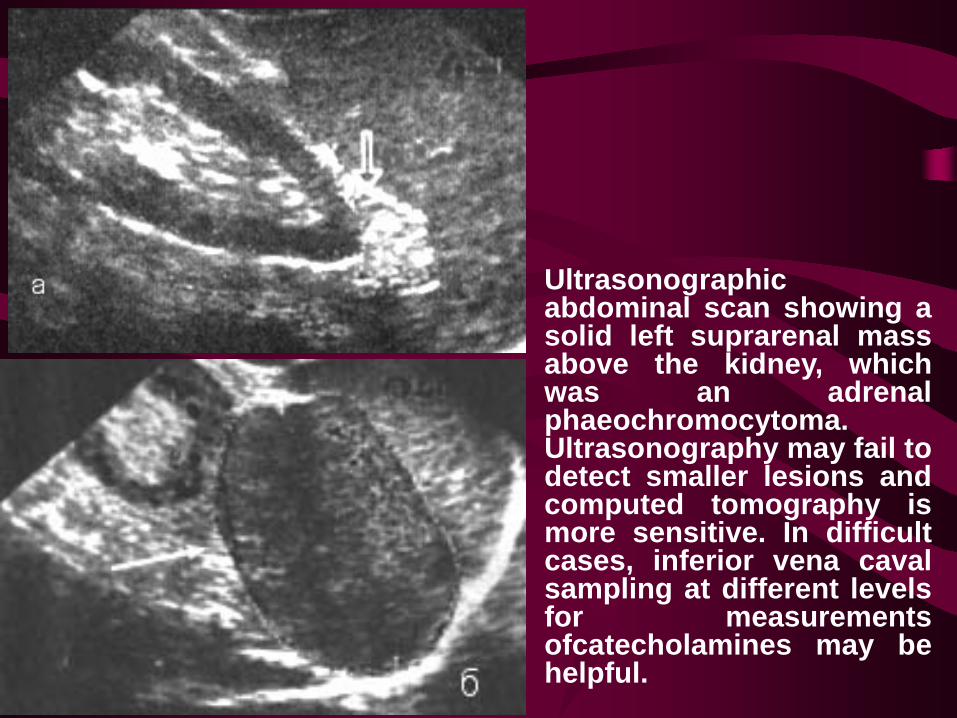
Ultrasonographic abdominal scan showing a solid left suprarenal mass above the kidney, which was an adrenal phaeochromocytoma. Ultrasonography may fail to detect smaller lesions and computed tomography is more sensitive. In difficult cases, inferior vena caval sampling at different levels for measurements ofcatecholamines may be helpful.
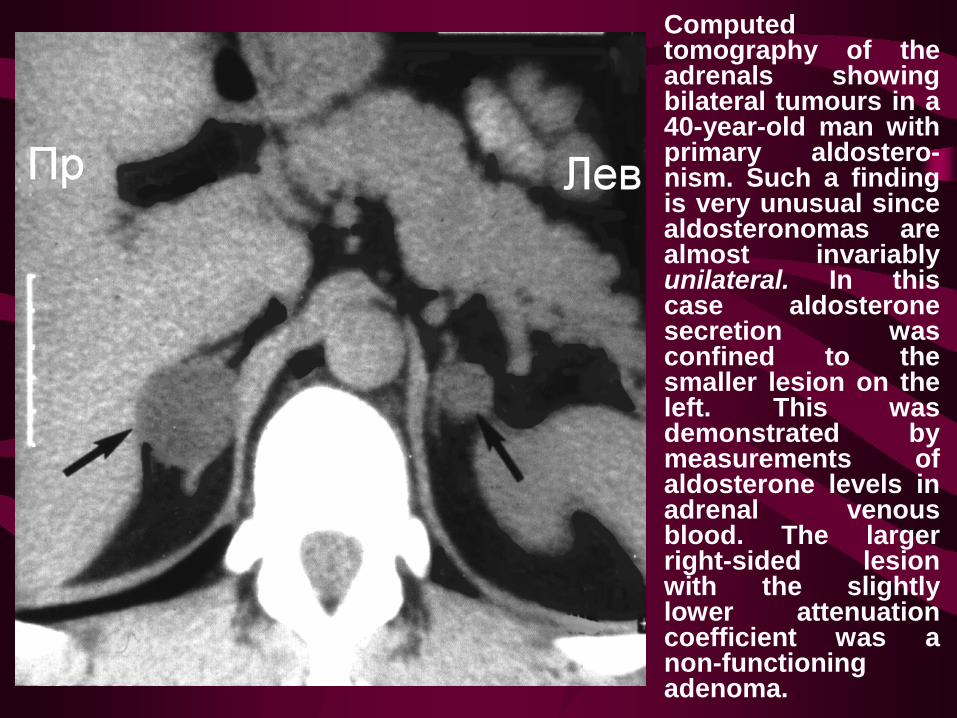
Computed tomography of the adrenals showing bilateral tumours in a 40-year-old man with primary aldostero-nism. Such a finding is very unusual since aldosteronomas are almost invariably unilateral. In this case aldosterone secretion was confined to the smaller lesion on the left. This was demonstrated by measurements of aldosterone levels in adrenal venous blood. The larger right-sided lesion with the slightly lower attenuation coefficient was a non-functioning adenoma.
Nonpharmacological Therapy Of Hypertension Smoking per se does not cause hypertension. However, smokers do have a higher incidence of malignant hypertension, and smoking is a major risk factor for coronary heart disease. Hypertensive patients should stop smoking. Reduction of Body Weight. Obesity and hypertension are closely associated, and the degree of obesity is positively correlated with the incidence of hypertension. Obese hypertensives may lower their blood pressure by losing weight regardless of a change in salt consumption. Sodium Restriction. High salt diets are associated with a high prevalence of hypertension. Moderate restriction of salt intake to approximately 5 g per day will, on average, lower blood pressure by 12 mm Hg systolic and 6 mm Hg diastolic. An additional benefit of salt restriction is improved responsiveness to some antihypertensive drugs.
Alcohol Restriction. Consumption of alcohol can raise blood pressure. Heavy consumption of alcohol increases the risk of cerebrovascular accidents but not coronary heart disease. All hypertensive patients should be advised to restrict consumption of ethanol to no more than 30 ml per day. Physical Exercise. Increased physical activity lowers rates of cardiovascular disease in men. Lack of physical activity is associated with a higher incidence of hypertension. Relaxation and Biofeedback Therapy. The fact that long-term stressful stimuli can cause sustained hypertension in animals has given credence to the possibility that relaxation therapy will lower blood pressure in some hypertensive patients. Potassium Therapy. In mildly hypertensive patients, oral K+ supplements of 48 mmol per day reduce both systolic and diastolic blood pressure.
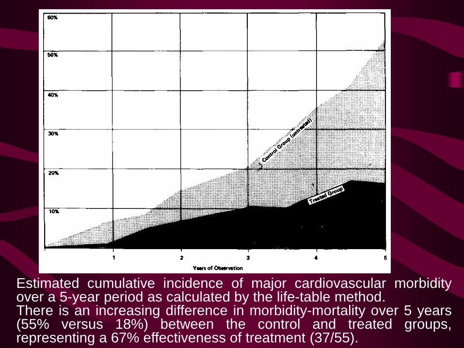
Estimated cumulative incidence of major cardiovascular morbidity over a 5-year period as calculated by the life-table method. There is an increasing difference in morbidity-mortality over 5 years (55% versus 18%) between the control and treated groups, representing a 67% effectiveness of treatment (37/55).
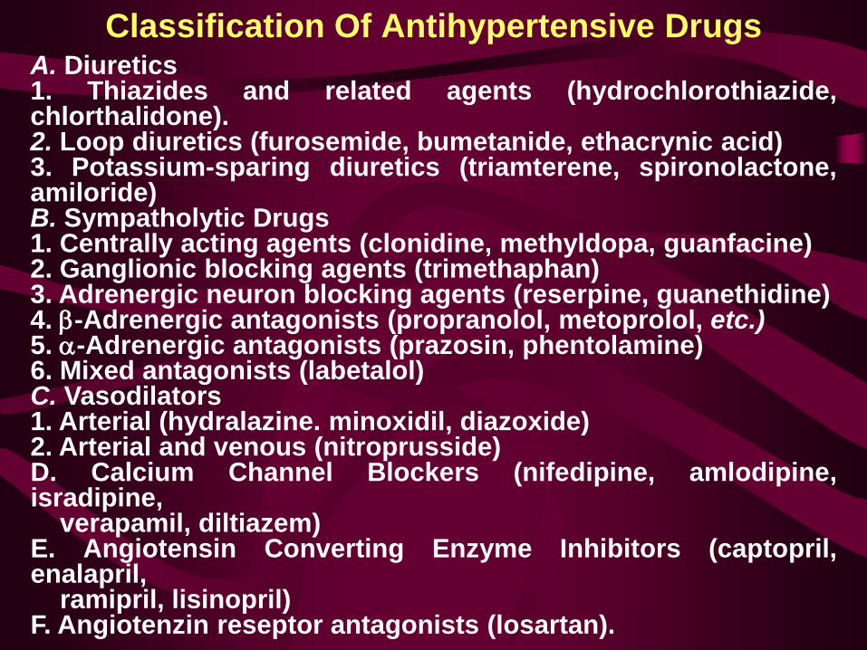
Classification Of Antihypertensive Drugs A. Diuretics 1. Thiazides and related agents (hydrochlorothiazide, chlorthalidone). 2. Loop diuretics (furosemide, bumetanide, ethacrynic acid) 3. Potassium-sparing diuretics (triamterene, spironolactone, amiloride) B. Sympatholytic Drugs 1. Centrally acting agents (clonidine, methyldopa, guanfacine) 2. Ganglionic blocking agents (trimethaphan) 3. Adrenergic neuron blocking agents (reserpine, guanethidine) 4. β-Adrenergic antagonists (propranolol, metoprolol, etc.) 5. α-Adrenergic antagonists (prazosin, phentolamine) 6. Mixed antagonists (labetalol) C. Vasodilators 1. Arterial (hydralazine. minoxidil, diazoxide) 2. Arterial and venous (nitroprusside) D. Calcium Channel Blockers (nifedipine, amlodipine, isradipine, verapamil, diltiazem) E. Angiotensin Converting Enzyme Inhibitors (captopril, enalapril, ramipril, lisinopril) F. Angiotenzin reseptor antagonists (losartan).
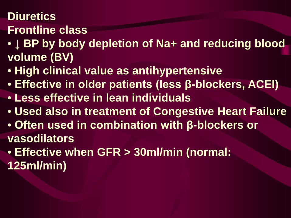
Diuretics Frontline class • ↓ BP by body depletion of Na+ and reducing blood volume (BV) • High clinical value as antihypertensive • Effective in older patients (less β-blockers, ACEI) • Less effective in lean individuals • Used also in treatment of Congestive Heart Failure • Often used in combination with β-blockers or vasodilators • Effective when GFR > 30ml/min (normal: 125ml/min)
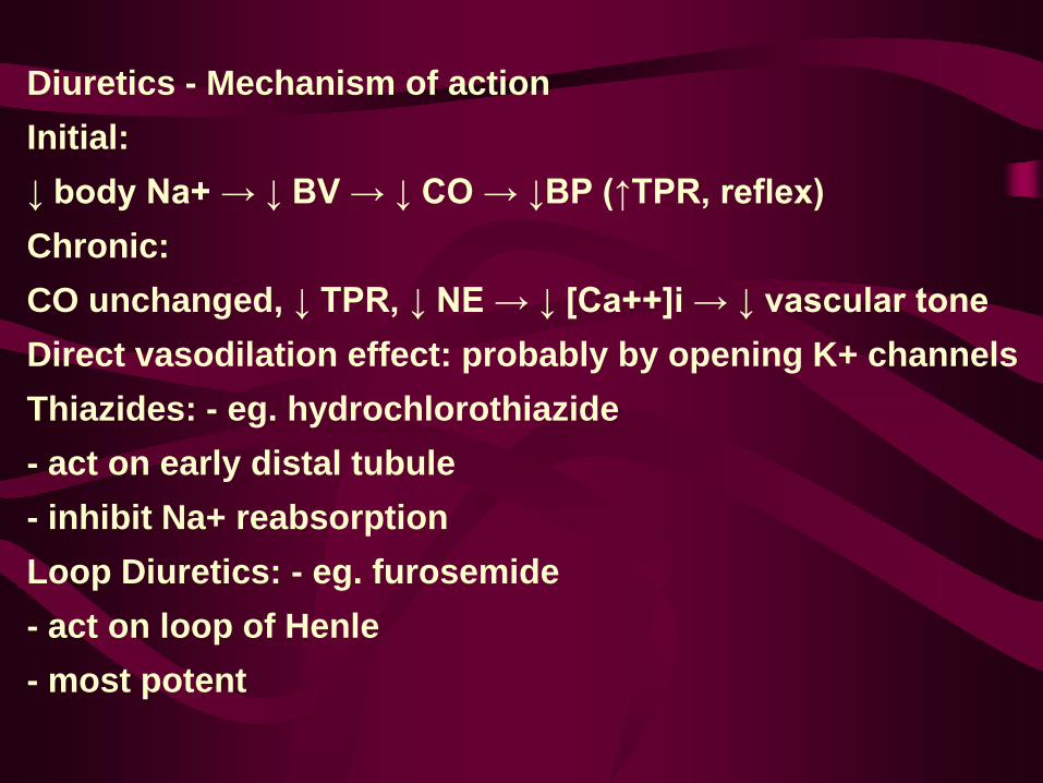
Diuretics - Mechanism of action Initial: ↓ body Na+ → ↓ BV → ↓ CO → ↓BP (↑TPR, reflex) Chronic: CO unchanged, ↓ TPR, ↓ NE → ↓ [Ca++]i → ↓ vascular tone Direct vasodilation effect: probably by opening K+ channels Thiazides: - eg. hydrochlorothiazide - act on early distal tubule - inhibit Na+ reabsorption Loop Diuretics: - eg. furosemide - act on loop of Henle - most potent
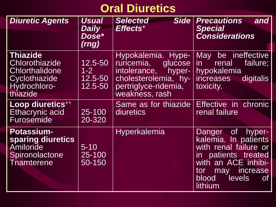
Oral Diuretics Diuretic Agents Usual
Daily Dose* (rng)
Selected Side Effects+
Precautions and Special Considerations
Thiazide Chlorothiazide Chlorthalidone Cyclothiazide Hydrochloro-thiazide
12.5-50 1-2 12.5-50 12.5-50
Hypokalemia. Hype-ruricemia, glucose intolerance, hyper-cholesterolemia, hy-pertriglyce-ridemia, weakness, rash
May be ineffective in renal failure; hypokalemia increases digitalis toxicity.
Loop diuretics++ Ethacrynic acid Furosemide
25-100 20-320
Same as for thiazide diuretics
Effective in chronic renal failure
Potassium-sparing diuretics Amiloride Spironolactone Tnamterene
5-10 25-100 50-150
Hyperkalemia Danger of hyper-kalemia. In patients with renal failure or in patients treated with an ACE inhibi-tor may increase blood levels of lithium
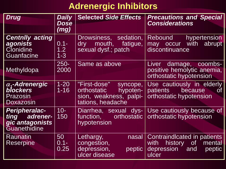
Adrenergic Inhibitors Drug Daily
Dose(mg)
Selected Side Effects Precautions and Special Considerations
Centnlly acting agonists Clonidine Guanfacine
0.1-1.2 1-3
Drowsiness, sedation, dry mouth, fatigue, sexual dysf.; patch
Rebound hypertension may occur with abrupt discontinuance
Methyldopa
250-2000
Same as above Liver damage, coombs-positive hemolytic anemia, orthostatic hypotension
α1-Adrenergic blockers Prazosin Doxazosin
1-20 1-16
"First-dose" syncope, orthostatic hypoten-sion, weakness, palpi-tations, headache
Use cautiously in elderly patients because of orthostatic hypotension
Peripheralac-ting adrener-gic antagonists Guanethidine
10-150
Diarrhea, sexual dys-function, orthostatic hypotension
Use cautiously because of orthostatic hypotension
Raunatin Reserpine
50 0.1-0.25
Lethargy, nasal congestion, depression, peptic ulcer disease
Contraindlcated in patients with history of mental depression and peptic ulcer
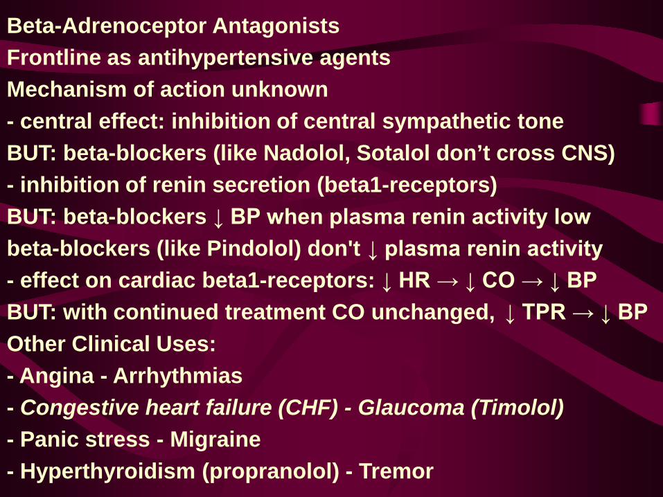
Beta-Adrenoceptor Antagonists Frontline as antihypertensive agents Mechanism of action unknown - central effect: inhibition of central sympathetic tone BUT: beta-blockers (like Nadolol, Sotalol don’t cross CNS) - inhibition of renin secretion (beta1-receptors) BUT: beta-blockers ↓ BP when plasma renin activity low beta-blockers (like Pindolol) don't ↓ plasma renin activity - effect on cardiac beta1-receptors: ↓ HR → ↓ CO → ↓ BP BUT: with continued treatment CO unchanged, ↓ TPR → ↓ BP Other Clinical Uses: - Angina - Arrhythmias - Congestive heart failure (CHF) - Glaucoma (Timolol) - Panic stress - Migraine - Hyperthyroidism (propranolol) - Tremor
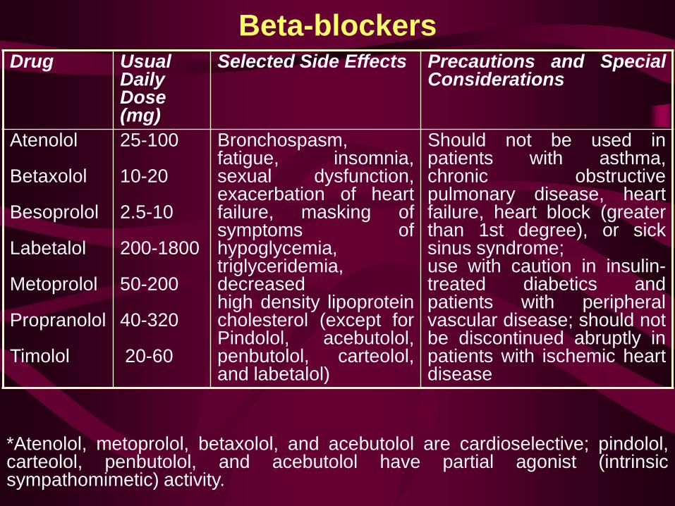
Beta-blockers Drug Usual
Daily Dose (mg)
Selected Side Effects Precautions and Special Considerations
Atenolol Betaxolol Besoprolol Labetalol Metoprolol Propranolol Timolol
25-100 10-20 2.5-10 200-1800 50-200 40-320 20-60
Bronchospasm, fatigue, insomnia, sexual dysfunction, exacerbation of heart failure, masking of symptoms of hypoglycemia, triglyceridemia, decreased high density lipoprotein cholesterol (except for Pindolol, acebutolol, penbutolol, carteolol, and labetalol)
Should not be used in patients with asthma, chronic obstructive pulmonary disease, heart failure, heart block (greater than 1st degree), or sick sinus syndrome; use with caution in insulin-treated diabetics and patients with peripheral vascular disease; should not be discontinued abruptly in patients with ischemic heart disease
*Atenolol, metoprolol, betaxolol, and acebutolol are cardioselective; pindolol, carteolol, penbutolol, and acebutolol have partial agonist (intrinsic sympathomimetic) activity.
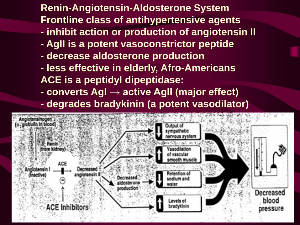
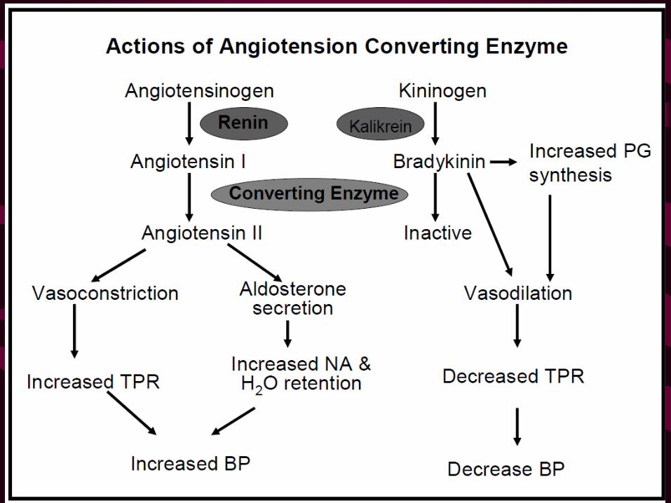
Renin-Angiotensin-Aldosterone System Frontline class of antihypertensive agents - inhibit action or production of angiotensin II - AgII is a potent vasoconstrictor peptide - decrease aldosterone production - less effective in elderly, Afro-Americans ACE is a peptidyl dipeptidase: - converts AgI → active AgII (major effect) - degrades bradykinin (a potent vasodilator)
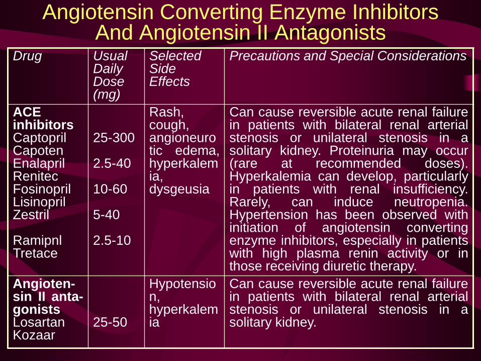
Angiotensin Converting Enzyme Inhibitors And Angiotensin II Antagonists
Drug Usual Daily Dose (mg)
Selected Side Effects
Precautions and Special Considerations
ACE inhibitors Captopril Capoten Enalapril Renitec Fosinopril Lisinopril Zestril Ramipnl Tretace
25-300 2.5-40 10-60 5-40 2.5-10
Rash, cough, angioneurotic edema, hyperkalemia, dysgeusia
Can cause reversible acute renal failure in patients with bilateral renal arterial stenosis or unilateral stenosis in a solitary kidney. Proteinuria may occur (rare at recommended doses). Hyperkalemia can develop, particularly in patients with renal insufficiency. Rarely, can induce neutropenia. Hypertension has been observed with initiation of angiotensin converting enzyme inhibitors, especially in patients with high plasma renin activity or in those receiving diuretic therapy.
Angioten-sin II anta-gonists Losartan Kozaar
25-50
Hypotension, hyperkalemia
Can cause reversible acute renal failure in patients with bilateral renal arterial stenosis or unilateral stenosis in a solitary kidney.
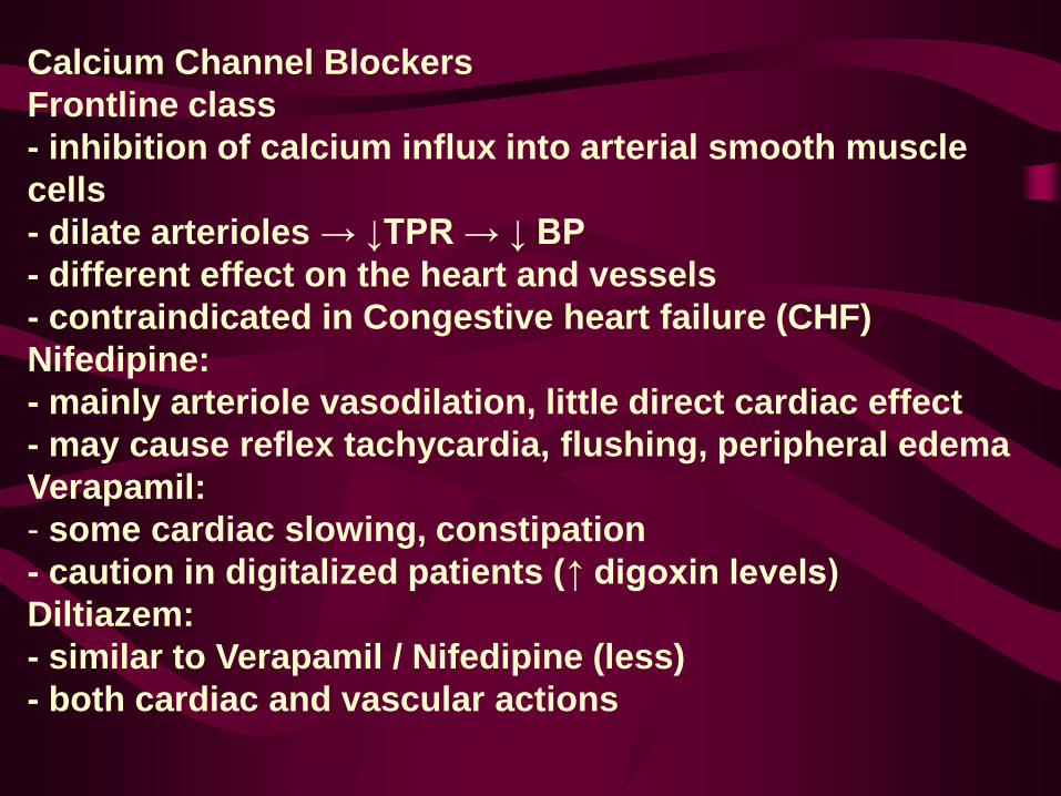
Calcium Channel Blockers Frontline class - inhibition of calcium influx into arterial smooth muscle cells - dilate arterioles → ↓TPR → ↓ BP - different effect on the heart and vessels - contraindicated in Congestive heart failure (CHF) Nifedipine: - mainly arteriole vasodilation, little direct cardiac effect - may cause reflex tachycardia, flushing, peripheral edema Verapamil: - some cardiac slowing, constipation - caution in digitalized patients (↑ digoxin levels) Diltiazem: - similar to Verapamil / Nifedipine (less) - both cardiac and vascular actions
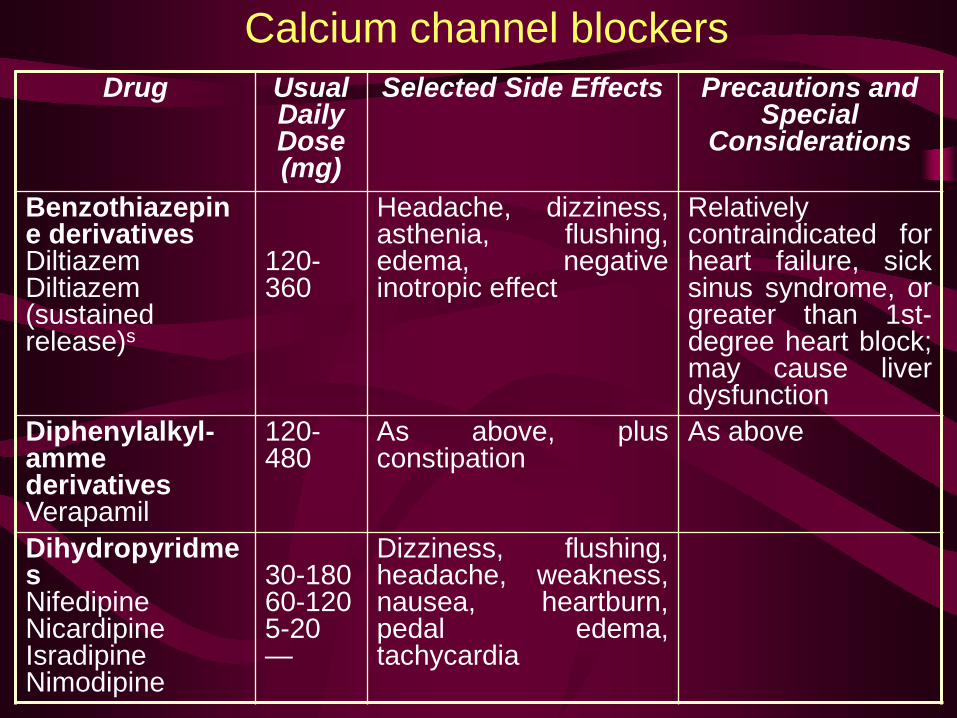
Calcium channel blockers Drug Usual
Daily Dose (mg)
Selected Side Effects Precautions and Special
Considerations
Benzothiazepine derivatives Diltiazem Diltiazem (sustained release)s
120-360
Headache, dizziness, asthenia, flushing, edema, negative inotropic effect
Relatively contraindicated for heart failure, sick sinus syndrome, or greater than 1st-degree heart block; may cause liver dysfunction
Diphenylalkyl-amme derivatives Verapamil
120-480
As above, plus constipation
As above
Dihydropyridmes Nifedipine Nicardipine Isradipine Nimodipine
30-180 60-120 5-20 —
Dizziness, flushing, headache, weakness, nausea, heartburn, pedal edema, tachycardia
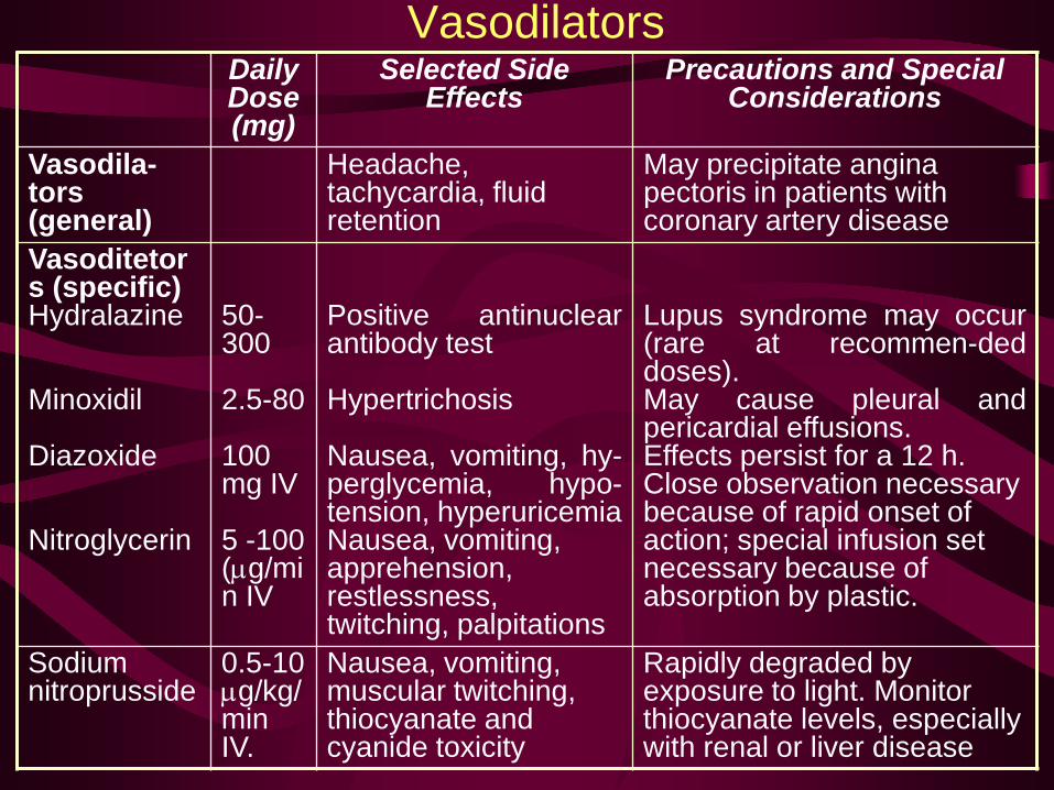
Vasodilators Daily Dose (mg)
Selected Side Effects
Precautions and Special Considerations
Vasodila-tors (general)
Headache, tachycardia, fluid retention
May precipitate angina pectoris in patients with coronary artery disease
Vasoditetors (specific) Hydralazine Minoxidil Diazoxide Nitroglycerin
50-300 2.5-80 100 mg IV 5 -100 (µg/min IV
Positive antinuclear antibody test Hypertrichosis Nausea, vomiting, hy-perglycemia, hypo-tension, hyperuricemia Nausea, vomiting, apprehension, restlessness, twitching, palpitations
Lupus syndrome may occur (rare at recommen-ded doses). May cause pleural and pericardial effusions. Effects persist for a 12 h. Close observation necessary because of rapid onset of action; special infusion set necessary because of absorption by plastic.
Sodium nitroprusside
0.5-10 µg/kg/min IV.
Nausea, vomiting, muscular twitching, thiocyanate and cyanide toxicity
Rapidly degraded by exposure to light. Monitor thiocyanate levels, especially with renal or liver disease
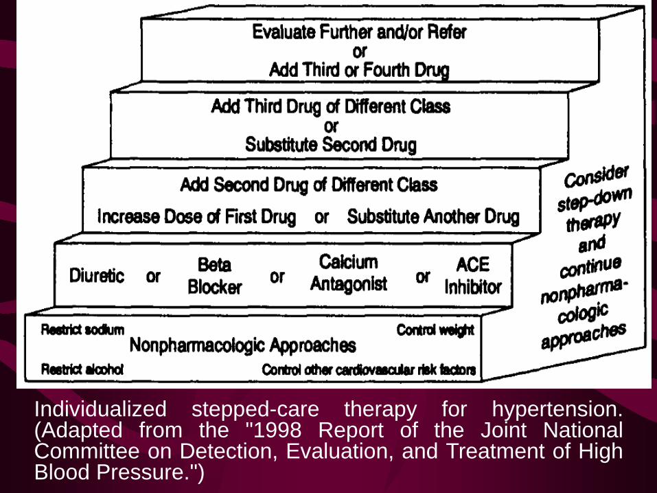
Individualized stepped-care therapy for hypertension. (Adapted from the "1998 Report of the Joint National Committee on Detection, Evaluation, and Treatment of High Blood Pressure.")
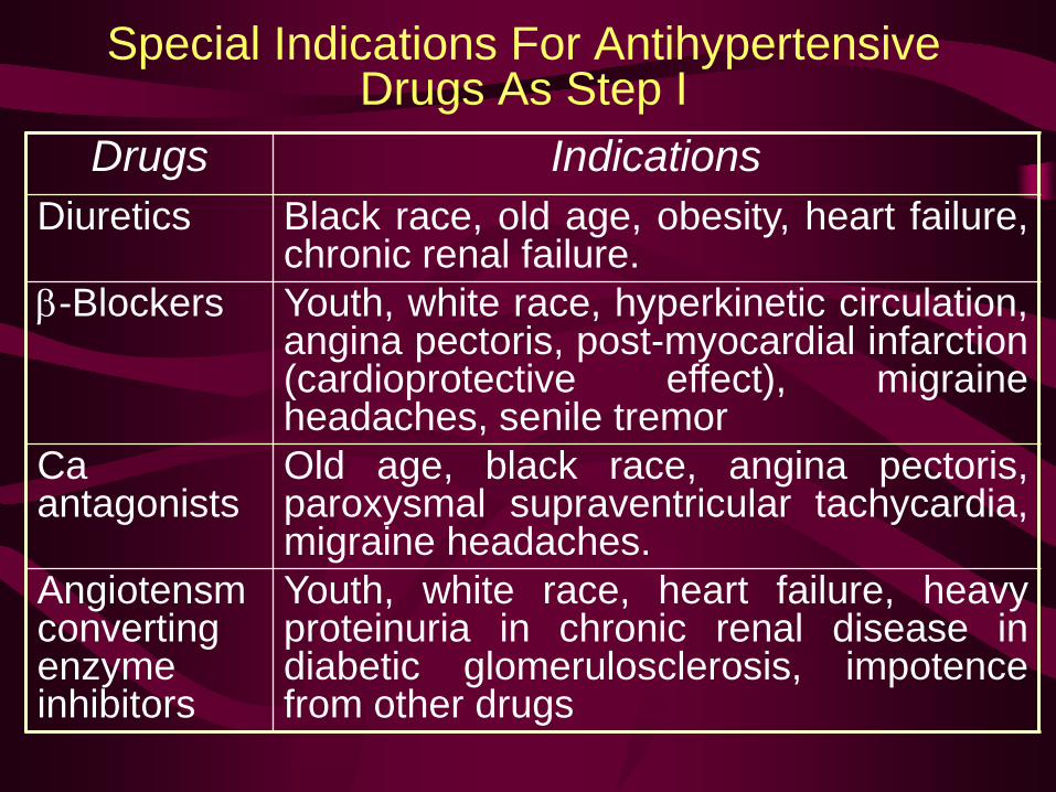
Special Indications For Antihypertensive Drugs As Step I
Drugs Indications Diuretics Black race, old age, obesity, heart failure,
chronic renal failure. β-Blockers Youth, white race, hyperkinetic circulation,
angina pectoris, post-myocardial infarction (cardioprotective effect), migraine headaches, senile tremor
Ca antagonists
Old age, black race, angina pectoris, paroxysmal supraventricular tachycardia, migraine headaches.
Angiotensm converting enzyme inhibitors
Youth, white race, heart failure, heavy proteinuria in chronic renal disease in diabetic glomerulosclerosis, impotence from other drugs
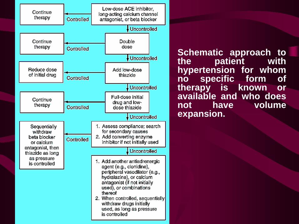
Schematic approach to the patient with hypertension for whom no specific form of therapy is known or available and who does not have volume expansion.
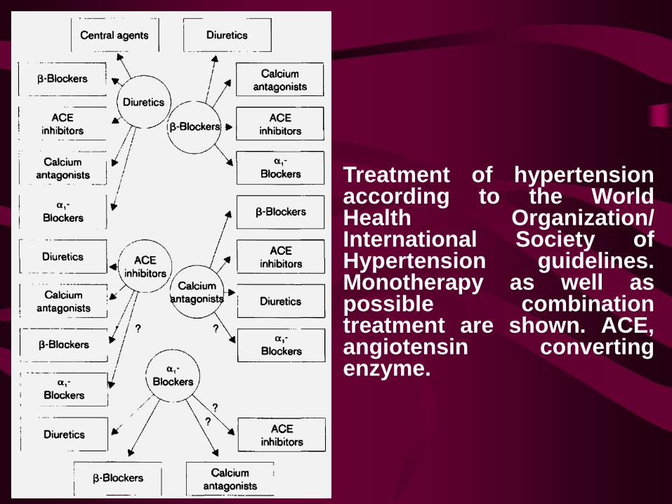
Treatment of hypertension according to the World Health Organization/ International Society of Hypertension guidelines. Monotherapy as well as possible combination treatment are shown. ACE, angiotensin converting enzyme.
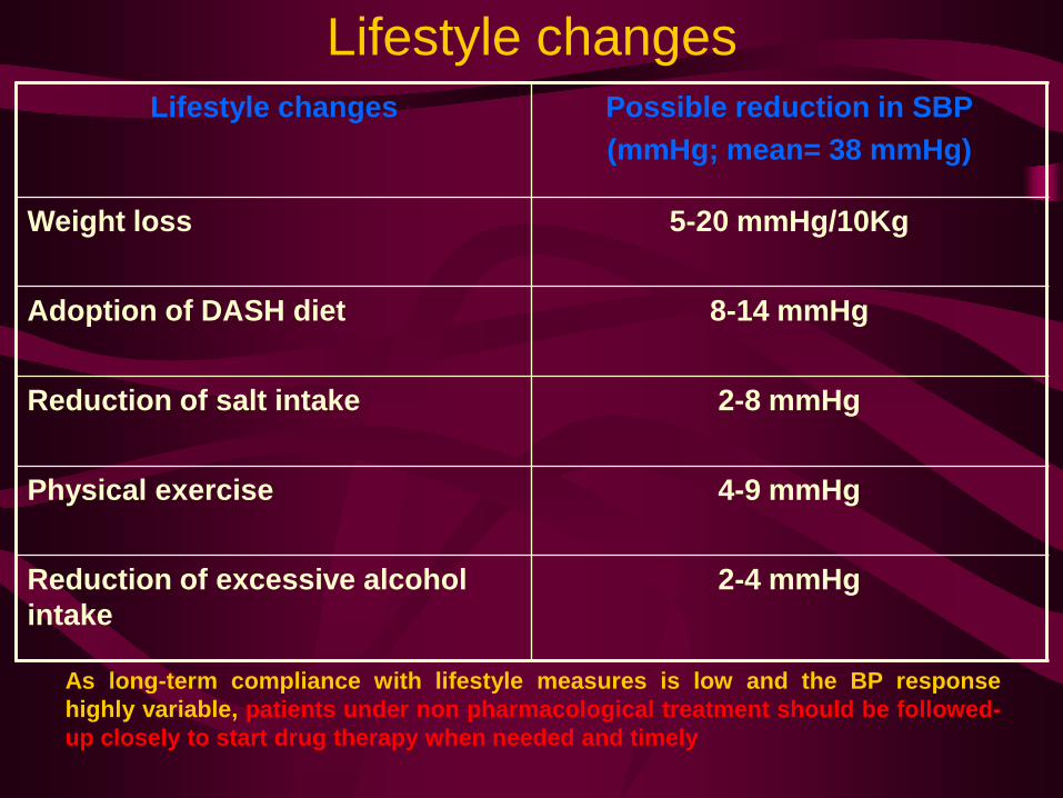
Lifestyle changes Lifestyle changes Possible reduction in SBP
(mmHg; mean= 38 mmHg)
Weight loss 5-20 mmHg/10Kg
Adoption of DASH diet 8-14 mmHg
Reduction of salt intake 2-8 mmHg
Physical exercise 4-9 mmHg
Reduction of excessive alcohol intake
2-4 mmHg
As long-term compliance with lifestyle measures is low and the BP response highly variable, patients under non pharmacological treatment should be followed-up closely to start drug therapy when needed and timely
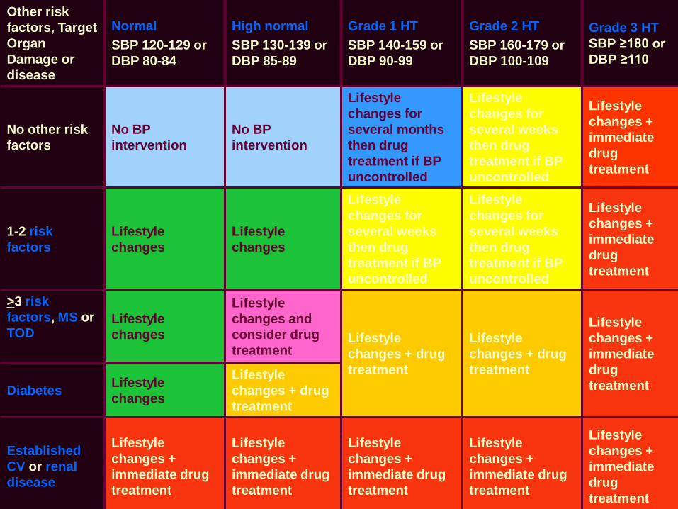
Initiation of antihypertensive treatment Other risk factors, Target Organ Damage or disease
Normal SBP 120-129 or DBP 80-84
High normal SBP 130-139 or DBP 85-89
Grade 1 HT SBP 140-159 or DBP 90-99
Grade 2 HT SBP 160-179 or DBP 100-109
Grade 3 HT SBP ≥180 or DBP ≥110
No other risk factors
No BP intervention
No BP intervention
Lifestyle changes for several months then drug treatment if BP uncontrolled
Lifestyle changes for several weeks then drug treatment if BP uncontrolled
Lifestyle changes + immediate drug treatment
1-2 risk factors
Lifestyle changes
Lifestyle changes
Lifestyle changes for several weeks then drug treatment if BP uncontrolled
Lifestyle changes for several weeks then drug treatment if BP uncontrolled
Lifestyle changes + immediate drug treatment
>3 risk factors, MS or TOD
Lifestyle changes
Lifestyle changes and consider drug treatment
Lifestyle changes + drug treatment
Lifestyle changes + drug treatment
Lifestyle changes + immediate drug treatment Diabetes Lifestyle
changes
Lifestyle changes + drug treatment
Established CV or renal disease
Lifestyle changes + immediate drug treatment
Lifestyle changes + immediate drug treatment
Lifestyle changes + immediate drug treatment
Lifestyle changes + immediate drug treatment
Lifestyle changes + immediate drug treatment
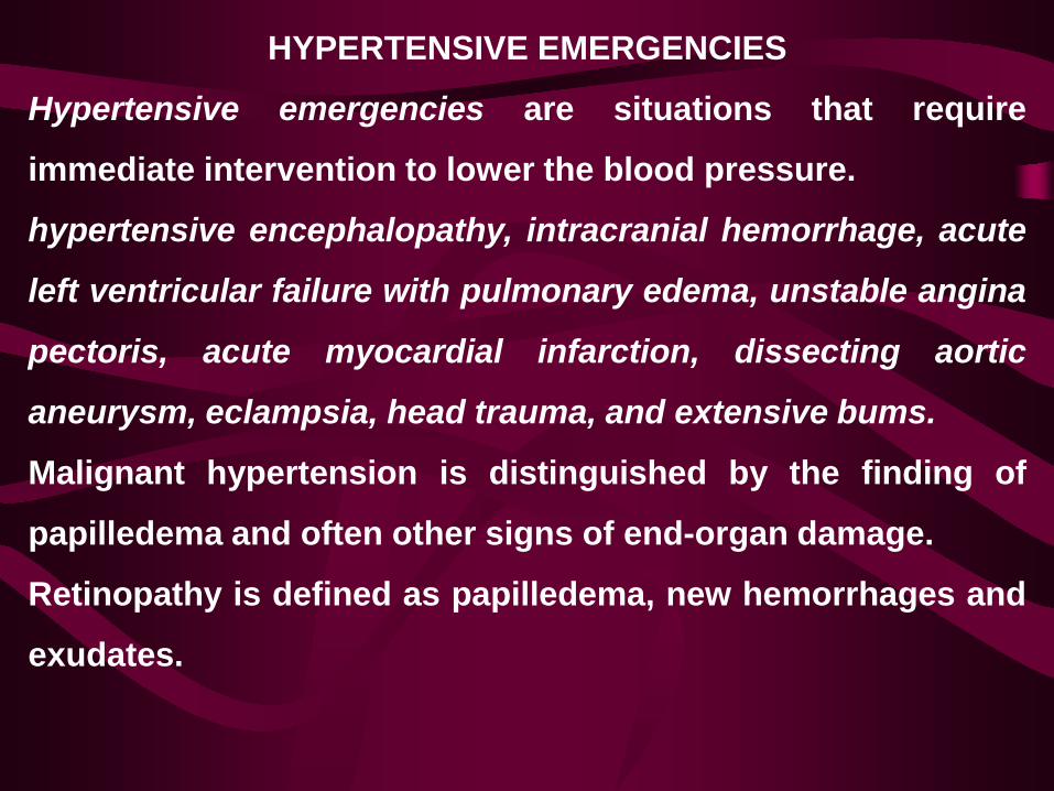
HYPERTENSIVE EMERGENCIES
Hypertensive emergencies are situations that require
immediate intervention to lower the blood pressure.
hypertensive encephalopathy, intracranial hemorrhage, acute
left ventricular failure with pulmonary edema, unstable angina
pectoris, acute myocardial infarction, dissecting aortic
aneurysm, eclampsia, head trauma, and extensive bums.
Malignant hypertension is distinguished by the finding of
papilledema and often other signs of end-organ damage.
Retinopathy is defined as papilledema, new hemorrhages and
exudates.
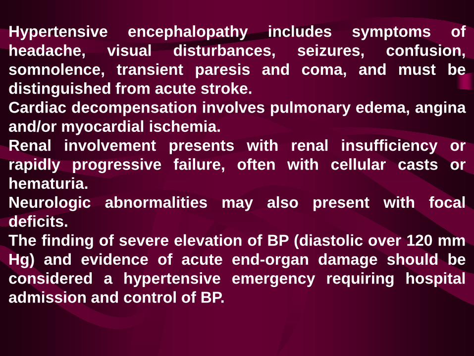
Hypertensive encephalopathy includes symptoms of headache, visual disturbances, seizures, confusion, somnolence, transient paresis and coma, and must be distinguished from acute stroke. Cardiac decompensation involves pulmonary edema, angina and/or myocardial ischemia. Renal involvement presents with renal insufficiency or rapidly progressive failure, often with cellular casts or hematuria. Neurologic abnormalities may also present with focal deficits. The finding of severe elevation of BP (diastolic over 120 mm Hg) and evidence of acute end-organ damage should be considered a hypertensive emergency requiring hospital admission and control of BP.
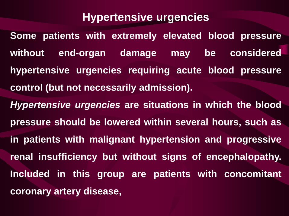
Hypertensive urgencies
Some patients with extremely elevated blood pressure
without end-organ damage may be considered
hypertensive urgencies requiring acute blood pressure
control (but not necessarily admission).
Hypertensive urgencies are situations in which the blood
pressure should be lowered within several hours, such as
in patients with malignant hypertension and progressive
renal insufficiency but without signs of encephalopathy.
Included in this group are patients with concomitant
coronary artery disease,
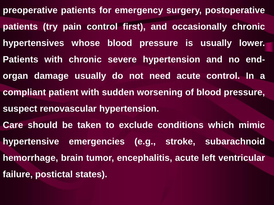
preoperative patients for emergency surgery, postoperative
patients (try pain control first), and occasionally chronic
hypertensives whose blood pressure is usually lower.
Patients with chronic severe hypertension and no end-
organ damage usually do not need acute control. In a
compliant patient with sudden worsening of blood pressure,
suspect renovascular hypertension.
Care should be taken to exclude conditions which mimic
hypertensive emergencies (e.g., stroke, subarachnoid
hemorrhage, brain tumor, encephalitis, acute left ventricular
failure, postictal states).
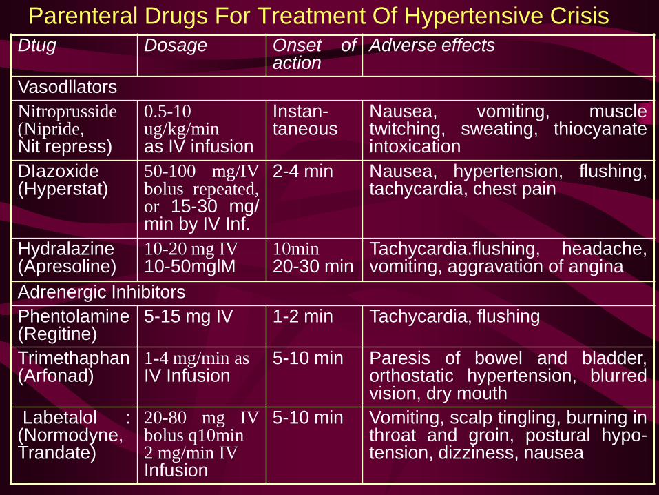
Parenteral Drugs For Treatment Of Hypertensive Crisis Dtug Dosage Onset of
action Adverse effects
Vasodllators Nitroprusside (Nipride, Nit repress)
0.5-10 ug/kg/min as IV infusion
Instan-taneous
Nausea, vomiting, muscle twitching, sweating, thiocyanate intoxication
DIazoxide (Hyperstat)
50-100 mg/IV bolus repeated, or 15-30 mg/ min by IV Inf.
2-4 min Nausea, hypertension, flushing, tachycardia, chest pain
Hydralazine (Apresoline)
10-20 mg IV 10-50mglM
10min 20-30 min
Tachycardia.flushing, headache, vomiting, aggravation of angina
Adrenergic Inhibitors Phentolamine (Regitine)
5-15 mg IV 1-2 min Tachycardia, flushing
Trimethaphan (Arfonad)
1-4 mg/min as IV Infusion
5-10 min Paresis of bowel and bladder, orthostatic hypertension, blurred vision, dry mouth
Labetalol : (Normodyne, Trandate)
20-80 mg IV bolus q10min 2 mg/min IV Infusion
5-10 min Vomiting, scalp tingling, burning in throat and groin, postural hypo-tension, dizziness, nausea