Embed Size (px)
Citation preview

See discussions, stats, and author profiles for this publication at: https://www.researchgate.net/publication/332546446
Learning to detect anatomical landmarks of the pelvis in X-rays from
arbitrary views
Article in International Journal of Computer Assisted Radiology and Surgery · April 2019
DOI: 10.1007/s11548-019-01975-5
CITATIONS
0READS
51
11 authors, including:
Some of the authors of this publication are also working on these related projects:
iPV4.0 View project
Interactive Medical Image Segmentation View project
Bastian Bier
Friedrich-Alexander-University of Erlangen-Nürnberg
28 PUBLICATIONS 45 CITATIONS
SEE PROFILE
Javad Fotouhi
Johns Hopkins University
35 PUBLICATIONS 136 CITATIONS
SEE PROFILE
Greg Osgood
Johns Hopkins University
62 PUBLICATIONS 266 CITATIONS
SEE PROFILE
All content following this page was uploaded by Mathias Unberath on 21 April 2019.
The user has requested enhancement of the downloaded file.
Noname manuscript No.(will be inserted by the editor)
Learning to Detect Anatomical Landmarks of thePelvis in X-rays From Arbitrary Views
Bastian Bier1,3 · Florian Goldmann1,3
Jan-Nico Zaech1,3 · Javad Fotouhi1,2 ·Rachel Hegeman4 · Robert Grupp2
Mehran Armand4,5 · Greg Osgood5 ·Nassir Navab1,2 · Andreas Maier3 ·Mathias Unberath1,2
Received: date / Accepted: date
AbstractPurpose Minimally invasive alternatives are now available for many complex surg-eries. These approaches are enabled by the increasing availability of intra-operativeimage guidance. Yet, fluoroscopic X-rays suffer from projective transformation, andthus cannot provide direct views onto anatomy. Surgeons could highly benefit fromadditional information, such as the anatomical landmark locations in the projec-tions, to support intra-operative decision making. However, detecting landmarks ischallenging since the viewing direction changes substantially between views lead-ing to varying appearance of the same landmark. Therefore, and to the best of ourknowledge, view-independent anatomical landmark detection has not been inves-tigated yet.Methods In this work, we propose a novel approach to detect multiple anatom-ical landmarks in X-ray images from arbitrary viewing directions. To this end,a sequential prediction framework based on convolutional neural networks is em-ployed to simultaneously regress all landmark locations. For training, syntheticX-rays are generated with a physically accurate forward model that allows directapplication of the trained model to real X-ray images of the pelvis. View invari-ance is achieved via data augmentation by sampling viewing angles on a sphericalsegment of 120◦ × 90◦.Results On synthetic data, a mean prediction error of 5.6 ± 4.5 mm is achieved.Further, we demonstrate that the trained model can be directly applied to realX-rays, and show that these detections define correspondences to a respective CT
B. BierE-mail: [email protected]
M. UnberathE-mail: [email protected]
1 Computer Aided Medical Procedures, Johns Hopkins University, Baltimore, USA2 Department of Computer Science, Johns Hopkins University, Baltimore, USA3 Pattern Recognition Lab, Friedrich-Alexander-Universitat Erlangen-Nurnberg, Erlangen,Germany4 Applied Physics Laboratory, Johns Hopkins University, Baltimore, USA5 Department of Orthopedic Surgery, Johns Hopkins Hospital, Baltimore, USA
2 Bier et al.
volume, which allows for analytic estimation of the 11 degree of freedom projectivemapping.Conclusion We present the first tool to detect anatomical landmarks in X-rayimages independent of their viewing direction. Access to this information duringsurgery may benefit decision making and constitutes a first step towards globalinitialization of 2D/3D registration without the need of calibration. As such, theproposed concept has a strong prospect to facilitate and enhance applications andmethods in the realm of image-guided surgery.
Keywords Anatomical Landmarks · Convolutional Neural Networks · 2D/3DRegistration · Landmark Detection
1 Introduction
In recent years, the increasing availability of intra-operative image guidance hasenabled percutaneous alternatives to complex procedures. This is beneficial forthe patient since minimally invasive surgeries are associated with a reduced riskof infection, less blood loss, and an overall decrease of discomfort. However, thiscomes at the cost of increased task-load for the surgeon, who has no direct viewonto the patient’s anatomy but has to rely on indirect feedback through X-rayimages. These suffer from projective transformation; in particular the absence ofdepth cues and, depending on the viewing direction, vanishing anatomical land-marks. One of these procedures is percutaneous pelvis fracture fixation. Pelvisfractures may be complex with a variety of fracture patterns. In order to fixatepelvic fractures internally, K-wires must be guided through narrow bone corridors.Numerous X-ray images from different views may be required to ensure a correcttool trajectory [2,24]. One possibility to support the surgeon during these pro-cedures is to supply additional contextual information extracted from the image.Providing additional, ”implicit 3D” information during these interventions candrastically ease the mental mapping, where the surgeon has to register the tool inhis hand to the 3D patient anatomy using 2D X-ray images only [22,8]. In thiscase, implicit 3D information refers to data that is not 3D as such but providesmeaningful contextual information related to prior knowledge of the surgeon.
A promising candidate for implicit 3D information are the positions of anatom-ical landmarks in the X-ray images. Anatomical landmarks are biologically mean-ingful locations in anatomy that can be readily detected and enable correspondencebetween specimens and across domains. Inherently, the knowledge of landmark lo-cations exhibits helpful properties: (1) context is provided, which supports intra-operative decision making, (2) they supply semantic information, which definescorrespondences across multiple images, and (3) they might foster machine under-standing. For these reasons, anatomical landmarks are widely used in medicine andmedical imaging, where they serve as orientation in diagnostic and interventionalradiology [7]. They deliver a better interpretation of the patients’ anatomy [28]and are also of interest for image processing tasks as prerequisite to initialize orconstrain mathematical models [27]. A non-exhaustive review reveals that anatom-ical landmarks have been used to guide and model segmentation tasks [31,10], toperform image registration [12], to extract relevant clinical quantitative measure-ments [16], to plan therapies [14], or to initialize further image processing [17].

Detect Anatomical Landmarks in X-rays From Arbitrary Views 3
Often, knowing the exact location of landmarks is mandatory for the desiredapplication suggesting that landmarks must be labeled manually [28]. Manuallabeling is time consuming, interrupts the clinical workflow, and is subjective,which yields rater dependent results. Although important, anatomical landmarkdetection is a challenging task due to patient specific variations and ambiguousanatomical structures. At the same time, automatic algorithms should be fast,robust, reliable, and accurate.
Landmark detection methods have been developed for various imaging modal-ities and for 2D or 3D image data [6,7]. In the following overview we focus on 2DX-ray images. Landmark or key point detection is well understood in computervision, where robust feature descriptors disambiguate correspondences betweenmultiple 2D images, finally enabling purely image-based pose retrieval. Unfortu-nately, the above concept defined for reflection imaging does not translate di-rectly to transmission imaging. For the latter, image and landmark appearancecan change fundamentally depending on the viewing direction since the whole 3Dobject contributes to the resulting detector measurement.
Most of the current landmark detection approaches either predict the land-mark positions on the input image directly, or combine these initial estimatessubsequently with a parametric or graphical model fitting step [27]. Constrainingdetection results by models that encode prior knowledge can disambiguate falsepositive responses. Alternatively, priors can be incorporated implicitly, if multiplelandmarks are detected simultaneously by reducing the search space to possibleconfigurations [15]. Wang et al. summarized several landmark detection methodscompeting in a Grand Challenge [28], where 19 landmarks have to be detectedin 2D cephalometric X-ray images of the craniofacial area, a task necessary formodern orthodontics. Mader et al. used a U-net to localize ribs in chest radio-graphs [17]. They solved the problem of ambiguities in the local image informa-tion (false responses) using a conditional random field. This second step assessesspatial information between the landmarks and also refines the hypotheses gen-erated by the U-net. Sa et al. detected intervertebral discs in X-ray images ofthe spine to predict a bounding box of the respective vertebrae, by using a pre-trained Faster-RNN and refining its weights [21]. Payer et al. evaluated differentCNN architectures to detect multiple landmark locations in X-ray images of handX-rays by regressing a single heat map for each landmark [19]. In a similar task,another approach used random forests to detect 37 anatomical landmark in handradiographs. The initial estimates were subsequently combined with prior knowl-edge given by possible landmark configurations. [23]. For each landmark a uniquerandom regression forest is trained. In Xie et al., anatomical landmarks were aprerequisite for the segmentation of the pelvis in anterior-posterior radiographs inorder to create a 3D patient specific pelvis model for surgical planning. The shapemodel utilized for this purpose is based on anatomical landmarks [30].
All the presented approaches above assume a single, predefined view onto theanatomy. This assumption is valid for certain applications, where radiographic im-ages in a diagnostic setup are often acquired in standardized views, but is stronglyviolated when view changes continuously e.g. in interventional applications or forprojection data acquired on trajectories, scenarios in which the view changes con-tinuously. To the best of our knowledge, there exists no approach that is able todetect anatomical landmarks in X-ray images independent of the viewing direction.The view independence substantially complicates the landmark detection problem

4 Bier et al.
C9×9128
Input Image615×479
P2
C9×9128
P2
C9×9128
P2
C5×532
C9×9512
C1×1512
C1×123
b1p
76×59 23
C9×9128
Input Image615×479
P2
C9×9128
P2
C9×9128
P2
C5×532
C11×11128
C11×11128
C11×11128
C11×11128
C1×123
wp
w1
Stage 1 Stage >= 2
btp
76×59 23
C/P : convolution/pooling9×9 : filter size128 : filter number
Fig. 1 Schematic representation of the convolutional neural network used in this work. A singleinput image is processed by multiple stages of convolutional and pooling layers, resulting in astack of belief maps, where each map corresponds to a landmark location. During the stage-wiseapplication, these belief maps are refined.
in X-ray images since object edges vanish and anatomical structures overlap due tothe effect of transmission imaging. X-ray transform invariant landmark detection,therefore, bears great potential to aid fluoroscopic guidance.
In contrast to the landmark detection approaches that deliver implicit 3D in-formation, several approaches exist that introduce explicit 3D information. Thesesolutions rely on external markers to track the tools or the patient in 3D [18], con-sistency conditions to estimate relative pose between X-ray images [1], or 2D/3Dregistration of pre-operative CT to intra-operative X-ray to render multiple viewssimultaneously [18,25]. While these approaches have proven helpful, they are notwidely accepted in clinical practice. The primary reasons are disruptions to thesurgical workflow [8], as well as susceptibility to both truncation and initializationdue to the low capture range of the optimization target [11].
In this work, we propose an automatic, purely image-based method to detectmultiple anatomical landmarks in X-ray images independent of the viewing direc-tion. Landmarks are detected using a sequential prediction framework [29] trainedon synthetically generated images. Based on landmark knowledge, we can a) iden-tify corresponding landmarks between arbitrary views of the same anatomy andb) estimate pose relative to a pre-procedurally acquired volume without the needfor any calibration. We evaluate our approach on synthetic data and demonstratethat it generalizes to unseen clinical X-rays of the pelvis without the need for re-training. Further, we argue that the accuracy of our detections in clinical X-raysmay benefit the initialization of 2D/3D registration. This paper is an extendedversion of the work presented at the MICCAI 2018 conference [4] and providesa broader background on existing landmark detection research, a comprehensivequantitative analysis of the view invariance on synthetic data, and a quantitativeevaluation on real X-ray images of cadaveric specimens.
2 Materials and Methods
2.1 Network Architecture
The sequential prediction framework used in this work has been initially developedfor human pose estimation [29]. In the original application, the machine learningtask is to detect multiple human joint positions in RGB images. The architectureis abstractly depicted in Figure 1. Given a single RGB input image, the network

Detect Anatomical Landmarks in X-rays From Arbitrary Views 5
predicts multiple belief maps bpt for each joint position p ∈ [1, ..., P ] at the end of
every stage t ∈ [1, ..., T ] of the network. In the first stage, initial belief maps bp1
are predicted based only on local image information. Image features are extractedusing a stack of convolutional and pooling layers with Rectified Linear Units (Re-LUs) as activation functions, described by weights w1. In following stages t ≥ 2,the predicted belief maps bp
t are obtained by combining local image informationextracted by the layers with weights wp and the prediction results of the precedingstage. Note that this combination is implemented using a concatenation operation.The weights wp are shared for all stages t ≥ 2. The cost function C is the sumof the L2-losses between the predicted belief maps bp
t and the ground truth beliefmaps b∗
t :
C =T∑
t=1
P∑p=1
||bpt − b∗
t ||22 (1)
The ground truth belief maps b∗t contain a normal distribution, centered at
the ground truth joint position. By design, the network imposes several properties:The key element of the architecture is that the belief maps are predicted based onlocal image information as well as the results of the preceding stage. This enablesthe model to learn long-range contextual dependencies of landmark configurations.The belief maps of the first stage bp
1 are predicted only on local image informa-tion, which leads to false positive responses due to ambiguities in the local imageappearance. The stage wise application resolves these by implicitly incorporatingthe characteristic configuration of the landmark positions. Furthermore, the net-work has a very large receptive field that also increases over stages, which enablesthe learning of spatial dependencies over long distances. Lastly, the loss over allintermediate predictions bp
t is computed, which counteracts the vanishing gradienteffect and simultaneously guides the network to focus early on the detection task.A drawback of this architecture is the small size of the output belief maps thatare downsampled by a factor of around eight compared to the input size.
2.2 Landmark Detection
We exploit the aforementioned advantages of sequential prediction frameworksfor the detection of anatomical landmarks in X-ray images independent of theirviewing direction. Our assumption is that anatomical landmarks exhibit strongconstraints and thus characteristic patterns even in presence of arbitrary viewingangles. In fact, this assumption may be even stronger compared to human poseestimation if limited anatomy, such as the pelvis, is considered due to rigidity.Within this paper and as a first proof-of-concept, we study anatomical landmarkson the pelvis. We devise a network adapted from [29] with six stages to simultane-ously predict 23 belief maps per X-ray image that are used for landmark locationextraction, as shown in Figure 1.
In order to obtain the predicted landmark positions, all predicted belief mapsbpt are averaged over all stages prior to estimating the position of the landmarks
yielding the averaged belief map bp. We then define the landmark position lp as theposition with the highest response in bp. Since the belief maps are downsampled,

6 Bier et al.
1
3
711
15
19
21
6
5
2
20
22
128
4
18
17
23
1314
9 10
Fig. 2 The pelvis bone of a CT of the used data set is rendered with the corresponding 3Dlandmark labels that have been labeled manually. Orange dots with numbers indicate visiblelandmarks. Landmarks hidden due to the rendering are marked with a grey box and number(e.g. the tip of the right femoral head, landmark #13).
the maximum location is computed in a sub pixel accuracy by a Maximum Like-lihood Estimation of the Gaussian estimate. If the maximum response in a beliefmap is below 0.4, the landmarks are discarded since they may be outside the fieldof view or are not reliably recognized. The implementation was done in Pythonand Tensorflow. The hyperparameters for the network training are set to 10−6 forthe learning rate and a batch size of one. Optimization was performed using Adamover 30 epochs until convergence in the validation set had been reached.
2.3 Data Generation
The network training requires a data set of X-ray images with corresponding land-mark positions. Manual labeling is infeasible for various reasons: First of all, thelabeling process to obtain the required amount of training data is time costly.However, and more importantly, an accurate and consistent labeling cannot beguaranteed in the 2D projection images due to the discussed properties of trans-mission imaging (vanishing edges, superimposed anatomy). Therefore, we synthet-ically generated the training data from full body CTs of the NIH Cancer ImagingArchive [20]. In total, 23 landmark positions were manually labeled in 20 CTs ofmale and female patients using 3D volume renderings in 3D Slicer [5]. The land-mark positions have been selected to be clinically meaningful, to have a good visi-bility in the projection images, and to be consistently identifiable on the anatomy.The selected landmarks are depicted in Figure 2.
Subsequently, data was obtained by forward projection of the volume and therespective 3D labels with the same X-ray imaging geometry, resulting in a setof X-ray images with corresponding landmark positions. The synthetic X-ray im-ages had a size of 615 × 479 pixels with an isotropic pixel spacing of 0.616 mm.

Detect Anatomical Landmarks in X-rays From Arbitrary Views 7
The corresponding ground truth belief maps were downsampled by a factor ofabout eight and had a size of 76 × 59. For data generation, two factors are im-portant to emphasize: (1) The augmentation of training data in order to obtainview-invariance is crucial. To this end, we applied random translation to the CTvolume, varied the source-to-isocenter distance, applied flipping on the detector,and most importantly, varied the angular range of the X-ray source position ona spherical segment of 120◦ in LAO/RAO and in 90◦ in CRAN/CAUD, centeredaround an AP view of the pelvis. This range approximates the range of variationin X-ray images during surgical procedures on the pelvis [13]. (2) A realistic for-ward projector that accounts for physically accurate image formation, while beingcapable of fast data generation was used to obtain realistic synthetic training data.This allows direct application of the network model to real clinical X-ray images.The forward projector computes material-dependent attenuation images that areconverted into synthetic X-rays [26]. In total, 20000 X-rays were generated andsplit 18× 1× 1-fold into training, validation, and testing, where we ensured thatimages of one patient are not shared among these sets.
2.4 2D/3D Registration
As motivated previously, the detected anatomical landmarks offer a range of pos-sible applications. In this work, we focus on the example of initializing 2D/3Dregistration. To this end, 2D landmark positions are automatically extracted fromX-ray images while 3D points are obtained from a manually labeled pre-operativeCT acquisition of the same patient. Since the landmark detections supply semanticinformation, correspondences between the 2D and 3D points are defined, which en-ables the computation of the projection matrix P ∈ R3×4 in closed form across thetwo domains [9]. The set of 2D detections are expressed as homogeneous vectorsas dn ∈ R3 with n ∈ [1, . . . , N ]. Each point contains the entries dn = (xn.yn, wn).The set of corresponding 3D points are denoted as homogeneous vectors rn ∈ R4.Following the direct linear transform, each correspondence yields two linearly in-dependent equations [9, p.178]:
[0T −wir
Ti yir
Ti
wirTi 0T −xir
Ti
]p1
p2
p3
= 0. (2)
With N being the number of corresponding points, these rows are stacked intoa measurement matrix that results in a size of 2N×12. p1, p2, and p3 are vectors∈ R4 that contain the entries of the projection matrix P. These are obtainedsubsequently by computing the null space of the measurement matrix.
3 Experiments and Results
The result section is split into two parts: In the first part, the results of thelandmark detection on the synthetic data set are presented. In the second part,the network trained on synthetic data is used to predict anatomical landmarksin real X-ray images acquired using a clinical C-arm X-ray system of cadavericspecimens. Note, that the network has not been re-trained for this purpose. Forboth cases, the results are presented qualitatively and quantitatively.

8 Bier et al.C
RA
45°
CA
U 4
5°
RAO 60° LAO 60°
Fig. 3 Predicted landmark positions for example projection images sampled across the sam-pled spherical segment of the synthetic test data set. Ground truth positions are marked withblue labels and automatic detection with red labels. Note, that each projection image is pro-cessed independently.
Fig. 4 Accuracy depending on the viewing direction of the X-ray source. In average thedetection result from central views is superior to the ones at the border of the sphere. Theaccuracy is defined as the ratio of landmarks that have an error below 15 pixels in the respectiveview.
3.1 Synthetic Data
For the evaluation of the view-invariant landmark detection, we created X-rayimages of the testing CT data set that were uniformly sampled across the wholespherical segment with an angular spacing of 5◦ in both dimensions. A standardsetting for the geometry with 750 mm source-to-isocenter distance and 1,200 mmsource-to-detector distance was used. These distances are not varied in the evalu-ation, since the focus is the angular dependent detectability of landmarks.

Detect Anatomical Landmarks in X-rays From Arbitrary Views 9
Table 1 Individual landmark belief and error. Average Belief is the average of the highestresponses in the belief maps for a certain landmark. Average Error is the average distancebetween the landmark detection and its ground truth location. The columns Q1, Q2, Q3 andQ4 also contain the average error, but evaluated only in a particular quadrant of the sphericalsegment to indicate detectability changes of certain landmarks across the spherical segment.Error values are given in pixels.
Landmark # Average Belief Average Error [pixel] Q1 Q2 Q3 Q4
1 0.79 7.60 9.42 5.35 7.13 7.892 0.84 6.68 5.66 7.67 6.63 6.053 0.83 6.86 9.13 7.81 5.07 5.264 0.87 7.69 8.79 10.2 7.11 4.705 0.85 7.53 8.11 8.47 6.63 6.626 0.82 5.63 4.72 5.21 5.97 6.357 0.78 7.90 7.96 7.48 8.29 7.998 0.77 10.1 5.87 12.1 7.70 15.39 0.90 5.26 5.15 5.08 5.55 5.0710 0.88 7.19 7.60 6.90 5.80 8.4111 0.89 6.43 5.77 5.99 6.86 6.8312 0.91 7.78 8.96 7.23 5.71 8.5513 0.92 4.47 5.64 4.10 4.67 3.2414 0.90 5.64 3.70 7.00 5.24 6.1815 0.85 9.04 8.77 9.54 7.75 10.316 0.82 7.23 6.55 6.95 7.26 8.1817 0.81 19.9 20.0 24.2 15.2 21.118 0.80 15.3 11.2 16.6 14.5 19.319 0.74 9.56 10.4 10.4 9.80 7.0920 0.77 8.59 5.78 12.9 6.83 8.9121 0.51 9.40 14.3 6.86 13.9 8.5122 0.44 13.7 9.73 25.0 10.1 16.223 0.51 26.0 24.2 17.6 39.3 29.8
Average 9.10 ± 7.38
In Figure 3, the detection results are presented qualitatively and comparedto the ground truth positions. Overall, the qualitative agreement between groundtruth locations and predictions is very good. Quantitatively, the average distancebetween ground truth positions and detection across all projections and landmarksis 9.1 ± 7.4 pixels (5.6 ± 4.5 mm). Note that, as motivated previously, belief mapresponses lower than 0.4 are considered as landmark not detected and the cor-responding landmark are excluded from the statistics. Graphically, the detectionaccuracy is plotted across all viewing directions in Figure 4. We define the detec-tion accuracy as the percentage of landmarks that have an error smaller than adistance threshold of 15 pixels in the respective view. The detection accuracy isalso plotted against this threshold in Figure 8.
In Table 1, a more detailed analysis of the error across the different landmarksand the view position is presented. For each landmark, the average maximum be-lief, the average error across all projections, as well as the error across quadrants isshown. For the latter, the spherical segment is subdivided into four areas, centeredat the AP position with two perpendicular divisions across the CRAN/CAUD andRAO/LAO axis. This reveals three interesting observations: first, some landmarkshave an overall lower error (e.g. landmark #9 with an average error of 5.26 pixels),while others are detected poorly (e.g. landmark #23 with an average error of

10 Bier et al.
0 0.2 0.4 0.6 0.8 10
20
40
60
80
100
Belief
Distance
Error[pixel]
Fig. 5 The error of a landmark detection is plotted onto the belief of the correspondinglandmark detection. A correlation can be observed: higher beliefs indicate lower detectionerrors.
Fig. 6 Maximum belief depending on the viewing direction for two landmarks (#11 and #19).While landmark #11 (left) is equally well visible across views, the belief for landmark #19(right) changes substantially across views.
26.05 pixels). Second, there exists a correlation between the average maximum be-lief response and the average error: the higher the response in a belief map, thelower the error. This observation is also supported by the scatter plot presentedin Figure 5, where for each prediction the detection error is plotted over the corre-sponding maximum belief map response. Third, some landmarks can be detectedequally well, independently of the viewing direction (e.g. landmark #11), while forothers, the detectability highly varies across the quadrants (e.g. landmark #19).This observation is graphically well visible for these two landmarks, as shown inFigure 6.
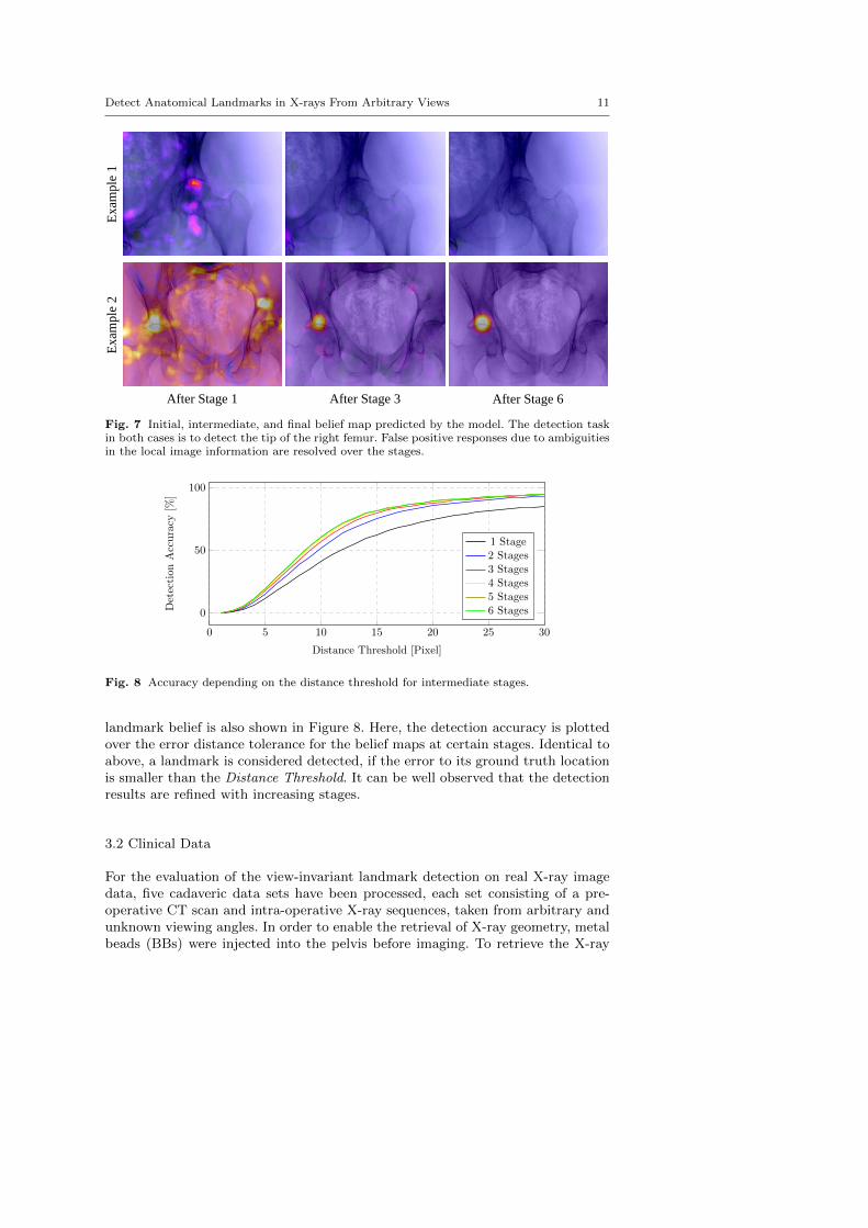
We further investigated how the belief map response develops over the stages ofthe network and how ambiguities in the early stages are resolved. In Figure 7, twoexample projections are shown, overlain by their corresponding belief maps at therespective stage. In the first row, the landmark of interest (tip of the right femoralhead) is outside the field of view. However, a false position response appears afterthe first stage due to the similar appearance of the anatomy. With further stages,this ambiguity gets resolved. In the second row, a similar behavior is visible and arefinement of the prediction accuracy is clearly observable. The development of a
Detect Anatomical Landmarks in X-rays From Arbitrary Views 11E
xam
ple
2E
xam
ple
1
After Stage 6After Stage 1 After Stage 3
Fig. 7 Initial, intermediate, and final belief map predicted by the model. The detection taskin both cases is to detect the tip of the right femur. False positive responses due to ambiguitiesin the local image information are resolved over the stages.
0 5 10 15 20 25 30
0
50
100
Distance Threshold [Pixel]
Detection
Accuracy
[%]
1 Stage2 Stages3 Stages4 Stages5 Stages6 Stages
Fig. 8 Accuracy depending on the distance threshold for intermediate stages.
landmark belief is also shown in Figure 8. Here, the detection accuracy is plottedover the error distance tolerance for the belief maps at certain stages. Identical toabove, a landmark is considered detected, if the error to its ground truth locationis smaller than the Distance Threshold. It can be well observed that the detectionresults are refined with increasing stages.
3.2 Clinical Data
For the evaluation of the view-invariant landmark detection on real X-ray imagedata, five cadaveric data sets have been processed, each set consisting of a pre-operative CT scan and intra-operative X-ray sequences, taken from arbitrary andunknown viewing angles. In order to enable the retrieval of X-ray geometry, metalbeads (BBs) were injected into the pelvis before imaging. To retrieve the X-ray

12 Bier et al.
projection geometry, first BB correspondences are established between individualimages of the intra-operative X-ray sequence. Then, the fundamental matrix iscomputed for each image pair allowing for the 3D reconstruction of the BB po-sitions [9]. This 3D reconstruction was then registered to the 3D BB locationsextracted from the CT volume, allowing for an exact registration of each BB in2D space to its corresponding location in 3D space. With these correspondencesestablished, the projection matrices for each X-ray image was then calculated inclosed form solution as in Equation 2. To evaluate the reprojection error of theseprojection matrices (which defines a lower bound on the accuracy achievable usinganatomical landmarks), the 3D BB coordinates as per the CT scan are forwardprojected into 2D space. Table 2 shows the reprojection error between the forwardprojection and the real X-ray images for the X-ray sequences. Note that one ofthis sequences has a tool in the field of view (sequence #3), while another showsa fracture of the pelvis (sequence #2). The low reprojection error of 2–5 px isin line with our expectations, and suggests that the resulting projection matricesare an appropriate reference when evaluating the performance of our proposedview-invariant landmark detection.
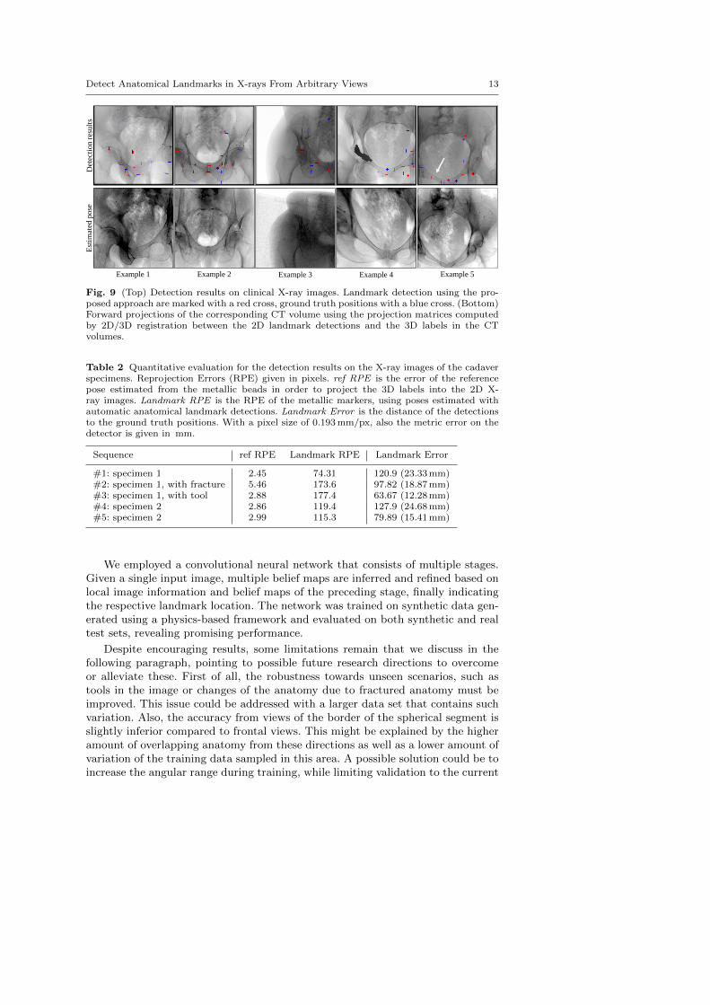
In the top row of Figure 9, example landmark detection results in X-ray imagesof these sequences are shown. Automatic detections and ground truth locationsare indicated with red and blue crosses, respectively. Overall, the well agreementbetween the automatic detections and the ground truth positions can be appreci-ated for various poses and truncations. In complicated situations when tools arepresent in the image, the landmark detection approach fails to detect the sur-rounding landmarks, as can be seen in example 4. However, this is not surprisingsince such situations were not part of the training data set. Small unknown ob-jects, such as the metallic beads on the bone, seem to only have a limited influenceon performance. Furthermore, example 5 depicts an image of the sequence wherea fracture is present in the X-ray image, indicated with the white arrow. Qualita-tively, this did not influence the detection substantially. Quantitatively, the overalldeviation between true and predicted landmark locations averaged over the total106 real images is shown in Table 2 in column titled Landmark Error.
The bottom row in Figure 9 shows digitally reconstructed radiographs (DRRs)of the CT volumes belonging to the real X-ray image of the same patient shownabove. The geometry for generating the DRRs has been obtained in closed form2D/3D registration of the detected landmarks to the 3D labels in the CT volume, asdescribed in Section 2.4. For these various poses, the landmark detection accuracyproves sufficient to achieve views that are very similar to the target X-ray image,suggesting successful initialization.
4 Discussion and Conclusion
We presented a novel approach to detect anatomical landmarks in X-ray imagesindependent of the viewing direction. The landmark locations supply additionalinformation for the surgeon and enable various applications, including global ini-tialization of 2D/3D registration in closed form. Due to the characteristics oftransmission imaging, landmark appearances change substantially with the view-ing direction making anatomical landmark detection a challenging task that has,to the best of our knowledge, not previously been addressed.
Detect Anatomical Landmarks in X-rays From Arbitrary Views 13D
etec
tio
n r
esu
lts
Est
imat
ed p
ose
Example 1 Example 2 Example 3 Example 4 Example 5
Fig. 9 (Top) Detection results on clinical X-ray images. Landmark detection using the pro-posed approach are marked with a red cross, ground truth positions with a blue cross. (Bottom)Forward projections of the corresponding CT volume using the projection matrices computedby 2D/3D registration between the 2D landmark detections and the 3D labels in the CTvolumes.
Table 2 Quantitative evaluation for the detection results on the X-ray images of the cadaverspecimens. Reprojection Errors (RPE) given in pixels. ref RPE is the error of the referencepose estimated from the metallic beads in order to project the 3D labels into the 2D X-ray images. Landmark RPE is the RPE of the metallic markers, using poses estimated withautomatic anatomical landmark detections. Landmark Error is the distance of the detectionsto the ground truth positions. With a pixel size of 0.193 mm/px, also the metric error on thedetector is given in mm.
Sequence ref RPE Landmark RPE Landmark Error
#1: specimen 1 2.45 74.31 120.9 (23.33 mm)#2: specimen 1, with fracture 5.46 173.6 97.82 (18.87 mm)#3: specimen 1, with tool 2.88 177.4 63.67 (12.28 mm)#4: specimen 2 2.86 119.4 127.9 (24.68 mm)#5: specimen 2 2.99 115.3 79.89 (15.41 mm)
We employed a convolutional neural network that consists of multiple stages.Given a single input image, multiple belief maps are inferred and refined based onlocal image information and belief maps of the preceding stage, finally indicatingthe respective landmark location. The network was trained on synthetic data gen-erated using a physics-based framework and evaluated on both synthetic and realtest sets, revealing promising performance.
Despite encouraging results, some limitations remain that we discuss in thefollowing paragraph, pointing to possible future research directions to overcomeor alleviate these. First of all, the robustness towards unseen scenarios, such astools in the image or changes of the anatomy due to fractured anatomy must beimproved. This issue could be addressed with a larger data set that contains suchvariation. Also, the accuracy from views of the border of the spherical segment isslightly inferior compared to frontal views. This might be explained by the higheramount of overlapping anatomy from these directions as well as a lower amount ofvariation of the training data sampled in this area. A possible solution could be toincrease the angular range during training, while limiting validation to the current

14 Bier et al.
range Further, the network architecture in its current state yields belief maps thatare downsampled by a factor of around eight compared to the input image. Thisdownsampling inherently limits the accuracy of the detection results. While thisaccuracy may have been sufficient for the initial purpose of human pose estimation,in medical imaging, higher accuracy is desirable. Possible improvements that aresubject to future work could be achieved by competing network architectures basedon an encoder-decoder design with skip connections in order to preserve resolu-tion of the output images. Alternatively, test-time augmentation could be appliedby processing slightly altered versions of the input image with the same networkduring application. The results of these multiple outputs could subsequently beaveraged, which might yield higher accuracy. Furthermore, the robustness as wellas the overall accuracy could benefit by providing prior knowledge in the form of amodel-based post-processing step. A possible source of error might be introducedby the labeling of the landmarks in the 3D volume that, since manual, is inherentlyprone to errors. Ideally, an unsupervised landmark or keypoint selection processwould be of great benefit for this approach. As a possible application, we showedthat an initialization of 2D/3D registration based on the automatic detections issuccessful without the need for additional calibration. In this work, we relied ona closed form solution to estimate the image pose which is compelling due to itssimplicity, yet, a more sophisticated approach based on maximum likelihood wouldcertainly yield superior results in presence of statistical outliers. In this task wealso showed that considering the maximum belief is powerful for selecting reliablydetected landmarks. This additional information can be used as a confidence mea-sure for further processing tasks. Recently, the proposed concept of view-invariantanatomical landmark detection has been transferred to projection images of kneesin an attempt to estimate involuntary motion during scans [3]
In conclusion, detecting anatomical landmarks has grown to be an essential toolin automatic image parsing in diagnostic imaging, suggesting similar importancefor image-guided interventions. The implementation of anatomical landmarks asa powerful concept for aiding image-guided interventions will be pushed continu-ously as new approaches, such as this one, strive to achieve clinically acceptableperformance.
Acknowledgements We gratefully acknowledge the support of NIH/NIBIB R01 EB023939,R21 EB020113, R01 EB016703, R01 EB0223939, and the NVIDIA Corporation with the do-nation of the GPUs used for this research. Further, the authors acknowledge funding supportfrom NIH 5R01AR065248-03.
Conflict of Interest The authors declare that they have no conflict of interest.
References
1. Aichert, A., Berger, M., Wang, J., Maass, N., Doerfler, A., Hornegger, J., Maier, A.K.:Epipolar consistency in transmission imaging. IEEE Trans. Med. Imag. 34(11), 2205–2219(2015)
2. Baumgartner, R., Libuit, K., Ren, D., Bakr, O., Singh, N., Kandemir, U., Marmor, M.T.,Morshed, S.: Reduction of radiation exposure from c-arm fluoroscopy during orthopaedictrauma operations with introduction of real-time dosimetry. Journal of OrthopaedicTrauma 3(2), e53e58 (2016)
3. Bier, B., Aschoff, K., Syben, C., Unberath, M., Levenston, M., Gold, G., Fahrig, R., Maier,A.: Detecting anatomical landmarks for motion estimation in weight-bearing imaging of

Detect Anatomical Landmarks in X-rays From Arbitrary Views 15
knees. In: International Workshop on Machine Learning for Medical Image Reconstruction,pp. 83–90. Springer (2018)
4. Bier, B., Unberath, M., Zaech, J.N., Fotouhi, J., Armand, M., Osgood, G., Navab, N.,Maier, A.: X-ray-transform invariant anatomical landmark detection for pelvic traumasurgery. In: International Conference on Medical Image Computing and Computer-Assisted Intervention, pp. 55–63. Springer (2018)
5. Fedorov, A., Beichel, R., Kalpathy-Cramer, J., Finet, J., Fillion-Robin, J.C., Pujol, S.,Bauer, C., Jennings, D., Fennessy, F., Sonka, M., Buatti, J., Aylward, S., Miller, J., Pieper,S., Kikinis, R.: 3d slicer as an image computing platform for the quantitative imagingnetwork. Magnetic resonance imaging 30(9), 1323–1341 (2012)
6. Ghesu, F.C., Georgescu, B., Mansi, T., Neumann, D., Hornegger, J., Comaniciu, D.: Anartificial agent for anatomical landmark detection in medical images. In: MICCAI, pp.229–237. Springer (2016)
7. Ghesu, F.C., Georgescu, B., Zheng, Y., Grbic, S., Maier, A., Hornegger, J., Comaniciu, D.:Multi-scale deep reinforcement learning for real-time 3d-landmark detection in ct scans.IEEE Transactions on Pattern Analysis and Machine Intelligence (2017)
8. Hartl, R., Lam, K.S., Wang, J., Korge, A., Audige, F.K.L.: Worldwide survey on the useof navigation in spine surgery. World Neurosurg 379(1), 162172 (2013)
9. Hartley, R.I., Zisserman, A.: Multiple View Geometry in Computer Vision. CambridgeUniversity Press, ISBN: 0521540518 (2004)
10. Heimann, T., Meinzer, H.P.: Statistical shape models for 3d medical image segmentation:a review. Medical image analysis 13(4), 543–563 (2009)
11. Hou, B., Alansary, A., McDonagh, S., Davidson, A., Rutherford, M., Hajnal, J.V., Rueck-ert, D., Glocker, B., Kainz, B.: Predicting slice-to-volume transformation in presence ofarbitrary subject motion. In: MICCAI, pp. 296–304. Springer (2017)
12. Johnson, H.J., Christensen, G.E.: Consistent landmark and intensity-based image regis-tration. IEEE transactions on medical imaging 21(5), 450–461 (2002)
13. Khurana, B., Sheehan, S.E., Sodickson, A.D., Weaver, M.J.: Pelvic ring fractures: whatthe orthopedic surgeon wants to know. Radiographics 34(5), 1317–1333 (2014)
14. Litjens, G., Kooi, T., Bejnordi, B.E., Setio, A.A.A., Ciompi, F., Ghafoorian, M., van derLaak, J.A., Van Ginneken, B., Sanchez, C.I.: A survey on deep learning in medical imageanalysis. Medical image analysis 42, 60–88 (2017)
15. Liu, D., Zhou, K.S., Bernhardt, D., Comaniciu, D.: Search strategies for multiple landmarkdetection by submodular maximization. In: Computer Vision and Pattern Recognition(CVPR), 2010 IEEE Conference on, pp. 2831–2838. IEEE (2010)
16. M Pouch, A., A Yushkevich, P., M Jackson, B., S Jassar, A., Vergnat, M., H Gorman, J.,C Gorman, R., M Sehgal, C.: Development of a semi-automated method for mitral valvemodeling with medial axis representation using 3d ultrasound. Medical physics 39(2),933–950 (2012)
17. Mader, A.O., von Berg, J., Fabritz, A., Lorenz, C., Meyer, C.: Localization and labelingof posterior ribs in chest radiographs using a crf-regularized fcn with local refinement. In:International Conference on Medical Image Computing and Computer-Assisted Interven-tion, pp. 562–570. Springer (2018)
18. Markelj, P., Tomazevic, D., Likar, B., Pernus, F.: A review of 3d/2d registration methodsfor image-guided interventions. Med Image Anal 16(3), 642–661 (2012)
19. Payer, C., Stern, D., Bischof, H., Urschler, M.: Regressing heatmaps for multiple landmarklocalization using cnns. In: International Conference on Medical Image Computing andComputer-Assisted Intervention, pp. 230–238. Springer (2016)
20. Roth, H., Lu, L., Seff, A., Cherry, K.M., Hoffman, J., Wang, S., Summers, R.M.: A new2.5 d representation for lymph node detection in ct. The Cancer Imaging Archive (2015)
21. Sa, R., Owens, W., Wiegand, R., Studin, M., Capoferri, D., Barooha, K., Greaux, A.,Rattray, R., Hutton, A., Cintineo, J., Chaudhary, V.: Intervertebral disc detection in x-ray images using faster r-cnn. In: Engineering in Medicine and Biology Society (EMBC),2017 39th Annual International Conference of the IEEE, pp. 564–567. IEEE (2017)
22. Starr, R., Jones, A., Reinert, C., Borer, D.: Preliminary results and complications follow-ing limited open reduction and percutaneous screw fixation of displaced fractures of theacetabulum. Injury 32, SA45–50 (2001)
23. Stern, D., Ebner, T., Urschler, M.: From local to global random regression forests: ex-ploring anatomical landmark localization. In: International Conference on Medical ImageComputing and Computer-Assisted Intervention, pp. 221–229. Springer (2016)

16 Bier et al.
24. Stockle, U., Schaser, K., Konig, B.: Image guidance in pelvic and acetabular surgeryex-pectations, success and limitations. Injury 38(4), 450462 (2007)
25. Tucker, E., Fotouhi, J., Lee, S., Unberath, M., Fuerst, B., Johnson, A., Armand, M.,Osgood, G., Navab, N.: Towards clinical translation of augmented orthopedic surgery:from pre-op ct to intra-op x-ray via rgbd sensing. In: SPIE Medical Imaging (2018)
26. Unberath, M., Zaech, J.N., Lee, S.C., Bier, B., Fotouhi, J., Armand, M., Navab, N.:Deepdrr–a catalyst for machine learning in fluoroscopy-guided procedures. In: Inter-national Conference on Medical Image Computing and Computer-Assisted Intervention.Springer (2018)
27. Urschler, M., Ebner, T., Stern, D.: Integrating geometric configuration and appearanceinformation into a unified framework for anatomical landmark localization. Medical imageanalysis 43, 23–36 (2018)
28. Wang, C.W., Huang, C.T., Hsieh, M.C., Li, C.H., Chang, S.W., Li, W.C., Vandaele, R.,Maree, R., Jodogne, S., Geurts, P., Chen, C., Zhen, G., chu, C., Mirzaalian, H., Vrtovec,T., Ibragimov, B.: Evaluation and comparison of anatomical landmark detection methodsfor cephalometric x-ray images: A grand challenge. IEEE transactions on medical imaging34(9), 1890–1900 (2015)
29. Wei, S.E., Ramakrishna, V., Kanade, T., Sheikh, Y.: Convolutional pose machines. In:CVPR, pp. 4724–4732 (2016)
30. Xie, W., Franke, J., Chen, C., Grutzner, P.A., Schumann, S., Nolte, L.P., Zheng, G.:A complete-pelvis segmentation framework for image-free total hip arthroplasty (tha):methodology and clinical study. The International Journal of Medical Robotics and Com-puter Assisted Surgery 11(2), 166–180 (2015)
31. Zheng, Y., Barbu, A., Georgescu, B., Scheuering, M., Comaniciu, D.: Four-chamber heartmodeling and automatic segmentation for 3-d cardiac ct volumes using marginal spacelearning and steerable features. IEEE transactions on medical imaging 27(11), 1668–1681(2008)
View publication statsView publication stats


![Ultrasound guidance versus anatomical landmarks for ...€¦ · [Intervention Review] Ultrasound guidance versus anatomical landmarks for internal jugular vein catheterization Patrick](https://img.pdfslide.us/doc/110x75/5f9beef95154c7333f47d212/ultrasound-guidance-versus-anatomical-landmarks-for-intervention-review-ultrasound.jpg)