Embed Size (px)
Citation preview
195
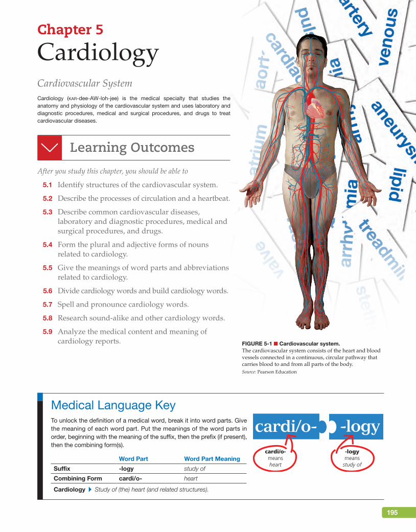
Figure 5-1 ■ Cardiovascular system.The cardiovascular system consists of the heart and blood vessels connected in a continuous, circular pathway that carries blood to and from all parts of the body.Source: Pearson Education
Chapter 5
CardiologyCardiovascular SystemCardiology (kar-dee-AW-loh-jee) is the medical specialty that studies the anatomy and physiology of the cardiovascular system and uses laboratory and diagnostic procedures, medical and surgical procedures, and drugs to treat cardiovascular diseases.
Learning Outcomes
After you study this chapter, you should be able to
5.1 Identify structures of the cardiovascular system.
5.2 Describe the processes of circulation and a heartbeat.
5.3 Describe common cardiovascular diseases, laboratory and diagnostic procedures, medical and surgical procedures, and drugs.
5.4 Form the plural and adjective forms of nouns related to cardiology.
5.5 Give the meanings of word parts and abbreviations related to cardiology.
5.6 Divide cardiology words and build cardiology words.
5.7 Spell and pronounce cardiology words.
5.8 Research sound-alike and other cardiology words.
5.9 Analyze the medical content and meaning of cardiology reports.
To unlock the definition of a medical word, break it into word parts. Give the meaning of each word part. Put the meanings of the word parts in order, beginning with the meaning of the suffix, then the prefix (if present), then the combining form(s).
Word Part Word Part Meaning
Suffix -logy study of
Combining Form cardi/o- heart
Cardiology ▸ Study of (the) heart (and related structures).
Medical Language Key
cardi/o-meansheart
cardi/o- -logy-logymeans
study of
M05_TURL8127_04_SE_C05.indd 195 14/12/15 10:48 pm
196 Chapter 5 Cardiology
Anatomy and Physiology
The cardiovascular system is a continuous, circular body system that includes the heart and the vascular structures (blood vessels such as arteries, capillar-ies, and veins) (see Figure 5-1 ■). It is also known as the circulatory system. To
study the cardiovascular system, you can begin with the heart or you can begin with the capillaries, the tiniest blood vessels in the farthest parts of the body. Beginning at either starting point, you can go through every part of the cardiovascular system and arrive back where you began. The purpose of the cardiovascular system is to move (circulate) the blood to every part of the body as it transports oxygen, carbon dioxide, nutrients, and wastes. The blood is discussed in “Hematology and Immu-nology,” Chapter 6.
Anatomy of the Cardiovascular SystemHeartThe heart is perhaps the best-known organ in the body and certainly one of the most important. It is a muscular organ that contracts at least once every second to pump blood throughout the body. It also has an extensive electrical system that initiates and coordinates its contractions.
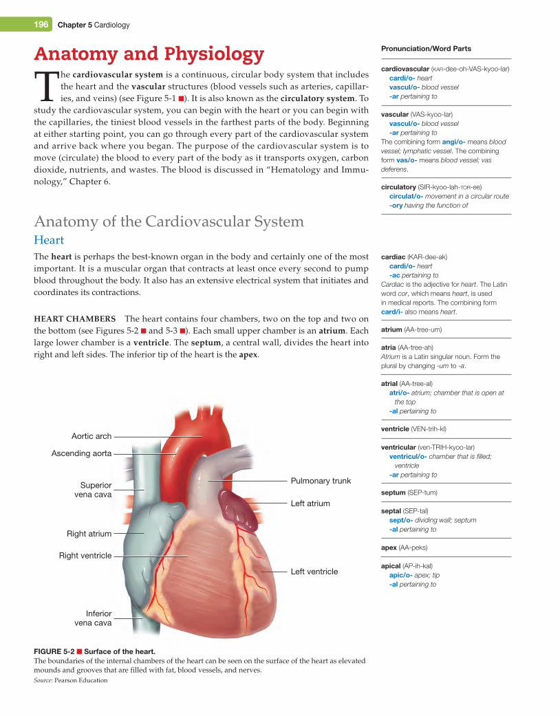
Heart CHambers The heart contains four chambers, two on the top and two on the bottom (see Figures 5-2 ■ and 5-3 ■). Each small upper chamber is an atrium. Each large lower chamber is a ventricle. The septum, a central wall, divides the heart into right and left sides. The inferior tip of the heart is the apex.
Figure 5-2 ■ Surface of the heart.The boundaries of the internal chambers of the heart can be seen on the surface of the heart as elevated mounds and grooves that are filled with fat, blood vessels, and nerves.Source: Pearson Education
Ascending aorta
Pulmonary trunkSuperiorvena cava
Inferiorvena cava
Right ventricle
Right atrium
Aortic arch
Left atrium
Left ventricleapical (AP-ih-kal)
apic/o- apex; tip-al pertaining to
septal (SEP-tal)sept/o- dividing wall; septum-al pertaining to
apex (AA-peks)
ventricular (ven-TRIH-kyoo-lar)ventricul/o- chamber that is filled;
ventricle-ar pertaining to
septum (SEP-tum)
Pronunciation/Word Parts
cardiovascular (kar-dee-oh-VAS-kyoo-lar)cardi/o- heartvascul/o- blood vessel-ar pertaining to
atrial (AA-tree-al)atri/o- atrium; chamber that is open at
the top-al pertaining to
ventricle (VEN-trih-kl)
atrium (AA-tree-um)
atria (AA-tree-ah)Atrium is a Latin singular noun. Form the plural by changing -um to -a.
cardiac (KAR-dee-ak)cardi/o- heart-ac pertaining to
Cardiac is the adjective for heart. The Latin word cor, which means heart, is used in medical reports. The combining form card/i- also means heart.
vascular (VAS-kyoo-lar)vascul/o- blood vessel-ar pertaining to
The combining form angi/o- means blood vessel; lymphatic vessel. The combining form vas/o- means blood vessel; vas deferens.
circulatory (SIR-kyoo-lah-tor-ee)circulat/o- movement in a circular route-ory having the function of
M05_TURL8127_04_SE_C05.indd 196 14/12/15 10:48 pm
Chapter 5 Cardiology 197
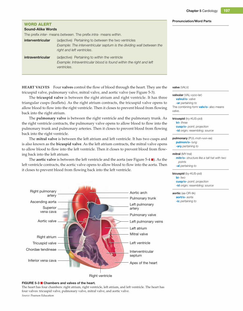
Heart ValVes Four valves control the flow of blood through the heart. They are the tricuspid valve, pulmonary valve, mitral valve, and aortic valve (see Figure 5-3).
The tricuspid valve is between the right atrium and right ventricle. It has three triangular cusps (leaflets). As the right atrium contracts, the tricuspid valve opens to allow blood to flow into the right ventricle. Then it closes to prevent blood from flowing back into the right atrium.
The pulmonary valve is between the right ventricle and the pulmonary trunk. As the right ventricle contracts, the pulmonary valve opens to allow blood to flow into the pulmonary trunk and pulmonary arteries. Then it closes to prevent blood from flowing back into the right ventricle.
The mitral valve is between the left atrium and left ventricle. It has two cusps and is also known as the bicuspid valve. As the left atrium contracts, the mitral valve opens to allow blood to flow into the left ventricle. Then it closes to prevent blood from flow-ing back into the left atrium.
The aortic valve is between the left ventricle and the aorta (see Figure 5-4 ■). As the left ventricle contracts, the aortic valve opens to allow blood to flow into the aorta. Then it closes to prevent blood from flowing back into the left ventricle.
bicuspid (by-KUS-pid)bi- twocusp/o- point; projection-id origin; resembling; source
aortic (aa-OR-tik)aort/o- aorta-ic pertaining to
pulmonary (PUL-moh-nair-ee)pulmon/o- lung-ary pertaining to
mitral (MY-tral)mitr/o- structure like a tall hat with two
points-al pertaining to
valvular (VAL-vyoo-lar)valvul/o- valve-ar pertaining to
The combining form valv/o- also means valve.
tricuspid (try-KUS-pid)tri- threecusp/o- point; projection-id origin; resembling; source
Pronunciation/Word Parts
Figure 5-3 ■ Chambers and valves of the heart.The heart has four chambers: right atrium, right ventricle, left atrium, and left ventricle. The heart has four valves: tricuspid valve, pulmonary valve, mitral valve, and aortic valve.Source: Pearson Education
Aortic valve
Right atrium
Right ventricle
Interventricularseptum
Apex of the heart
Mitral valve
Tricuspid valve
Chordae tendineae
Left atrium
Left ventricle
Ascending aorta
Right pulmonaryartery
Superiorvena cava
Inferior vena cava
Pulmonary valve
Left pulmonaryartery
Left pulmonary veins
Aortic arch
Pulmonary trunk
Word AlertSound-Alike WordsThe prefix inter- means between. The prefix intra- means within.
interventricular (adjective) Pertaining to between the two ventriclesExample: The interventricular septum is the dividing wall between the right and left ventricles.
intraventricular (adjective) Pertaining to within the ventricleExample: Intraventricular blood is found within the right and left ventricles.
valve (VALV)
M05_TURL8127_04_SE_C05.indd 197 14/12/15 10:48 pm
198 Chapter 5 Cardiology
The tricuspid and mitral valves have chordae tendineae, rope-like strands attached to their valve leaflets (see Figure 5-3). The other end of the chordae tendineae is anchored to small muscles on the wall of the ventricles. When the ventricles contract, these small muscles also contract and pull on the chordae tendineae. This stabilizes the valve leaflets and keeps them firmly sealed together to keep blood from flowing back into the atria, even during the strong force of a ventricular contraction.
The sounds of the valves closing are commonly known as “lubb-dupp” (a phonetic approximation of the actual sounds). The “lubb” is made as the tricuspid and mitral valves close. This first heart sound is abbreviated as S1. The “dupp” is made as the pul-monary and aortic valves close. This second heart sound is abbreviated as S2.
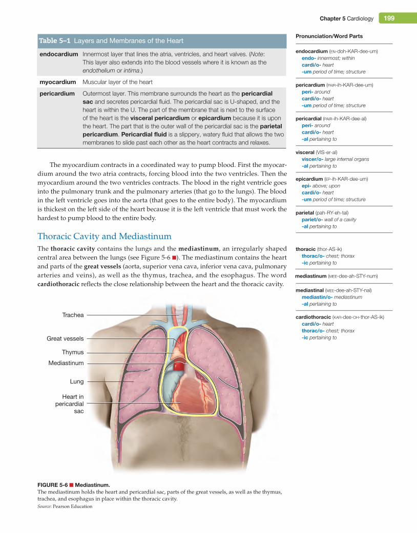
Heart musCle The myocardium is the muscular layer of the heart (see Figure 5-5 ■ and Table 5-1 ■). The myocardium is composed of cardiac muscle. Its muscle fibers (muscle cells) respond to electrical impulses generated by a node within the right atrium. This process is discussed in a later section.
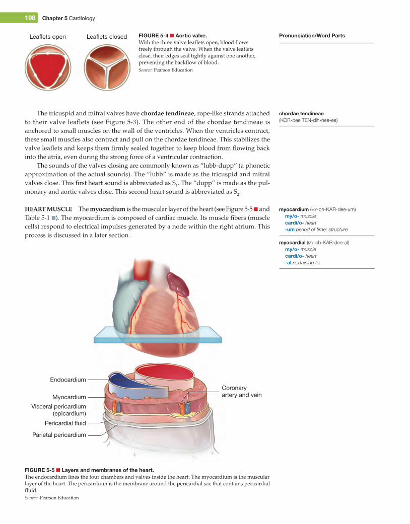
Figure 5-4 ■ Aortic valve.With the three valve leaflets open, blood flows freely through the valve. When the valve leaflets close, their edges seal tightly against one another, preventing the backflow of blood.Source: Pearson Education
Leaflets open Leaflets closed
Figure 5-5 ■ Layers and membranes of the heart.The endocardium lines the four chambers and valves inside the heart. The myocardium is the muscular layer of the heart. The pericardium is the membrane around the pericardial sac that contains pericardial fluid.Source: Pearson Education
Parietal pericardium
Pericardial fluid
Visceral pericardium(epicardium)
Myocardium
Coronaryartery and vein
Endocardium
Pronunciation/Word Parts
myocardium (my-oh-KAR-dee-um)my/o- musclecardi/o- heart-um period of time; structure
myocardial (my-oh-KAR-dee-al)my/o- musclecardi/o- heart-al pertaining to
chordae tendineae (KOR-dee TEN-dih-nee-ee)
M05_TURL8127_04_SE_C05.indd 198 14/12/15 10:48 pm
Chapter 5 Cardiology 199
The myocardium contracts in a coordinated way to pump blood. First the myocar-dium around the two atria contracts, forcing blood into the two ventricles. Then the myocardium around the two ventricles contracts. The blood in the right ventricle goes into the pulmonary trunk and the pulmonary arteries (that go to the lungs). The blood in the left ventricle goes into the aorta (that goes to the entire body). The myocardium is thickest on the left side of the heart because it is the left ventricle that must work the hardest to pump blood to the entire body.
Thoracic Cavity and MediastinumThe thoracic cavity contains the lungs and the mediastinum, an irregularly shaped central area between the lungs (see Figure 5-6 ■). The mediastinum contains the heart and parts of the great vessels (aorta, superior vena cava, inferior vena cava, pulmonary arteries and veins), as well as the thymus, trachea, and the esophagus. The word cardiothoracic reflects the close relationship between the heart and the thoracic cavity.
endocardium Innermost layer that lines the atria, ventricles, and heart valves. (Note: This layer also extends into the blood vessels where it is known as the endothelium or intima.)
myocardium Muscular layer of the heart
pericardium Outermost layer. This membrane surrounds the heart as the pericardial sac and secretes pericardial fluid. The pericardial sac is U-shaped, and the heart is within the U. The part of the membrane that is next to the surface of the heart is the visceral pericardium or epicardium because it is upon the heart. The part that is the outer wall of the pericardial sac is the parietal pericardium. Pericardial fluid is a slippery, watery fluid that allows the two membranes to slide past each other as the heart contracts and relaxes.
Table 5–1 Layers and Membranes of the Heart
cardiothoracic (kar-dee-oh-thor-AS-ik)cardi/o- heartthorac/o- chest; thorax-ic pertaining to
mediastinum (mee-dee-ah-STY-num)
mediastinal (mee-dee-ah-STY-nal)mediastin/o- mediastinum-al pertaining to
parietal (pah-RY-eh-tal)pariet/o- wall of a cavity-al pertaining to
thoracic (thor-AS-ik)thorac/o- chest; thorax-ic pertaining to
visceral (VIS-er-al)viscer/o- large internal organs-al pertaining to
epicardium (ep-ih-KAR-dee-um)epi- above; uponcardi/o- heart-um period of time; structure
pericardium (pair-ih-KAR-dee-um)peri- aroundcardi/o- heart-um period of time; structure
pericardial (pair-ih-KAR-dee-al)peri- aroundcardi/o- heart-al pertaining to
Pronunciation/Word Parts
endocardium (en-doh-KAR-dee-um)endo- innermost; withincardi/o- heart-um period of time; structure
Figure 5-6 ■ Mediastinum.The mediastinum holds the heart and pericardial sac, parts of the great vessels, as well as the thymus, trachea, and esophagus in place within the thoracic cavity.Source: Pearson Education
Great vessels
Lung
Thymus
Heart inpericardial
sac
Trachea
Mediastinum
M05_TURL8127_04_SE_C05.indd 199 14/12/15 10:48 pm
200 Chapter 5 Cardiology
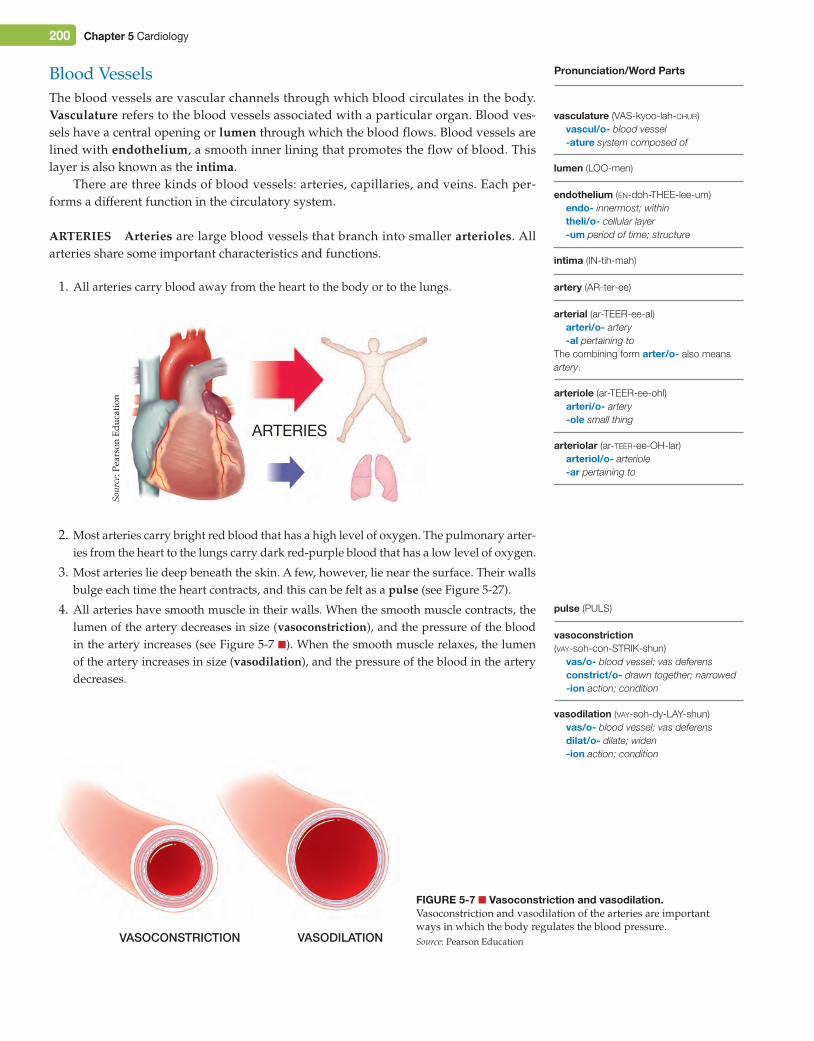
Blood VesselsThe blood vessels are vascular channels through which blood circulates in the body. Vasculature refers to the blood vessels associated with a particular organ. Blood ves-sels have a central opening or lumen through which the blood flows. Blood vessels are lined with endothelium, a smooth inner lining that promotes the flow of blood. This layer is also known as the intima.
There are three kinds of blood vessels: arteries, capillaries, and veins. Each per-forms a different function in the circulatory system.
arteries arteries are large blood vessels that branch into smaller arterioles. All arteries share some important characteristics and functions.
1. All arteries carry blood away from the heart to the body or to the lungs.
Figure 5-7 ■ Vasoconstriction and vasodilation.Vasoconstriction and vasodilation of the arteries are important ways in which the body regulates the blood pressure.Source: Pearson EducationVASOCONSTRICTION VASODILATION
vasoconstriction (vay-soh-con-STRIK-shun)
vas/o- blood vessel; vas deferensconstrict/o- drawn together; narrowed-ion action; condition
vasodilation (vay-soh-dy-LAY-shun)vas/o- blood vessel; vas deferensdilat/o- dilate; widen-ion action; condition
arteriolar (ar-teer-ee-OH-lar)arteriol/o- arteriole-ar pertaining to
pulse (PULS)
arterial (ar-TEER-ee-al)arteri/o- artery-al pertaining to
The combining form arter/o- also means artery.
arteriole (ar-TEER-ee-ohl)arteri/o- artery-ole small thing
intima (IN-tih-mah)
artery (AR-ter-ee)
lumen (LOO-men)
endothelium (en-doh-THEE-lee-um)endo- innermost; withintheli/o- cellular layer-um period of time; structure
Pronunciation/Word Parts
ARTERIES
2. Most arteries carry bright red blood that has a high level of oxygen. The pulmonary arter-ies from the heart to the lungs carry dark red-purple blood that has a low level of oxygen.
3. Most arteries lie deep beneath the skin. A few, however, lie near the surface. Their walls bulge each time the heart contracts, and this can be felt as a pulse (see Figure 5-27).
4. All arteries have smooth muscle in their walls. When the smooth muscle contracts, the lumen of the artery decreases in size (vasoconstriction), and the pressure of the blood in the artery increases (see Figure 5-7 ■). When the smooth muscle relaxes, the lumen of the artery increases in size (vasodilation), and the pressure of the blood in the artery decreases.
vasculature (VAS-kyoo-lah-chur)vascul/o- blood vessel-ature system composed of
Sour
ce: P
ears
on E
duca
tion
M05_TURL8127_04_SE_C05.indd 200 14/12/15 10:48 pm
Chapter 5 Cardiology 201
Capillaries Capillaries are the smallest blood vessels in the body. The lumen of a capillary is so small that blood cells must pass through in single file. A network of capil-laries connects the arterioles and venules. An arteriole branches into a network of capil-laries that reaches each cell in the body and then the capillaries merge into a venule.
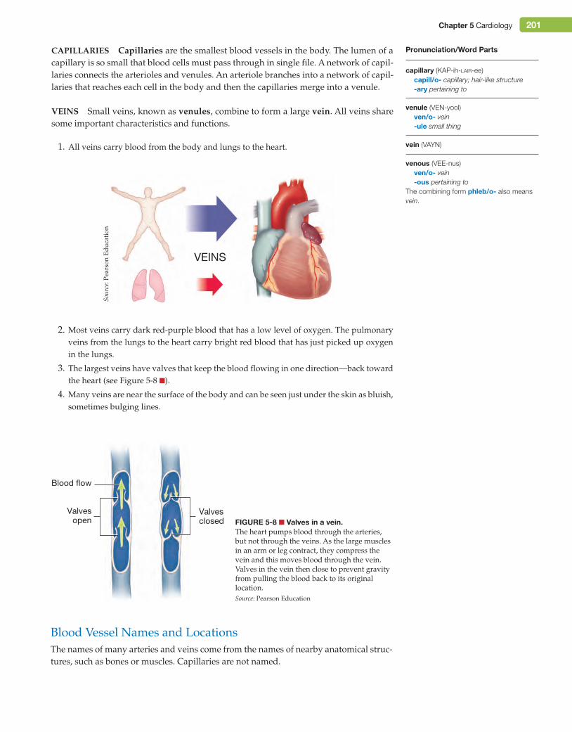
Veins Small veins, known as venules, combine to form a large vein. All veins share some important characteristics and functions.
1. All veins carry blood from the body and lungs to the heart.
Figure 5-8 ■ Valves in a vein.The heart pumps blood through the arteries, but not through the veins. As the large muscles in an arm or leg contract, they compress the vein and this moves blood through the vein. Valves in the vein then close to prevent gravity from pulling the blood back to its original location.Source: Pearson Education
Valvesopen
Blood flow
Valvesclosed
vein (VAYN)
venous (VEE-nus)ven/o- vein-ous pertaining to
The combining form phleb/o- also means vein.
Pronunciation/Word Parts
capillary (KAP-ih-lair-ee)capill/o- capillary; hair-like structure-ary pertaining to
venule (VEN-yool)ven/o- vein-ule small thing
VEINS
2. Most veins carry dark red-purple blood that has a low level of oxygen. The pulmonary veins from the lungs to the heart carry bright red blood that has just picked up oxygen in the lungs.
3. The largest veins have valves that keep the blood flowing in one direction—back toward the heart (see Figure 5-8 ■).
4. Many veins are near the surface of the body and can be seen just under the skin as bluish, sometimes bulging lines.
Blood Vessel Names and LocationsThe names of many arteries and veins come from the names of nearby anatomical struc-tures, such as bones or muscles. Capillaries are not named.
Sour
ce: P
ears
on E
duca
tion
M05_TURL8127_04_SE_C05.indd 201 14/12/15 10:48 pm
202 Chapter 5 Cardiology
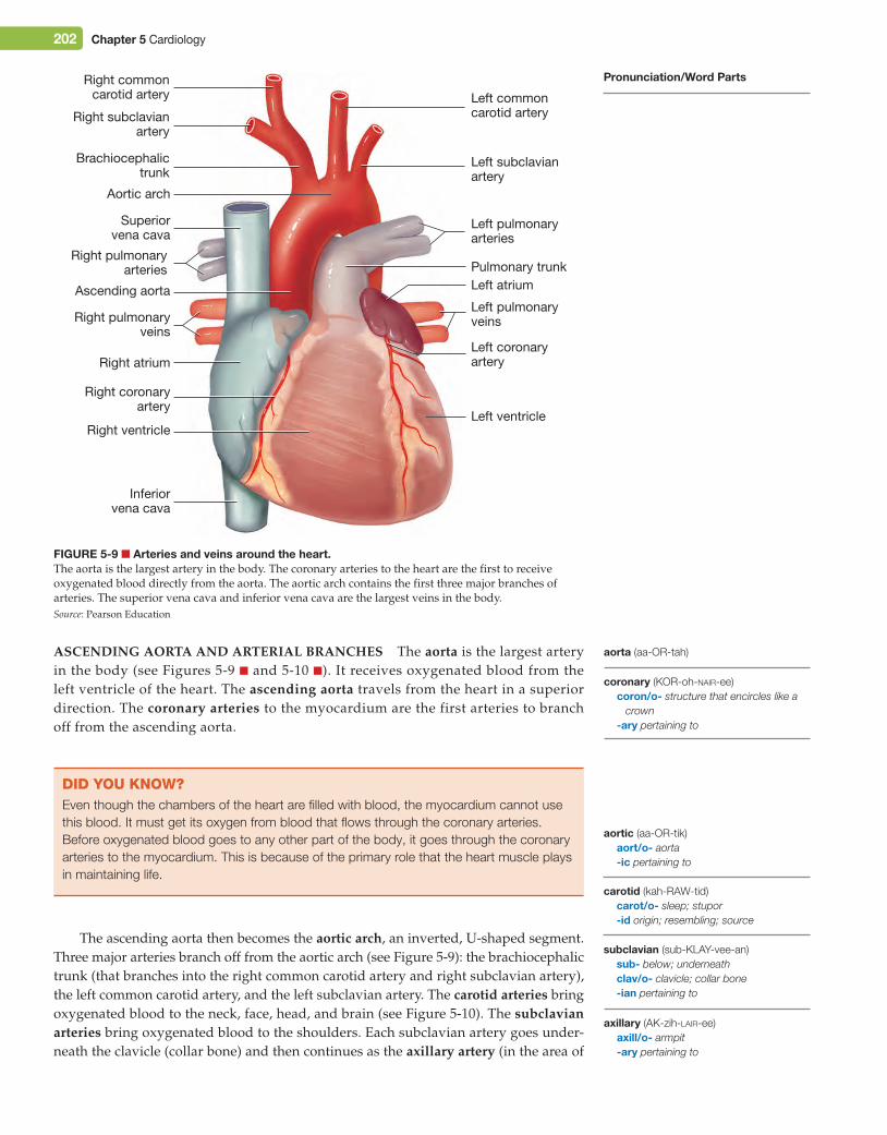
asCending aorta and arterial branCHes The aorta is the largest artery in the body (see Figures 5-9 ■ and 5-10 ■). It receives oxygenated blood from the left ventricle of the heart. The ascending aorta travels from the heart in a superior direction. The coronary arteries to the myocardium are the first arteries to branch off from the ascending aorta.
Figure 5-9 ■ Arteries and veins around the heart.The aorta is the largest artery in the body. The coronary arteries to the heart are the first to receive oxygenated blood directly from the aorta. The aortic arch contains the first three major branches of arteries. The superior vena cava and inferior vena cava are the largest veins in the body.Source: Pearson Education
Ascending aorta
Right pulmonaryveins
Pulmonary trunk
Superiorvena cava
Inferiorvena cava
Right ventricle
Right atrium
Right coronaryartery
Aortic arch
Left atrium
Left ventricle
Brachiocephalictrunk
Left subclavianartery
Left commoncarotid arteryRight subclavian
artery
Right commoncarotid artery
Left pulmonaryarteries
Left pulmonaryveins
Left coronaryartery
Right pulmonaryarteries
did You KnoW?Even though the chambers of the heart are filled with blood, the myocardium cannot use this blood. It must get its oxygen from blood that flows through the coronary arteries. Before oxygenated blood goes to any other part of the body, it goes through the coronary arteries to the myocardium. This is because of the primary role that the heart muscle plays in maintaining life.
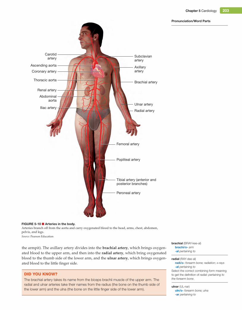
The ascending aorta then becomes the aortic arch, an inverted, U-shaped segment. Three major arteries branch off from the aortic arch (see Figure 5-9): the brachiocephalic trunk (that branches into the right common carotid artery and right subclavian artery), the left common carotid artery, and the left subclavian artery. The carotid arteries bring oxygenated blood to the neck, face, head, and brain (see Figure 5-10). The subclavian arteries bring oxygenated blood to the shoulders. Each subclavian artery goes under-neath the clavicle (collar bone) and then continues as the axillary artery (in the area of
axillary (AK-zih-lair-ee)axill/o- armpit-ary pertaining to
carotid (kah-RAW-tid)carot/o- sleep; stupor-id origin; resembling; source
subclavian (sub-KLAY-vee-an)sub- below; underneathclav/o- clavicle; collar bone-ian pertaining to
coronary (KOR-oh-nair-ee)coron/o- structure that encircles like a
crown-ary pertaining to
aortic (aa-OR-tik)aort/o- aorta-ic pertaining to
Pronunciation/Word Parts
aorta (aa-OR-tah)
M05_TURL8127_04_SE_C05.indd 202 14/12/15 10:48 pm
Chapter 5 Cardiology 203
the armpit). The axillary artery divides into the brachial artery, which brings oxygen-ated blood to the upper arm, and then into the radial artery, which bring oxygenated blood to the thumb side of the lower arm, and the ulnar artery, which brings oxygen-ated blood to the little finger side.
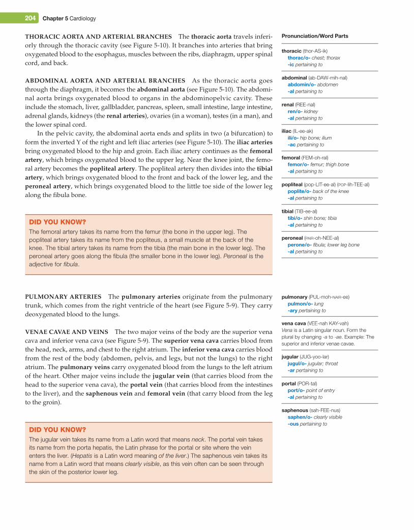
Figure 5-10 ■ Arteries in the body.Arteries branch off from the aorta and carry oxygenated blood to the head, arms, chest, abdomen, pelvis, and legs.Source: Pearson Education
Carotidartery
Thoracic aorta
Abdominalaorta
Femoral artery
Popliteal artery
Peroneal artery
Subclavianartery
Axillaryartery
Brachial artery
Renal artery
Radial arteryIliac artery
Ulnar artery
Ascending aorta
Coronary artery
Tibial artery (anterior andposterior branches)
radial (RAY-dee-al)radi/o- forearm bone; radiation; x-rays-al pertaining to
Select the correct combining form meaning to get the definition of radial: pertaining to the forearm bone.
ulnar (UL-nar)uln/o- forearm bone; ulna-ar pertaining to
Pronunciation/Word Parts
did You KnoW?The brachial artery takes its name from the biceps brachii muscle of the upper arm. The radial and ulnar arteries take their names from the radius (the bone on the thumb side of the lower arm) and the ulna (the bone on the little finger side of the lower arm).
brachial (BRAY-kee-al)brachi/o- arm-al pertaining to
M05_TURL8127_04_SE_C05.indd 203 14/12/15 10:48 pm
204 Chapter 5 Cardiology
tHoraCiC aorta and arterial branCHes The thoracic aorta travels inferi-orly through the thoracic cavity (see Figure 5-10). It branches into arteries that bring oxygenated blood to the esophagus, muscles between the ribs, diaphragm, upper spinal cord, and back.
abdominal aorta and arterial branCHes As the thoracic aorta goes through the diaphragm, it becomes the abdominal aorta (see Figure 5-10). The abdomi-nal aorta brings oxygenated blood to organs in the abdominopelvic cavity. These include the stomach, liver, gallbladder, pancreas, spleen, small intestine, large intestine, adrenal glands, kidneys (the renal arteries), ovaries (in a woman), testes (in a man), and the lower spinal cord.
In the pelvic cavity, the abdominal aorta ends and splits in two (a bifurcation) to form the inverted Y of the right and left iliac arteries (see Figure 5-10). The iliac arteries bring oxygenated blood to the hip and groin. Each iliac artery continues as the femoral artery, which brings oxygenated blood to the upper leg. Near the knee joint, the femo-ral artery becomes the popliteal artery. The popliteal artery then divides into the tibial artery, which brings oxygenated blood to the front and back of the lower leg, and the peroneal artery, which brings oxygenated blood to the little toe side of the lower leg along the fibula bone.
did You KnoW?The femoral artery takes its name from the femur (the bone in the upper leg). The popliteal artery takes its name from the popliteus, a small muscle at the back of the knee. The tibial artery takes its name from the tibia (the main bone in the lower leg). The peroneal artery goes along the fibula (the smaller bone in the lower leg). Peroneal is the adjective for fibula.
pulmonary arteries The pulmonary arteries originate from the pulmonary trunk, which comes from the right ventricle of the heart (see Figure 5-9). They carry deoxygenated blood to the lungs.
Venae CaVae and Veins The two major veins of the body are the superior vena cava and inferior vena cava (see Figure 5-9). The superior vena cava carries blood from the head, neck, arms, and chest to the right atrium. The inferior vena cava carries blood from the rest of the body (abdomen, pelvis, and legs, but not the lungs) to the right atrium. The pulmonary veins carry oxygenated blood from the lungs to the left atrium of the heart. Other major veins include the jugular vein (that carries blood from the head to the superior vena cava), the portal vein (that carries blood from the intestines to the liver), and the saphenous vein and femoral vein (that carry blood from the leg to the groin).
did You KnoW?The jugular vein takes its name from a Latin word that means neck. The portal vein takes its name from the porta hepatis, the Latin phrase for the portal or site where the vein enters the liver. (Hepatis is a Latin word meaning of the liver.) The saphenous vein takes its name from a Latin word that means clearly visible, as this vein often can be seen through the skin of the posterior lower leg.
jugular (JUG-yoo-lar)jugul/o- jugular; throat-ar pertaining to
portal (POR-tal)port/o- point of entry-al pertaining to
pulmonary (PUL-moh-nair-ee)pulmon/o- lung-ary pertaining to
vena cava (VEE-nah KAY-vah)Vena is a Latin singular noun. Form the plural by changing -a to -ae. Example: The superior and inferior venae cavae.
tibial (TIB-ee-al)tibi/o- shin bone; tibia-al pertaining to
peroneal (pair-oh-NEE-al)perone/o- fibula; lower leg bone-al pertaining to
femoral (FEM-oh-ral)femor/o- femur; thigh bone-al pertaining to
popliteal (pop-LIT-ee-al) (pop-lih-TEE-al)poplite/o- back of the knee-al pertaining to
renal (REE-nal)ren/o- kidney-al pertaining to
iliac (IL-ee-ak)ili/o- hip bone; ilium-ac pertaining to
Pronunciation/Word Parts
thoracic (thor-AS-ik)thorac/o- chest; thorax-ic pertaining to
abdominal (ab-DAW-mih-nal)abdomin/o- abdomen-al pertaining to
saphenous (sah-FEE-nus)saphen/o- clearly visible-ous pertaining to
M05_TURL8127_04_SE_C05.indd 204 14/12/15 10:48 pm
Chapter 5 Cardiology 205
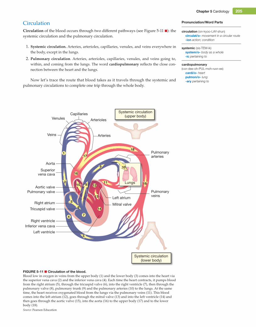
CirculationCirculation of the blood occurs through two different pathways (see Figure 5-11 ■): the systemic circulation and the pulmonary circulation.
1. systemic circulation. Arteries, arterioles, capillaries, venules, and veins everywhere in the body, except in the lungs.
2. pulmonary circulation. Arteries, arterioles, capillaries, venules, and veins going to, within, and coming from the lungs. The word cardiopulmonary reflects the close con-nection between the heart and the lungs.
Now let’s trace the route that blood takes as it travels through the systemic and pulmonary circulations to complete one trip through the whole body.
Figure 5-11 ■ Circulation of the blood.Blood low in oxygen in veins from the upper body (1) and the lower body (3) comes into the heart via the superior vena cava (2) and the inferior vena cava (4). Each time the heart contracts, it pumps blood from the right atrium (5), through the tricuspid valve (6), into the right ventricle (7), then through the pulmonary valve (8), pulmonary trunk (9) and the pulmonary arteries (10) to the lungs. At the same time, the heart receives oxygenated blood from the lungs via the pulmonary veins (11). This blood comes into the left atrium (12), goes through the mitral valve (13) and into the left ventricle (14) and then goes through the aortic valve (15), into the aorta (16) to the upper body (17) and to the lower body (18).Source: Pearson Education
Superiorvena cava
2
4
5
6
8
3
10
11
9
7
12
16
14
1
13
15
17
18
18
Aorta
Right atrium
Pulmonary valve
Tricuspid valve
Right ventricleInferior vena cava
Mitral valve
Aortic valve
Left atrium
Arteries
ArteriolesVenules
Veins
Capillaries
Left ventricle
Pulmonaryveins
Pulmonaryarteries
Lungs
Systemic circulation(upper body)
Systemic circulation(lower body)
systemic (sis-TEM-ik)system/o- body as a whole-ic pertaining to
Pronunciation/Word Parts
circulation (sir-kyoo-LAY-shun)circulat/o- movement in a circular route-ion action; condition
cardiopulmonary (kar-dee-oh-PUL-moh-nair-ee)
cardi/o- heartpulmon/o- lung-ary pertaining to
M05_TURL8127_04_SE_C05.indd 205 14/12/15 10:48 pm
206 Chapter 5 Cardiology
systemiC CirCulation tHrougH tHe Veins Blood coming from the cells is dark red-purple in color because it has a low level of oxygen. Blood coming from cells in the upper body (1) travels through capillaries, venules, and veins to the superior vena cava (2). Blood coming from cells in the lower body (3) travels through capillar-ies, venules, and veins to the inferior vena cava (4). Then this blood travels through the right atrium (5), tricuspid valve (6), and right ventricle (7).
pulmonary CirCulation At this point, the blood enters the pulmonary circula-tion. The blood travels through the pulmonary valve (8), pulmonary trunk (9), and pul-monary arteries (10) and arterioles to the capillaries in the lungs. In a capillary beside an alveolus, the blood releases carbon dioxide, picks up oxygen, and becomes bright red in color. The blood then travels through the pulmonary veins (11) to the left atrium (12) of the heart.
systemiC CirCulation tHrougH tHe arteries At this point, the blood is back in the systemic circulation. From the left atrium (12), the blood travels through the mitral valve (13) and left ventricle (14). The blood then travels through the aortic valve (15) and into the aorta (16) to the upper body (17) and the lower body (18). The arteries, arterioles, and capillaries distribute this oxygenated blood to every part of the body. In a capillary beside a body cell, the blood releases oxygen, and picks up carbon dioxide, and becomes dark red-purple in color. This completes one trip around the circulatory system.
node (NOHD)
conduction (con-DUK-shun)conduct/o- carrying; conveying-ion action; condition
sinoatrial (sy-noh-AA-tree-al)sin/o- channel; hollow cavityatri/o- atrium; chamber that is open at
the top-al pertaining to
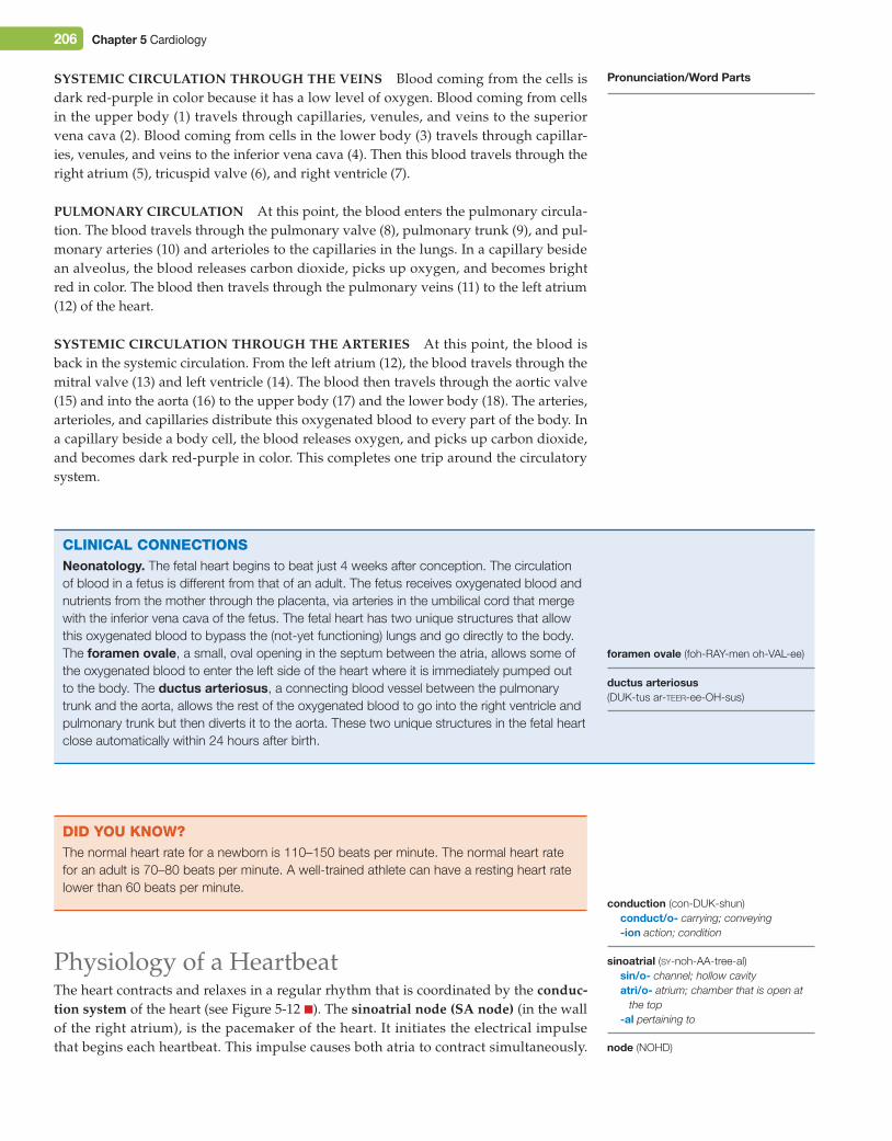
CliniCAl ConneCtionsNeonatology. The fetal heart begins to beat just 4 weeks after conception. The circulation of blood in a fetus is different from that of an adult. The fetus receives oxygenated blood and nutrients from the mother through the placenta, via arteries in the umbilical cord that merge with the inferior vena cava of the fetus. The fetal heart has two unique structures that allow this oxygenated blood to bypass the (not-yet functioning) lungs and go directly to the body. The foramen ovale, a small, oval opening in the septum between the atria, allows some of the oxygenated blood to enter the left side of the heart where it is immediately pumped out to the body. The ductus arteriosus, a connecting blood vessel between the pulmonary trunk and the aorta, allows the rest of the oxygenated blood to go into the right ventricle and pulmonary trunk but then diverts it to the aorta. These two unique structures in the fetal heart close automatically within 24 hours after birth.
Pronunciation/Word Parts
ductus arteriosus (DUK-tus ar-teer-ee-OH-sus)
did You KnoW?The normal heart rate for a newborn is 110–150 beats per minute. The normal heart rate for an adult is 70–80 beats per minute. A well-trained athlete can have a resting heart rate lower than 60 beats per minute.
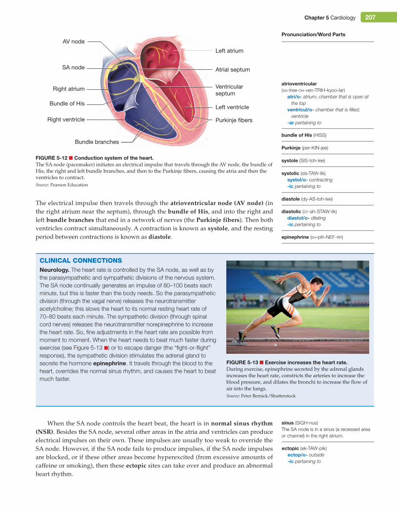
Physiology of a HeartbeatThe heart contracts and relaxes in a regular rhythm that is coordinated by the conduc-tion system of the heart (see Figure 5-12 ■). The sinoatrial node (sa node) (in the wall of the right atrium), is the pacemaker of the heart. It initiates the electrical impulse that begins each heartbeat. This impulse causes both atria to contract simultaneously.
foramen ovale (foh-RAY-men oh-VAL-ee)
M05_TURL8127_04_SE_C05.indd 206 14/12/15 10:48 pm
Chapter 5 Cardiology 207
The electrical impulse then travels through the atrioventricular node (aV node) (in the right atrium near the septum), through the bundle of His, and into the right and left bundle branches that end in a network of nerves (the purkinje fibers). Then both ventricles contract simultaneously. A contraction is known as systole, and the resting period between contractions is known as diastole.
Bundle of His
Bundle branches
Right ventricle Purkinje fibers
Left ventricle
Left atrium
SA node
AV node
Right atrium
Atrial septum
Ventricularseptum
Figure 5-12 ■ Conduction system of the heart.The SA node (pacemaker) initiates an electrical impulse that travels through the AV node, the bundle of His, the right and left bundle branches, and then to the Purkinje fibers, causing the atria and then the ventricles to contract.Source: Pearson Education
epinephrine (eh-pih-NEF-rin)
diastole (dy-AS-toh-lee)
diastolic (dy-ah-STAW-lik)diastol/o- dilating-ic pertaining to
systole (SIS-toh-lee)
systolic (sis-TAW-lik)systol/o- contracting-ic pertaining to
bundle of His (HISS)
Purkinje (per-KIN-jee)
Pronunciation/Word Parts
CliniCAl ConneCtionsNeurology. The heart rate is controlled by the SA node, as well as by the parasympathetic and sympathetic divisions of the nervous system. The SA node continually generates an impulse of 80–100 beats each minute, but this is faster than the body needs. So the parasympathetic division (through the vagal nerve) releases the neurotransmitter acetylcholine; this slows the heart to its normal resting heart rate of 70–80 beats each minute. The sympathetic division (through spinal cord nerves) releases the neurotransmitter norepinephrine to increase the heart rate. So, fine adjustments in the heart rate are possible from moment to moment. When the heart needs to beat much faster during exercise (see Figure 5-13 ■) or to escape danger (the “fight-or-flight” response), the sympathetic division stimulates the adrenal gland to secrete the hormone epinephrine. It travels through the blood to the heart, overrides the normal sinus rhythm, and causes the heart to beat much faster.
Figure 5-13 ■ exercise increases the heart rate.During exercise, epinephrine secreted by the adrenal glands increases the heart rate, constricts the arteries to increase the blood pressure, and dilates the bronchi to increase the flow of air into the lungs.Source: Peter Bernick/Shutterstock
When the SA node controls the heart beat, the heart is in normal sinus rhythm (nsr). Besides the SA node, several other areas in the atria and ventricles can produce electrical impulses on their own. These impulses are usually too weak to override the SA node. However, if the SA node fails to produce impulses, if the SA node impulses are blocked, or if these other areas become hyperexcited (from excessive amounts of caffeine or smoking), then these ectopic sites can take over and produce an abnormal heart rhythm.
ectopic (ek-TAW-pik)ectop/o- outside-ic pertaining to
sinus (SIGH-nus)The SA node is in a sinus (a recessed area or channel) in the right atrium.
atrioventricular (aa-tree-oh-ven-TRIH-kyoo-lar)
atri/o- atrium; chamber that is open at the top
ventricul/o- chamber that is filled; ventricle
-ar pertaining to
M05_TURL8127_04_SE_C05.indd 207 14/12/15 10:48 pm
208 Chapter 5 Cardiology
A Closer looKelectrical Activity of the Heart. On a molecular level, an elegant and intricate system allows the heart to contract tirelessly, approximately 100,000 times each day. An electrical impulse from the SA node changes the permeability of a myocardial cell. Sodium ions (Na+) outside the cell move through the cell membrane, followed by calcium ions (Ca++). This gives the inside of the cell a positive electrical charge, which triggers the release of calcium ions stored inside the cell. This process is known as depolarization because it reverses the normal, slightly negative electrical state of the cell. The calcium ions cause the myocardial cell to contract. As one cell depolarizes and contracts, it triggers the next myocardial cell to do the same.
A contraction ends when potassium ions (K+) move out of the cell, while tiny molecular pumps move sodium ions and some calcium ions out of the cell and move the rest of the calcium ions back into storage within the cell. This process is known as repolarization. This restores the normal, slightly negative electrical state of a resting myocardial cell. The myocardial cell is now ready for another impulse from the SA node.
A myocardial cell cannot respond to another electrical impulse from the SA node until the full cycle of depolarization and repolarization is complete. This very short period of unresponsiveness is known as the refractory period.
Pronunciation/Word Parts
depolarization (dee-poh-lar-ih-ZAY-shun)de- reversal of; withoutpolar/o- negative state; positive state-ization process of creating; process
of inserting; process of making
repolarization (ree-poh-lar-ih-ZAY-shun)re- again and again; backward;
unable topolar/o- negative state; positive state-ization process of creating; process
of inserting; process of making
refractory (ree-FRAK-tor-ee)re- again and again; backward;
unable tofract/o- bend; break up-ory having the function of
Select the correct prefix meaning to get the definition of refractory: having the function of (being) unable to break up.
Word Alertcardia (noun) Small region of the stomach where the esophagus enters
Example: The cardia is the first part of the stomach to receive food from the esophagus.
cardiac (adjective) Pertaining to the heartExample: During a cardiac arrest, the heart stops beating.
cardiac valve (noun) Structure between two chambers of the heart (or between a heart chamber and a blood vessel). It opens and closes to regulate the flow of blood.
Example: A stethoscope allows you to hear the sound that a cardiac valve makes as it opens and closes.
M05_TURL8127_04_SE_C05.indd 208 14/12/15 10:48 pm
Chapter 5 Cardiology 209
Vocabulary ReviewAnatomy and Physiology
Word or Phrase Description Combining Forms
cardiopulmonary Pertaining to the heart and lungs cardi/o- heartpulmon/o- lung
cardiothoracic Pertaining to the heart and thoracic cavity cardi/o- heartthorac/o- chest; thorax
cardiovascular system
Body system that includes the heart and the blood vessels (vascular structures)
cardi/o- heartvascul/o- blood vessel
circulatory system
Continuous, circular pathway that the blood takes as it moves through the body. Circulation is the process of moving the blood through the system. The circulatory system consists of the systemic circulation and the pulmonary circulation.
circulat/o- movement in a circular route
mediastinum Irregularly shaped, central area in the thoracic cavity that lies between the lungs. It contains the heart, parts of the great vessels, as well as the thymus, trachea, and esophagus.
mediastin/o- mediastinumthorac/o- chest; thorax
pulmonary circulation
The arteries, arterioles, capillaries, venules, and veins going to, within, and coming from the lungs
pulmon/o- lung
systemic circulation
The arteries, arterioles, capillaries, venules, and veins everywhere in the body, except in the lungs
system/o- body as a whole
Heartaortic valve Heart valve between the left ventricle and the aorta aort/o- aorta
valvul/o- valve
atrium Each of the two upper chambers of the heart atri/o- atrium; chamber that is open at the top
chordae tendineae
Rope-like strands that support the tricuspid and mitral valve leaflets and keep them tightly closed when the ventricles are contracting
ductus arteriosus Temporary blood vessel in the fetal heart that connects the pulmonary trunk to the aorta. It closes within 24 hours after birth.
endocardium Innermost layer that lines the atria, ventricles, and valves of the heart cardi/o- heart
foramen ovale Temporary, oval-shaped opening in the interatrial septum of the fetal heart. It closes within 24 hours after birth.
heart Organ that pumps blood throughout the body. It contains four chambers, the septum (a center wall), and four valves. The lower tip of the heart is the apex. The adjective for heart is cardiac.
cardi/o- heartcard/i- heartsept/o- dividing wall; septumapic/o- apex; tip
mitral valve Heart valve between the left atrium and the left ventricle. It is also known as the bicuspid valve. It has two (bi-) leaflets or cusps.
mitr/o- structure like a tall hat with two points
valvul/o- valvecusp/o- point; projection
myocardium Muscular layer of the heart my/o- musclecardi/o- heart
M05_TURL8127_04_SE_C05.indd 209 14/12/15 10:48 pm
210 Chapter 5 Cardiology
Word or Phrase Description Combining Forms
pericardium Membrane that surrounds the heart as the pericardial sac and is filled with pericardial fluid. The part of the membrane next to the surface of the heart is the visceral pericardium or epicardium. The part in the outer wall of the pericardial sac is the parietal pericardium.
cardi/o- heartviscer/o- large internal organspariet/o- wall of a cavity
pulmonary valve Heart valve between the right ventricle and the pulmonary trunk pulmon/o- lungvalvul/o- valve
tricuspid valve Heart valve between the right atrium and right ventricle. It has three (tri-) leaflets or cusps.
cusp/o- point; projectionvalvul/o- valve
valve Structure that opens and closes to control the flow of blood. Heart valves include the tricuspid valve, pulmonary valve, mitral valve, and aortic valve. There are also valves in some of the large veins to prevent backflow of blood.
valvul/o- valvevalv/o- valve
ventricle Each of the two large, lower chambers of the heart ventricul/o- chamber that is filled; ventricle
Blood Vesselsaorta Largest artery. It receives oxygenated blood from the left ventricle. It
includes the ascending aorta, the aortic arch, the thoracic aorta, and the abdominal aorta.
aort/o- aortathorac/o- chest; thoraxabdomin/o- abdomen
arteriole Smallest branch of an artery arteriol/o- arteriole
artery Blood vessel that carries oxygenated blood away from the heart to the body. This bright red blood has a high level of oxygen. (The pulmonary arteries carry blood from the heart to the lungs. They carry dark red-purple blood with a low level of oxygen.)
arteri/o- arteryarter/o- artery
axillary artery Artery that carries oxygenated blood to the axilla (armpit) area axill/o- armpit
blood vessels Large and small channels through which the blood circulates throughout the body. These include arteries, arterioles, capillaries, venules, and veins that are also known as vascular structures. The lumen is the central opening inside a blood vessel through which the blood flows.
angi/o- blood vessel; lymphatic vessel
vascul/o- blood vesselvas/o- blood vessel; vas deferens
brachial artery Artery that carries oxygenated blood to the upper arm brachi/o- arm
capillary Smallest blood vessel in the body. A capillary network connects the arterioles to the venules. The exchange of oxygen and carbon dioxide takes place in the capillaries.
capill/o- capillary; hair-like structure
carotid artery Artery that carries oxygenated blood to the neck, face, head, and brain. If these arteries are compressed, the lack of blood to the brain will cause a person to become unconscious.
carot/o- sleep; stupor
coronary artery Artery that carries oxygenated blood to the myocardium (heart muscle) coron/o- structure that encircles like a crown
endothelium Smooth layer that lines the inner wall of a blood vessel. It is also known as the intima.
theli/o- cellular layer
femoral artery Artery that carries oxygenated blood to the upper leg femor/o- femur; thigh bone
great vessels Collective phrase for the aorta (the largest artery), the superior and inferior venae cavae (the largest veins), and the pulmonary trunk, pulmonary arteries, and pulmonary veins
M05_TURL8127_04_SE_C05.indd 210 14/12/15 10:48 pm
Chapter 5 Cardiology 211
Word or Phrase Description Combining Forms
iliac artery Artery that carries oxygenated blood to the hip and groin area ili/o- hip bone; ilium
jugular vein Vein that carries blood from the head to the superior vena cava jugul/o- jugular; throat
peroneal artery Artery that carries oxygenated blood to the little toe side of the lower leg (along the fibula bone)
perone/o- fibula; lower leg bone
popliteal artery Artery that carries oxygenated blood to the back of the knee and then branches into the tibial and peroneal arteries
poplite/o- back of the knee
portal vein Vein that carries blood from the intestines to the liver port/o- point of entry
pulmonary artery Artery that carries blood away from the heart to the lungs. The pulmonary artery is the only artery that carries blood that has a low level of oxygen.
pulmon/o- lung
pulmonary vein Vein that carries oxygenated blood from the lungs to the heart. The pulmonary vein is the only vein that carries blood that has a high level of oxygen.
pulmon/o- lung
pulse The bulging of the wall of an artery located near the surface as blood is pumped by the heart
radial artery Artery that carries oxygenated blood to the thumb side of the lower arm (along the radius bone)
radi/o- forearm bone; radiation; x-rays
renal artery Artery that carries oxygenated blood to the kidney ren/o- kidney
saphenous vein Vein that carries blood from the leg to the groin saphen/o- clearly visible
subclavian artery Artery that carries oxygenated blood to the shoulder. It goes underneath (sub-) the clavicle (collar bone).
clav/o- clavicle; collar bone
tibial artery Artery that carries oxygenated blood to the front and back of the lower leg
tibi/o- shin bone; tibia
ulnar artery Artery that carries oxygenated blood to the little finger side of the lower arm (along the ulna bone)
uln/o- forearm bone; ulna
vasculature Blood vessels associated with a particular organ vascul/o- blood vessel
vasoconstriction Constriction of smooth muscle in the wall of a blood vessel that causes the lumen to decrease in size
vas/o- blood vessel; vas deferensconstrict/o- drawn together;
narrowed
vasodilation Relaxation of smooth muscle in the wall of a blood vessel that causes the lumen to increase in size
vas/o- blood vessel; vas deferensdilat/o- dilate; widen
vein Blood vessel that carries blood from the body back to the heart. This blood has a low level of oxygen and a high level of carbon dioxide and waste products of cellular metabolism from the cells. The exception is the pulmonary veins that carry blood that has a high level of oxygen from the lungs back to the heart.
ven/o- veinphleb/o- vein
venae cavae The two major veins. The superior vena cava carries blood from the head, neck, arms, and chest back to the right atrium of the heart. The inferior vena cava carries blood from the abdomen, pelvis, and legs back to the right atrium.
venule Smallest branch of a vein ven/o- vein
M05_TURL8127_04_SE_C05.indd 211 14/12/15 10:48 pm
212 Chapter 5 Cardiology
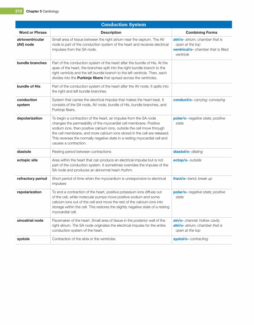
Conduction SystemWord or Phrase Description Combining Forms
atrioventricular (AV) node
Small area of tissue between the right atrium near the septum. The AV node is part of the conduction system of the heart and receives electrical impulses from the SA node.
atri/o- atrium; chamber that is open at the top
ventricul/o- chamber that is filled; ventricle
bundle branches Part of the conduction system of the heart after the bundle of His. At the apex of the heart, the branches split into the right bundle branch to the right ventricle and the left bundle branch to the left ventricle. Then, each divides into the Purkinje fibers that spread across the ventricles.
bundle of His Part of the conduction system of the heart after the AV node. It splits into the right and left bundle branches.
conduction system
System that carries the electrical impulse that makes the heart beat. It consists of the SA node, AV node, bundle of His, bundle branches, and Purkinje fibers.
conduct/o- carrying; conveying
depolarization To begin a contraction of the heart, an impulse from the SA node changes the permeability of the myocardial cell membrane. Positive sodium ions, then positive calcium ions, outside the cell move through the cell membrane, and more calcium ions stored in the cell are released. This reverses the normally negative state in a resting myocardial cell and causes a contraction.
polar/o- negative state; positive state
diastole Resting period between contractions diastol/o- dilating
ectopic site Area within the heart that can produce an electrical impulse but is not part of the conduction system. It sometimes overrides the impulse of the SA node and produces an abnormal heart rhythm.
ectop/o- outside
refractory period Short period of time when the myocardium is unresponsive to electrical impulses
fract/o- bend; break up
repolarization To end a contraction of the heart, positive potassium ions diffuse out of the cell, while molecular pumps move positive sodium and some calcium ions out of the cell and move the rest of the calcium ions into storage within the cell. This restores the slightly negative state of a resting myocardial cell.
polar/o- negative state; positive state
sinoatrial node Pacemaker of the heart. Small area of tissue in the posterior wall of the right atrium. The SA node originates the electrical impulse for the entire conduction system of the heart.
sin/o- channel; hollow cavityatri/o- atrium; chamber that is
open at the top
systole Contraction of the atria or the ventricles systol/o- contracting
M05_TURL8127_04_SE_C05.indd 212 14/12/15 10:48 pm
Chapter 5 Cardiology 213
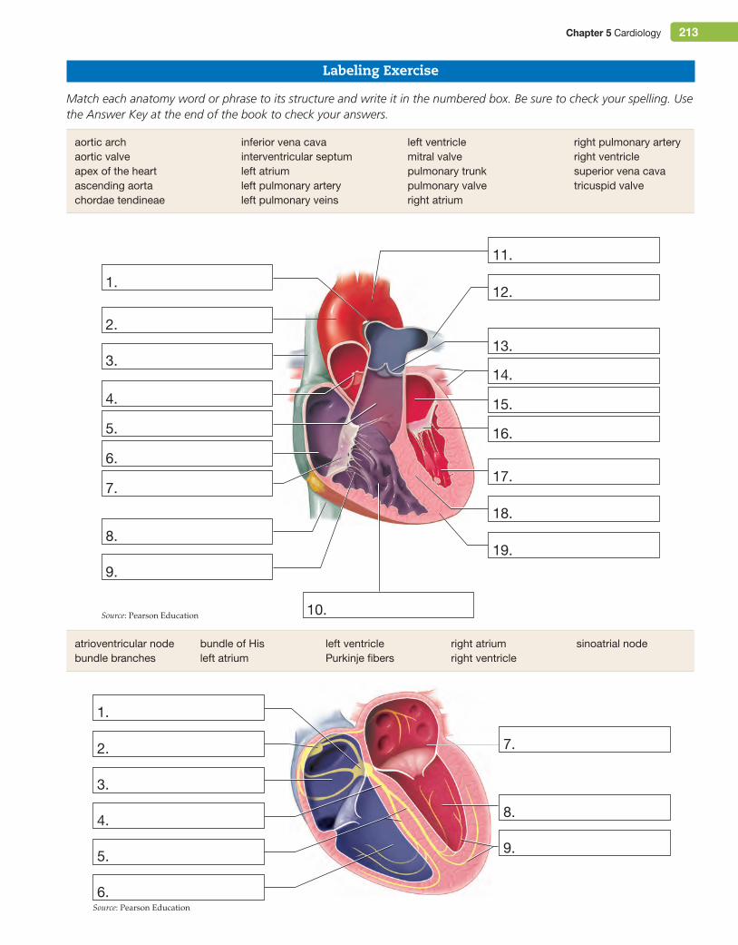
Labeling Exercise
Match each anatomy word or phrase to its structure and write it in the numbered box. Be sure to check your spelling. Use the Answer Key at the end of the book to check your answers.
3.
4.
2.
1.
11.
12.
13.
14.
15.
16.
17.
18.
19.
10.
5.
6.
7.
8.
9.
3.
2.
1.
4.
5.
6.
7.
8.
9.
atrioventricular nodebundle branches
bundle of Hisleft atrium
left ventriclePurkinje fibers
right atriumright ventricle
sinoatrial node
aortic archaortic valveapex of the heartascending aortachordae tendineae
inferior vena cavainterventricular septumleft atriumleft pulmonary arteryleft pulmonary veins
left ventriclemitral valvepulmonary trunkpulmonary valveright atrium
right pulmonary arteryright ventriclesuperior vena cavatricuspid valve
Source: Pearson Education
Source: Pearson Education
M05_TURL8127_04_SE_C05.indd 213 14/12/15 10:48 pm
214 Chapter 5 Cardiology
5.
6.
4.
7.
8.
9.
10.
11.
12.
13.
2.
1.
3.
14.
16.
15.
abdominal aortaascending aortaaxillary arterybrachial artery
carotid arterycoronary arteryfemoral artery
iliac arteryperoneal arterypopliteal artery
radial arteryrenal arterysubclavian artery
tibial arterythoracic aortaulnar artery
Source: Pearson Education
M05_TURL8127_04_SE_C05.indd 214 14/12/15 10:48 pm
Chapter 5 Cardiology 215
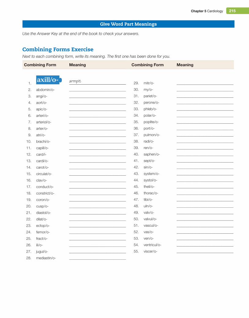
Give Word Part Meanings
Use the Answer Key at the end of the book to check your answers.
Combining Forms ExerciseNext to each combining form, write its meaning. The first one has been done for you.
Combining Form Meaning
1. axill/o- armpit
2. abdomin/o- _______________________________
3. angi/o- _______________________________
4. aort/o- _______________________________
5. apic/o- _______________________________
6. arteri/o- _______________________________
7. arteriol/o- _______________________________
8. arter/o- _______________________________
9. atri/o- _______________________________
10. brachi/o- _______________________________
11. capill/o- _______________________________
12. card/i- _______________________________
13. cardi/o- _______________________________
14. carot/o- _______________________________
15. circulat/o- _______________________________
16. clav/o- _______________________________
17. conduct/o- _______________________________
18. constrict/o- _______________________________
19. coron/o- _______________________________
20. cusp/o- _______________________________
21. diastol/o- _______________________________
22. dilat/o- _______________________________
23. ectop/o- _______________________________
24. femor/o- _______________________________
25. fract/o- _______________________________
26. ili/o- _______________________________
27. jugul/o- _______________________________
28. mediastin/o- _______________________________
Combining Form Meaning
29. mitr/o- _______________________________
30. my/o- _______________________________
31. pariet/o- _______________________________
32. perone/o- _______________________________
33. phleb/o- _______________________________
34. polar/o- _______________________________
35. poplite/o- _______________________________
36. port/o- _______________________________
37. pulmon/o- _______________________________
38. radi/o- _______________________________
39. ren/o- _______________________________
40. saphen/o- _______________________________
41. sept/o- _______________________________
42. sin/o- _______________________________
43. system/o- _______________________________
44. systol/o- _______________________________
45. theli/o- _______________________________
46. thorac/o- _______________________________
47. tibi/o- _______________________________
48. uln/o- _______________________________
49. valv/o- _______________________________
50. valvul/o- _______________________________
51. vascul/o- _______________________________
52. vas/o- _______________________________
53. ven/o- _______________________________
54. ventricul/o- _______________________________
55. viscer/o- _______________________________
M05_TURL8127_04_SE_C05.indd 215 14/12/15 10:48 pm
216 Chapter 5 Cardiology
Build Medical Words
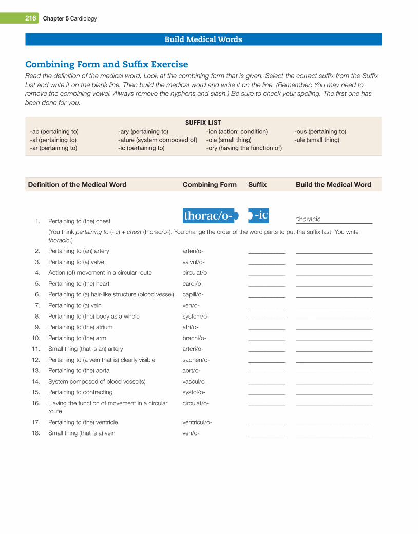
Combining Form and Suffix ExerciseRead the definition of the medical word. Look at the combining form that is given. Select the correct suffix from the Suffix List and write it on the blank line. Then build the medical word and write it on the line. (Remember: You may need to remove the combining vowel. Always remove the hyphens and slash.) Be sure to check your spelling. The first one has been done for you.
-ac (pertaining to)-al (pertaining to)-ar (pertaining to)
-ary (pertaining to)-ature (system composed of)-ic (pertaining to)
-ion (action; condition)-ole (small thing)-ory (having the function of)
-ous (pertaining to)-ule (small thing)
Suffix LiSt
Definition of the Medical Word Combining Form Suffix Build the Medical Word
1. Pertaining to (the) chestthorac/o- -ic thoracic
(You think pertaining to (-ic) + chest (thorac/o-). You change the order of the word parts to put the suffix last. You write thoracic.)
2. Pertaining to (an) artery arteri/o- ____________ _________________________
3. Pertaining to (a) valve valvul/o- ____________ _________________________
4. Action (of) movement in a circular route circulat/o- ____________ _________________________
5. Pertaining to (the) heart cardi/o- ____________ _________________________
6. Pertaining to (a) hair-like structure (blood vessel) capill/o- ____________ _________________________
7. Pertaining to (a) vein ven/o- ____________ _________________________
8. Pertaining to (the) body as a whole system/o- ____________ _________________________
9. Pertaining to (the) atrium atri/o- ____________ _________________________
10. Pertaining to (the) arm brachi/o- ____________ _________________________
11. Small thing (that is an) artery arteri/o- ____________ _________________________
12. Pertaining to (a vein that is) clearly visible saphen/o- ____________ _________________________
13. Pertaining to (the) aorta aort/o- ____________ _________________________
14. System composed of blood vessel(s) vascul/o- ____________ _________________________
15. Pertaining to contracting systol/o- ____________ _________________________
16. Having the function of movement in a circular route
circulat/o- ____________ _________________________
17. Pertaining to (the) ventricle ventricul/o- ____________ _________________________
18. Small thing (that is a) vein ven/o- ____________ _________________________
M05_TURL8127_04_SE_C05.indd 216 14/12/15 10:48 pm
Chapter 5 Cardiology 217
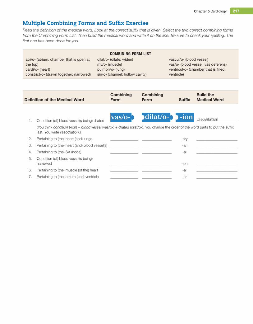
Multiple Combining Forms and Suffix ExerciseRead the definition of the medical word. Look at the correct suffix that is given. Select the two correct combining forms from the Combining Form List. Then build the medical word and write it on the line. Be sure to check your spelling. The first one has been done for you.
Combining form LiSt atri/o- (atrium; chamber that is open at the top)cardi/o- (heart)constrict/o- (drawn together; narrowed)
dilat/o- (dilate; widen)my/o- (muscle)pulmon/o- (lung)sin/o- (channel; hollow cavity)
vascul/o- (blood vessel)vas/o- (blood vessel; vas deferens)ventricul/o- (chamber that is filled; ventricle)
Definition of the Medical WordCombining Form
Combining Form Suffix
Build the Medical Word
1. Condition (of) blood vessel(s being) dilatedvas/o- dilat/o- -ion vasodilation
(You think condition (-ion) + blood vessel (vas/o-) + dilated (dilat/o-). You change the order of the word parts to put the suffix last. You write vasodilation.)
2. Pertaining to (the) heart (and) lungs _______________ ________________ -ary ______________________
3. Pertaining to (the) heart (and) blood vessel(s) _______________ ________________ -ar ______________________
4. Pertaining to (the) SA (node) _______________ ________________ -al ______________________
5. Condition (of) blood vessel(s being) narrowed _______________ ________________ -ion ______________________
6. Pertaining to (the) muscle (of the) heart _______________ ________________ -al ______________________
7. Pertaining to (the) atrium (and) ventricle _______________ ________________ -ar ______________________
M05_TURL8127_04_SE_C05.indd 217 14/12/15 10:48 pm
218 Chapter 5 Cardiology
DiseasesMyocardium
Word or Phrase Description Pronunciation/Word Parts
acute coronary syndrome
Syndrome that includes acute ischemia of the myocardium (because of a blood clot or atherosclerosis blocking blood flow through a coronary artery) and unstable angina pectoris. Treatment: Nitroglycerin drug, thrombolytic drug, oxygen therapy.
angina pectoris Mild-to-severe chest pain caused by ischemia of the myocardium. Atherosclerosis blocks the flow of oxygenated blood through the coronary arteries to the myocardium. Anginal pain is a crushing, squeezing, heaviness, or pressure-like sensation in the chest, with pain extending up into the jaw, teeth, neck, or down the left arm, often with extreme sweating (diaphoresis) and a sense of doom. Angina pectoris can occur during exercise, stress, after a heavy meal, or while resting. It is a warning sign of an impending myocardial infarction. Treatment: Nitroglycerin drug, oxygen therapy.
cardiomegaly Enlargement of the heart, usually due to congestive heart failure. Treatment: Correct the underlying cause.
cardiomyopathy Any disease condition of the heart muscle that includes heart enlargement and heart failure. In dilated cardiomyopathy, the left ventricle is dilated and the myocardium is so stretched that it can no longer contract to pump blood. idiopathic cardiomyopathy has an unknown cause. Treatment: Correct the underlying cause, if known.
did You KnoW?For many years, newspaper and magazine articles described the classic symptoms of angina pectoris in order to raise public awareness and encourage those with angina to promptly seek medical help. Now it is known that those symptoms occur in men, but women most often experience angina as indigestion, nausea, anxiety, extreme fatigue, or trouble sleeping.
idiopathic (id-ee-oh-PATH-ik)idi/o- individual; unknownpath/o- disease-ic pertaining to
cardiomegaly (kar-dee-oh-MEG-ah-lee)
cardi/o- heart-megaly enlargement
cardiomyopathy (kar-dee-oh-my-AW-pah-thee)
cardi/o- heartmy/o- muscle-pathy disease
pectoris (PEK-toh-ris)The combining form pector/o- means chest.
anginal (AN-jih-nal) (an-JY-nal)angin/o- angina-al pertaining to
ischemia (is-KEE-mee-ah)isch/o- block; keep back-emia condition of the blood;
substance in the blood
angina (AN-jih-nah) (an-JY-nah)
M05_TURL8127_04_SE_C05.indd 218 14/12/15 10:48 pm
Chapter 5 Cardiology 219
Word or Phrase Description Pronunciation/Word Parts
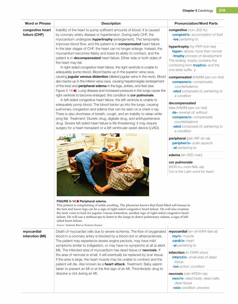
congestive heart failure (CHF)
Inability of the heart to pump sufficient amounts of blood. It is caused by coronary artery disease or hypertension. During early CHF, the myocardium undergoes hypertrophy (enlargement). This temporarily improves blood flow, and the patient is in compensated heart failure. In the later stages of CHF, the heart can no longer enlarge. Instead, the myocardium becomes flabby and loses its ability to contract, and the patient is in decompensated heart failure. Either side or both sides of the heart may fail.
In right-sided congestive heart failure, the right ventricle is unable to adequately pump blood. Blood backs up in the superior vena cava, causing jugular venous distention (dilated jugular veins in the neck). Blood also backs up in the inferior vena cava, causing hepatomegaly (enlargement of the liver) and peripheral edema in the legs, ankles, and feet (see Figure 5-14 ■). Lung disease and increased pressure in the lungs cause the right ventricle to become enlarged; this condition is cor pulmonale.
In left-sided congestive heart failure, the left ventricle is unable to adequately pump blood. The blood backs up into the lungs, causing pulmonary congestion and edema that can be seen on a chest x-ray. There is also shortness of breath, cough, and an inability to sleep while lying flat. Treatment: Diuretic drug, digitalis drug, and antihypertensive drug. Severe left-sided heart failure is life-threatening; it may require surgery for a heart transplant or a left ventricular assist device (LVAD).
myocardial infarction (Mi)
Death of myocardial cells due to severe ischemia. The flow of oxygenated blood in a coronary artery is blocked by a blood clot or atherosclerosis. The patient may experience severe angina pectoris, may have mild symptoms similar to indigestion, or may have no symptoms at all (a silent MI). The infarcted area of myocardium has dead tissue or necrosis. If the area of necrosis is small, it will eventually be replaced by scar tissue. If the area is large, the heart muscle may be unable to contract and the patient will die. Also known as a heart attack. Treatment: Baby aspirin taken to prevent an MI or at the first sign of an MI. Thrombolytic drug to dissolve a clot during an MI. necrosis (neh-KROH-sis)
necr/o- dead body; dead cells; dead tissue
-osis condition; process
myocardial (my-oh-KAR-dee-al)my/o- musclecardi/o- heart-al pertaining to
infarction (in-FARK-shun)infarct/o- small area of dead
tissue-ion action; condition
edema (eh-DEE-mah)
cor pulmonale (KOR pul-moh-NAL-ee)Cor is the Latin word for heart.
decompensated (dee-KAWM-pen-say-ted)
de- reversal of; withoutcompens/o- compensate;
counterbalance-ated composed of; pertaining to
a condition
peripheral (peh-RIF-eh-ral)peripher/o- outer aspects-al pertaining to
hypertrophy (hy-PER-troh-fee)hyper- above; more than normal-trophy process of development
The ending -trophy contains the combining form troph/o- and the one-letter suffix -y.
compensated (KAWM-pen-say-ted)compens/o- compensate;
counterbalance-ated composed of; pertaining to
a condition
congestive (con-JES-tiv)congest/o- accumulation of fluid-ive pertaining to
Figure 5-14 ■ Peripheral edema.This patient is complaining of ankle swelling. The physician knows that fluid-filled soft tissues in the feet and lower legs can be a sign of right-sided congestive heart failure. He will also examine the neck veins to look for jugular venous distention, another sign of right-sided congestive heart failure. He will use a stethoscope to listen to the lungs to detect pulmonary edema, a sign of left-sided heart failure.Source: Antonia Reeve/Science Source
M05_TURL8127_04_SE_C05.indd 219 14/12/15 10:48 pm
220 Chapter 5 Cardiology
regurgitation (ree-ger-jih-TAY-shun)regurgitat/o- backward flow-ion action; condition
subacute (sub-ah-KYOOT)
prolapse (PROH-laps)
endocarditis (en-doh-kar-DY-tis)endo- innermost; withincard/i- heart-itis infection of; inflammation of
The combining vowel i of card/i- is deleted before it is joined to the suffix -itis.
Heart Valves and Layers of the HeartWord or Phrase Description Pronunciation/Word Parts
endocarditis Inflammation and bacterial infection of the endocardium lining a heart valve. This occurs in patients who have a structural defect of the valve. Bacteria from an infection elsewhere in the body travel through the blood, are trapped by the structural defect, and cause infection. Acute endocarditis causes a high fever and shock, while subacute bacterial endocarditis (SBe) causes fever, fatigue, and aching muscles. Treatment: Antibiotic drug.
mitral valve prolapse (MVP)
Structural abnormality in which the leaflets of the mitral valve do not close tightly. This can be a congenital condition or can occur if the valve is damaged by infection. There is regurgitation as blood flows back into the left atrium with each contraction. A slight prolapse is a common condition and does not require treatment. Treatment: Valvoplasty, mitral valve ring implant, or valve replacement surgery.
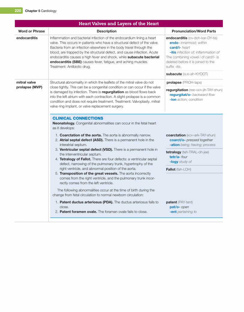
CliniCAl ConneCtionsNeonatology. Congenital abnormalities can occur in the fetal heart as it develops:
1. Coarctation of the aorta. The aorta is abnormally narrow.2. Atrial septal defect (ASD). There is a permanent hole in the
interatrial septum.3. Ventricular septal defect (VSD). There is a permanent hole in
the interventricular septum.4. Tetralogy of Fallot. There are four defects: a ventricular septal
defect, narrowing of the pulmonary trunk, hypertrophy of the right ventricle, and abnormal position of the aorta.
5. Transposition of the great vessels. The aorta incorrectly comes from the right ventricle, and the pulmonary trunk incor-rectly comes from the left ventricle.
The following abnormalities occur at the time of birth during the change from fetal circulation to normal newborn circulation:
1. Patent ductus arteriosus (PDA). The ductus arteriosus fails to close.
2. Patent foramen ovale. The foramen ovale fails to close.
tetralogy (teh-TRAL-oh-jee)tetr/a- four-logy study of
Fallot (fah-LOH)
patent (PAY-tent)pat/o- open-ent pertaining to
coarctation (koh-ark-TAY-shun)coarct/o- pressed together-ation being; having; process
M05_TURL8127_04_SE_C05.indd 220 14/12/15 10:48 pm
Chapter 5 Cardiology 221
Word or Phrase Description Pronunciation/Word Parts
murmur Abnormal heart sound created by turbulence as blood leaks through a defective heart valve. Murmurs are described according to their volume (soft or loud), their sound, and when they occur. Functional murmurs are mild murmurs that are not associated with disease and are not clinically significant. Treatment: Surgery to correct a severely defective heart valve (valvuloplasty).
pericarditis Inflammation or infection of the pericardial sac with an excessive accumulation of pericardial fluid. When the fluid compresses the heart and prevents it from beating, this is cardiac tamponade. Treatment: Antibiotic drug. Surgery to remove the fluid (pericardiocentesis).
rheumatic heart disease
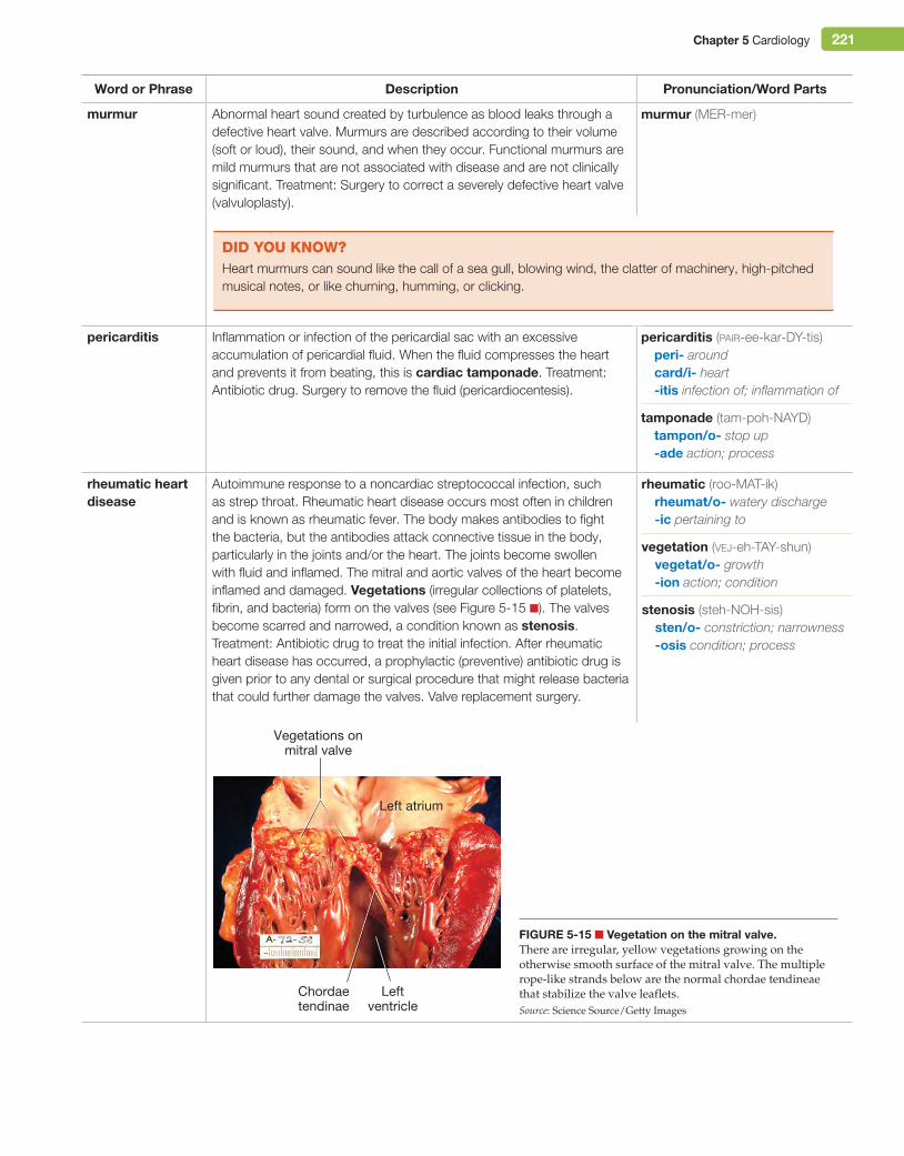
Autoimmune response to a noncardiac streptococcal infection, such as strep throat. Rheumatic heart disease occurs most often in children and is known as rheumatic fever. The body makes antibodies to fight the bacteria, but the antibodies attack connective tissue in the body, particularly in the joints and/or the heart. The joints become swollen with fluid and inflamed. The mitral and aortic valves of the heart become inflamed and damaged. Vegetations (irregular collections of platelets, fibrin, and bacteria) form on the valves (see Figure 5-15 ■). The valves become scarred and narrowed, a condition known as stenosis. Treatment: Antibiotic drug to treat the initial infection. After rheumatic heart disease has occurred, a prophylactic (preventive) antibiotic drug is given prior to any dental or surgical procedure that might release bacteria that could further damage the valves. Valve replacement surgery.
pericarditis (pair-ee-kar-DY-tis)peri- aroundcard/i- heart-itis infection of; inflammation of
tamponade (tam-poh-NAYD)tampon/o- stop up-ade action; process
murmur (MER-mer)
did You KnoW?Heart murmurs can sound like the call of a sea gull, blowing wind, the clatter of machinery, high-pitched musical notes, or like churning, humming, or clicking.
stenosis (steh-NOH-sis)sten/o- constriction; narrowness-osis condition; process
rheumatic (roo-MAT-ik)rheumat/o- watery discharge-ic pertaining to
vegetation (vej-eh-TAY-shun)vegetat/o- growth-ion action; condition
Vegetations onmitral valve
Leftventricle
Chordaetendinae
Left atrium
Figure 5-15 ■ Vegetation on the mitral valve.There are irregular, yellow vegetations growing on the otherwise smooth surface of the mitral valve. The multiple rope-like strands below are the normal chordae tendineae that stabilize the valve leaflets.Source: Science Source/Getty Images
M05_TURL8127_04_SE_C05.indd 221 14/12/15 10:48 pm
222 Chapter 5 Cardiology
Conduction SystemWord or Phrase Description Pronunciation/Word Parts
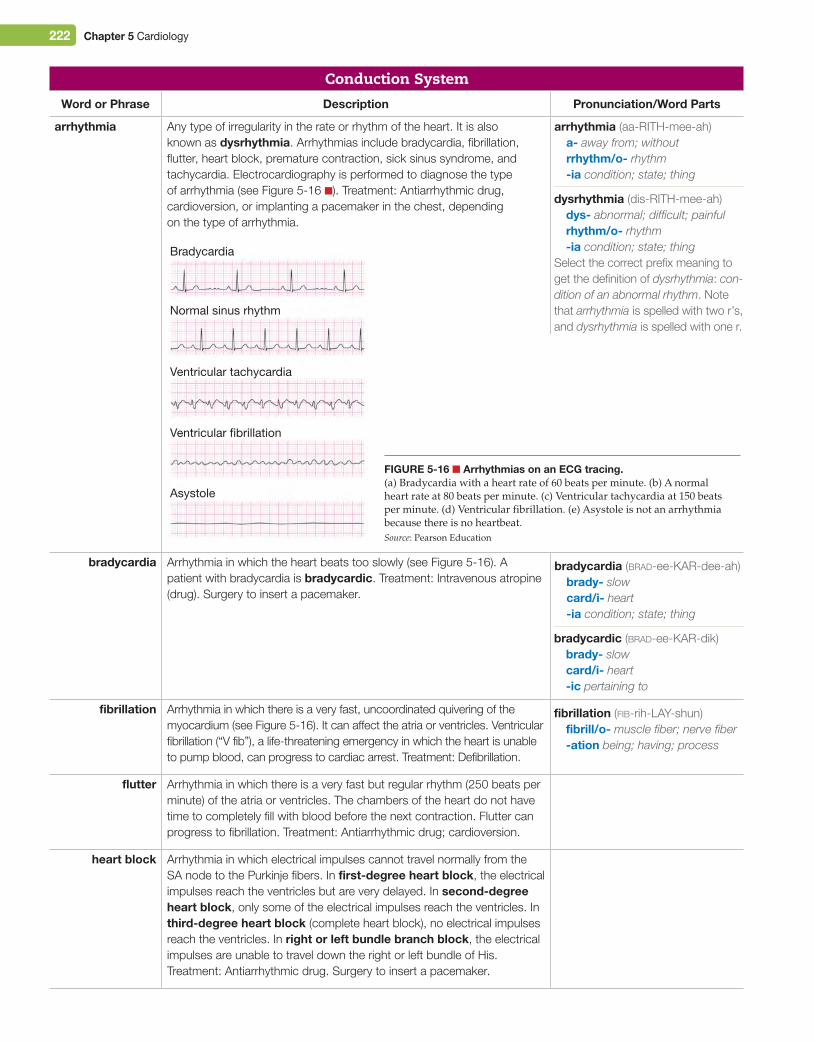
arrhythmia Any type of irregularity in the rate or rhythm of the heart. It is also known as dysrhythmia. Arrhythmias include bradycardia, fibrillation, flutter, heart block, premature contraction, sick sinus syndrome, and tachycardia. Electrocardiography is performed to diagnose the type of arrhythmia (see Figure 5-16 ■). Treatment: Antiarrhythmic drug, cardioversion, or implanting a pacemaker in the chest, depending on the type of arrhythmia.
bradycardia Arrhythmia in which the heart beats too slowly (see Figure 5-16). A patient with bradycardia is bradycardic. Treatment: Intravenous atropine (drug). Surgery to insert a pacemaker.
fibrillation Arrhythmia in which there is a very fast, uncoordinated quivering of the myocardium (see Figure 5-16). It can affect the atria or ventricles. Ventricular fibrillation (“V fib”), a life-threatening emergency in which the heart is unable to pump blood, can progress to cardiac arrest. Treatment: Defibrillation.
flutter Arrhythmia in which there is a very fast but regular rhythm (250 beats per minute) of the atria or ventricles. The chambers of the heart do not have time to completely fill with blood before the next contraction. Flutter can progress to fibrillation. Treatment: Antiarrhythmic drug; cardioversion.
heart block Arrhythmia in which electrical impulses cannot travel normally from the SA node to the Purkinje fibers. In first-degree heart block, the electrical impulses reach the ventricles but are very delayed. In second-degree heart block, only some of the electrical impulses reach the ventricles. In third-degree heart block (complete heart block), no electrical impulses reach the ventricles. In right or left bundle branch block, the electrical impulses are unable to travel down the right or left bundle of His. Treatment: Antiarrhythmic drug. Surgery to insert a pacemaker.
Bradycardia
Normal sinus rhythm
Ventricular tachycardia
Ventricular fibrillation
Asystole
Figure 5-16 ■ Arrhythmias on an eCg tracing.(a) Bradycardia with a heart rate of 60 beats per minute. (b) A normal heart rate at 80 beats per minute. (c) Ventricular tachycardia at 150 beats per minute. (d) Ventricular fibrillation. (e) Asystole is not an arrhythmia because there is no heartbeat.Source: Pearson Education
arrhythmia (aa-RITH-mee-ah)a- away from; withoutrrhythm/o- rhythm-ia condition; state; thing
dysrhythmia (dis-RITH-mee-ah)dys- abnormal; difficult; painfulrhythm/o- rhythm-ia condition; state; thing
Select the correct prefix meaning to get the definition of dysrhythmia: con-dition of an abnormal rhythm. Note that arrhythmia is spelled with two r’s, and dysrhythmia is spelled with one r.
fibrillation (fib-rih-LAY-shun)fibrill/o- muscle fiber; nerve fiber-ation being; having; process
bradycardia (brad-ee-KAR-dee-ah)brady- slowcard/i- heart-ia condition; state; thing
bradycardic (brad-ee-KAR-dik)brady- slowcard/i- heart-ic pertaining to
M05_TURL8127_04_SE_C05.indd 222 14/12/15 10:48 pm
Chapter 5 Cardiology 223
Word or Phrase Description Pronunciation/Word Parts
arrhythmia (continued) premature
contraction
Arrhythmia in which there are one or more extra contractions in between systole and diastole. This is also known as an extrasystole. There are two types of premature contractions: premature atrial contractions (PACs) and premature ventricular contractions (PVCs). A repeating pattern of one premature contraction followed by one normal contraction is bigeminy. A repeating pattern of one premature contraction followed by two normal contractions is trigeminy. Two premature contractions occurring together is a couplet. Treatment: Antiarrhythmic drug. Surgery to insert a pacemaker.
sick sinus syndrome
Arrhythmia in which bradycardia alternates with tachycardia. It occurs when the sinoatrial node and an ectopic site elsewhere in the myocardium take turns being the heart’s pacemaker. Treatment: Antiarrhythmic drug. Surgery to insert a pacemaker.
tachycardia Arrhythmia in which there is a fast but regular rhythm (up to 200 beats/minute) (see Figure 5-16). A patient with tachycardia is tachycardic. Sinus tachycardia occurs because of an abnormality in the sinoatrial (SA) node. Atrial tachycardia occurs when an ectopic site somewhere in the atrium produces an electrical impulse that overrides the SA node rhythm. Supraventricular tachycardia occurs when an ectopic site above (superior to) the ventricles produces an electrical impulse. Paroxysmal tachycardia is an episode of tachycardia that occurs suddenly and then goes away without treatment. Treatment: Antiarrhythmic drug. Cardioversion. Surgery to insert a pacemaker.
asystole Complete absence of a heartbeat (see Figure 5-16). This is also known as cardiac arrest. Treatment: Cardiopulmonary resuscitation (CPR).
palpitation An uncomfortable sensation felt in the chest during a premature contraction of the heart. It is often described as a “thump.” Treatment: None, unless it becomes an arrhythmia.
tachycardic (tak-ih-KAR-dik)tachy- fastcard/i- heart-ic pertaining to
tachycardia (tak-ih-KAR-dee-ah)tachy- fastcard/i- heart-ia condition; state; thing
trigeminy (try-JEM-ih-nee)The prefix tri- means three.
extrasystole (eks-trah-SIS-toh-lee)extra- outside-systole contraction
The ending -systole contains the combining form systol/o- and the one-letter suffix -e.
bigeminy (by-JEM-ih-nee)The prefix bi- means two.
supraventricular (soo-prah-ven-TRIH-kyoo-lar)
supra- aboveventricul/o- chamber that is
filled; ventricle-ar pertaining to
palpitation (pal-pih-TAY-shun)palpit/o- throb-ation being; having; process
asystole (aa-SIS-toh-lee)a- away from; without-systole contraction
paroxysmal (pair-awk-SIZ-mal)
Word AlertSound-Alike Wordspalpation (noun) A process of touching and feeling.
Example: Palpation allowed the physician to identify a tumor in the abdomen.
palpitation (noun) Being or having (the heart) throb
Example: Her occasional palpitations concerned the patient until the physician reassured her.
contraction (con-TRAK-shun)contract/o- pull together-ion action; condition
M05_TURL8127_04_SE_C05.indd 223 14/12/15 10:48 pm
224 Chapter 5 Cardiology
Blood VesselsWord or Phrase Description Pronunciation/Word Parts
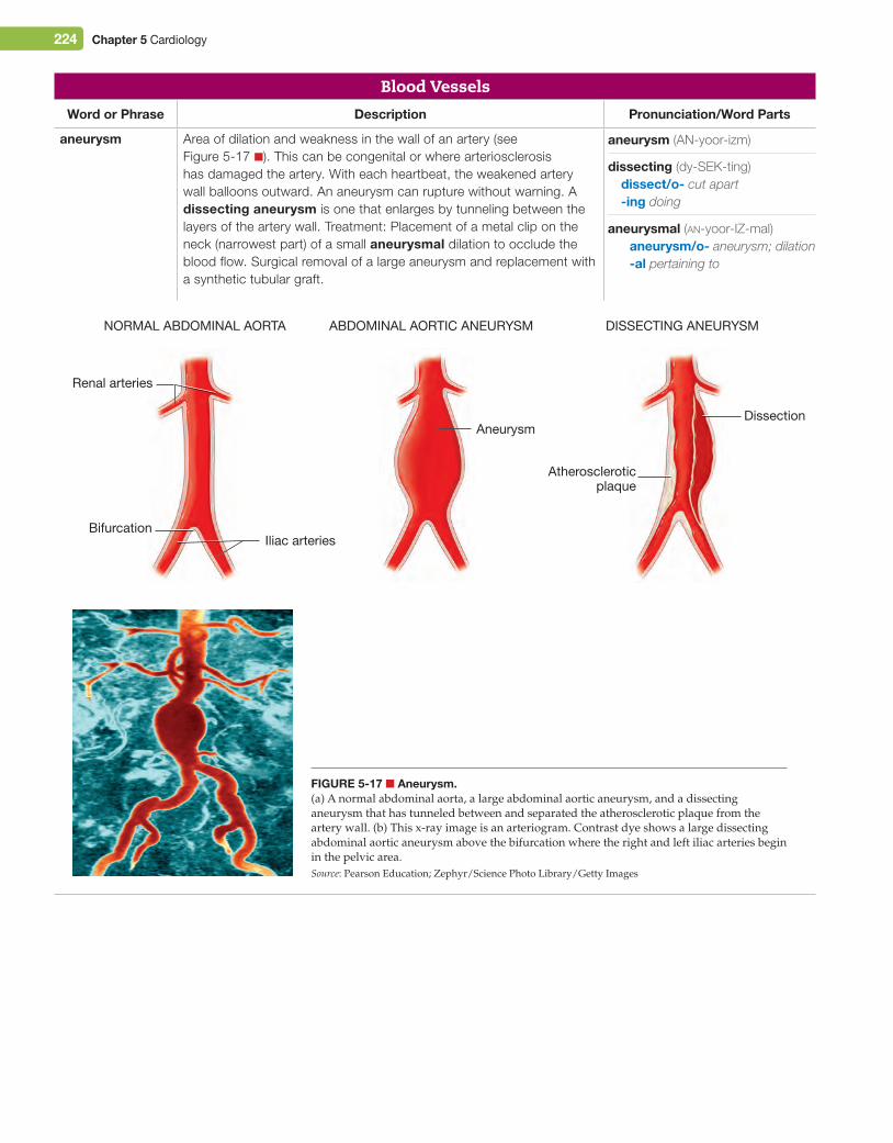
aneurysm Area of dilation and weakness in the wall of an artery (see Figure 5-17 ■). This can be congenital or where arteriosclerosis has damaged the artery. With each heartbeat, the weakened artery wall balloons outward. An aneurysm can rupture without warning. A dissecting aneurysm is one that enlarges by tunneling between the layers of the artery wall. Treatment: Placement of a metal clip on the neck (narrowest part) of a small aneurysmal dilation to occlude the blood flow. Surgical removal of a large aneurysm and replacement with a synthetic tubular graft.
Figure 5-17 ■ Aneurysm.(a) A normal abdominal aorta, a large abdominal aortic aneurysm, and a dissecting aneurysm that has tunneled between and separated the atherosclerotic plaque from the artery wall. (b) This x-ray image is an arteriogram. Contrast dye shows a large dissecting abdominal aortic aneurysm above the bifurcation where the right and left iliac arteries begin in the pelvic area.Source: Pearson Education; Zephyr/Science Photo Library/Getty Images
NORMAL ABDOMINAL AORTA DISSECTING ANEURYSMABDOMINAL AORTIC ANEURYSM
Renal arteries
BifurcationIliac arteries
Atheroscleroticplaque
AneurysmDissection
dissecting (dy-SEK-ting)dissect/o- cut apart-ing doing
aneurysmal (an-yoor-IZ-mal)aneurysm/o- aneurysm; dilation-al pertaining to
aneurysm (AN-yoor-izm)
M05_TURL8127_04_SE_C05.indd 224 14/12/15 10:48 pm
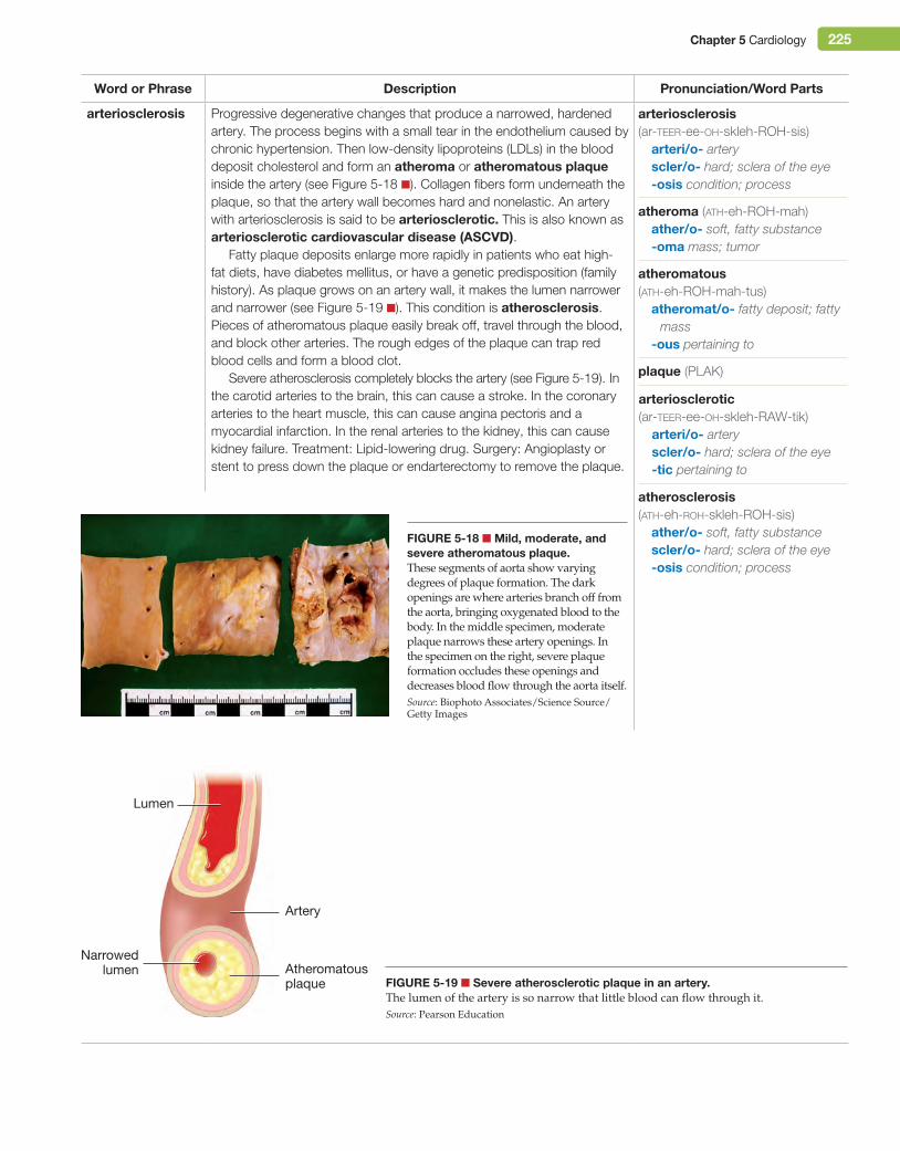
Chapter 5 Cardiology 225
Word or Phrase Description Pronunciation/Word Parts
arteriosclerosis Progressive degenerative changes that produce a narrowed, hardened artery. The process begins with a small tear in the endothelium caused by chronic hypertension. Then low-density lipoproteins (LDLs) in the blood deposit cholesterol and form an atheroma or atheromatous plaque inside the artery (see Figure 5-18 ■). Collagen fibers form underneath the plaque, so that the artery wall becomes hard and nonelastic. An artery with arteriosclerosis is said to be arteriosclerotic. This is also known as arteriosclerotic cardiovascular disease (ASCVD).
Fatty plaque deposits enlarge more rapidly in patients who eat high-fat diets, have diabetes mellitus, or have a genetic predisposition (family history). As plaque grows on an artery wall, it makes the lumen narrower and narrower (see Figure 5-19 ■). This condition is atherosclerosis. Pieces of atheromatous plaque easily break off, travel through the blood, and block other arteries. The rough edges of the plaque can trap red blood cells and form a blood clot.
Severe atherosclerosis completely blocks the artery (see Figure 5-19). In the carotid arteries to the brain, this can cause a stroke. In the coronary arteries to the heart muscle, this can cause angina pectoris and a myocardial infarction. In the renal arteries to the kidney, this can cause kidney failure. Treatment: Lipid-lowering drug. Surgery: Angioplasty or stent to press down the plaque or endarterectomy to remove the plaque.
arteriosclerotic (ar-teer-ee-oh-skleh-RAW-tik)
arteri/o- arteryscler/o- hard; sclera of the eye-tic pertaining to
atherosclerosis (ath-eh-roh-skleh-ROH-sis)
ather/o- soft, fatty substancescler/o- hard; sclera of the eye-osis condition; process
atheromatous (ath-eh-ROH-mah-tus)
atheromat/o- fatty deposit; fatty mass
-ous pertaining to
plaque (PLAK)
arteriosclerosis (ar-teer-ee-oh-skleh-ROH-sis)
arteri/o- arteryscler/o- hard; sclera of the eye-osis condition; process
atheroma (ath-eh-ROH-mah)ather/o- soft, fatty substance-oma mass; tumor
Figure 5-18 ■ Mild, moderate, and severe atheromatous plaque.These segments of aorta show varying degrees of plaque formation. The dark openings are where arteries branch off from the aorta, bringing oxygenated blood to the body. In the middle specimen, moderate plaque narrows these artery openings. In the specimen on the right, severe plaque formation occludes these openings and decreases blood flow through the aorta itself.Source: Biophoto Associates/Science Source/Getty Images
Figure 5-19 ■ Severe atherosclerotic plaque in an artery.The lumen of the artery is so narrow that little blood can flow through it.Source: Pearson Education
Atheromatousplaque
Lumen
Artery
narrowedlumen
M05_TURL8127_04_SE_C05.indd 225 14/12/15 10:48 pm
226 Chapter 5 Cardiology
Word or Phrase Description Pronunciation/Word Parts
arteriosclerosis (continued)
bruit A harsh, rushing sound made by blood passing through an artery narrowed and roughened by atherosclerosis. The bruit can be heard when a stethoscope is placed over the artery. Treatment: Correct the underlying cause.
coronary artery disease (CAD)
Arteriosclerosis of the coronary arteries. They are filled with atheromatous plaque, and their narrowed lumens cannot carry enough oxygenated blood to the myocardium. This results in angina pectoris. Severe atherosclerosis (or a blood clot that forms on an atherosclerotic plaque) can completely block the lumen of a coronary artery. This causes a myocardial infarction. Treatment: Lipid-lowering drug. Surgery: Percutaneous transluminal coronary angioplasty (PTCA) or coronary artery bypass grafting (CABG).
hyperlipidemia Elevated levels of lipids (fats) in the blood. Lipids include cholesterol and triglycerides. Hypercholesterolemia is an elevated level of cholesterol in the blood. Hypertriglyceridemia is an elevated level of triglycerides in the blood. Normal levels are below 200 mg/dL for cholesterol and below 150 mg/dL for triglycerides. Treatment: Lipid-lowering drug.
bruit (BROO-ee)
CliniCAl ConneCtionsDietetics. The body produces its own supply of cholesterol to make bile, neurotransmitters, and male and female sexual hormones. The diet contains additional cholesterol in foods from animal sources. An excessive amount of animal fat in the diet increases the cholesterol level in the blood. An excessive amount of sugar in the diet is converted by the body to triglycerides, and this causes an increased triglyceride level in the blood and increased storage as adipose tissue (fat).
Lipoproteins are carrier molecules produced in the liver. They transport lipids (fats such as cholesterol and triglycerides) in the blood. There are three types of lipoproteins. High-density lipoprotein (HDL) carries cholesterol to the liver where it is excreted in the bile. HDL is known by laypersons as “good cholesterol,” and an increased level of HDL is beneficial. Low-density lipoprotein (LDL) carries cholesterol but deposits it on the walls of the arteries, and so it is known as “bad cholesterol.” Very low-density lipoprotein (VLDL) carries triglyc-erides and deposits them on the walls of the arteries.
hypercholesterolemia (hy-per-koh-les-ter-awl-EE-mee-ah)
hyper- above; more than normalcholesterol/o- cholesterol-emia condition of the blood;
substance in the blood
hypertriglyceridemia (hy-per-try-glis-eh-ry-DEE-mee-ah)
hyper- above; more than normaltriglycerid/o- triglyceride-emia condition of the blood;
substance in the blood
CliniCAl ConneCtionsPublic Health. There are many factors that contribute to the development of coronary artery disease. These are known as cardiac risk factors. They include demographic factors (heredity, gender, age), medical factors (hypertension, hypercholesterolemia, diabetes mellitus, obesity), and lifestyle factors (smoking, lack of exercise, poor diet, stress, alcoholism).
hyperlipidemia (hy-per-lip-ih-DEE-mee-ah)
hyper- above; more than normallipid/o- fat; lipid-emia condition of the blood;
substance in the blood
M05_TURL8127_04_SE_C05.indd 226 14/12/15 10:48 pm
Chapter 5 Cardiology 227
Word or Phrase Description Pronunciation/Word Parts
hypertension (HTN)
Elevated blood pressure. A normal blood pressure reading in an adult is less than 120/80 mm Hg. Readings between 120/80 mm Hg and 140/90 mm Hg are categorized as prehypertension. Blood pressures above 140/90 mm Hg are categorized as hypertension, and the patient is said to be hypertensive. Several blood pressure readings, not just one, are needed to make a diagnosis. Essential hypertension, the most common type of hypertension, is one in which the exact cause is not known. Secondary hypertension has a known cause, such as kidney disease. Treatment: Lifestyle changes (decreased salt intake, increased exercise, weight loss) followed by an antihypertensive drug.
hypotension Blood pressure lower than 90/60 mm Hg, usually because of a loss of blood volume. A patient with hypotension is hypotensive. Orthostatic hypotension is the sudden, temporary, but self-correcting decrease in systolic blood pressure that occurs when the patient changes from a lying to a standing position and experiences lightheadedness. Treatment: Change positions slowly to avoid dizziness.
peripheral artery disease (PAD)
Atherosclerosis of the arteries in the legs. Blood flow (perfusion) to the extremities is poor, and there is ischemia of the tissues. While walking, the patient experiences pain in the calf (intermittent claudication). In severe PAD, the feet and toes remain cool and cyanotic and may become necrotic as the tissues die. Treatment: Lipid-lowering drug. Surgery: Angioplasty and stent in the iliac or femoral artery. Possible amputation of the foot.
peripheral vascular disease (PVD)
Any disease of the arteries of the extremities. It includes peripheral artery disease as well as Raynaud’s disease. Treatment: See peripheral artery disease and Raynaud’s disease.
necrotic (neh-KRAW-tik)necr/o- dead body; dead cells;
dead tissue-tic pertaining to
claudication (klaw-dih-KAY-shun)claudicat/o- limping pain-ion action; condition
peripheral (peh-RIF-eh-ral)peripher/o- outer aspects-al pertaining to
peripheral (peh-RIF-eh-ral)peripher/o- outer aspects-al pertaining to
perfusion (per-FYOO-zhun)per- through; throughoutfus/o- pouring-ion action; condition
hypotensive (hy-poh-TEN-siv)hypo- below; deficienttens/o- pressure; tension-ive pertaining to
orthostatic (or-thoh-STAT-ik)orth/o- straightstat/o- standing still; staying in
one place-ic pertaining to
hypotension (hy-poh-TEN-shun)hypo- below; deficienttens/o- pressure; tension-ion action; condition
hypertensive (hy-per-TEN-siv)hyper- above; more than normaltens/o- pressure; tension-ive pertaining to
hypertension (hy-per-TEN-shun)hyper- above; more than normaltens/o- pressure; tension-ion action; condition
prehypertension (pree-hy-per-TEN-shun)
pre- before; in front ofhyper- above; more than normaltens/o- pressure; tension-ion action; condition
did You KnoW?Some people have increased blood pressure readings just because they are nervous about being in a doctor’s office. This is known as white-coat hypertension. This is not a true hypertension because as soon as they leave the doctor’s office, their blood pressure returns to normal.
M05_TURL8127_04_SE_C05.indd 227 14/12/15 10:48 pm
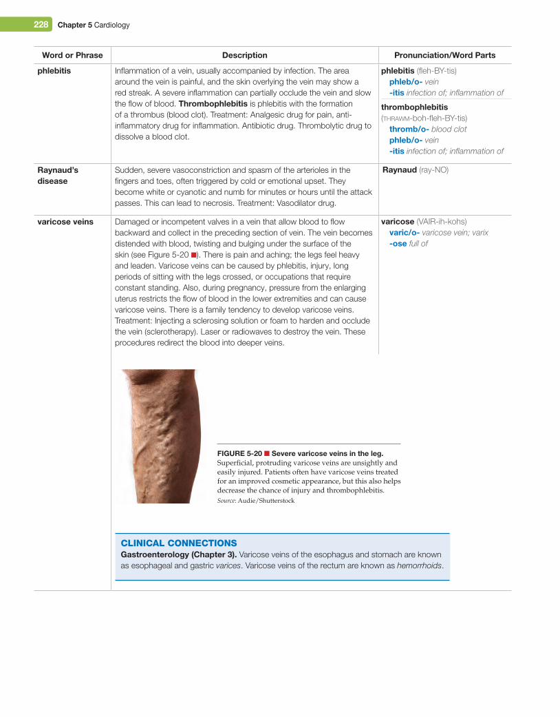
228 Chapter 5 Cardiology
Word or Phrase Description Pronunciation/Word Parts
phlebitis Inflammation of a vein, usually accompanied by infection. The area around the vein is painful, and the skin overlying the vein may show a red streak. A severe inflammation can partially occlude the vein and slow the flow of blood. Thrombophlebitis is phlebitis with the formation of a thrombus (blood clot). Treatment: Analgesic drug for pain, anti-inflammatory drug for inflammation. Antibiotic drug. Thrombolytic drug to dissolve a blood clot.
raynaud’s disease
Sudden, severe vasoconstriction and spasm of the arterioles in the fingers and toes, often triggered by cold or emotional upset. They become white or cyanotic and numb for minutes or hours until the attack passes. This can lead to necrosis. Treatment: Vasodilator drug.
varicose veins Damaged or incompetent valves in a vein that allow blood to flow backward and collect in the preceding section of vein. The vein becomes distended with blood, twisting and bulging under the surface of the skin (see Figure 5-20 ■). There is pain and aching; the legs feel heavy and leaden. Varicose veins can be caused by phlebitis, injury, long periods of sitting with the legs crossed, or occupations that require constant standing. Also, during pregnancy, pressure from the enlarging uterus restricts the flow of blood in the lower extremities and can cause varicose veins. There is a family tendency to develop varicose veins. Treatment: Injecting a sclerosing solution or foam to harden and occlude the vein (sclerotherapy). Laser or radiowaves to destroy the vein. These procedures redirect the blood into deeper veins.
Figure 5-20 ■ Severe varicose veins in the leg.Superficial, protruding varicose veins are unsightly and easily injured. Patients often have varicose veins treated for an improved cosmetic appearance, but this also helps decrease the chance of injury and thrombophlebitis.Source: Audie/Shutterstock
CliniCAl ConneCtionsgastroenterology (Chapter 3). Varicose veins of the esophagus and stomach are known as esophageal and gastric varices. Varicose veins of the rectum are known as hemorrhoids.
thrombophlebitis (thrawm-boh-fleh-BY-tis)
thromb/o- blood clotphleb/o- vein-itis infection of; inflammation of
raynaud (ray-NO)
phlebitis (fleh-BY-tis)phleb/o- vein-itis infection of; inflammation of
varicose (VAIR-ih-kohs)varic/o- varicose vein; varix-ose full of
M05_TURL8127_04_SE_C05.indd 228 14/12/15 10:48 pm
Chapter 5 Cardiology 229
Laboratory and Diagnostic ProceduresBlood Tests
Word or Phrase Description Pronunciation/Word Parts
cardiac enzymes Test to measure the levels of enzymes that are released into the blood when myocardial cells die during a myocardial infarction. (These enzymes are not released during angina pectoris.) The higher the levels, the more severe the myocardial infarction and the larger the area of infarct. Creatine kinase (CK) is found in all muscle cells, but a specific form of it (CK-MB) is only found in myocardial cells. The CK-MB level begins to rise 2–6 hours after a myocardial infarction. It is also known as creatine phosphokinase (CPK). Lactate dehydrogenase (LDH) is found in many different cells, including the heart. The LDH level begins to rise 12 hours after a myocardial infarction. An elevated LDH can support the CK-MB results but cannot be the only basis for a diagnosis of myocardial infarction. Cardiac enzymes are measured every few hours for several days. This test is done in conjunction with troponin.
C-reactive protein (CrP)
Test to measure the level of inflammation in the body. Inflammation from sites other than the cardiovascular system (such as inflammation of the gums or from a chronic urinary tract infection) can produce inflammation of the walls of the blood vessels. This can lead to blood clot formation and a myocardial infarction. The high-sensitivity CRP test can detect a lower blood level of CRP and is used to predict a healthy person’s risk of developing cardiovascular disease.
homocysteine Test included as part of a cardiac risk assessment. This amino acid damages the blood vessel walls. An elevated level increases the patient’s risk of arteriosclerosis and a blood clot that can cause a heart attack or stroke.
lipid profile Test that provides a comprehensive picture of the blood levels of cholesterol and triglycerides and their lipoprotein carriers (HDL, LDL, VLDL).
troponin Test to measure the level of two proteins that are released into the blood when myocardial cells die. Troponin I and troponin T are only found in the myocardium. The troponin levels begin to rise 4–6 hours after a myocardial infarction. More importantly, they remain elevated for up to 10 days, so they can be used to diagnose a myocardial infarction many days after it occurred. Troponin levels are done in conjunction with cardiac enzyme levels.
Diagnostic Procedurescardiac catheterization
Procedure performed to study the anatomy and pressures in the heart. During a right heart catheterization, a catheter is inserted into the femoral or brachial vein and threaded to the right atrium. The catheter is used to record right heart pressures. Then a radiopaque contrast dye is injected through the catheter to outline the chambers of the heart to diagnose congenital heart defects. During a left heart catheterization, a catheter is inserted into the femoral or brachial artery and threaded to the left atrium. Then radiopaque contrast dye is injected to outline the coronary arteries and show narrowed or blocked areas. If blockage of a coronary artery is present, an angioplasty can be performed at that time. This procedure is also referred to as a cardiac cath.
catheterization (kath-eh-ter-ih-ZAY-shun)
catheter/o- catheter-ization process of creating;
process of inserting; process of making
troponin (troh-POH-nin)
lipoprotein (lih-poh-PROH-teen)
lipid (LIP-id)lip/o- fat; lipid-id origin; resembling; source
lactate dehydrogenase (LAK-tayt dee-HY-droh-jeh-nays)
homocysteine (hoh-moh-SIS-teen)
creatine kinase (KREE-ah-teen KY-nays)
creatine phosphokinase (KREE-ah-teen faws-foh-KY-nays)
enzyme (EN-zime)The suffix -ase means enzyme.
M05_TURL8127_04_SE_C05.indd 229 14/12/15 10:48 pm
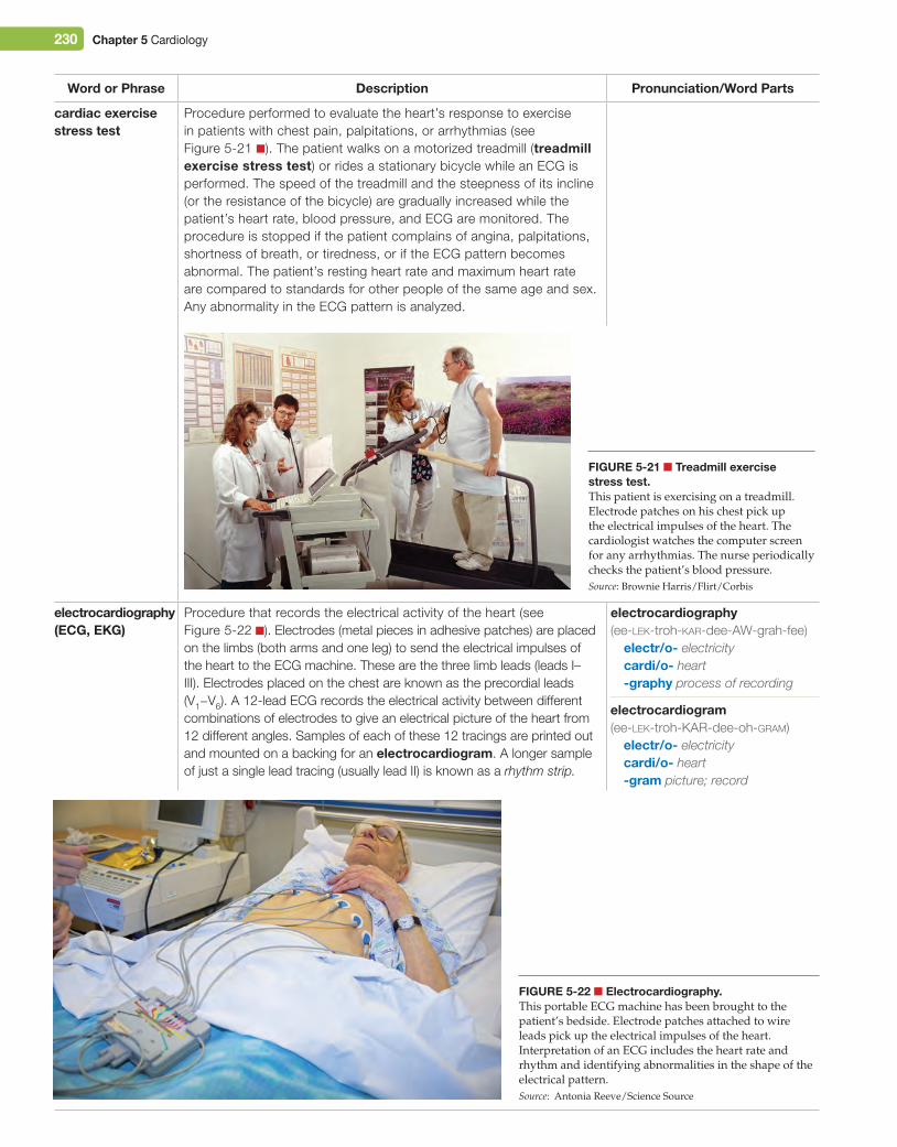
230 Chapter 5 Cardiology
Word or Phrase Description Pronunciation/Word Parts
cardiac exercise stress test
Procedure performed to evaluate the heart’s response to exercise in patients with chest pain, palpitations, or arrhythmias (see Figure 5-21 ■). The patient walks on a motorized treadmill (treadmill exercise stress test) or rides a stationary bicycle while an ECG is performed. The speed of the treadmill and the steepness of its incline (or the resistance of the bicycle) are gradually increased while the patient’s heart rate, blood pressure, and ECG are monitored. The procedure is stopped if the patient complains of angina, palpitations, shortness of breath, or tiredness, or if the ECG pattern becomes abnormal. The patient’s resting heart rate and maximum heart rate are compared to standards for other people of the same age and sex. Any abnormality in the ECG pattern is analyzed.
electrocardiography (eCg, eKg)
Procedure that records the electrical activity of the heart (see Figure 5-22 ■). Electrodes (metal pieces in adhesive patches) are placed on the limbs (both arms and one leg) to send the electrical impulses of the heart to the ECG machine. These are the three limb leads (leads I–III). Electrodes placed on the chest are known as the precordial leads (V1−V6). A 12-lead ECG records the electrical activity between different combinations of electrodes to give an electrical picture of the heart from 12 different angles. Samples of each of these 12 tracings are printed out and mounted on a backing for an electrocardiogram. A longer sample of just a single lead tracing (usually lead II) is known as a rhythm strip.
electrocardiography (ee-lek-troh-kar-dee-AW-grah-fee)
electr/o- electricitycardi/o- heart-graphy process of recording
Figure 5-21 ■ Treadmill exercise stress test.This patient is exercising on a treadmill. Electrode patches on his chest pick up the electrical impulses of the heart. The cardiologist watches the computer screen for any arrhythmias. The nurse periodically checks the patient’s blood pressure.Source: Brownie Harris/Flirt/Corbis
electrocardiogram (ee-lek-troh-KAR-dee-oh-gram)
electr/o- electricitycardi/o- heart-gram picture; record
Figure 5-22 ■ electrocardiography.This portable ECG machine has been brought to the patient’s bedside. Electrode patches attached to wire leads pick up the electrical impulses of the heart. Interpretation of an ECG includes the heart rate and rhythm and identifying abnormalities in the shape of the electrical pattern.Source: Antonia Reeve/Science Source
M05_TURL8127_04_SE_C05.indd 230 14/12/15 10:48 pm
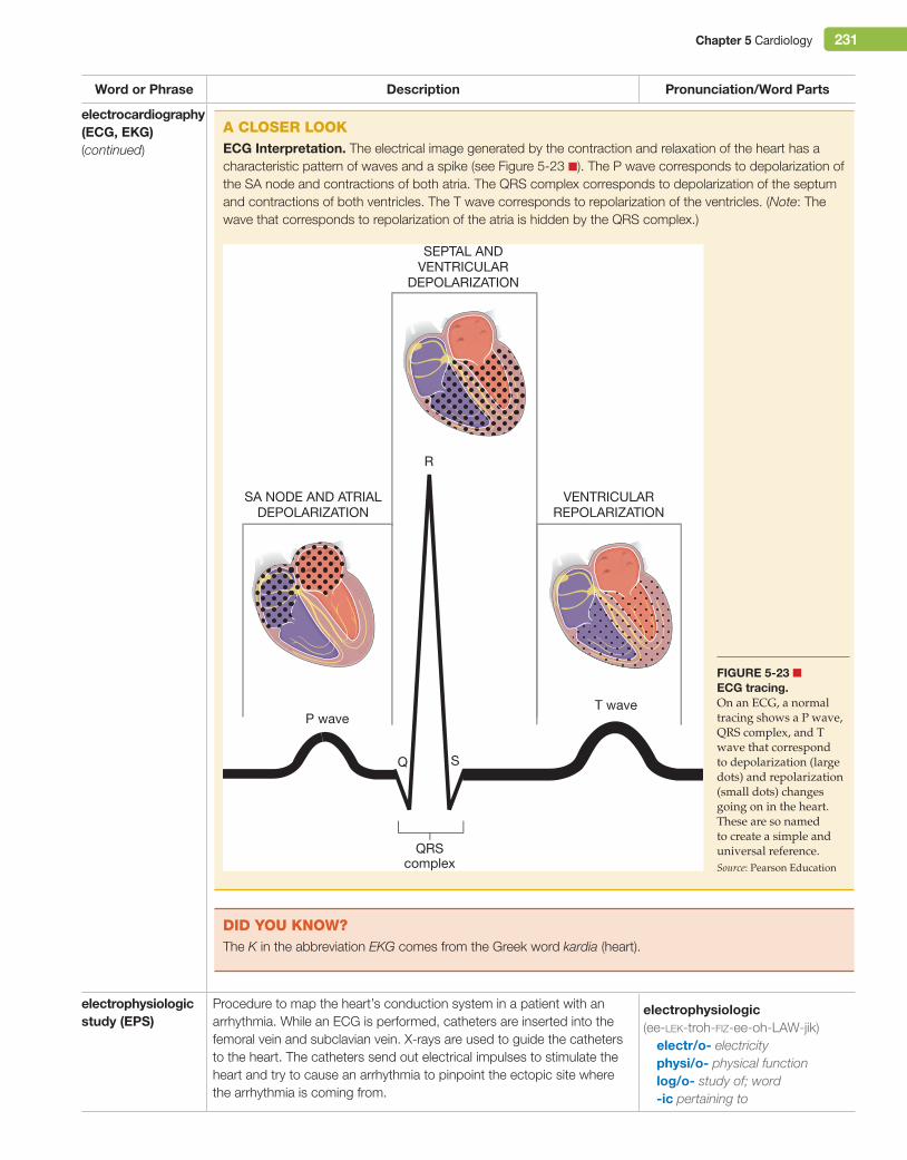
Chapter 5 Cardiology 231
Word or Phrase Description Pronunciation/Word Parts
electrocardiography (eCg, eKg)(continued)
electrophysiologic study (ePS)
Procedure to map the heart’s conduction system in a patient with an arrhythmia. While an ECG is performed, catheters are inserted into the femoral vein and subclavian vein. X-rays are used to guide the catheters to the heart. The catheters send out electrical impulses to stimulate the heart and try to cause an arrhythmia to pinpoint the ectopic site where the arrhythmia is coming from.
electrophysiologic (ee-lek-troh-fiz-ee-oh-LAW-jik)
electr/o- electricityphysi/o- physical functionlog/o- study of; word-ic pertaining to
A Closer looKeCg interpretation. The electrical image generated by the contraction and relaxation of the heart has a characteristic pattern of waves and a spike (see Figure 5-23 ■). The P wave corresponds to depolarization of the SA node and contractions of both atria. The QRS complex corresponds to depolarization of the septum and contractions of both ventricles. The T wave corresponds to repolarization of the ventricles. (Note: The wave that corresponds to repolarization of the atria is hidden by the QRS complex.)
Figure 5-23 ■ eCg tracing.On an ECG, a normal tracing shows a P wave, QRS complex, and T wave that correspond to depolarization (large dots) and repolarization (small dots) changes going on in the heart. These are so named to create a simple and universal reference.Source: Pearson Education
P waveT wave
R
QRScomplex
Q S
SEPTAL ANDVENTRICULAR
DEPOLARIZATION
VENTRICULARREPOLARIZATION
SA NODE AND ATRIALDEPOLARIZATION
did You KnoW?The K in the abbreviation EKG comes from the Greek word kardia (heart).
M05_TURL8127_04_SE_C05.indd 231 14/12/15 10:48 pm
232 Chapter 5 Cardiology
Word or Phrase Description Pronunciation/Word Parts
Holter monitor Procedure during which the patient’s heart rate and rhythm are continuously monitored as an outpatient for 24 hours. The patient wears electrodes attached to a small portable ECG monitor (carried in a vest or placed in a pocket). The patient also keeps a diary of activities, meals, and symptoms. A Holter monitor procedure is used to document infrequently occurring arrhythmias and to link them to activities or symptoms such as chest pain.
pharmacologic stress test
Procedure performed instead of a cardiac stress test for patients who cannot exercise vigorously. The vasodilator drug dipyridamole (Persantine) is given to cause normal coronary arteries to dilate. Occluded arteries cannot dilate, and this stresses the heart and causes angina in a way that is similar to an exercise stress test.
telemetry Procedure to monitor a patient’s heart rate and rhythm in the hospital. The patient wears electrodes connected to a device that continuously transmits an ECG tracing to a central monitoring station in the coronary care unit or intensive care unit. A nurse at the station constantly watches all of the patients’ cardiac monitors.
Radiology and Nuclear Medicine Proceduresangiography Procedure in which radiopaque contrast dye is injected into a blood
vessel to fill and outline it. In arteriography, it is injected into an artery to show blockage, narrowed areas, or aneurysms (see Figure 5-17). In venography, it is injected into a vein to show weakened valves and dilated walls. The x-ray image is an angiogram or, more specifically, an arteriogram or venogram.
In coronary angiography, a catheter is inserted into the femoral artery and threaded to the aorta. The radiopaque contrast dye is injected to outline the coronary arteries and show narrowing or blockage. The x-ray is a coronary angiogram.
In rotational angiography, multiple x-rays are taken as the x-ray machine goes around the patient. This technique is particularly helpful in documenting tortuous blood vessels in three dimensions.
Digital subtraction angiography (DSA) combines two x-ray images, one taken without radiopaque contrast dye and a second image taken after radiopaque contrast dye has been injected to outline the blood vessel. A computer compares the two images and digitally “subtracts” or removes the soft tissues, bones, and muscles, leaving just the image of the arteries.
telemetry (teh-LEM-eh-tree)tele/o- distance-metry process of measuring
Holter (HOL-ter)
pharmacologic (far-mah-koh-LAW-jik)
pharmac/o- drug; medicinelog/o- study of; word-ic pertaining to
arteriogram (ar-TEER-ee-oh-gram)arteri/o- artery-gram picture; record
venogram (VEE-noh-gram)ven/o- vein-gram picture; record
angiogram (AN-jee-oh-gram)angi/o- blood vessel; lymphatic
vessel-gram picture; record
arteriography (ar-teer-ee-AW-grah-fee)
arteri/o- artery-graphy process of recording
venography (vee-NAW-grah-fee)ven/o- vein-graphy process of recording
angiography (an-jee-AW-grah-fee)angi/o- blood vessel; lymphatic
vessel-graphy process of recording
M05_TURL8127_04_SE_C05.indd 232 14/12/15 10:48 pm
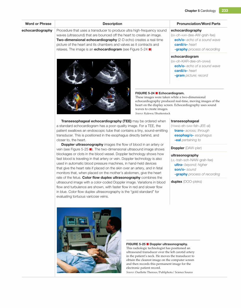
Chapter 5 Cardiology 233
Word or Phrase Description Pronunciation/Word Parts
echocardiography Procedure that uses a transducer to produce ultra high-frequency sound waves (ultrasound) that are bounced off the heart to create an image. Two-dimensional echocardiography (2-D echo) creates a real-time picture of the heart and its chambers and valves as it contracts and relaxes. The image is an echocardiogram (see Figure 5-24 ■)
Transesophageal echocardiography (Tee) may be ordered when a standard echocardiogram has a poor-quality image. For a TEE, the patient swallows an endoscopic tube that contains a tiny, sound-emitting transducer. This is positioned in the esophagus directly behind, and closer to, the heart.
Doppler ultrasonography images the flow of blood in an artery or vein (see Figure 5-25 ■). The two-dimensional ultrasound image shows blockages or clots in the blood vessel. Doppler technology shows how fast blood is traveling in that artery or vein. Doppler technology is also used in automatic blood pressure machines, in hand-held devices that give the heart rate if placed on the skin over an artery, and in fetal monitors that, when placed on the mother’s abdomen, give the heart rate of the fetus. Color flow duplex ultrasonography combines the ultrasound image with a color-coded Doppler image. Variations in blood flow and turbulence are shown, with faster flow in red and slower flow in blue. Color flow duplex ultrasonography is the “gold standard” for evaluating tortuous varicose veins.
Figure 5-24 ■ echocardiogram.These images were taken while a two-dimensional echocardiography produced real-time, moving images of the heart on the display screen. Echocardiography uses sound waves to create images.Source: Kalewa/Shutterstock
echocardiography (ek-oh-kar-dee-AW-grah-fee)
ech/o- echo of a sound wavecardi/o- heart-graphy process of recording
echocardiogram (ek-oh-KAR-dee-oh-gram)
ech/o- echo of a sound wavecardi/o- heart-gram picture; record
ultrasonography (ul-trah-soh-NAW-grah-fee)
ultra- beyond; higherson/o- sound-graphy process of recording
duplex (DOO-pleks)
transesophageal (trans-eh-saw-fah-JEE-al)
trans- across; throughesophag/o- esophagus-eal pertaining to
Doppler (DAW-pler)
Figure 5-25 ■ Doppler ultrasonography.This radiologic technologist has positioned an ultrasound transducer over the left carotid artery in the patient’s neck. He moves the transducer to obtain the clearest image on the computer screen and then records this permanent image for the electronic patient record.Source: Ouellette Theroux/Publiphoto/ Science Source
M05_TURL8127_04_SE_C05.indd 233 14/12/15 10:48 pm
234 Chapter 5 Cardiology
Word or Phrase Description Pronunciation/Word Parts
multiple-gated acquisition (MugA) scan
Nuclear medicine procedure that uses the radioactive tracer technetium-99m. First, pyrophosphate is injected intravenously to allow red blood cells to bind with technetium-99m. Then technetium-99m is injected. A gamma camera records gamma rays emitted by the technetium-99m bound to red blood cells. The camera is coordinated (gated) with the patient’s ECG so that images of the heart chambers (with blood—and red blood cells—in them) are taken at various times. A MUGA scan also calculates the ejection fraction (how much blood the ventricle can eject with one contraction). The ejection fraction is the most accurate indicator of overall heart function. This procedure is also known as a radionuclide ventriculography (rNV) or gated blood pool scan.
myocardial perfusion scan
Nuclear medicine procedure that combines a cardiac exercise stress test with intravenous injections of a radioactive tracer. The radioactive tracer collects in those parts of the myocardium that have the best perfusion (blood flow). A gamma camera records gamma rays emitted by the radioactive tracer and creates a two-dimensional image of the heart. Areas of decreased uptake (“cold spots”) indicate poor perfusion from a blocked coronary artery. The artery must be about 70% blocked before any abnormality is evident on the image. Areas of no uptake indicate dead tissue from a previous myocardial infarction. Technetium-99m is joined to a synthetic molecule (sestamibi). The combination of technetium-99m with sestamibi is the drug Cardiolite, so this test is also known as a Cardiolite stress test. In a thallium stress test, thallium-201 is the radioactive tracer, or thallium-201 and technetium-99m can be used. Myocardial perfusion PET scans are used to image the metabolism of the heart.
single-photon emission computed tomography (SPeCT) scan
Procedure that is a variation of a myocardial perfusion scan or a MUGA scan. Instead of being stationary above the patient’s chest, the gamma camera is moved in a circle around the patient. The computer creates many individual images or “slices” (tomography) and compiles them into a three-dimensional image of the heart. tomography (toh-MAW-grah-fee)
tom/o- cut; layer; slice-graphy process of recording
photon (FOH-tawn)A photon is another name for a gamma ray.
perfusion (per-FYOO-zhun)per- through; throughoutfus/o- pouring-ion action; condition
Cardiolite (KAR-dee-oh-lite)
thallium (THAL-ee-um)
radionuclide (ray-dee-oh-NOO-klide)
radi/o- forearm bone; radiation; x-rays
nucle/o- nucleus of an atom; nucleus of a cell
-ide chemically modified structure
ventriculography (ven-trih-kyoo-LAW-grah-fee)
ventricul/o- chamber that is filled; ventricle
-graphy process of recording
stethoscope (STETH-oh-skohp)steth/o- chest-scope instrument used to
examine
auscultation (aws-kul-TAY-shun)auscult/o- listening-ation being; having; process
Medical and Surgical Procedures Medical Procedures
Word or Phrase Description Pronunciation/Word Parts
auscultation Procedure that uses a stethoscope to listen to the heart sounds. The stethoscope is placed at the point of maximum impulse (PMi), which is at the apex of the heart. Auscultation can determine the heart rate and detect arrhythmias and murmurs.
M05_TURL8127_04_SE_C05.indd 234 14/12/15 10:48 pm
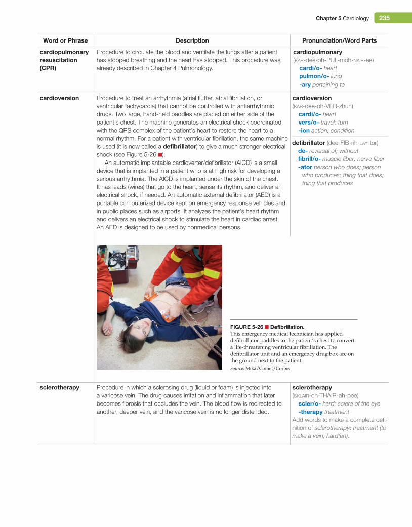
Chapter 5 Cardiology 235
Word or Phrase Description Pronunciation/Word Parts
cardiopulmonary resuscitation (CPr)
Procedure to circulate the blood and ventilate the lungs after a patient has stopped breathing and the heart has stopped. This procedure was already described in Chapter 4 Pulmonology.
cardiopulmonary (kar-dee-oh-PUL-moh-nair-ee)
cardi/o- heartpulmon/o- lung-ary pertaining to
cardioversion Procedure to treat an arrhythmia (atrial flutter, atrial fibrillation, or ventricular tachycardia) that cannot be controlled with antiarrhythmic drugs. Two large, hand-held paddles are placed on either side of the patient’s chest. The machine generates an electrical shock coordinated with the QRS complex of the patient’s heart to restore the heart to a normal rhythm. For a patient with ventricular fibrillation, the same machine is used (it is now called a defibrillator) to give a much stronger electrical shock (see Figure 5-26 ■).
An automatic implantable cardioverter/defibrillator (AICD) is a small device that is implanted in a patient who is at high risk for developing a serious arrhythmia. The AICD is implanted under the skin of the chest. It has leads (wires) that go to the heart, sense its rhythm, and deliver an electrical shock, if needed. An automatic external defibrillator (AED) is a portable computerized device kept on emergency response vehicles and in public places such as airports. It analyzes the patient’s heart rhythm and delivers an electrical shock to stimulate the heart in cardiac arrest. An AED is designed to be used by nonmedical persons.
sclerotherapy Procedure in which a sclerosing drug (liquid or foam) is injected into a varicose vein. The drug causes irritation and inflammation that later becomes fibrosis that occludes the vein. The blood flow is redirected to another, deeper vein, and the varicose vein is no longer distended.
defibrillator (dee-FIB-rih-lay-tor)de- reversal of; withoutfibrill/o- muscle fiber; nerve fiber-ator person who does; person
who produces; thing that does; thing that produces
sclerotherapy (sklair-oh-THAIR-ah-pee)
scler/o- hard; sclera of the eye-therapy treatment
Add words to make a complete defi-nition of sclerotherapy: treatment (to make a vein) hard(en).
cardioversion (kar-dee-oh-VER-zhun)
cardi/o- heartvers/o- travel; turn-ion action; condition
Figure 5-26 ■ Defibrillation.This emergency medical technician has applied defibrillator paddles to the patient’s chest to convert a life-threatening ventricular fibrillation. The defibrillator unit and an emergency drug box are on the ground next to the patient.Source: Mika/Comet/Corbis
M05_TURL8127_04_SE_C05.indd 235 14/12/15 10:48 pm
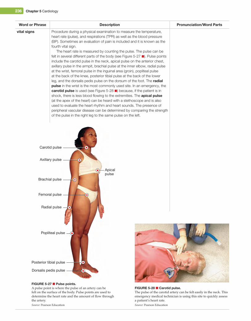
236 Chapter 5 Cardiology
Word or Phrase Description Pronunciation/Word Parts
vital signs Procedure during a physical examination to measure the temperature, heart rate (pulse), and respirations (TPR) as well as the blood pressure (BP). Sometimes an evaluation of pain is included and it is known as the fourth vital sign.
The heart rate is measured by counting the pulse. The pulse can be felt in several different parts of the body (see Figure 5-27 ■). Pulse points include the carotid pulse in the neck, apical pulse on the anterior chest, axillary pulse in the armpit, brachial pulse at the inner elbow, radial pulse at the wrist, femoral pulse in the inguinal area (groin), popliteal pulse at the back of the knee, posterior tibial pulse at the back of the lower leg, and the dorsalis pedis pulse on the dorsum of the foot. The radial pulse in the wrist is the most commonly used site. In an emergency, the carotid pulse is used (see Figure 5-28 ■) because, if the patient is in shock, there is less blood flowing to the extremities. The apical pulse (at the apex of the heart) can be heard with a stethoscope and is also used to evaluate the heart rhythm and heart sounds. The presence of peripheral vascular disease can be determined by comparing the strength of the pulse in the right leg to the same pulse on the left.
Figure 5-27 ■ Pulse points.A pulse point is where the pulse of an artery can be felt on the surface of the body. Pulse points are used to determine the heart rate and the amount of flow through the artery.Source: Pearson Education
Carotid pulse
Axillary pulse
Apicalpulse
Brachial pulse
Femoral pulse
Radial pulse
Popliteal pulse
Posterior tibial pulse
Dorsalis pedis pulse
Figure 5-28 ■ Carotid pulse.The pulse of the carotid artery can be felt easily in the neck. This emergency medical technician is using this site to quickly assess a patient’s heart rate.Source: Pearson Education
M05_TURL8127_04_SE_C05.indd 236 14/12/15 10:48 pm
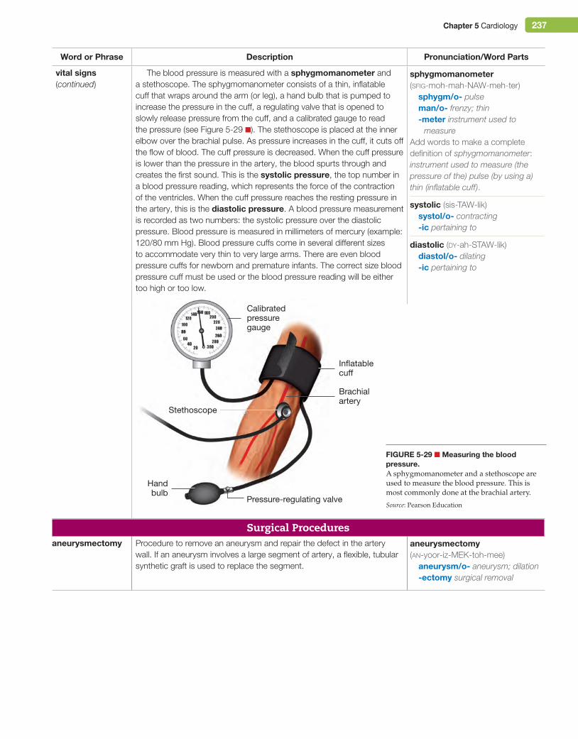
Chapter 5 Cardiology 237
Word or Phrase Description Pronunciation/Word Parts
vital signs (continued)
The blood pressure is measured with a sphygmomanometer and a stethoscope. The sphygmomanometer consists of a thin, inflatable cuff that wraps around the arm (or leg), a hand bulb that is pumped to increase the pressure in the cuff, a regulating valve that is opened to slowly release pressure from the cuff, and a calibrated gauge to read the pressure (see Figure 5-29 ■). The stethoscope is placed at the inner elbow over the brachial pulse. As pressure increases in the cuff, it cuts off the flow of blood. The cuff pressure is decreased. When the cuff pressure is lower than the pressure in the artery, the blood spurts through and creates the first sound. This is the systolic pressure, the top number in a blood pressure reading, which represents the force of the contraction of the ventricles. When the cuff pressure reaches the resting pressure in the artery, this is the diastolic pressure. A blood pressure measurement is recorded as two numbers: the systolic pressure over the diastolic pressure. Blood pressure is measured in millimeters of mercury (example: 120/80 mm Hg). Blood pressure cuffs come in several different sizes to accommodate very thin to very large arms. There are even blood pressure cuffs for newborn and premature infants. The correct size blood pressure cuff must be used or the blood pressure reading will be either too high or too low.
Surgical Proceduresaneurysmectomy Procedure to remove an aneurysm and repair the defect in the artery
wall. If an aneurysm involves a large segment of artery, a flexible, tubular synthetic graft is used to replace the segment.
sphygmomanometer (sfig-moh-mah-NAW-meh-ter)
sphygm/o- pulseman/o- frenzy; thin-meter instrument used to
measureAdd words to make a complete definition of sphygmomanometer: instrument used to measure (the pressure of the) pulse (by using a) thin (inflatable cuff).
systolic (sis-TAW-lik)systol/o- contracting-ic pertaining to
diastolic (dy-ah-STAW-lik)diastol/o- dilating-ic pertaining to
aneurysmectomy (an-yoor-iz-MEK-toh-mee)
aneurysm/o- aneurysm; dilation-ectomy surgical removal
Calibratedpressuregauge
Inflatablecu�
Handbulb
Brachialartery
Pressure-regulating valve
Stethoscope
Figure 5-29 ■ Measuring the blood pressure.A sphygmomanometer and a stethoscope are used to measure the blood pressure. This is most commonly done at the brachial artery.
Source: Pearson Education
M05_TURL8127_04_SE_C05.indd 237 14/12/15 10:48 pm
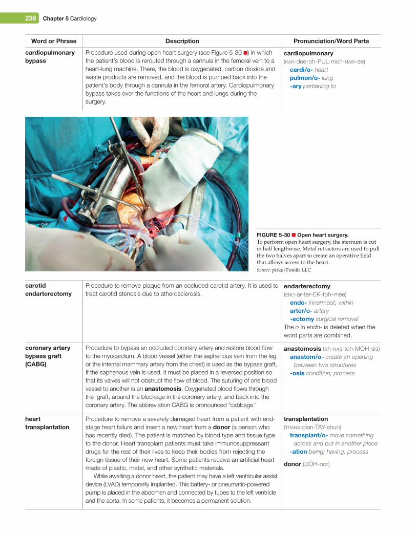
238 Chapter 5 Cardiology
Word or Phrase Description Pronunciation/Word Parts
cardiopulmonary bypass
Procedure used during open heart surgery (see Figure 5-30 ■) in which the patient’s blood is rerouted through a cannula in the femoral vein to a heart-lung machine. There, the blood is oxygenated, carbon dioxide and waste products are removed, and the blood is pumped back into the patient’s body through a cannula in the femoral artery. Cardiopulmonary bypass takes over the functions of the heart and lungs during the surgery.
carotid endarterectomy
Procedure to remove plaque from an occluded carotid artery. It is used to treat carotid stenosis due to atherosclerosis.
coronary artery bypass graft (CABg)
Procedure to bypass an occluded coronary artery and restore blood flow to the myocardium. A blood vessel (either the saphenous vein from the leg or the internal mammary artery from the chest) is used as the bypass graft. If the saphenous vein is used, it must be placed in a reversed position so that its valves will not obstruct the flow of blood. The suturing of one blood vessel to another is an anastomosis. Oxygenated blood flows through the graft, around the blockage in the coronary artery, and back into the coronary artery. The abbreviation CABG is pronounced “cabbage.”
heart transplantation
Procedure to remove a severely damaged heart from a patient with end-stage heart failure and insert a new heart from a donor (a person who has recently died). The patient is matched by blood type and tissue type to the donor. Heart transplant patients must take immunosuppressant drugs for the rest of their lives to keep their bodies from rejecting the foreign tissue of their new heart. Some patients receive an artificial heart made of plastic, metal, and other synthetic materials.
While awaiting a donor heart, the patient may have a left ventricular assist device (LVAD) temporarily implanted. This battery- or pneumatic-powered pump is placed in the abdomen and connected by tubes to the left ventricle and the aorta. In some patients, it becomes a permanent solution.
cardiopulmonary (kar-dee-oh-PUL-moh-nair-ee)
cardi/o- heartpulmon/o- lung-ary pertaining to
endarterectomy (end-ar-ter-EK-toh-mee)
endo- innermost; withinarter/o- artery-ectomy surgical removal
The o in endo- is deleted when the word parts are combined.
anastomosis (ah-nas-toh-MOH-sis)anastom/o- create an opening
between two structures-osis condition; process
transplantation (trans-plan-TAY-shun)
transplant/o- move something across and put in another place
-ation being; having; process
donor (DOH-nor)
Figure 5-30 ■ Open heart surgery.To perform open heart surgery, the sternum is cut in half lengthwise. Metal retractors are used to pull the two halves apart to create an operative field that allows access to the heart.Source: pirke/Fotolia LLC
M05_TURL8127_04_SE_C05.indd 238 14/12/15 10:48 pm
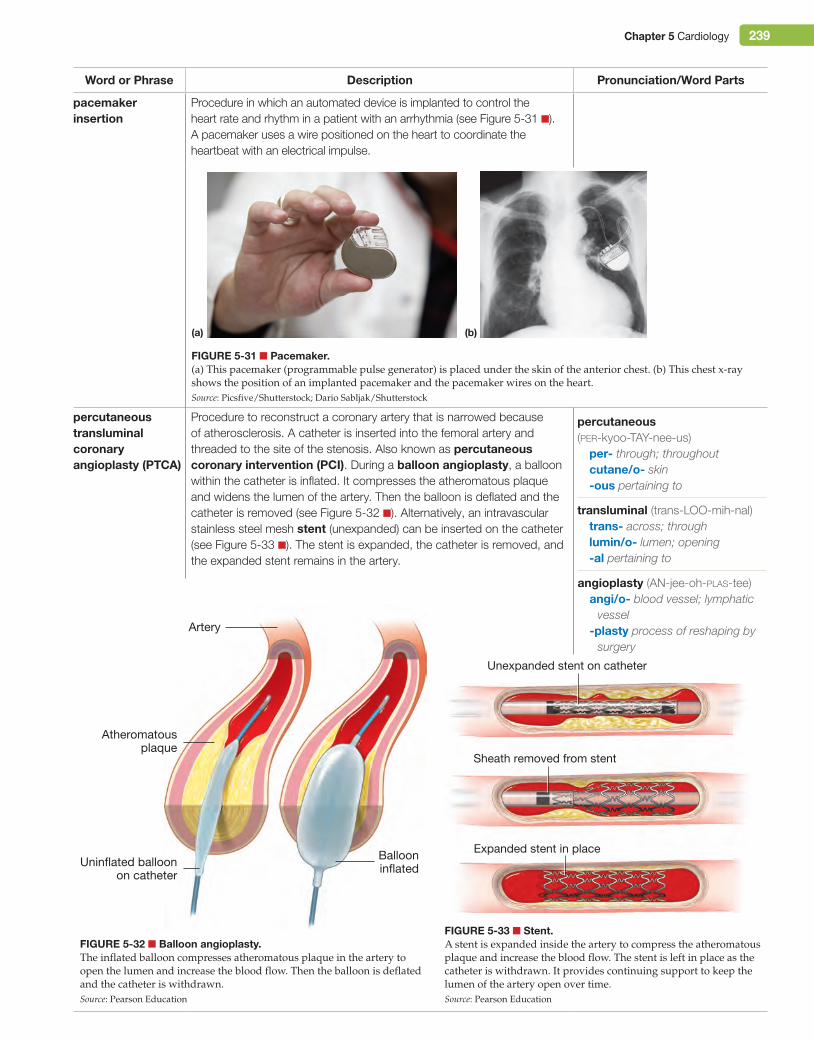
Chapter 5 Cardiology 239
Word or Phrase Description Pronunciation/Word Parts
pacemaker insertion
Procedure in which an automated device is implanted to control the heart rate and rhythm in a patient with an arrhythmia (see Figure 5-31 ■). A pacemaker uses a wire positioned on the heart to coordinate the heartbeat with an electrical impulse.
percutaneous transluminal coronary angioplasty (PTCA)
Procedure to reconstruct a coronary artery that is narrowed because of atherosclerosis. A catheter is inserted into the femoral artery and threaded to the site of the stenosis. Also known as percutaneous coronary intervention (PCi). During a balloon angioplasty, a balloon within the catheter is inflated. It compresses the atheromatous plaque and widens the lumen of the artery. Then the balloon is deflated and the catheter is removed (see Figure 5-32 ■). Alternatively, an intravascular stainless steel mesh stent (unexpanded) can be inserted on the catheter (see Figure 5-33 ■). The stent is expanded, the catheter is removed, and the expanded stent remains in the artery.
Figure 5-31 ■ Pacemaker.(a) This pacemaker (programmable pulse generator) is placed under the skin of the anterior chest. (b) This chest x-ray shows the position of an implanted pacemaker and the pacemaker wires on the heart.Source: Picsfive/Shutterstock; Dario Sabljak/Shutterstock
(a) (b)
percutaneous (per-kyoo-TAY-nee-us)
per- through; throughoutcutane/o- skin-ous pertaining to
transluminal (trans-LOO-mih-nal)trans- across; throughlumin/o- lumen; opening-al pertaining to
angioplasty (AN-jee-oh-plas-tee)angi/o- blood vessel; lymphatic
vessel-plasty process of reshaping by
surgery
Figure 5-32 ■ Balloon angioplasty.The inflated balloon compresses atheromatous plaque in the artery to open the lumen and increase the blood flow. Then the balloon is deflated and the catheter is withdrawn.Source: Pearson Education
BallooninflatedUninflated balloon
on catheter
Atheromatousplaque
Artery
Figure 5-33 ■ Stent.A stent is expanded inside the artery to compress the atheromatous plaque and increase the blood flow. The stent is left in place as the catheter is withdrawn. It provides continuing support to keep the lumen of the artery open over time.Source: Pearson Education
Unexpanded stent on catheter
Sheath removed from stent
Expanded stent in place
M05_TURL8127_04_SE_C05.indd 239 14/12/15 10:48 pm
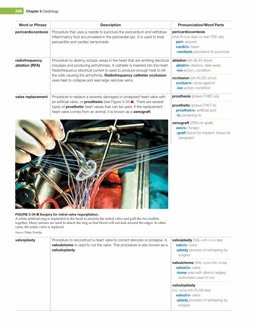
240 Chapter 5 Cardiology
Word or Phrase Description Pronunciation/Word Parts
pericardiocentesis Procedure that uses a needle to puncture the pericardium and withdraw inflammatory fluid accumulated in the pericardial sac. It is used to treat pericarditis and cardiac tamponade.
radiofrequency ablation (rFA)
Procedure to destroy ectopic areas in the heart that are emitting electrical impulses and producing arrhythmias. A catheter is inserted into the heart. Radiofrequency electrical current is used to produce enough heat to kill the cells causing the arrhythmia. radiofrequency catheter occlusion uses heat to collapse and seal large varicose veins.
valve replacement Procedure to replace a severely damaged or prolapsed heart valve with an artificial valve, or prosthesis (see Figure 5-34 ■). There are several types of prosthetic heart valves that can be used. If the replacement heart valve comes from an animal, it is known as a xenograft.
valvoplasty Procedure to reconstruct a heart valve to correct stenosis or prolapse. A valvulotome is used to cut the valve. This procedure is also known as a valvuloplasty.
pericardiocentesis (pair-ih-kar-dee-oh-sen-TEE-sis)
peri- aroundcardi/o- heart-centesis procedure to puncture
ablation (ah-BLAY-shun)ablat/o- destroy; take away-ion action; condition
prosthetic (praws-THET-ik)prosthet/o- artificial part-ic pertaining to
xenograft (ZEN-oh-graft)xen/o- foreign-graft tissue for implant; tissue for
transplant
valvulotome (VAL-vyoo-loh-tohm)valvul/o- valve-tome area with distinct edges;
instrument used to cut
valvuloplasty (val-vyoo-loh-PLAS-tee)
valvul/o- valve-plasty process of reshaping by
surgery
occlusion (oh-KLOO-zhun)occlus/o- close against-ion action; condition
valvoplasty (VAL-voh-plas-tee)valv/o- valve-plasty process of reshaping by
surgery
prosthesis (praws-THEE-sis)
Figure 5-34 ■ Surgery for mitral valve regurgitation.A white artificial ring is implanted in the heart to encircle the mitral valve and pull the two leaflets together. Many sutures are used to attach the ring so that blood will not leak around the edges. In other cases, the entire valve is replaced.
Source: Pirke/Fotolia
M05_TURL8127_04_SE_C05.indd 240 14/12/15 10:49 pm
Chapter 5 Cardiology 241
DrugsThese drug categories and drugs are used to treat cardiovascular diseases. The most common generic and trade name drugs in each category are listed.
Category indication examples Pronunciation/Word Parts
ACe (angiotensin-converting enzyme) inhibitor drugs
Treat congestive heart failure and hypertension. ACE inhibitor drugs produce vasodilation and decrease the blood pressure by blocking an enzyme that converts angiotensin I to angiotensin II (a vasoconstrictor).
captopril (Capoten), lisinopril (Prinivil, Zestril), trandolapril (Mavik)
antiarrhythmic drugs
Treat arrhythmias Intravenous atropine for heart block, intravenous lidocaine (Xylocaine) for ventricular fibrillation. Some beta-blocker drugs and calcium channel blocker drugs are used to treat ventricular tachycardia.
anticoagulant drugs
Prevent a blood clot from forming in patients with arteriosclerosis, atrial fibrillation, previous myocardial infarction, or an artificial heart valve
heparin, warfarin (Coumadin), clopidogrel (Plavix)
antihypertensive drugs
Treat hypertension See ACE inhibitor drugs, be ta-blocker drugs, calcium channel blocker drugs, and diuretic drugs.
aspirin Prevents heart attacks. Prevents blood clots from forming by keeping platelets from sticking together.
aspirin (81 mg)
beta-blocker drugs
Treat angina pectoris and hypertension. Beta-blocker drugs decrease the heart rate and dilate the arteries by blocking beta receptors.
atenolol (Tenormin), nadolol (Corgard), propranolol (Inderal), metoprolol (Lopressor)
calcium channel blocker drugs
Treat angina pectoris and hypertension. These drugs block the movement of calcium ions into myocardial cells and smooth muscle cells of the artery walls, causing the heart rate and blood pressure to decrease.
amlodipine (Norvasc), diltiazem (Cardizem), nifedipine (Adalat, Procardia), verapamil (Calan)
angiotensin (an-jee-oh-TEN-sin)angi/o- blood vessel;
lymphatic vesseltens/o- pressure; tension-in substance
antiarrhythmic (an-tee-aa-RITH-mik)
anti- againsta- away from; withoutrrhythm/o- rhythm-ic pertaining to
anticoagulant (an-tee-koh-AG-yoo-lant)(an-tih-koh-AG-yoo-lant)
anti- againstcoagul/o- clotting-ant pertaining to
antihypertensive (an-tee-hy-per-TEN-siv)
anti- againsthyper- above; more than
normaltens/o- pressure; tension-ive pertaining to
M05_TURL8127_04_SE_C05.indd 241 14/12/15 10:49 pm
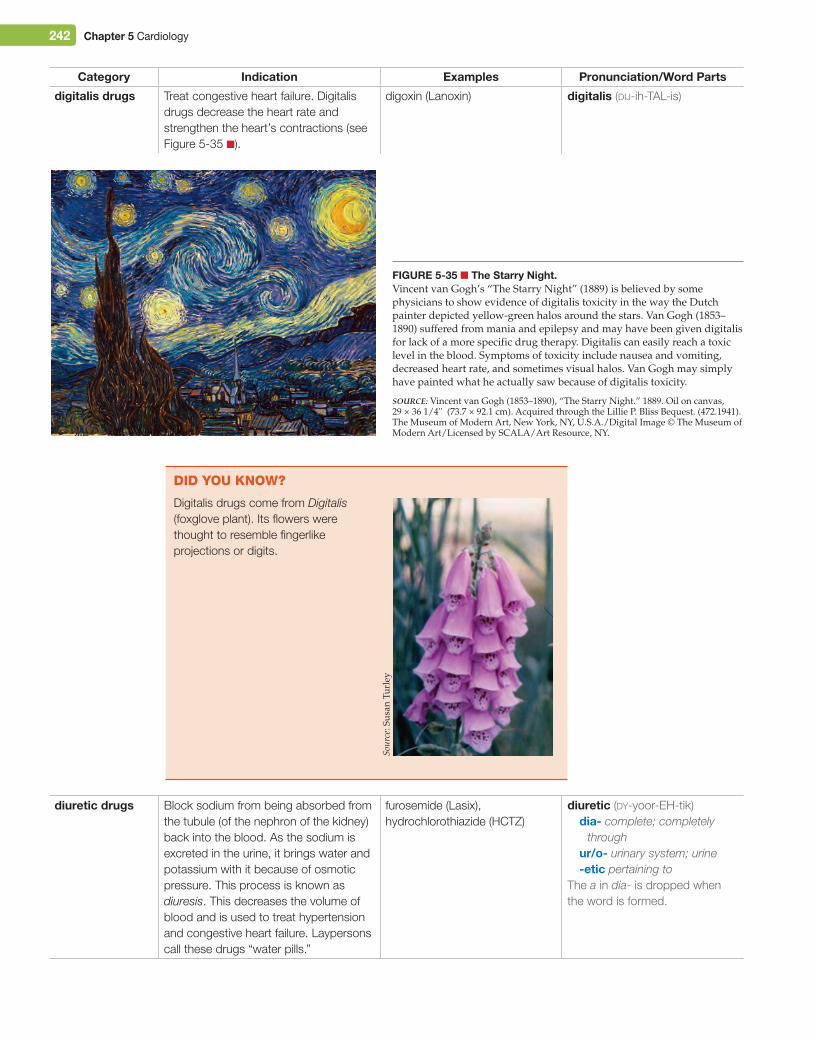
242 Chapter 5 Cardiology
Category indication examples Pronunciation/Word Parts
digitalis drugs Treat congestive heart failure. Digitalis drugs decrease the heart rate and strengthen the heart’s contractions (see Figure 5-35 ■).
digoxin (Lanoxin)
diuretic drugs Block sodium from being absorbed from the tubule (of the nephron of the kidney) back into the blood. As the sodium is excreted in the urine, it brings water and potassium with it because of osmotic pressure. This process is known as diuresis. This decreases the volume of blood and is used to treat hypertension and congestive heart failure. Laypersons call these drugs “water pills.”
furosemide (Lasix), hydrochlorothiazide (HCTZ)
did You KnoW?
Digitalis drugs come from Digitalis (foxglove plant). Its flowers were thought to resemble fingerlike projections or digits.
digitalis (dij-ih-TAL-is)
diuretic (dy-yoor-EH-tik)dia- complete; completely
throughur/o- urinary system; urine-etic pertaining to
The a in dia- is dropped when the word is formed.
Figure 5-35 ■ The Starry Night.Vincent van Gogh’s “The Starry Night” (1889) is believed by some physicians to show evidence of digitalis toxicity in the way the Dutch painter depicted yellow-green halos around the stars. Van Gogh (1853–1890) suffered from mania and epilepsy and may have been given digitalis for lack of a more specific drug therapy. Digitalis can easily reach a toxic level in the blood. Symptoms of toxicity include nausea and vomiting, decreased heart rate, and sometimes visual halos. Van Gogh may simply have painted what he actually saw because of digitalis toxicity.
Source: Vincent van Gogh (1853–1890), “The Starry Night.” 1889. Oil on canvas, 29 × 36 1/4'' (73.7 × 92.1 cm). Acquired through the Lillie P. Bliss Bequest. (472.1941). The Museum of Modern Art, New York, NY, U.S.A./Digital Image © The Museum of Modern Art/Licensed by SCALA/Art Resource, NY.
Sour
ce: S
usan
Tur
ley
M05_TURL8127_04_SE_C05.indd 242 14/12/15 10:49 pm
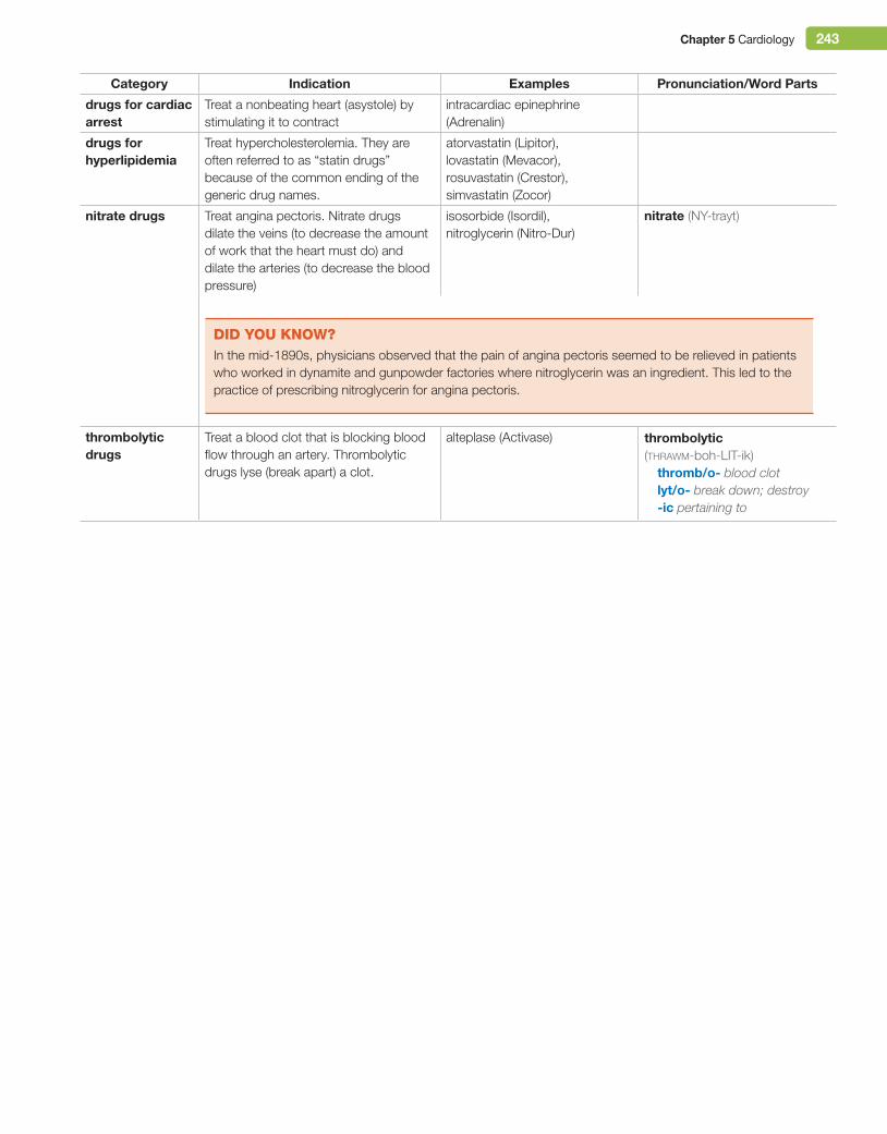
Chapter 5 Cardiology 243
Category indication examples Pronunciation/Word Parts
drugs for cardiac arrest
Treat a nonbeating heart (asystole) by stimulating it to contract
intracardiac epinephrine (Adrenalin)
drugs for hyperlipidemia
Treat hypercholesterolemia. They are often referred to as “statin drugs” because of the common ending of the generic drug names.
atorvastatin (Lipitor), lovastatin (Mevacor), rosuvastatin (Crestor), simvastatin (Zocor)
nitrate drugs Treat angina pectoris. Nitrate drugs dilate the veins (to decrease the amount of work that the heart must do) and dilate the arteries (to decrease the blood pressure)
isosorbide (Isordil), nitroglycerin (Nitro-Dur)
thrombolytic drugs
Treat a blood clot that is blocking blood flow through an artery. Thrombolytic drugs lyse (break apart) a clot.
alteplase (Activase)
nitrate (NY-trayt)
thrombolytic (thrawm-boh-LIT-ik)
thromb/o- blood clotlyt/o- break down; destroy-ic pertaining to
did You KnoW?In the mid-1890s, physicians observed that the pain of angina pectoris seemed to be relieved in patients who worked in dynamite and gunpowder factories where nitroglycerin was an ingredient. This led to the practice of prescribing nitroglycerin for angina pectoris.
M05_TURL8127_04_SE_C05.indd 243 14/12/15 10:49 pm
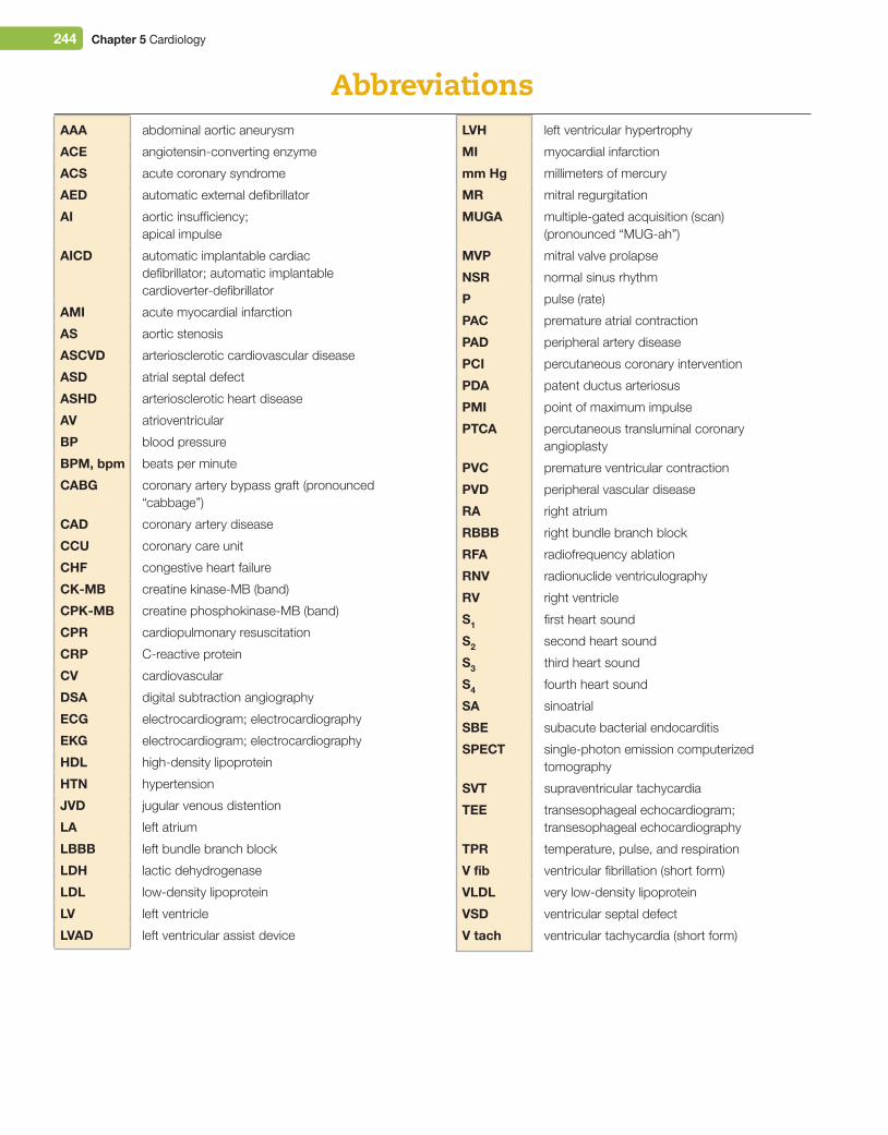
244 Chapter 5 Cardiology
AbbreviationsAAA abdominal aortic aneurysm
ACe angiotensin-converting enzyme
ACS acute coronary syndrome
AeD automatic external defibrillator
Ai aortic insufficiency; apical impulse
AiCD automatic implantable cardiac defibrillator; automatic implantable cardioverter-defibrillator
AMi acute myocardial infarction
AS aortic stenosis
ASCVD arteriosclerotic cardiovascular disease
ASD atrial septal defect
ASHD arteriosclerotic heart disease
AV atrioventricular
BP blood pressure
BPM, bpm beats per minute
CABg coronary artery bypass graft (pronounced “cabbage”)
CAD coronary artery disease
CCu coronary care unit
CHF congestive heart failure
CK-MB creatine kinase-MB (band)
CPK-MB creatine phosphokinase-MB (band)
CPr cardiopulmonary resuscitation
CrP C-reactive protein
CV cardiovascular
DSA digital subtraction angiography
eCg electrocardiogram; electrocardiography
eKg electrocardiogram; electrocardiography
HDL high-density lipoprotein
HTN hypertension
JVD jugular venous distention
LA left atrium
LBBB left bundle branch block
LDH lactic dehydrogenase
LDL low-density lipoprotein
LV left ventricle
LVAD left ventricular assist device
LVH left ventricular hypertrophy
Mi myocardial infarction
mm Hg millimeters of mercury
Mr mitral regurgitation
MugA multiple-gated acquisition (scan) (pronounced “MUG-ah”)
MVP mitral valve prolapse
NSr normal sinus rhythm
P pulse (rate)
PAC premature atrial contraction
PAD peripheral artery disease
PCi percutaneous coronary intervention
PDA patent ductus arteriosus
PMi point of maximum impulse
PTCA percutaneous transluminal coronary angioplasty
PVC premature ventricular contraction
PVD peripheral vascular disease
rA right atrium
rBBB right bundle branch block
rFA radiofrequency ablation
rNV radionuclide ventriculography
rV right ventricle
S1 first heart sound
S2 second heart sound
S3 third heart sound
S4 fourth heart sound
SA sinoatrial
SBe subacute bacterial endocarditis
SPeCT single-photon emission computerized tomography
SVT supraventricular tachycardia
Tee transesophageal echocardiogram; transesophageal echocardiography
TPr temperature, pulse, and respiration
V fib ventricular fibrillation (short form)
VLDL very low-density lipoprotein
VSD ventricular septal defect
V tach ventricular tachycardia (short form)
M05_TURL8127_04_SE_C05.indd 244 14/12/15 10:49 pm
Chapter 5 Cardiology 245
Word AlertAbbreviations
Abbreviations are commonly used in all types of medical documents; however, they can mean different things to different people and their meanings can be misinterpreted. Always verify the meaning of an abbreviation.
AI means aortic insufficiency and apical impulse, but it also means artificial insemination and artificial intelligence.
RA means right atrium, but it also means rheumatoid arthritis or room air.
Do not confuse the abbreviation CPR (cardiopulmonary resuscitation) with CRP (C-reactive protein).
it’s GreeK to Me!Did you notice that some words have two different combining forms? Combining forms from both Greek and Latin remain a part of medical language today.
Word greek Latin Medical Word examples
blood vessel angi/o- vas/o-, vascul/o- angiography, vasoconstriction, vasodilator vascular, vasculature
heart card/i-, cardi/o- [cor, Latin word] bradycardia, endocarditis, cor pulmonale, cardiac, cardiopulmonary
vein phleb/o- ven/o- phlebitis, thrombophlebitis, venous, venography
CAreer FoCusMeet Laurie, a cardiac stress test technologist in a hospital
“Cardiology is a very important department. I use medical terminology dur-ing all aspects of my job. My daughter was born with a heart defect. Want-ing to know more information about what was happening to her, I started to take a class here and a class there, and then I just wound up in a certificate program. The most rewarding part of my job is if I can get a patient through the test and at the end of the test they say, ‘You made that so much easier for me.”’
Cardiac stress test technologists are allied health professionals who perform ECGs, Holter monitor tests, and cardiac stress tests in a hospital setting or a cardiologist’s office.
Cardiologists are physicians who practice in the medical specialty of cardiology. They diagnose and treat patients with diseases of the heart and circulatory system. When cardiologists perform surgery, they are known as heart surgeons, cardiothoracic surgeons, or cardiovascular surgeons. Physicians can take additional training and become board certified in the subspecialty of pediatric cardiology. Cancerous tumors of the heart or blood vessels are treated medically by an oncologist or surgically by a cardiothoracic or cardiovascular surgeon.
To see Laurie’s complete video profile, log into MyMedicalTerminologyLab and navigate to the Multimedia Library for Chapter 5. Check the Video box, and then click the Career Focus - Cardiac Stress Tech link.
technologist (tek-NAW-loh-jist)techn/o- technical skilllog/o- study of; word-ist person who specializes in
cardiologist (kar-dee-AW-loh-jist)cardi/o- heartlog/o- study of; word-ist person who specializes in
Sour
ce: P
ears
on E
duca
tion
M05_TURL8127_04_SE_C05.indd 245 14/12/15 10:49 pm
246 Chapter 5 Cardiology
true or FAlse exerCise
Indicate whether each statement is true or false by writing T or F on the line.
1. ______ The aorta is the largest vein in the body.
2. ______ The adrenal glands secrete epinephrine, which increases the heart rate and blood pressure.
3. ______ Blood flows from the inferior vena cava to the right atrium to the right ventricle and to the pulmonary veins to the lungs.
4. ______ The refractory period is the time during which the ventricles contract.
5. ______ The smallest veins are known as capillaries.
6. ______ The interventricular septum is within a ventricle.
7. ______ The epicardium is the part of the pericardium that lies on the surface of the heart.
8. ______ All arteries carry blood away from the heart.
9. ______ The subclavian artery carries blood to the leg.
10. ______ The peroneal artery carries blood to the lateral aspect of the lower leg in the area of the fibula bone.
11. ______ The blood in most veins is a dark red–purple color because it has a low level of oxygen.
12. ______ The systemic circulation carries blood to the whole body with the exception of the lungs.
13. ______ Vasodilation is the opposite of vasoconstriction.
14. ______ The electrical impulse of a heart beat travels through the AV node and then into the diastole.
CHAPTER REViEW ExERCiSES Test your knowledge of the chapter by completing these review exercises. Use the Answer Key at the end of the book to check your answers. Note: Each of the numbered exercise headers corresponds to a numbered learning outcome on the first page of the chapter. Headers that include a number with an A or with a B after it show that there are two different parts to that learning outcome.
5.1 identify Anatomical Structures
5.2 Describe PhysiologyMAtChinG exerCise
Match each word or phrase to its description.
1. mediastinum
2. pericardium
3. myocardium
4. ventricle
5. tricuspid valve
6. septum
7. aortic valve
8. vasculature
9. bicuspid valve
10. chordae tendineae
______ Another name for the mitral valve
______ Network of blood vessels related to a particular organ
______ Dividing wall between the atria and ventricles
______ Double-layered membrane around the heart
______ Bottom chamber of the heart
______ Area between the lungs that contains the heart
______ Valve between the right atrium and right ventricle
______ Heart muscle
______ Valve that blood flows through as it leaves the left ventricle
______ Rope-like strands that strengthen the tricuspid and mitral valves
M05_TURL8127_04_SE_C05.indd 246 14/12/15 10:49 pm
Chapter 5 Cardiology 247
sequenCinG exerCise
Beginning with blood entering the right atrium, write each structure of the circulatory system in the order in which blood moves through it.
Structure Correct Orderaortic valve and aorta 1. right atrium
arteries and arterioles 2. _______________________
capillaries 3. _______________________
left atrium 4. _______________________
left ventricle 5. _______________________
lungs 6. _______________________
mitral valve 7. _______________________
pulmonary valve, pulmonary trunk, and pulmonary arteries 8. _______________________
pulmonary veins 9. _______________________
right atrium 10. _______________________
right ventricle 11. _______________________
superior and inferior venae cavae 12. _______________________
tricuspid valve 13. _______________________
venules and veins 14. _______________________
CirCle exerCise
Circle the correct word from the choices given.
1. The great vessels include the superior and inferior venae cavae and the (aorta, artery, mediastinum).
2. The vascular structures of the body include all of the following except (arteries, capillaries, heart valves, veins).
3. Listening to a patient’s heart sounds with a stethoscope is known as (auscultation, diastole, repolarization).
4. What unique structure is found only in the fetal heart? (apex, foramen ovale, vasculature).
5. This vein brings blood from the head to the superior vena cava: (jugular, portal, saphenous).
6. The (AV node, Purkinje fiber, SA node) is the pacemaker of the heart.
7. Depolarization involves the movement of sodium and (calcium, epinephrine, electrical) ions into the myocardial cell.
8. Which of these can take over and produce its own abnormal heart rhythm? (foramen ovale, ectopic site, intima)
5.3A Describe DiseasesFill in the BlAnK exerCise
Fill in the blank with the correct word from the word list.
arrhythmiaasystoleatheroma
cardiomegalyclaudicationcoarctation
necrosispalpitation
tetralogy of Fallotthrombus
1. Calf pain caused by peripheral artery disease _________________________________________________________________________
2. Fatty deposit on the wall of an artery _________________________________________________________________________________
3. Another name for a dysrhythmia _____________________________________________________________________________________
4. Chest sensation during a premature contraction _______________________________________________________________________
5. Cell death _______________________________________________________________________________________________________
6. Cardiac arrest with no heart activity seen on an ECG ___________________________________________________________________
7. Blood clot ________________________________________________________________________________________________________
M05_TURL8127_04_SE_C05.indd 247 14/12/15 10:49 pm
248 Chapter 5 Cardiology
8. Abnormal narrowing of the aorta in the fetal heart ______________________________________________________________________
9. Enlargement of the heart ___________________________________________________________________________________________
10. Set of four different defects in the fetal heart __________________________________________________________________________
CirCle exerCise
Circle the correct word from the choices given.
1. (Asystole, Bradycardia, Fibrillation) is an abnormally slow heart rate.
2. Narrowing of an artery is known as (arteriosclerosis, endocarditis, stenosis).
3. A weakness in the wall of an artery is known as a/an (aneurysm, couplet, varicose vein).
4. (Patent foramen ovale, Heart block, Aneurysm) is a congenital heart defect.
true or FAlse exerCise
Indicate whether each statement is true or false by writing T or F on the line.
1. ______ Angina pectoris is chest pain that means that myocardial cells have died.
2. ______ Prolapse of a valve is when the cusps do not close completely.
3. ______ Raynaud’s disease is severe vasoconstriction of the extremities triggered by cold or emotional stress.
4. ______ Hyperlipidemia includes both hypercholesterolemia and hypertriglyceridemia.
5. ______ Regurgitation is an infection of the heart caused by bacteria.
6. ______ To check for atherosclerosis in the arteries of the legs, you would feel the pulse in the radial artery.
7. ______ Auscultation is using a stethoscope to listen to the heart sounds.
8. ______ S1 and S2 are abnormal heart sounds.
9. ______ In a patient with right-sided congestive heart failure, the neck may show signs of jugular venous distention.
10. ______ A sphygmomanometer measures the blood pressure in millimeters of mercury (mm Hg).
5.3B Describe Laboratory, Radiology, Surgery, and DrugslABorAtorY test exerCise
You need to order the following laboratory tests to be done for a cardiology patient. Find each of these tests on the laboratory form and put a checkmark in the box next to it.
C-reactive proteincholesterolcreatine kinase, MB
digoxin drug levelHDL lipoproteinLDL lipoprotein
lipid paneltriglyceridestroponin
venipunctureVLDL lipoprotein
CPT CODe PANeLS AND PrOFiLeS80047 Metabolic Panel, Basic80051 Electrolyte Panel80053 Metabolic Panel, Comprehensive80055 Obstetric Panel80061 Lipid Panel80069 Renal Function Panel80076 Hepatic Function Panel80100 Drug Screening PanelCPT CODe BLOOD TeSTS80162 Digoxin Drug Level82040 Albumin82055 Blood Alcohol (ETOH)
82247 Bilirubin, Total82310 Calcium, Total Blood82375 Carboxyhemoglobin82378 Carcinoembryonic Antigen (CEA)82435 Chloride82465 Cholesterol82553 Creatine Kinase, MB82565 Creatinine, Blood82575 Creatinine Clearance82947 Glucose, Fasting82951 Glucose Tolerance Test (GTT)83036 Hemoglobin A1c83550 Iron, TIBC
M05_TURL8127_04_SE_C05.indd 248 14/12/15 10:49 pm
Chapter 5 Cardiology 249
CPT CODe PANeLS AND PrOFiLeS83655 Lead, Blood83661 Fetal Lung Maturity (L/S Ratio)83718 HDL Lipoprotein83719 VLDL Lipoprotein83721 LDL Lipoprotein83735 Magnesium84075 Alkaline Phosphatase84144 Progesterone84146 Prolactin84152 Prostate-specific Antigen (PSA)CPT CODe BLOOD TeSTS84295 Sodium84443 Thyroid-stimulating Hormone (TSH)84436 Thyroxine (T4), Total84450 Aspartate Transaminase (AST)84460 Alanine Transaminase (ALT)84478 Triglycerides84480 Triiodothyronine (T3), Total84484 Troponin84520 Blood Urea Nitrogen (BUN)84550 Uric Acid84702 Human Chorionic Gonadotropin (hCG), Serum85004 White Blood Cell (WBC) Count with Differential
85014 Hematocrit (HCT)85018 Hemoglobin (Hgb)85025 Complete Blood Count (CBC)85049 Platelet Count85610 Prothrombin Time (PT)85730 Partial Thromboplastin Time (PTT)86038 Antinuclear Antibody (ANA)86140 C-Reactive Protein86592 Syphilis Blood Test (VDRL or RPR)86762 Rubella Antibody86812 Human Leukocyte Antigen (HLA) Typing86900 Blood Type87340 Hepatitis B Surface AntigenCPT CODe OTHer TeSTS AND PrOCeDureS36415 Venipuncture81001 Urinalysis, Automated82270 Stool Occult Blood87040 Blood Culture87045 Stool Culture87086 Urine Culture87177 Ova and Parasites89220 Sputum Analysis89300 Semen Analysis
true or FAlse exerCise
Indicate whether each statement is true or false by writing T or F on the line.
1. ______ Antiarrhythmic drugs are used to treat hypertension.
2. ______ Thrombolytic drugs break apart blood clots.
3. ______ An artificial valve is also known as a prosthesis.
4. ______ A stent is inserted during a MUGA scan.
5. ______ Sclerotherapy is used to treat arteriosclerosis (hardening of the arteries).
6. ______ Auscultation of the heart uses a stethoscope.
7. ______ Cardiopulmonary bypass is used during open heart surgery.
8. ______ A sphygmomanometer is used to perform a heart transplantation.
5.4 Form Plurals and AdjectivesPlurAl noun And AdjeCtive exerCise
Read the noun and write its plural form and/or adjective form. Be sure to check your spelling. The first one has been done for you.
Singular Noun Plural Noun Adjective Form 1. pericardium pericardial 2. artery _______________ _________________
3. atrium _______________ _________________
4. ventricle _______________ _________________
5. septum _________________
6. myocardium _________________
Singular Noun Plural Noun Adjective Form 7. valve _______________ _________________
8. aorta _________________
9. vein _______________ _________________
10. heart _________________
11. arteriole _______________ _________________
12. diastole _________________
M05_TURL8127_04_SE_C05.indd 249 14/12/15 10:49 pm
250 Chapter 5 Cardiology
5.5A Give Word Part MeaningsWord PArts exerCise
Next to each word part, write its meaning. The first one has been done for you.
Word Part Meaning 1. angin/o- angina 2. a- ______________________________
3. abdomin/o- ______________________________
4. ablat/o- ______________________________
5. -ac ______________________________
6. -ade ______________________________
7. -al ______________________________
8. anastom/o- ______________________________
9. aneurysm/o- ______________________________
10. angi/o- ______________________________
11. anti- ______________________________
12. aort/o- ______________________________
13. apic/o- ______________________________
14. -ar ______________________________
15. arteri/o- ______________________________
16. arteriol/o- ______________________________
17. arter/o- ______________________________
18. -ary ______________________________
19. -ase ______________________________
20. -ated ______________________________
21. ather/o- ______________________________
22. atheromat/o- ______________________________
23. -ation ______________________________
24. -ator ______________________________
25. atri/o- ______________________________
26. -ature ______________________________
27. auscult/o- ______________________________
28. axill/o- ______________________________
29. bi- ______________________________
30. brachi/o- ______________________________
31. brady- ______________________________
32. capill/o- ______________________________
33. card/i- ______________________________
34. cardi/o- ______________________________
35. carot/o- ______________________________
36. catheter/o- ______________________________
37. -centesis ______________________________
38. cholesterol/o- ______________________________
39. circulat/o- ______________________________
40. claudicat/o- ______________________________
41. clav/o- ______________________________
Word Part Meaning42. coagul/o- ______________________________
43. coarct/o- ______________________________
44. compens/o- ______________________________
45. conduct/o- ______________________________
46. congest/o- ______________________________
47. constrict/o- ______________________________
48. contract/o- ______________________________
49. coron/o- ______________________________
50. cusp/o- ______________________________
51. cutane/o- ______________________________
52. de- ______________________________
53. dia- ______________________________
54. diastol/o- ______________________________
55. dilat/o- ______________________________
56. dissect/o- ______________________________
57. dys- ______________________________
58. -eal ______________________________
59. ech/o- ______________________________
60. -ectomy ______________________________
61. ectop/o- ______________________________
62. electr/o- ______________________________
63. -emia ______________________________
64. endo- ______________________________
65. -ent ______________________________
66. epi- ______________________________
67. -etic ______________________________
68. extra- ______________________________
69. femor/o- ______________________________
70. fibrill/o- ______________________________
71. fract/o- ______________________________
72. fus/o- ______________________________
73. -graft ______________________________
74. -gram ______________________________
75. -graphy ______________________________
76. hyper- ______________________________
77. hypo- ______________________________
78. -ia ______________________________
79. -ian ______________________________
80. -ic ______________________________
81. -id ______________________________
82. idi/o- ______________________________
M05_TURL8127_04_SE_C05.indd 250 14/12/15 10:49 pm
Chapter 5 Cardiology 251
Word Part Meaning83. ili/o- ______________________________
84. -in ______________________________
85. infarct/o- ______________________________
86. -ion ______________________________
87. isch/o- ______________________________
88. -ist ______________________________
89. -itis ______________________________
90. -ive ______________________________
91. -ization ______________________________
92. jugul/o- ______________________________
93. lipid/o- ______________________________
94. lip/o- ______________________________
95. log/o- ______________________________
96. -logy ______________________________
97. lumin/o- ______________________________
98. lyt/o- ______________________________
99. man/o- ______________________________
100. mediastin/o- ______________________________
101. -megaly ______________________________
102. -meter ______________________________
103. -metry ______________________________
104. mitr/o- ______________________________
105. my/o- ______________________________
106. necr/o- ______________________________
107. occlus/o- ______________________________
108. -ole ______________________________
109. -oma ______________________________
110. -ory ______________________________
111. -ose ______________________________
112. -osis ______________________________
113. -ous ______________________________
114. palpit/o- ______________________________
115. pariet/o- ______________________________
116. path/o- ______________________________
117. -pathy ______________________________
118. pat/o- ______________________________
119. pector/o- ______________________________
120. per- ______________________________
121. peri- ______________________________
122. peripher/o- ______________________________
123. perone/o- ______________________________
124. pharmac/o- ______________________________
125. phleb/o- ______________________________
126. physi/o- ______________________________
127. -plasty ______________________________
128. polar/o- ______________________________
Word Part Meaning129. poplite/o- ______________________________
130. port/o- ______________________________
131. pre- ______________________________
132. prosthet/o- ______________________________
133. pulmon/o- ______________________________
134. radi/o- ______________________________
135. re- ______________________________
136. regurgitat/o- ______________________________
137. ren/o- ______________________________
138. rheumat/o- ______________________________
139. rrhythm/o- ______________________________
140. saphen/o- ______________________________
141. scler/o- ______________________________
142. -scope ______________________________
143. sept/o- ______________________________
144. sin/o- ______________________________
145. son/o- ______________________________
146. sphygm/o- ______________________________
147. sten/o- ______________________________
148. sub- ______________________________
149. supra- ______________________________
150. system/o- ______________________________
151. -systole ______________________________
152. systol/o- ______________________________
153. tachy- ______________________________
154. tampon/o- ______________________________
155. techn/o- ______________________________
156. tele/o- ______________________________
157. tens/o- ______________________________
158. tetr/a- ______________________________
159. theli/o- ______________________________
160. -therapy ______________________________
161. thorac/o- ______________________________
162. thromb/o- ______________________________
163. tibi/o- ______________________________
164. -tic ______________________________
165. -tome ______________________________
166. tom/o- ______________________________
167. trans- ______________________________
168. transplant/o- ______________________________
169. tri- ______________________________
170. triglycerid/o- ______________________________
171. -trophy ______________________________
172. -ule ______________________________
173. uln/o- ______________________________
174. ultra- ______________________________
M05_TURL8127_04_SE_C05.indd 251 14/12/15 10:49 pm
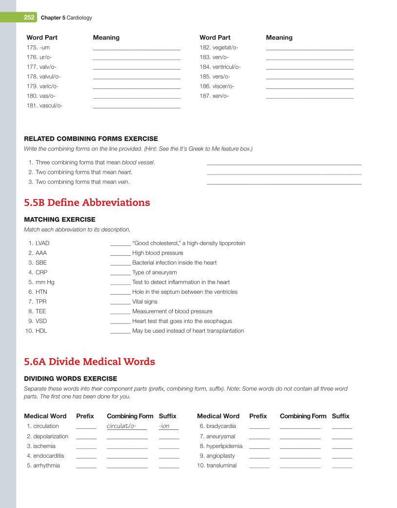
252 Chapter 5 Cardiology
_______ “Good cholesterol,” a high-density lipoprotein
_______ High blood pressure
_______ Bacterial infection inside the heart
_______ Type of aneurysm
_______ Test to detect inflammation in the heart
_______ Hole in the septum between the ventricles
_______ Vital signs
_______ Measurement of blood pressure
_______ Heart test that goes into the esophagus
_______ May be used instead of heart transplantation
relAted CoMBininG ForMs exerCise
Write the combining forms on the line provided. (Hint: See the It’s Greek to Me feature box.)
1. Three combining forms that mean blood vessel. _____________________________________________________
2. Two combining forms that mean heart. _____________________________________________________
3. Two combining forms that mean vein. _____________________________________________________
5.5B Define AbbreviationsMAtChinG exerCise
Match each abbreviation to its description.
1. LVAD
2. AAA
3. SBE
4. CRP
5. mm Hg
6. HTN
7. TPR
8. TEE
9. VSD
10. HDL
5.6A Divide Medical WordsdividinG Words exerCise
Separate these words into their component parts (prefix, combining form, suffix). Note: Some words do not contain all three word parts. The first one has been done for you.
Medical Word Prefix Combining Form Suffix 1. circulation _______ circulat/o- -ion 2. depolarization _______ ______________ _______
3. ischemia _______ ______________ _______
4. endocarditis _______ ______________ _______
5. arrhythmia _______ ______________ _______
Medical Word Prefix Combining Form Suffix 6. bradycardia _______ ______________ _______
7. aneurysmal _______ ______________ _______
8. hyperlipidemia _______ ______________ _______
9. angioplasty _______ ______________ _______
10. transluminal _______ ______________ _______
Word Part Meaning175. -um ______________________________
176. ur/o- ______________________________
177. valv/o- ______________________________
178. valvul/o- ______________________________
179. varic/o- ______________________________
180. vas/o- ______________________________
181. vascul/o- ______________________________
Word Part Meaning182. vegetat/o- ______________________________
183. ven/o- ______________________________
184. ventricul/o- ______________________________
185. vers/o- ______________________________
186. viscer/o- ______________________________
187. xen/o- ______________________________
M05_TURL8127_04_SE_C05.indd 252 14/12/15 10:49 pm
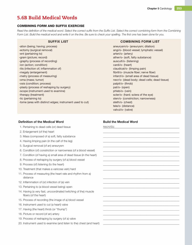
Chapter 5 Cardiology 253
5.6B Build Medical WordsCoMBininG ForM And suFFix exerCise
Read the definition of the medical word. Select the correct suffix from the Suffix List. Select the correct combining form from the Combining Form List. Build the medical word and write it on the line. Be sure to check your spelling. The first one has been done for you.
SuFFix LiST COMBiNiNg FOrM LiST-ation (being; having; process) aneurysm/o- (aneurysm; dilation)-ectomy (surgical removal) angi/o- (blood vessel; lymphatic vessel)-ent (pertaining to) arteri/o- (artery)-gram (picture; record) ather/o- (soft, fatty substance)-graphy (process of recording) auscult/o- (listening)-ion (action; condition) cardi/o- (heart)-itis (infection of; inflammation of) claudicat/o- (limping pain)-megaly (enlargement) fibrill/o- (muscle fiber; nerve fiber)-metry (process of measuring) infarct/o- (small area of dead tissue)-oma (mass; tumor) necr/o- (dead body; dead cells; dead tissue)-osis (condition; process) palpit/o- (throb)-plasty (process of reshaping by surgery) pat/o- (open)-scope (instrument used to examine) phleb/o- (vein)-therapy (treatment) scler/o- (hard; sclera of the eye)-tic (pertaining to) sten/o- (constriction; narrowness)-tome (area with distinct edges; instrument used to cut) steth/o- (chest)
tele/o- (distance)valvul/o- (valve)
Definition of the Medical Word Build the Medical Word
1. Pertaining to dead cells (or) dead tissue necrotic 2. Enlargement (of the) heart _______________________________________________________
3. Mass (composed of a) soft, fatty substance ______________________________________________________
4. Having limping pain (in the calf of the leg) ______________________________________________________
5. Surgical removal (of an) aneurysm ______________________________________________________
6. Condition (of) constriction or narrowness (of a blood vessel) ______________________________________________________
7. Condition (of having a) small area of dead tissue (in the heart) ______________________________________________________
8. Process of reshaping by surgery (of a) blood vessel ______________________________________________________
9. Process (of) listening (to the heart) ______________________________________________________
10. Treatment (that makes a varicose vein) hard ______________________________________________________
11. Process of measuring (the heart rate and rhythm from a) distance ______________________________________________________
12. Inflammation of (or) infection of (a) vein ______________________________________________________
13. Pertaining to (a blood vessel being) open ______________________________________________________
14. Having (a very fast, uncoordinated twitching of the) muscle fibers (of the heart) ______________________________________________________
15. Process of recording (the image of a) blood vessel ______________________________________________________
16. Instrument used to cut (a heart) valve ______________________________________________________
17. Having (the heart) throb (or “thump”) ______________________________________________________
18. Picture or record (of an) artery ______________________________________________________
19. Process of reshaping by surgery (of a) valve ______________________________________________________
20. Instrument used to examine (and listen to the) chest (and heart) ______________________________________________________
M05_TURL8127_04_SE_C05.indd 253 14/12/15 10:49 pm
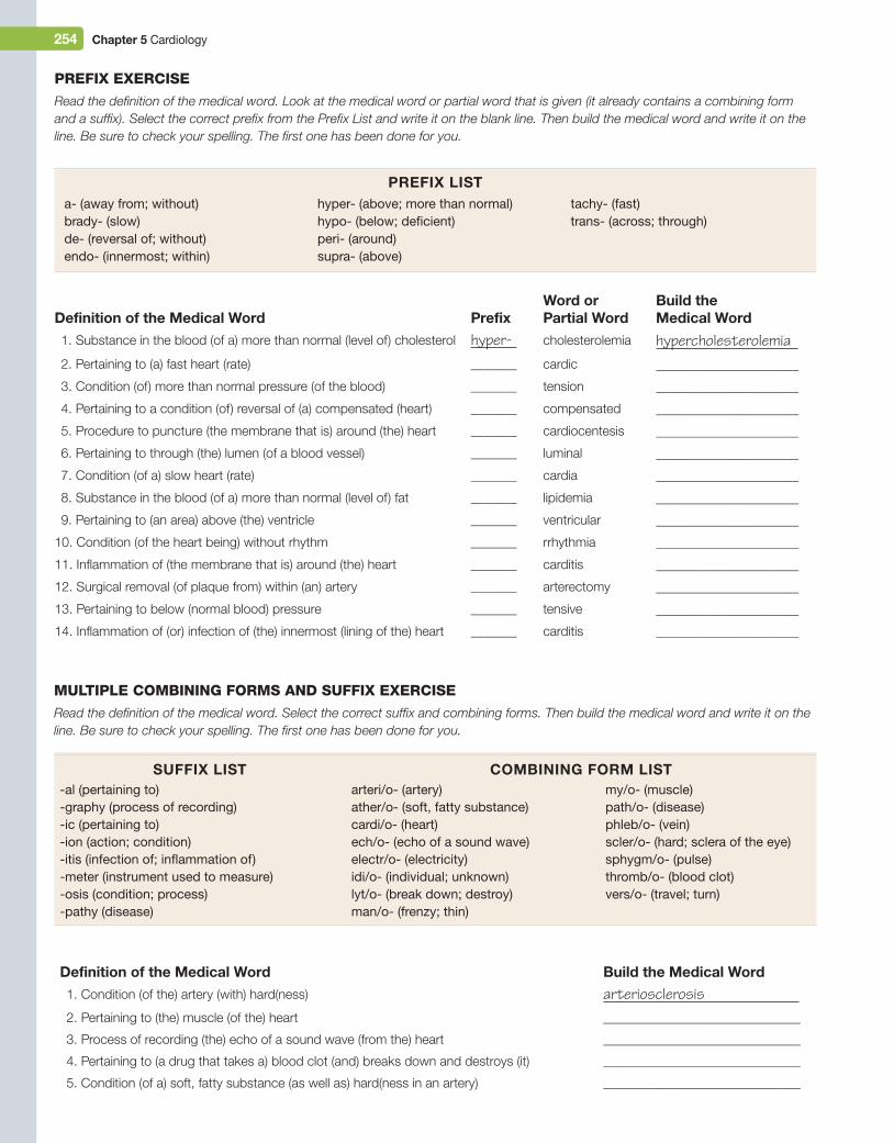
254 Chapter 5 Cardiology
MultiPle CoMBininG ForMs And suFFix exerCise
Read the definition of the medical word. Select the correct suffix and combining forms. Then build the medical word and write it on the line. Be sure to check your spelling. The first one has been done for you.
SuFFix LiST COMBiNiNg FOrM LiST-al (pertaining to) arteri/o- (artery) my/o- (muscle)-graphy (process of recording) ather/o- (soft, fatty substance) path/o- (disease)-ic (pertaining to) cardi/o- (heart) phleb/o- (vein)-ion (action; condition) ech/o- (echo of a sound wave) scler/o- (hard; sclera of the eye)-itis (infection of; inflammation of) electr/o- (electricity) sphygm/o- (pulse)-meter (instrument used to measure) idi/o- (individual; unknown) thromb/o- (blood clot)-osis (condition; process) lyt/o- (break down; destroy) vers/o- (travel; turn)-pathy (disease) man/o- (frenzy; thin)
Definition of the Medical Word PrefixWord or Partial Word
Build the Medical Word
1. Substance in the blood (of a) more than normal (level of) cholesterol hyper- cholesterolemia hypercholesterolemia 2. Pertaining to (a) fast heart (rate) _______ cardic ______________________
3. Condition (of) more than normal pressure (of the blood) _______ tension ______________________
4. Pertaining to a condition (of) reversal of (a) compensated (heart) _______ compensated ______________________
5. Procedure to puncture (the membrane that is) around (the) heart _______ cardiocentesis ______________________
6. Pertaining to through (the) lumen (of a blood vessel) _______ luminal ______________________
7. Condition (of a) slow heart (rate) _______ cardia ______________________
8. Substance in the blood (of a) more than normal (level of) fat _______ lipidemia ______________________
9. Pertaining to (an area) above (the) ventricle _______ ventricular ______________________
10. Condition (of the heart being) without rhythm _______ rrhythmia ______________________
11. Inflammation of (the membrane that is) around (the) heart _______ carditis ______________________
12. Surgical removal (of plaque from) within (an) artery _______ arterectomy ______________________
13. Pertaining to below (normal blood) pressure _______ tensive ______________________
14. Inflammation of (or) infection of (the) innermost (lining of the) heart _______ carditis ______________________
a- (away from; without)brady- (slow)de- (reversal of; without)endo- (innermost; within)
hyper- (above; more than normal)hypo- (below; deficient)peri- (around)supra- (above)
tachy- (fast)trans- (across; through)
PreFix exerCise
Read the definition of the medical word. Look at the medical word or partial word that is given (it already contains a combining form and a suffix). Select the correct prefix from the Prefix List and write it on the blank line. Then build the medical word and write it on the line. Be sure to check your spelling. The first one has been done for you.
PreFix LiST
Definition of the Medical Word Build the Medical Word 1. Condition (of the) artery (with) hard(ness) arteriosclerosis 2. Pertaining to (the) muscle (of the) heart ______________________________
3. Process of recording (the) echo of a sound wave (from the) heart ______________________________
4. Pertaining to (a drug that takes a) blood clot (and) breaks down and destroys (it) ______________________________
5. Condition (of a) soft, fatty substance (as well as) hard(ness in an artery) ______________________________
M05_TURL8127_04_SE_C05.indd 254 14/12/15 10:49 pm
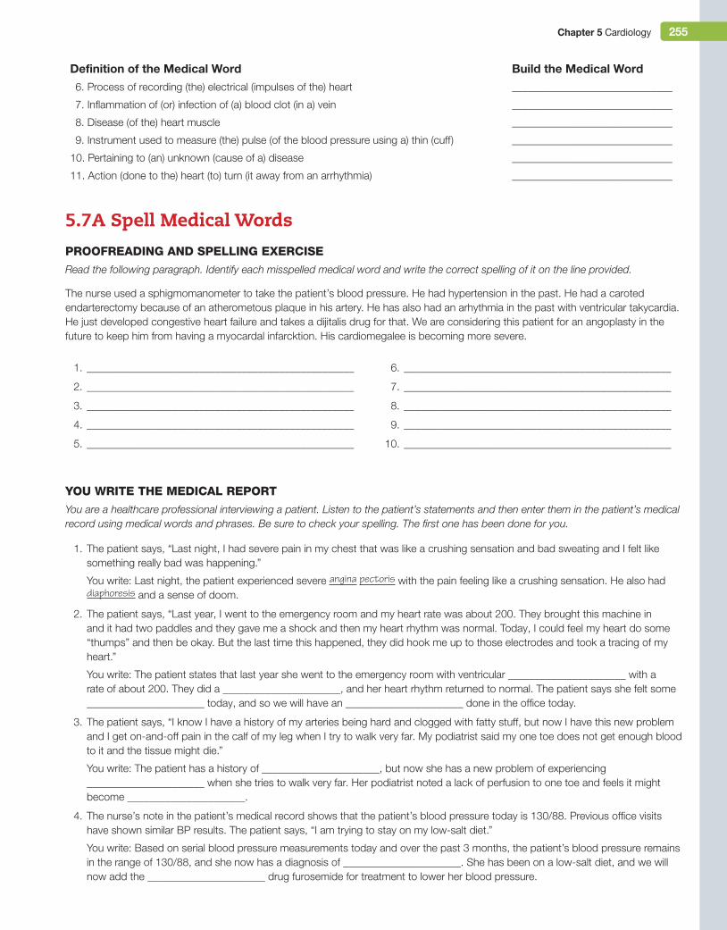
Chapter 5 Cardiology 255
You Write the MediCAl rePort
You are a healthcare professional interviewing a patient. Listen to the patient’s statements and then enter them in the patient’s medical record using medical words and phrases. Be sure to check your spelling. The first one has been done for you.
1. The patient says, “Last night, I had severe pain in my chest that was like a crushing sensation and bad sweating and I felt like something really bad was happening.”
You write: Last night, the patient experienced severe angina pectoris with the pain feeling like a crushing sensation. He also had diaphoresis and a sense of doom.
2. The patient says, “Last year, I went to the emergency room and my heart rate was about 200. They brought this machine in and it had two paddles and they gave me a shock and then my heart rhythm was normal. Today, I could feel my heart do some “thumps” and then be okay. But the last time this happened, they did hook me up to those electrodes and took a tracing of my heart.”
You write: The patient states that last year she went to the emergency room with ventricular ______________________ with a rate of about 200. They did a ______________________, and her heart rhythm returned to normal. The patient says she felt some ______________________ today, and so we will have an ______________________ done in the office today.
3. The patient says, “I know I have a history of my arteries being hard and clogged with fatty stuff, but now I have this new problem and I get on-and-off pain in the calf of my leg when I try to walk very far. My podiatrist said my one toe does not get enough blood to it and the tissue might die.”
You write: The patient has a history of ______________________, but now she has a new problem of experiencing ______________________ when she tries to walk very far. Her podiatrist noted a lack of perfusion to one toe and feels it might become ______________________.
4. The nurse’s note in the patient’s medical record shows that the patient’s blood pressure today is 130/88. Previous office visits have shown similar BP results. The patient says, “I am trying to stay on my low-salt diet.”
You write: Based on serial blood pressure measurements today and over the past 3 months, the patient’s blood pressure remains in the range of 130/88, and she now has a diagnosis of ______________________. She has been on a low-salt diet, and we will now add the ______________________ drug furosemide for treatment to lower her blood pressure.
1. __________________________________________________
2. __________________________________________________
3. __________________________________________________
4. __________________________________________________
5. __________________________________________________
6. __________________________________________________
7. __________________________________________________
8. __________________________________________________
9. __________________________________________________
10. __________________________________________________
5.7A Spell Medical WordsProoFreAdinG And sPellinG exerCise
Read the following paragraph. Identify each misspelled medical word and write the correct spelling of it on the line provided.
The nurse used a sphigmomanometer to take the patient’s blood pressure. He had hypertension in the past. He had a caroted endarterectomy because of an atherometous plaque in his artery. He has also had an arhythmia in the past with ventricular takycardia. He just developed congestive heart failure and takes a dijitalis drug for that. We are considering this patient for an angoplasty in the future to keep him from having a myocardal infarcktion. His cardiomegalee is becoming more severe.
Definition of the Medical Word Build the Medical Word 6. Process of recording (the) electrical (impulses of the) heart ______________________________
7. Inflammation of (or) infection of (a) blood clot (in a) vein ______________________________
8. Disease (of the) heart muscle ______________________________
9. Instrument used to measure (the) pulse (of the blood pressure using a) thin (cuff) ______________________________
10. Pertaining to (an) unknown (cause of a) disease ______________________________
11. Action (done to the) heart (to) turn (it away from an arrhythmia) ______________________________
M05_TURL8127_04_SE_C05.indd 255 14/12/15 10:49 pm
256 Chapter 5 Cardiology
5.7B Pronounce Medical WordsPronunCiAtion exerCise
Read the medical word and the syllables in its pronunciation. Circle the primary (main) accented syllable. The first one has been done for you.
1. cardiac (kar-dee-ac)
2. coronary (kor-oh-nair-ee)
3. vasodilation (vay-soh-dy-lay-shun)
4. cardiopulmonary (kar-dee-oh-pul-moh-nair-ee)
5. pericarditis (pair-ee-kar-dy-tis)
6. myocardial infarction (my-oh-kar-dee-al in-fark-shun)
7. fibrillation (fib-rih-lay-shun)
8. atherosclerosis (ath-eh-roh-skleh-roh-sis)
9. auscultation (aws-kul-tay-shun)
10. angioplasty (an-jee-oh-plas-tee)
5.8 Research Medical Wordssound-AliKe Words
Compare and contrast the meanings of these sound-alike cardiology words.
1. cardiac and cardia (Chapter 3)
2. stress test and nonstress test (Chapter 13)
3. palpation (Chapter 2) and palpitation
1. KAR-dee-ac cardiac2. AN-yoor-izm _______________________
3. kar-dee-oh-thor-as-ik _______________________
4. my-oh-KAR-dee-um _______________________
5. KOR-oh-nair-ee AR-ter-ee _______________________
6. vay-soh-con-STRIK-shun _______________________
7. kar-dee-oh-MEG-ah-lee _______________________
8. aa-RITH-mee-ah _______________________
9. ath-eh-roh-skleh-ROH-sis _______________________
10. ek-oh-KAR-dee-oh-gram _______________________
11. AN-jee-oh-plas-tee _______________________
12. sfig-moh-mah-NAW-meh-ter _______________________
heArinG MediCAl Words exerCise
You hear someone speaking the medical words given below. Read each pronunciation and then write the medical word it represents. Be sure to check your spelling. The first one has been done for you.
M05_TURL8127_04_SE_C05.indd 256 14/12/15 10:49 pm
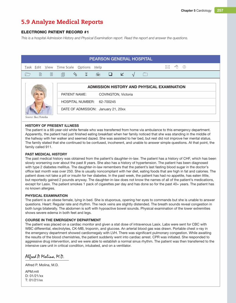
Chapter 5 Cardiology 257
5.9 Analyze Medical ReportseleCtroniC PAtient reCord #1
This is a hospital Admission History and Physical Examination report. Read the report and answer the questions.
Pearson General HosPital
Task Edit View Time Scale Options Help
ADMiSSiON HiSTOry AND PHySiCAL exAMiNATiON
PATienT nAMe: CoVinGTon, Victoria
HosPiTAL nuMBer: 62-700245
DATe oF ADMission: January 21, 20xx
HiSTOry OF PreSeNT iLLNeSSThe patient is a 66-year-old white female who was transferred from home via ambulance to this emergency department. Apparently, the patient had just finished eating breakfast when her family noticed that she was standing in the middle of the hallway with her walker and seemed dazed. she was assisted to her bed, but rest did not improve her mental status. The family stated that she continued to be confused, incoherent, and unable to answer simple questions. At that point, the family called 911.
PAST MeDiCAL HiSTOryThe past medical history was obtained from the patient’s daughter-in-law. The patient has a history of CHF, which has been slowly worsening over about the past 8 years. she also has a history of hypertension. The patient has been diagnosed with type 2 diabetes mellitus. The daughter-in-law remembers that the patient’s last fasting blood sugar in the doctor’s office last month was over 250. she is usually noncompliant with her diet, eating foods that are high in fat and calories. The patient does not take a pill or insulin for her diabetes. in the past week, the patient has had no appetite, has eaten little, but reportedly gained 2 pounds anyway. The daughter-in-law does not know the names of all of the patient’s medications, except for Lasix. The patient smokes 1 pack of cigarettes per day and has done so for the past 40+ years. The patient has no known allergies.
PHySiCAL exAMiNATiONThe patient is an obese female, lying in bed. she is stuporous, opening her eyes to commands but she is unable to answer questions. Heart: regular rate and rhythm. The neck veins are slightly distended. The breath sounds reveal congestion in both lungs bilaterally. The abdomen is soft with hypoactive bowel sounds. Physical examination of the lower extremities shows severe edema in both feet and legs.
COurSe iN THe eMergeNCy DePArTMeNTThe patient was placed on a cardiac monitor and given a stat dose of intravenous Lasix. Labs were sent for CBC with WBC differential, electrolytes, CK-MB, troponin, and glucose. An arterial blood gas was drawn. Portable chest x-ray in the emergency department showed cardiomegaly with LVH. There was significant pulmonary congestion. While awaiting the results of the blood chemistries, the patient suddenly went into cardiac arrest. CPr was initiated. she responded to aggressive drug intervention, and we were able to establish a normal sinus rhythm. The patient was then transferred to the intensive care unit in critical condition, intubated, and on a ventilator.
Alfred P. Molina, M.D.
Alfred P. Molina, M.D.
APM:mttD: 01/21/xxT: 01/21/xx
Source: Iko/Fotolia
M05_TURL8127_04_SE_C05.indd 257 14/12/15 10:49 pm
258 Chapter 5 Cardiology
1. What is the medical abbreviation for hypertension? ______________________
2. The patient has hypertension. If you wanted to use the adjective form of hypertension, you would say, “The patient is ______________________.”
3. What do these abbreviations stand for?
a. CHF _________________________________________________________________________________________________________
b. CK-MB _______________________________________________________________________________________________________
c. CPR _________________________________________________________________________________________________________
d. LVH __________________________________________________________________________________________________________
4. Divide vascular into its two word parts and give the meaning of each word part.
Word Part Meaning
_________________________________ _______________________________________________________________________
_________________________________ _______________________________________________________________________
5. Divide cardiomegaly into its two word parts and give the meaning of each word part.
Word Part Meaning
_________________________________ ______________________________________________________________________
_________________________________ ______________________________________________________________________
6. These medical words were not covered in this chapter, but you need to know their meanings in order to understand this medical report. Research these words and write their definitions.
Word Meaning
bilaterally _________________________________________________________________________________________________
incoherent __________________________________________________________________________________________________
stuporous __________________________________________________________________________________________________
7. What is the normal range of the heart rate in beats per minute for an adult?
_________________________________________________________________________________________________________________
8. Besides hypertension, what other two diagnoses did the patient have before this hospitalization?
a. ______________________________________________________________________________________________________________
b. ______________________________________________________________________________________________________________
9. Resuscitation was used to treat what condition? (Circle one)
cardiomegaly hypertension cardiac arrest diabetes mellitus
troponin portable chest x-ray blood glucose CK-MB intubation
congestive heart failure lack of appetite obesity confusion
10. Circle the two lab tests that were done to check to see if the patient had had a myocardial infarction.
11. The patient had a cardiac arrest. What is the medical word for having no heartbeat?
_________________________________________________________________________________________________________________
12. The severe edema in the patient’s lower extremities reflected backup of blood due to failure of which side of the heart?
_________________________________________________________________________________________________________________
13. The pulmonary congestion seen on the chest x-ray reflected failure of which side of the heart?
_________________________________________________________________________________________________________________
14. Lasix is a diuretic drug that removes fluid from the body by excreting it in the urine. For which of the patient’s medical conditions was this drug prescribed? (Circle one)
15. If the patient ate little food in the past week, why did she gain 2 pounds?
_________________________________________________________________________________________________________________
M05_TURL8127_04_SE_C05.indd 258 14/12/15 10:49 pm
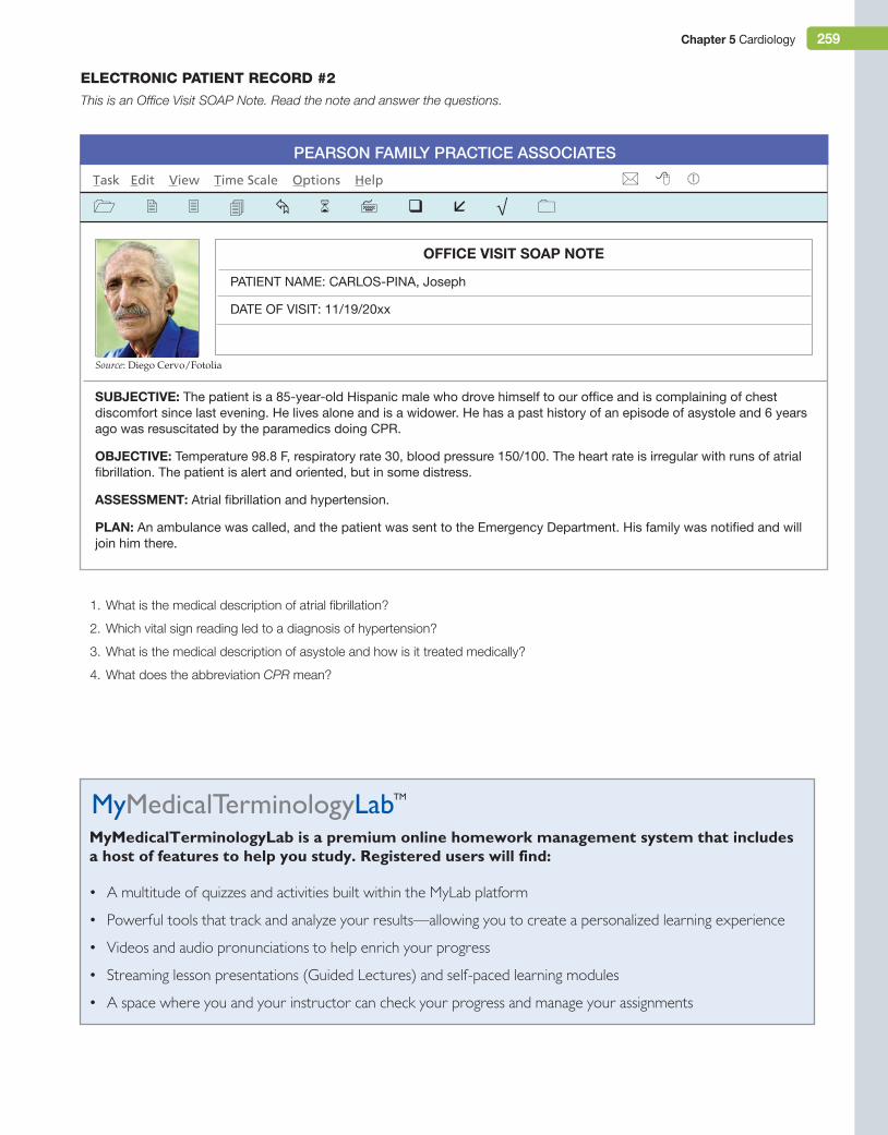
Chapter 5 Cardiology 259
Pearson Family Practice associates
Task Edit View Time Scale Options Help
OFFiCe ViSiT SOAP NOTe
PATienT nAMe: CArLos-PinA, Joseph
DATe oF VisiT: 11/19/20xx
SuBJeCTiVe: The patient is a 85-year-old Hispanic male who drove himself to our office and is complaining of chest discomfort since last evening. He lives alone and is a widower. He has a past history of an episode of asystole and 6 years ago was resuscitated by the paramedics doing CPr.
OBJeCTiVe: Temperature 98.8 F, respiratory rate 30, blood pressure 150/100. The heart rate is irregular with runs of atrial fibrillation. The patient is alert and oriented, but in some distress.
ASSeSSMeNT: Atrial fibrillation and hypertension.
PLAN: An ambulance was called, and the patient was sent to the emergency Department. His family was notified and will join him there.
eleCtroniC PAtient reCord #2
This is an Office Visit SOAP Note. Read the note and answer the questions.
MyMedicalTerminologyLab is a premium online homework management system that includes a host of features to help you study. Registered users will find:
• A multitude of quizzes and activities built within the mylab platform
• Powerful tools that track and analyze your results—allowing you to create a personalized learning experience
• Videos and audio pronunciations to help enrich your progress
• Streaming lesson presentations (Guided lectures) and self-paced learning modules
• A space where you and your instructor can check your progress and manage your assignments
1. What is the medical description of atrial fibrillation?
2. Which vital sign reading led to a diagnosis of hypertension?
3. What is the medical description of asystole and how is it treated medically?
4. What does the abbreviation CPR mean?
Source: Diego Cervo/Fotolia
M05_TURL8127_04_SE_C05.indd 259 14/12/15 10:49 pm


![Assessment Report on Curcuma Longa L. Rhizoma · : Curcuma, Indian saffron, Haridra (Sanskrit, Ayurvedic), Jianghuang (= yellow ginger in Chinese), Kyoo or Ukon (Japanese) [113]](https://img.pdfslide.us/doc/110x75/5ebc915f4571df749b0fad29/assessment-report-on-curcuma-longa-l-rhizoma-curcuma-indian-saffron-haridra.jpg)