Embed Size (px)
Citation preview

why this
matters
NEW! Chapter-opening Why This
Matters videos describe how the
material applies to your future
career. Scan the QR codes to see
brief videos of real health care
professionals discussing how they
use the chapter content every day
in the field. < <<
LEARN WHY THIS MATTERS
NEW! Key concept organization presents the material in manageable chunks and helps you easily navigate the chapter. Each section header states the key concept of that section.
SEE WHERE YOU
The variation in skin tone shown here is primarily due to varying concentrations of the pigment melanin.<
134
Would you be enticed by an ad for a coat that is waterproof, stretchable, washable, and air-conditioned, that automati-cally repairs small cuts, rips, and burns? How about one that’s
guaranteed to last a lifetime? Sounds too good to be true, but you already have such a coat—your skin.
The skin and its derivatives (sweat and oil glands, hairs, and nails) make up a complex set of organs that serves several functions, mostly protective. Together, these organs form the integumentary system (in-teg″u-men′tar-e).
5.1 The skin consists of two layers: the epidermis and dermis
Learning Objective List the two layers of skin and briefly describe subcutaneous
tissue.
The skin receives little respect from its inhabitants, but architectur-ally it is a marvel. It covers the entire body, has a surface area of 1.2 to 2.2 square meters, weighs 4 to 5 kilograms (4–5 kg = 9–11 lb), and accounts for about 7% of total body weight in the average adult. Also called the integument (“covering”), the skin multitasks. Its functions go well beyond serving as a bag for body contents. Pliable yet tough, it takes constant punishment from external agents. Without our skin, we would quickly fall prey to bacteria and perish from water and heat loss.
Varying in thickness from 1.5 to 4.0 millimeters (mm) or more in different parts of the body, the skin is composed of two distinct layers (Figure 5.1):● The epidermis (ep″ĭ-der′mis), composed of epithelial cells, is the outer-
most protective shield of the body (epi = upon).● The underlying dermis, making up the bulk of the skin, is a tough,
leathery layer composed mostly of dense connective tissue.
Only the dermis is vascularized. Nutrients reach the epidermis by diffusing through the tissue fluid from blood vessels in the dermis.
The Integumentary System5
why this
matters
5.1 The skin consists of two layers: the epidermis and dermis 134
5.2 The epidermis is a keratinized stratified squamous epithelium 135
5.3 The dermis consists of papillary and reticular layers 138
5.4 Melanin, carotene, and hemoglobin determine skin color 140
5.5 Hair consists of dead, keratinized cells 141
5.6 Nails are scale-like modifications of the epidermis 144
5.7 Sweat glands help control body temperature, and sebaceous glands secrete sebum 144
5.8 First and foremost, the skin is a barrier 146
5.9 Skin cancer and burns are major challenges to the body 148
Key ConCepTS
M05_MARI6415_10_SE_CH05_134-151.indd 134 30/06/15 3:14 PM
<<<
Overview of Key Concepts
Key Concept section header
Check Your Understanding questions end each section and allow you to assess your understanding of the concept before moving on.
Chapter 5 The Integumentary System 151
5
and accounts for about 7% of total body weight in the average adult. Also called the integument (“covering”), the skin multitasks. Its functions go well beyond serving as a bag for body contents. Pliable yet tough, it takes constant punishment from external agents. Without our skin, we would quickly fall prey to bacteria and perish from water and heat loss.
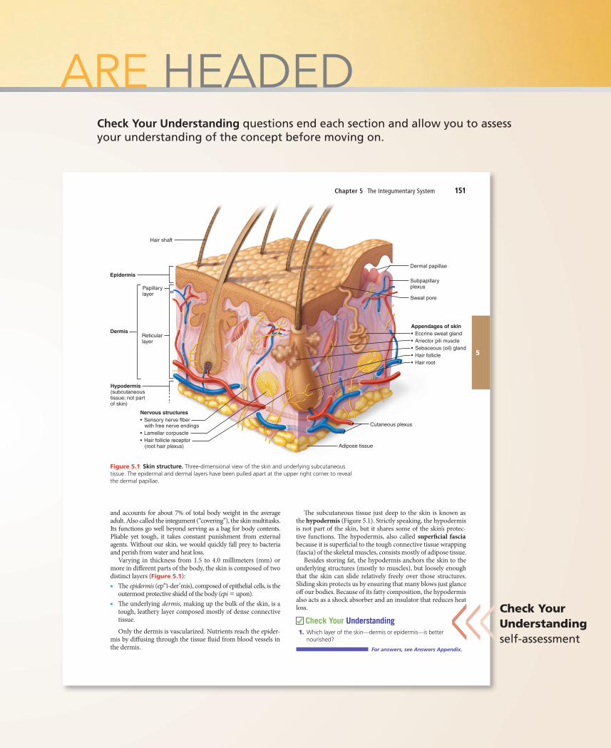
Varying in thickness from 1.5 to 4.0 millimeters (mm) or more in di�erent parts of the body, the skin is composed of two distinct layers (Figure 5.1):● �e epidermis (ep″ĭ-der′mis), composed of epithelial cells, is the
outermost protective shield of the body (epi = upon).● �e underlying dermis, making up the bulk of the skin, is a
tough, leathery layer composed mostly of dense connective tissue.
Only the dermis is vascularized. Nutrients reach the epider-mis by di�using through the tissue �uid from blood vessels in the dermis.
�e subcutaneous tissue just deep to the skin is known as the hypodermis (Figure 5.1). Strictly speaking, the hypodermis is not part of the skin, but it shares some of the skin’s protec-tive functions. �e hypodermis, also called super�cial fascia because it is super�cial to the tough connective tissue wrapping (fascia) of the skeletal muscles, consists mostly of adipose tissue.
Besides storing fat, the hypodermis anchors the skin to the underlying structures (mostly to muscles), but loosely enough that the skin can slide relatively freely over those structures. Sliding skin protects us by ensuring that many blows just glance o� our bodies. Because of its fatty composition, the hypodermis also acts as a shock absorber and an insulator that reduces heat loss.
Check Your Understanding1. Which layer of the skin—dermis or epidermis—is better
nourished?
For answers, see Answers Appendix.
Epidermis
Hair shaft
DermisReticularlayer
Papillarylayer
Hypodermis(subcutaneoustissue; not partof skin)
Dermal papillae
Sweat pore
Subpapillaryplexus
Appendages of skin• Eccrine sweat gland• Arrector pili muscle• Sebaceous (oil) gland• Hair follicle• Hair root
Nervous structures• Sensory nerve fiber with free nerve endings• Lamellar corpuscle• Hair follicle receptor (root hair plexus)
Cutaneous plexus
Adipose tissue
Figure 5.1 Skin structure. Three-dimensional view of the skin and underlying subcutaneous tissue. The epidermal and dermal layers have been pulled apart at the upper right corner to reveal the dermal papillae.
M05_MARI7040_10_SE_CH05_150-172.indd 151 21/07/14 10:04 AM
YOU ARE HEADED
Check Your Understanding self-assessment<<<
464 UNIT 3 Regulation and Integration of the Body
12
Cerebr�e single leading cauaccidents CVAs occur and brain tiblood suppl
�e mosa cerebral athe heart, foof a brain astrokes are
Many wbody (hemior have di�the picture of their lost branches thlost functioto prevent cles due to groups).
Not all reversible (TIAs), are characterizespeech. �etute “red �a
A CVA is blor that dothe coast latstroke is novessels in tneuron-killiwreak the
Experimglutamate, key role in lfunctions.
breakdown re�ects some change in the capillary endothelial cells or their tight junctions.
Check Your Understanding19. What is CSF? Where is it produced? What are its functions?20. A brain surgeon is about to make an incision. Name all the
tissue layers that she cuts through from the skin to the brain.
For answers, see Answers Appendix.
12.9 Brain injuries and disorders have devastating consequences
Learning ObjectivesDescribe the cause (if known) and major signs andsymptoms of cerebrovascular accidents, Alzheimer’s disease, Parkinson’s disease, and Huntington’s disease.List and explain several techniques used to diagnose brain disorders.
Brain dysfunctions are unbelievably varied and extensive. We have mentioned some of them already, but here we will focus on traumatic brain injuries, cerebrovascular accidents, and degen-erative brain disorders.
Traumatic Brain InjuriesHead injuries are a leading cause of accidental death in North America. Consider, for example, what happens if you forget to fasten your seat belt and then rear-end another car. Your head is moving and then stops suddenly as it hits the windshield. Brain damage is caused not only by localized injury at the site of the blow, but also by the ricocheting e�ect as the brain hits the opposite end of the skull.
A concussion is an alteration in brain function, usually tem-porary, following a blow to the head. �e victim may be dizzy or lose consciousness. Although typically mild and short-lived, even a seemingly mild concussion can be damaging, and multi-ple concussions over time produce cumulative damage.
-
CLINICAL
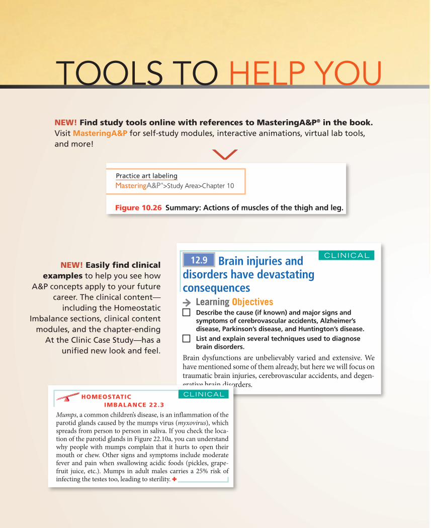
NEW! Find study tools online with references to MasteringA&P® in the book. Visit MasteringA&P for self-study modules, interactive animations, virtual lab tools, and more!
NEW! Easily find clinical examples to help you see how
A&P concepts apply to your future career. The clinical content—
including the Homeostatic Imbalance sections, clinical content
modules, and the chapter-ending At the Clinic Case Study—has a
unified new look and feel.
<Practice art labeling
>Study Area>Chapter 10
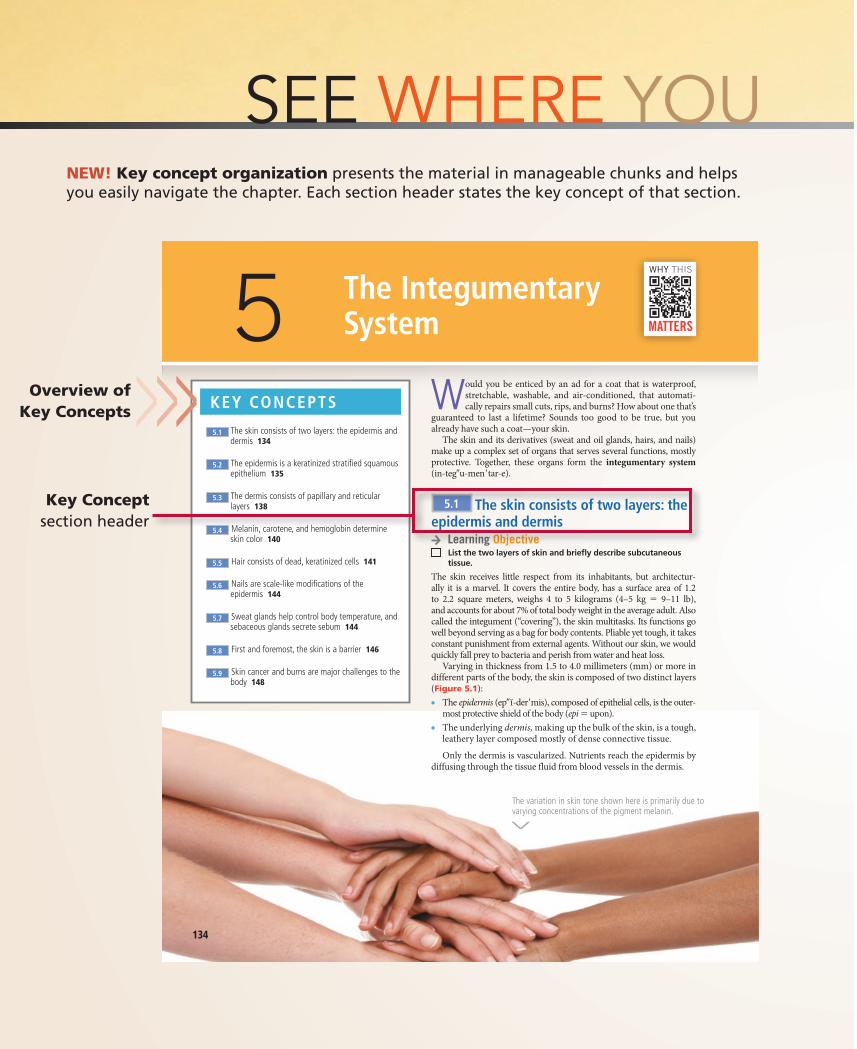
Figure 10.26 Summary: Actions of muscles of the thigh and leg.
Anterior compartment of leg(dorsiflexes foot, extends toes);innervated by deep fibular ner
Lateral compartment of leg(plantar flexes and everts foot);innervated by superficialfibular nerve
Tibialisanterior
Tibia
Fibularismuscles
(b) Muscles of the leg
compartmentmuscles
Anteriorcompartmentmuscles
Medialcompartmentmuscles of thigh and lateralcompartmentmuscles of leg
M10_MARI7040_09_SE_CH10_321-387.indd 383
TOOLS TO HELP YOU Chapter 22 The Digestive System 755
beneath the mucosa of the oral cavity floor and opens at the base of the lingual frenulum (see Figure 22.8b). The small, almond-shaped sublingual gland lies anterior to the subman-dibular gland under the tongue and opens via 10–20 ducts into the floor of the mouth (Figure 22.10a).
The salivary glands are composed of two types of secretory cells: serous and mucous (Figure 22.10b). Serous cells produce a watery secretion containing enzymes, ions, and a tiny bit of mucin, whereas mucous cells produce mucus, a stringy, vis-cous solution. The parotid and submandibular glands contain mostly serous cells. Buccal glands have approximately equal numbers of serous and mucous cells. The sublingual glands contain mostly mucous cells.
Composition of Saliva
Saliva is largely water—97 to 99.5%—and therefore is hypo-osmotic. Its osmolarity depends on the specific glands that are active and the stimulus for salivation. As a rule, saliva is slightly acidic (pH 6.75 to 7.00), but its pH may vary. Its solutes include:● Electrolytes (Na+, K+, Cl−, PO4
3−, and HCO3−)
● The digestive enzymes salivary amylase and lingual lipase (lingual lipase makes only a minor contribution to overall fat digestion)
● The proteins mucin, lysozyme, and IgA● Metabolic wastes (urea and uric acid)
When dissolved in water, the glycoprotein mucin forms thick mucus that lubricates the oral cavity and hydrates foodstuffs.
others) scattered throughout the oral cavity mucosa augment the output slightly.
The major salivary glands are paired compound tubulo-alveolar glands that develop from the oral mucosa and remain connected to it by ducts (Figure 22.10a). The large, roughly triangular parotid gland (pah-rot′id; par = near, oto = the ear) lies anterior to the ear between the masseter muscle and the skin. Its prominent duct parallels the zygomatic arch, pierces the buccinator muscle, and opens into the vestibule next to the second upper molar.
Branches of the facial nerve run through the parotid gland on their way to the muscles of facial expression. For this reason, surgery on this gland can result in facial paralysis.
Homeostatic imbalance 22.3
Mumps, a common children’s disease, is an inflammation of the parotid glands caused by the mumps virus (myxovirus), which spreads from person to person in saliva. If you check the loca-tion of the parotid glands in Figure 22.10a, you can understand why people with mumps complain that it hurts to open their mouth or chew. Other signs and symptoms include moderate fever and pain when swallowing acidic foods (pickles, grape-fruit juice, etc.). Mumps in adult males carries a 25% risk of infecting the testes too, leading to sterility. ✚
About the size of a walnut, the submandibular gland lies along the medial aspect of the mandibular body. Its duct runs
CliniCAl
View histology slides
>Study Area>Figure 22.10 The salivary glands. (a) The parotid, submandibular, and sublingual glands and their ducts. (b) Photomicrograph of the sublingual gland (150×), a mixed salivary gland containing mostly mucous cells (light blue) with a few serous cells (purple).
Teeth
Ducts ofsublingualgland
Sublingualgland
Submandibularduct
Posterior belly ofdigastric muscle
Parotid duct
Masseter muscle
Body of mandible(cut)
Parotid gland
Tongue
Submandibulargland
(a)
Frenulumof tongue
Mylohyoidmuscle (cut)
Anterior belly ofdigastric muscle Mucous cells
(b)
Serous cells
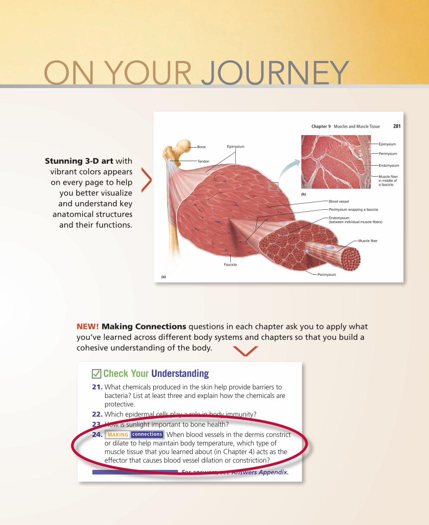
Stunning 3-D art with vibrant colors appears on every page to help
you better visualize and understand key
anatomical structures and their functions.
NEW! Making Connections questions in each chapter ask you to apply what you’ve learned across different body systems and chapters so that you build a cohesive understanding of the body. <
<
Chapter 9 Muscles and Muscle Tissue 281
9
Bone
Perimysium
Endomysium(between individual muscle fibers)
Muscle fiber
Perimysium wrapping a fascicle
Epimysium
Tendon
Epimysium
Muscle fiberin middle of a fascicle
Blood vessel
Perimysium
Endomysium
(a)
Fascicle
(b)
AttachmentsRecall from Chapter 8 that most skeletal muscles span joints and attach to bones (or other structures) in at least two places. When a muscle contracts, the movable bone, the muscle’s inser-tion, moves toward the immovable or less movable bone, the muscle’s origin. In the muscles of the limbs, the origin typically lies proximal to the insertion.
Muscle attachments, whether origin or insertion, may be direct or indirect.● In direct, or �eshy, attachments, the epimysium of the mus-
cle is fused to the periosteum of a bone or perichondrium of a cartilage.
● In indirect attachments, the muscle’s connective tissue wrappings extend beyond the muscle either as a ropelike tendon (Figure 9.1a) or as a sheetlike aponeurosis (ap″o-nu-ro′sis). �e tendon or aponeurosis anchors the muscle to the connective tissue covering of a skeletal element (bone or cartilage) or to the fascia of other muscles.
Indirect attachments are much more common because of their durability and small size. Tendons are mostly tough col-lagen �bers which can withstand the abrasion of rough bony projections that would tear apart the more delicate muscle tis-sues. Because of their relatively small size, more tendons than
Let’s consider these connective tissue sheaths from exter-nal to internal (see Figure 9.1 and the top three rows of Table 9.1).● Epimysium. �e epimysium (ep″ ĭ-mis′e-um; “outside the
muscle”) is an “overcoat” of dense irregular connective tissue that surrounds the whole muscle. Sometimes it blends with the deep fascia that lies between neighboring muscles or the super�cial fascia deep to the skin.
● Perimysium and fascicles. Within each skeletal muscle, the muscle �bers are grouped into fascicles (fas′ĭ-klz; “bundles”) that resemble bundles of sticks. Surrounding each fascicle is a layer of dense irregular connective tissue called perimy-sium (per″ ĭ-mis′e-um; “around the muscle”).
● Endomysium. �e endomysium (en″do-mis′e-um; “within the muscle”) is a wispy sheath of connective tissue that sur-rounds each individual muscle �ber. It consists of �ne areo-lar connective tissue.
As shown in Figure 9.1, all of these connective tissue sheaths are continuous with one another as well as with the tendons that join muscles to bones. When muscle �bers contract, they pull on these sheaths, which transmit the pulling force to the bone to be moved. �e sheaths contribute somewhat to the natural elas-ticity of muscle tissue, and also provide routes for the entry and exit of the blood vessels and nerve �bers that serve the muscle.
Figure 9.1 Connective tissue sheaths of skeletal muscle: epimysium, perimysium, and endomysium. (b) Photomicrograph of a cross section of part of a skeletal muscle (30×). (For a related image, see A Brief Atlas of the Human Body, Plate 29.)
Practice art labeling
>Study Area>Chapter 9
M09_MARI7040_10_SE_CH09_278-320.indd 281 25/07/14 10:15 AM
ovement of the Body
Excretion�e body eliminates limited amounts of nitrogen-containing wastes (ammonia, urea, and uric acid) in sweat, although most such wastes are excreted in urine. Profuse sweating is an impor-tant avenue for water and salt (sodium chloride) loss.
Check Your Understanding21. What chemicals produced in the skin help provide barriers to
bacteria? List at least three and explain how the chemicals are protective.
22. Which epidermal cells play a role in body immunity?23. How is sunlight important to bone health?24. MAKING connections When blood vessels in the dermis constrict
or dilate to help maintain body temperature, which type of muscle tissue that you learned about (in Chapter 4) acts as the effector that causes blood vessel dilation or constriction?
For answers, see Answers Appendix.
5.9 Skin cancer and burns are major challenges to the body
CLINICAL
passive heat loss from 24 discusses body tem-
ous sensory receptors, system. �e cutaneous rs (ek″ster-o-sep′torz)
g outside the body. For (in the dermal papil-
me aware of a caress or , whereas lamellar (also per dermis or hypoder-ving deep pressure. Hair g through our hair and
e endings that meander uli (irritating chemicals, efer detailed discussion
.us receptors mentioned h are found only in skin
re 5.2b.
ON YOUR JOURNEY
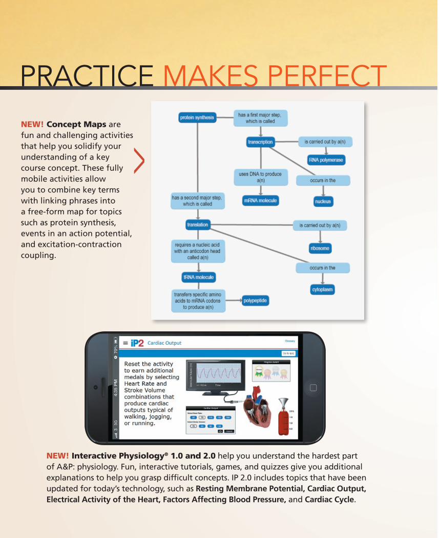
NEW! Concept Maps are fun and challenging activities that help you solidify your understanding of a key course concept. These fully mobile activities allow you to combine key terms with linking phrases into a free-form map for topics such as protein synthesis, events in an action potential, and excitation-contraction coupling.
NEW! Interactive Physiology® 1.0 and 2.0 help you understand the hardest part of A&P: physiology. Fun, interactive tutorials, games, and quizzes give you additional explanations to help you grasp difficult concepts. IP 2.0 includes topics that have been updated for today’s technology, such as Resting Membrane Potential, Cardiac Output, Electrical Activity of the Heart, Factors Affecting Blood Pressure, and Cardiac Cycle.
<
<
PRACTICE MAKES PERFECT
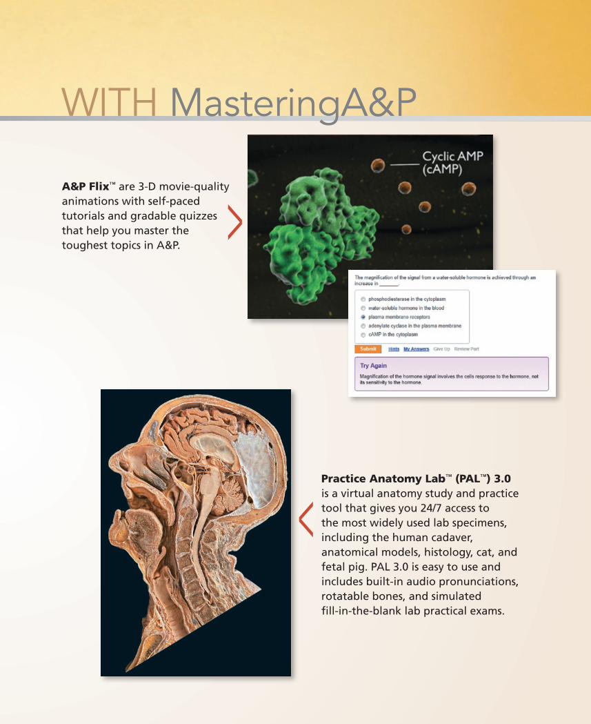
A&P Flix™ are 3-D movie-quality animations with self-paced tutorials and gradable quizzes that help you master the toughest topics in A&P.
Practice Anatomy Lab™ (PAL™) 3.0 is a virtual anatomy study and practice tool that gives you 24/7 access to the most widely used lab specimens, including the human cadaver, anatomical models, histology, cat, and fetal pig. PAL 3.0 is easy to use and includes built-in audio pronunciations, rotatable bones, and simulated fill-in-the-blank lab practical exams.
<<
WITH MasteringA&P
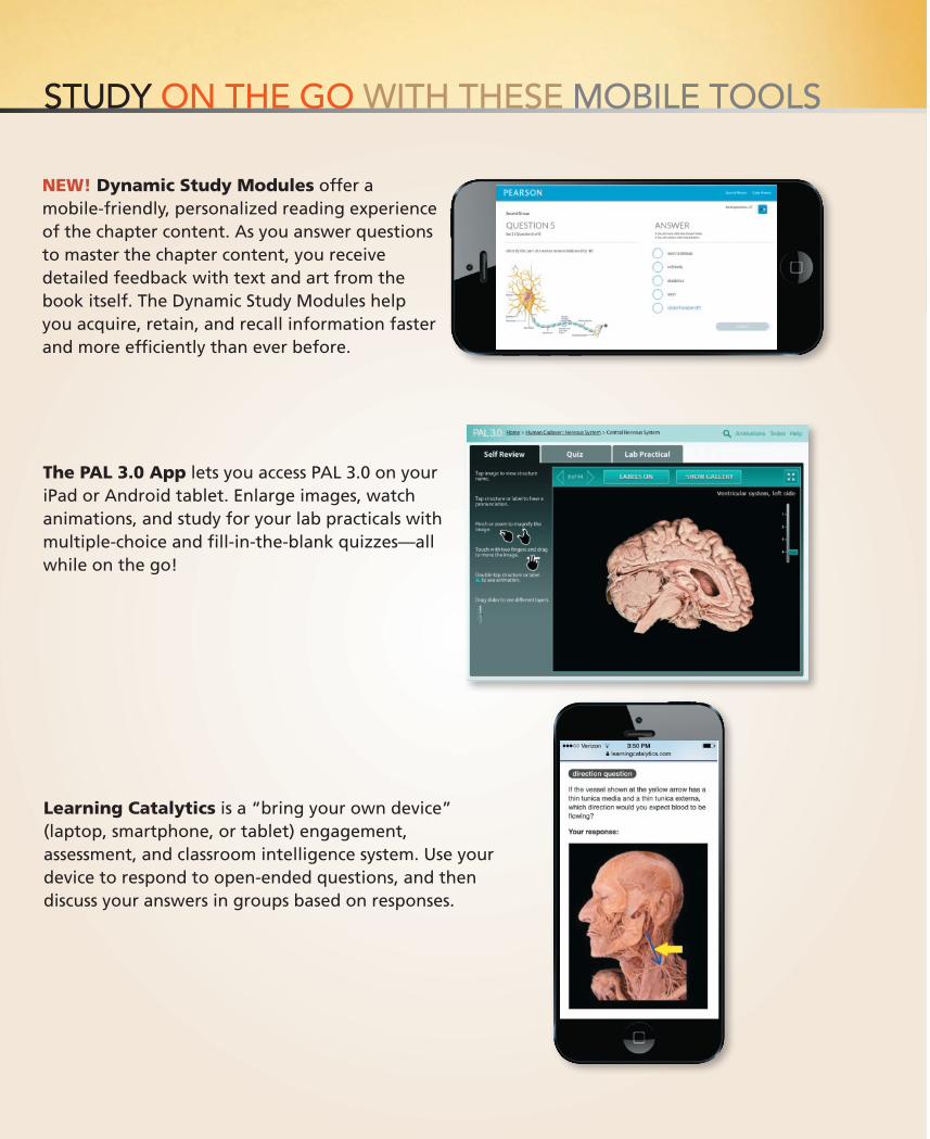
NEW! Dynamic Study Modules offer a mobile-friendly, personalized reading experience of the chapter content. As you answer questions to master the chapter content, you receive detailed feedback with text and art from the book itself. The Dynamic Study Modules help you acquire, retain, and recall information faster and more efficiently than ever before.
The PAL 3.0 App lets you access PAL 3.0 on your iPad or Android tablet. Enlarge images, watch animations, and study for your lab practicals with multiple-choice and fill-in-the-blank quizzes—all while on the go!
Learning Catalytics is a “bring your own device” (laptop, smartphone, or tablet) engagement, assessment, and classroom intelligence system. Use your device to respond to open-ended questions, and then discuss your answers in groups based on responses.
STUDY ON THE GO WITH THESE MOBILE TOOLS
Anatomy & PhysiologySixth Edition
Elaine N. Marieb, R.N., Ph.D.Holyoke Community College
Katja Hoehn, M.D., Ph.D.Mount Royal University
Editor-in-Chief: Serina BeauparlantSr. Acquisitions Editor: Brooke SuchomelProduction and Design Manager: Michele MangelliProgram Manager: Tiffany MokDevelopment Editor: Tanya MartinArt Development Manager: Laura SouthworthArt Development Editors: Laura Southworth, Elisheva MarcusEditorial Assistant: Nicky MontalvoDirector of Development: Barbara YienProgram Management Team Lead: Michael EarlyProject Management Team Lead: Nancy TaborCopyeditor: Anita HueftleCompositor: Cenveo® Publisher Services
Production Coordinator: David NovakArt Coordinator: Jean LakeProofreader: Martha GhentArt Proofreader: Betsy DietrichIndexer: Sallie SteeleCover and Interior Designer: Tandem Creative, Inc.Illustrators: Imagineering STA Media Services Inc.Text and Photo Permissions Management: Maya GomezPhoto Researcher: Kristin PiljaySr. Procurement Specialist: Stacey WeinbergerExec. Marketing Manager: Allison RonaSr. Anatomy & Physiology Specialist: Derek Perrigo
Cover Illustration: The plasma membrane, Imagineering STA Media Services/Precision Graphics
Copyright © 2017, 2014, 2011 Pearson Education, Inc. All Rights Reserved. Printed in the United States of America. This publication is protected by copyright, and permission should be obtained from the publisher prior to any prohibited reproduction, storage in a retrieval system, or transmission in any form or by any means, electronic, mechanical, photocopying, recording, or otherwise. For information regarding permissions, request forms and the appropriate contacts within the Pearson Education Global Rights & Permissions department, please visit www.pearsoned.com/permissions/.
Acknowledgments of third party content appear on pages C-1 and C-2 , which constitutes an extension of this copyright page.
PEARSON, ALWAYS LEARNING, MasteringA&P®, A&PFlixTM, and PALTM are exclusive trademarks in the U.S. and/or other countries owned by Pearson Education, Inc. or its affiliates.
Unless otherwise indicated herein, any third-party trademarks that may appear in this work are the property of their respective owners and any references to third-party trademarks, logos or other trade dress are for demonstrative or descriptive purposes only. Such references are not intended to imply any sponsorship, endorsement, authorization, or promotion of Pearson’s products by the owners of such marks, or any relationship between the owner and Pearson Education, Inc. or its affiliates, authors, licensees or distributors.
Library of Congress Cataloging-in-Publication Data
Marieb, Elaine Nicpon / Hoehn, Katja. Anatomy & physiology / Elaine N. Marieb, R.N., Ph.D. Holyoke CommunityCollege, Katja Hoehn, M.D., Ph.D., Mount Royal University.Anatomy and physiology Sixth edition. / San Francisco : Pearson Education, Inc., [2017] Includes index.LCCN 2015035194 / ISBN 9780134156415LCSH: Human physiology. / Human anatomy.LCC QP34.5 .M454 2017 / DDC 612—dc23LC record available at http://lccn.loc.gov/ 2015035194
ISBN 10: 0-13-415641-2; ISBN 13: 978-0-13-415641-5 (Student Edition)ISBN 10: 0-13-421336-X; ISBN 13: 978-0-13-421336-1 (Instructor’s Review Copy)
1 2 3 4 5 6 7 8 9 10—V357-19 18 17 16 15www.pearsonhighered.com
Elaine N. MariebFor Elaine N. Marieb, taking the student’s perspective into ac-count has always been an integral part of her teaching style. Dr. Marieb began her teaching career at Springfield College, where she taught anatomy and physiology to physical education majors. She then joined the faculty of the Biological Science Division of Holyoke Community College in 1969 after receiv-ing her Ph.D. in zoology from the University of Massachusetts at Amherst. While teaching at Holyoke Community College, where many of her students were pursuing nursing degrees, she developed a desire to better understand the relationship be-tween the scientific study of the human body and the clinical aspects of the nursing practice. To that end, while continuing to teach full time, Dr. Marieb pursued her nursing education, which culminated in a Master of Science degree with a clinical specialization in gerontology from the University of Massachu-setts. It is this experience that has informed the development of the unique perspective and accessibility for which her publica-tions are known.
Dr. Marieb has partnered with Benjamin Cummings for over 30 years. Her first work was Human Anatomy & Physiol-ogy Laboratory Manual (Cat Version), which came out in 1981. In the years since, several other lab manual versions and study guides, as well as the softcover Essentials of Human Anatomy & Physiology textbook, have hit the campus bookstores. This textbook, now in its 10th edition, made its appearance in 1989 and is the latest expression of her commitment to the needs of students studying human anatomy and physiology.
Dr. Marieb has given generously to colleges both near and far to provide opportunities for students to further their edu-cation. She contributes to the New Directions, New Careers Program at Holyoke Community College by funding a staffed drop-in center and by providing several full-tuition scholar-ships each year for women who are returning to college after
a hiatus or attending college for the first time and who would be unable to continue their studies without financial support. She funds the E. N. Marieb Science Research Awards at Mount Holyoke College, which promotes research by undergraduate science majors, and has underwritten renovation and updating of one of the biology labs in Clapp Laboratory at that college. Dr. Marieb also contributes to the University of Massachusetts at Amherst where she generously provided funding for recon-struction and instrumentation of a cutting-edge cytology re-search laboratory. Recognizing the severe national shortage of nursing faculty, she underwrites the Nursing Scholars of the Future Grant Program at the university.
In 1994, Dr. Marieb received the Benefactor Award from the National Council for Resource Development, American Association of Community Colleges, which recognizes her ongoing sponsorship of student scholarships, faculty teaching awards, and other academic contributions to Holyoke Com-munity College. In May 2000, the science building at Holyoke Community College was named in her honor.
Dr. Marieb is an active member of the Human Anatomy and Physiology Society (HAPS) and the American Association for the Advancement of Science (AAAS). Additionally, while actively engaged as an author, Dr. Marieb serves as a consultant for the Benjamin Cummings Interactive Physiology® CD-ROM series.
When not involved in academic pursuits, Dr. Marieb is a world traveler and has vowed to visit every country on this planet. Shorter term, she serves on the scholarship committee of the Women’s Resources Center and on the board of directors of several charitable institutions in Sarasota County. She is an enthusiastic supporter of the local arts and enjoys a competitive match of doubles tennis.
iii
We dedicate this work to our students both present and past, who always inspire us to “push the envelope.”
About the Authors
iv About the Authors
Katja HoehnDr. Katja Hoehn is a professor in the Department of Biology at Mount Royal University in Calgary, Canada. Dr. Hoehn’s first love is teaching. Her teaching excellence has been recognized by several awards during her 21 years at Mount Royal University. These include a PanCanadian Educational Technology Faculty Award (1999), a Teaching Excellence Award from the Students’ Association of Mount Royal (2001), and the Mount Royal Dis-tinguished Faculty Teaching Award (2004).
Dr. Hoehn received her M.D. (with Distinction) from the University of Saskatchewan, and her Ph.D. in Pharma-cology from Dalhousie University. In 1991, the Dalhousie Medical Research Foundation presented her with the Max Forman (Jr.) Prize for excellence in medical research. Dur-ing her Ph.D. and postdoctoral studies, she also pursued her passion for teaching by presenting guest lectures to first- and second-year medical students at Dalhousie University and at the University of Calgary.
Dr. Hoehn has been a contributor to several books and has written numerous research papers in Neuroscience and Phar-macology. She oversaw a recent revision of the Benjamin Cum-mings Interactive Physiology® CD-ROM series modules, and coauthored the newest module, The Immune System.
Following Dr. Marieb’s example, Dr. Hoehn provides fi-nancial support for students in the form of a scholarship that she established in 2006 for nursing students at Mount Royal University.
Dr. Hoehn is also actively involved in the Human Anatomy and Physiology Society (HAPS) and is a member of the Ameri-can Association of Anatomists. When not teaching, she likes to spend time outdoors with her husband and two sons, compete in triathlons, and play Irish flute.
As educators we continually make judgments about the enormous amount of information that besets us daily, so we can choose which morsels to pass on to our students.
Yet even this refined information avalanche challenges the learning student’s mind. What can we do to help students apply
the concepts they are faced with in our classrooms? We believe that this new edition of our textbook addresses that question by building on the strengths of previous editions while using new, innovative ways to help students visualize connections between various concepts.
Unifying ThemesThree unifying themes that have helped to organize and set the tone of this textbook continue to be valid and are retained in this edition. These themes are:
Interrelationships of body organ systems. This theme empha-sizes the fact that nearly all regulatory mechanisms have inter-actions with several organ systems. The respiratory system, for example, cannot carry out its role of gas exchange in the body if there are problems with the cardiovascular system that prevent the normal delivery of blood throughout the body.
Homeostasis. Homeostasis is the normal and most desirable condition of the body. Its loss is always associated with past or present pathology. This theme is not included to emphasize pathological conditions but rather to illustrate what happens in the body when homeostasis is lost.
Whenever students see a red balance beam symbol accompa-nied by an associated clinical topic, their understanding of how the body works to stay in balance is reinforced.
Complementarity of structure and function. This theme en-courages students to understand the structure of some bodily part (cell, bone, lung, etc.) in order to understand the function of that structure. For example, muscle cells can produce move-ment because they are contractile cells.
Changes Past and PresentMany of the changes made to the 5th edition have been retained and are reinforced in this 6th edition.
• There are more step-by-step blue texts accompanying certain pieces of art (blue text refers to the instructor’s voice).
• The many clinical features of the book have been clearly identified to help students understand why this material is important.
• The “Check Your Understanding” questions at the end of each module reinforce understanding throughout the chapter.
• We have improved a number of our Focus Figures. (Focus Figures are illustrations that use a “big picture” layout and dramatic art to walk the student through difficult processes in a step-by-step way.)
• MasteringA&P continues to provide text-integrated media of many types to aid learning. These include Interactive Phys-iology (IP) tutorials that help students to grasp difficult con-cepts, A&PFlix animations that help students visualize tough A&P topics, and the PAL (Practice Anatomy Lab) collection of virtual anatomy study and practice tools focusing on the most widely used lab specimens. These are by no means all of the helpful tools to which students have access. It’s just a smattering.
v
Preface
New to the Sixth EditionSo, besides these tools, what is really new to this textbook this time around? Each chapter’s first page has a “Why This Matters” icon and QR code that links to a video of a health-care profes-sional telling us why the chapter’s content is important for his or her work.
Other new features include (1) declarative headers at the be-ginning of each chapter module so that the student can quickly grasp the “big idea” for that module, (2) more modularization (chunking) of the text so that students can tackle manageable pieces of information as they read through the material, (3) in-creased readability of the text as a result of more bulleted lists and shorter paragraphs, (4) more summary tables to help stu-dents connect information, (5) improvements to many of the figures so that they teach even more effectively, and (6) “Mak-ing Connections” questions in each chapter that ask students to incorporate related information from earlier chapters or earlier modules in the same chapter, helping students to see the forest, not just the trees, as they study.
Chapter-by-Chapter Changes
Chapter 1 The Human Body: An Orientation• Updated Figure 1.8 for better teaching effectiveness.Chapter 2 Chemistry Comes Alive• Updated Figure 2.18 for better teaching effectiveness.Chapter 3 Cells: The Living Units• Updated statistics on Tay-Sachs disease.• Updated information about riboswitches and added infor-
mation about small interfering RNAs (siRNAs).• Added summary text to Figure 3.3 for better pedagogy.• Updated Focus Figure 3.4.Chapter 4 Tissue: The Living Fabric• New photos of simple columnar epithelium, pseudostrati-
fied ciliated columnar epithelium, cardiac muscle tissue, and smooth muscle tissue (Figures 4.3c, d and 4.9b, c).
Chapter 5 The Integumentary System• Added information about the role of tight junctions in skin.• New photo of stretch marks (Figure 5.5).• New photo of cradle cap (seborrhea) in a newborn
(Figure 5.9).• New photo of malignant melanoma (Figure 5.10).Chapter 6 Bones and Skeletal Tissues• Revised Figure 6.9 for improved teaching effectiveness.• New X rays showing Paget’s disease and normal bone
(Figure 6.16).Chapter 7 The Skeleton• Illustrated the skull bone table to facilitate student learning
(Table 7.1).• Added three new Check Your Understanding figure ques-
tions asking students to make anatomical identifications.• New photos of humerus, radius, and ulna (Figures 7.28 and
7.29).
Chapter 8 Joints• Updated statistics for osteoarthritis.• Updated figure showing movements allowed by synovial
joints (Figure 8.5).• New photos of special body movements (Figure 8.6).Chapter 9 Muscles and Muscle Tissue• Updated Table 9.2 information on sizes of skeletal muscle
fiber types in humans.Chapter 10 The Muscular System• New photos showing surface anatomy of muscles used in
seven facial expressions (Figure 10.7).Chapter 11 Fundamentals of the Nervous System and Nervous Tissue• Added overview figure of nervous system (Figure 11.2).• Improved Focus Figure 11.2 (Action Potential) for better stu-
dent understanding.• New image of a motor neuron based on a computerized 3-D
reconstruction of serial sections.• Converted Figure 11.17 to tabular head style to teach better.Chapter 12 The Central Nervous System• Updated mechanisms of Alzheimer’s disease to include
propagation of misfolded proteins.• Updated information about gender differences in the brain.• Streamlined discussion of sleep, memory, and stroke.• New figure to show distribution of gray and white matter
(Figure 12.3).• Functional neuroimaging of the cerebral cortex (Figure 12.6).• Improved reticular formation figure with “author’s voice”
blue text (Figure 12.18).• New figure showing decreased brain activity in Alzheimer’s
(Figure 12.26).Chapter 13 The Peripheral Nervous System and Reflex Activity• Updated description of cytostructure of human cochlear hair
cells (they have no kinocilia).• New data on the number of different odors that humans can
detect.• Reorganized discussion of sound transmission to the inner
ear. New numbered text improves text-art correlation.• New figure teaches the function of the basilar membrane
(Figure 13.26).• New figure on how the hairs on the cochlear hair cells trans-
duce sound (Figure 13.27).• New figure shows the structure and function of the macula
(Figure 13.28).• Updated and expanded description of axon regeneration (in
Figure 13.31).Chapter 14 The Autonomic Nervous System• Improved teaching effectiveness of Figure 14.3 (differences
in the parasympathetic and sympathetic nervous systems).• New summary table for autonomic ganglia (Table 14.2).Chapter 15 The Endocrine System• New information on actions of vitamin D and location of its
receptors.• New summary table showing differences between water-
soluble and lipid-soluble hormones (Table 15.1).
vi Preface
• New summary flowchart shows the signs and symptoms of diabetes mellitus (Figure 15.19).
Chapter 16 Blood• Improved teaching effectiveness of Figure 16.14 (intrinsic
and extrinsic clotting factors).Chapter 17 The Cardiovascular System: The Heart• Rearranged topics in this chapter for better flow.• New section and summary table (Table 17.1) teach key dif-
ferences between skeletal muscle and cardiac muscle.• New Making Connections figure question (students com-
pare three action potentials).• Rearranged material so that all electrical events are presented
in one module.• Added tabular headers, a photo, and bullets to more effec-
tively teach ECG abnormalities (Figure 17.18).• Streamlined figure showing effects of norepinephrine on
heart contractility (Figure 17.22).Chapter 18 The Cardiovascular System: Blood Vessels• New information about pericytes (now known to be stem
cells and generators of scar tissue in the CNS).• New information that the fenestrations in fenestrated capil-
laries are dynamic structures.• Rearranged topics in the physiology section of this chapter
for better flow.• New micrograph of artery and vein (Figure 18.2).• Revised Figure 18.3 (the structure of different types of capil-
laries), putting all of the information in one place.• New figure summarizes the major factors determining mean
arterial pressure to give a “big picture” view (Figure 18.9).• New figure illustrating active hyperemia (Figure 18.15).• Updated Focus Figure 18.1 (Bulk Flow across Capillary Walls).• New Homeostatic Imbalance feature on edema relates it
directly to the preceding Focus Figure 18.1) and incorporates information previously found in Chapter 25.
• New photos of pitting edema (Figure 18.18).Chapter 19 The Lymphatic System and Lymphoid Organs and Tissues• Updated statistics on survival of non-Hodgkin’s lymphoma
patients.• Updated figure to improve teaching of primary and second-
ary lymphoid organs (Figure 19.4).Chapter 20 The Immune System: Innate and Adaptive Body Defenses• Updated information on aging and the immune system, par-
ticularly with respect to chronic inflammation.• Added a new term, pattern recognition receptors, to help
describe how our innate defenses recognize pathogens.• Provided new research results updating the number of genes
in the human genome to about 20,000.Chapter 21 The Respiratory System• New Check Your Understanding question with graphs rein-
forces concepts learned in Focus Figure 21.1 (The Oxygen-Hemoglobin Dissociation Curve).
• New figure illustrating pneumothorax (Figure 21.14).
Chapter 22 The Digestive System• Updated information about the treatment of peptic ulcers.• Updated information about the types and locations of epi-
thelial cells of the small intestine.• New information about roles of our intestinal flora.• Updated hepatitis C treatment to include the new FDA-
approved drug sofosbuvir.• Added discussion of non-alcoholic fatty liver disease.• New information about fecal transplants to treat antibiotic-
associated diarrhea.• Updated figure that compares and contrasts peristalsis
and segmentation (Figure 22.3) for improved teaching effectiveness.
• Updated Figure 22.4 explaining the relationship between the peritoneum and the abdominal organs to improve teaching effectiveness.
• Enteric nervous system section rewritten and rearranged with new figure (Figure 22.6).
• Improved teaching effectiveness of Figure 22.14 (the steps of deglutition).
• Streamlined Figure 22.19 to enhance teaching of regulation of gastric secretion.
• Updated Figure 22.20 (the mechanism of HCl secretion by parietal cells) for improved teaching effectiveness.
• Improved the text flow by moving discussion of the liver, gallbladder, and pancreas before the small intestine.
• Improved teaching effectiveness of Figure 22.28 (mechanism promoting secretion and release of bile and pancreatic juice).
• Updated and revised sections about motility of the small and large intestines.
• Rearranged text to discuss digestion and absorption together for each nutrient. The figures for digestion and absorption of carbohydrates (Figure 22.35) and proteins (Figure 22.36) now parallel each other and appear together for easy comparison.
• Rearranged and rewrote lipid digestion and absorption text and updated Figure 22.37.
Chapter 23 Nutrition, Metabolism, and Energy Balance• Chapter title changed from Nutrition, Metabolism, and Body
Temperature Regulation in order to emphasize the concept of energy balance.
• Updated shape and mechanism of action of ATP synthase to reflect new research findings.
• Updated hypothalamic control of food intake per new research findings.
• Revised Figure 23.4 to enhance the ability of students to compare and contrast the mechanisms of phosphorylation that convert ADP to ATP.
• Revised figure describing ATP synthase structure and func-tion (Figure 23.10).
• Revised Figure 23.13 to help students compare and contrast glycogenesis and glycogenolysis (Figure 23.12).
• Three new figures help students grasp the terms for key pathways in carbohydrate, protein, and fat metabolism (Figures 23.12, 23.14, and 23.18).
Preface vii
Chapter 24 The Urinary System• New cadaver photo of urinary tract organs (Figure 24.2).• New Check Your Understanding question for nephron
labeling.• Improved Focus Figure 24.1 (Medullary Osmotic Gradient)
for better teaching effectiveness.• Added new illustrations to improve teaching effectiveness of
Figure 24.19 (the effects of ADH on the nephron).Chapter 25 Fluid, Electrolyte, and Acid-Base Balance• New Check Your Understanding figure question requires
students to integrate information.Chapter 26 The Reproductive System• Updated screening recommendations for prostate cancer, as
well as updated information on detection and treatment.
• Updated screening guidelines for cervical cancer.• Updated breast cancer statistics.• New Check Your Understanding figure labeling question.• New figure teaches independent assortment (Figure 26.8).• New photo of female pelvic organs (Figure 26.15c).• New photos of mammograms showing normal and cancer-
ous breast tissues (Figure 26.19).• Revised Figure 26.23 to reflect recent research about follicu-
lar development in humans.• Revised section describing the stages of follicle develop-
ment to facilitate student learning and to incorporate recent research.
Appendices• Added a table of the genetic code (Appendix B).
viii Preface
ix
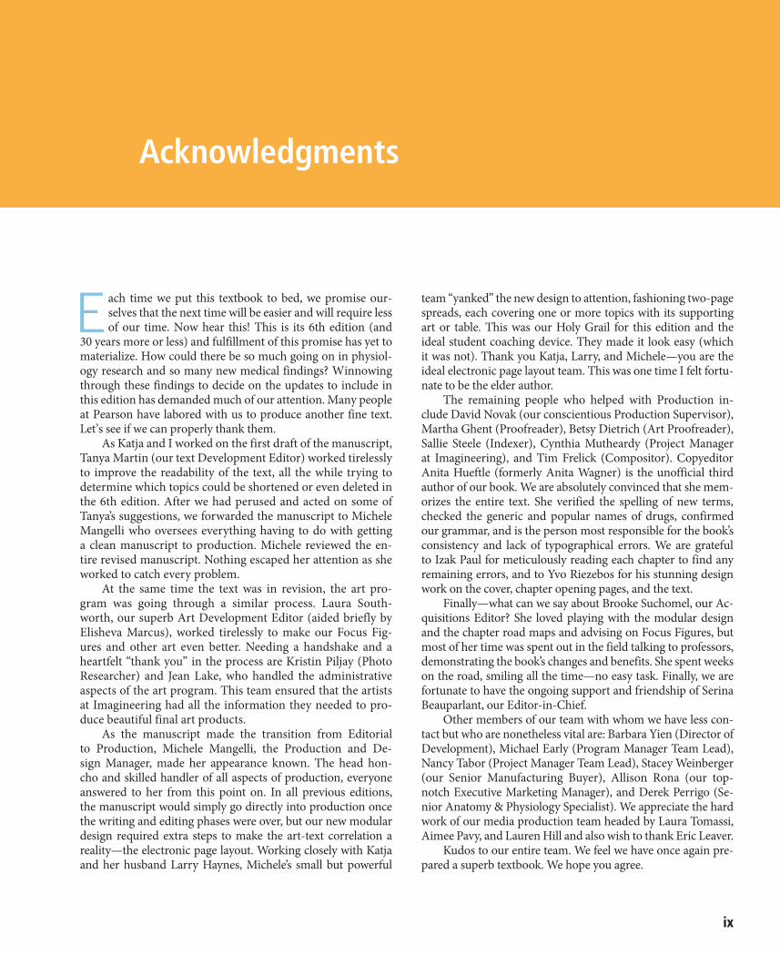
E ach time we put this textbook to bed, we promise our-selves that the next time will be easier and will require less of our time. Now hear this! This is its 6th edition (and
30 years more or less) and fulfillment of this promise has yet to materialize. How could there be so much going on in physiol-ogy research and so many new medical findings? Winnowing through these findings to decide on the updates to include in this edition has demanded much of our attention. Many people at Pearson have labored with us to produce another fine text. Let’s see if we can properly thank them.
As Katja and I worked on the first draft of the manuscript, Tanya Martin (our text Development Editor) worked tirelessly to improve the readability of the text, all the while trying to determine which topics could be shortened or even deleted in the 6th edition. After we had perused and acted on some of Tanya’s suggestions, we forwarded the manuscript to Michele Mangelli who oversees everything having to do with getting a clean manuscript to production. Michele reviewed the en-tire revised manuscript. Nothing escaped her attention as she worked to catch every problem.
At the same time the text was in revision, the art pro-gram was going through a similar process. Laura South-worth, our superb Art Development Editor (aided briefly by Elisheva Marcus), worked tirelessly to make our Focus Fig-ures and other art even better. Needing a handshake and a heartfelt “thank you” in the process are Kristin Piljay (Photo Researcher) and Jean Lake, who handled the administrative aspects of the art program. This team ensured that the artists at Imagineering had all the information they needed to pro-duce beautiful final art products.
As the manuscript made the transition from Editorial to Production, Michele Mangelli, the Production and De-sign Manager, made her appearance known. The head hon-cho and skilled handler of all aspects of production, everyone answered to her from this point on. In all previous editions, the manuscript would simply go directly into production once the writing and editing phases were over, but our new modular design required extra steps to make the art-text correlation a reality—the electronic page layout. Working closely with Katja and her husband Larry Haynes, Michele’s small but powerful
team “yanked” the new design to attention, fashioning two-page spreads, each covering one or more topics with its supporting art or table. This was our Holy Grail for this edition and the ideal student coaching device. They made it look easy (which it was not). Thank you Katja, Larry, and Michele—you are the ideal electronic page layout team. This was one time I felt fortu-nate to be the elder author.
The remaining people who helped with Production in-clude David Novak (our conscientious Production Supervisor), Martha Ghent (Proofreader), Betsy Dietrich (Art Proofreader), Sallie Steele (Indexer), Cynthia Mutheardy (Project Manager at Imagineering), and Tim Frelick (Compositor). Copyeditor Anita Hueftle (formerly Anita Wagner) is the unofficial third author of our book. We are absolutely convinced that she mem-orizes the entire text. She verified the spelling of new terms, checked the generic and popular names of drugs, confirmed our grammar, and is the person most responsible for the book’s consistency and lack of typographical errors. We are grateful to Izak Paul for meticulously reading each chapter to find any remaining errors, and to Yvo Riezebos for his stunning design work on the cover, chapter opening pages, and the text.
Finally—what can we say about Brooke Suchomel, our Ac-quisitions Editor? She loved playing with the modular design and the chapter road maps and advising on Focus Figures, but most of her time was spent out in the field talking to professors, demonstrating the book’s changes and benefits. She spent weeks on the road, smiling all the time—no easy task. Finally, we are fortunate to have the ongoing support and friendship of Serina Beauparlant, our Editor-in-Chief.
Other members of our team with whom we have less con-tact but who are nonetheless vital are: Barbara Yien (Director of Development), Michael Early (Program Manager Team Lead), Nancy Tabor (Project Manager Team Lead), Stacey Weinberger (our Senior Manufacturing Buyer), Allison Rona (our top-notch Executive Marketing Manager), and Derek Perrigo (Se-nior Anatomy & Physiology Specialist). We appreciate the hard work of our media production team headed by Laura Tomassi, Aimee Pavy, and Lauren Hill and also wish to thank Eric Leaver.
Kudos to our entire team. We feel we have once again pre-pared a superb textbook. We hope you agree.
Acknowledgments
There are many people who reviewed parts of this text—both professors and students, either individually or in focus groups, and we would like to thank them. Input from the fol-lowing reviewers has contributed to the continued excellence and accuracy of this text:
Matthew Abbott, Des Moines Area Community CollegeLynne Anderson, Meridian Community CollegeMartin W. Asobayire, Essex Community CollegeYvonne Baptiste-Szymanski, Niagara County Community
CollegeClaudia Barreto, University of New Mexico–ValenciaDiana Bourke, Community College of Allegheny CountySherry Bowen, Indian River State CollegeBeth Braun, Truman CollegeC. Steven Cahill, West Kentucky Community and
Technical CollegeBrandi Childress, Georgia Perimeter CollegeWilliam Michael Clark, Lone Star College–KingwoodTeresa Cowan, Baker College of Auburn HillsDonna Crapanzano, Stony Brook UniversityMaurice M. Culver, Florida State College at JacksonvilleSmruti A. Desai, Lone Star College–CyFairKaren Dunbar Kareiva, Ivy Tech Community CollegeElyce Ervin, University of ToledoMartha Eshleman, Pulaski Technical CollegeJuanita A. Forrester, Chattahoochee Technical CollegeReza Forough, Bellevue CollegeDean Furbish, Wake Technical Community CollegeEmily Getty, Ivy Tech Community CollegeAmy Giesecke, Chattahoochee Technical CollegeAbigail Goosie, Walters State Community CollegeMary Beth Hanlin, Des Moines Area Community CollegeHeidi Hawkins, College of Southern IdahoMartie Heath-Sinclair, Hawkeye Community CollegeNora Hebert, Red Rocks Community CollegeNadia Hedhli, Hudson County Community CollegeD.J. Hennager, Kirkwood Community CollegeShannon K. Hill, Temple CollegeMark Hollier, Georgia Perimeter CollegeH. Rodney Holmes, Waubonsee Community CollegeMark J. Hubley, Prince George’s Community CollegeJason Hunt, Brigham Young University–IdahoWilliam Karkow, University of DubuqueSuzanne Keller, Indian Hills Community CollegeMarta Klesath, North Carolina State UniversityNelson H. Kraus, University of IndianapolisSteven Lewis, Metropolitan Community College–Penn ValleyJerri K. Lindsey, Tarrant County College–NortheastChelsea Loafman, Central Texas College
Paul Luyster, Tarrant County College–SouthAbdallah M. Matari, Hudson County Community CollegeBhavya Mathur, Chattahoochee Technical CollegeTiffany Beth McFalls-Smith, Elizabethtown Community
and Technical CollegeTodd Miller, Hunter College of CUNYRegina Munro, Chandler-Gilbert Community CollegeNecia Nicholas, Calhoun Community CollegeEllen Ott-Reeves, Blinn College–BryanJessica Petersen, Pensacola State CollegeSarah A. Pugh, Shelton State Community CollegeRolando J. Ramirez, The University of AkronTerrence J. Ravine, University of South AlabamaLaura H. Ritt, Burlington County CollegeSusan Rohde, Triton CollegeBrian Sailer, Central New Mexico Community CollegeMark Schmidt, Clark State Community CollegeAmy Skibiel, Auburn UniversityLori Smith, American River CollegeAshley Spring-Beerensson, Eastern Florida State CollegeJustin R. St. Juliana, Ivy Tech Community CollegeLaura Steele, Ivy Tech Community CollegeShirley A. Whitescarver, Bluegrass Community and
Technical CollegePatricia Wilhelm, Johnson and Wales UniversityLuann Wilkinson, Marion Technical CollegePeggie Williamson, Central Texas CollegeMaryJo A. Witz, Monroe Community CollegeJames Robert Yount, Brevard Community College
Interactive Physiology 2.0 ReviewersLynne Anderson, Meridian Community CollegeJ. Gordon Betts, Tyler Junior CollegeMike Brady, Columbia Basin CollegeBetsy Brantley, Valencia CollegeTamyra Carmona, Cosumnes River CollegeAlexander G. Cheroske, Mesa Community College
at Red MountainSondra Dubowsky, McLennan Community CollegePaul Emerick, Monroe Community CollegeBrian D. Feige, Mott Community CollegeJohn E. Fishback, Ozarks Technical Community CollegeAaron Fried, Mohawk Valley Community CollegeJane E. Gavin, University of South DakotaGary Glaser, Genesee Community CollegeMary E. Hanlin, Des Moines Area Community CollegeMark Hubley, Prince George’s Community CollegeWilliam Karkow, University of DubuqueMichael Kielb, Eastern Michigan UniversityPaul Luyster, Tarrant County College–South
x Acknowledgments
Acknowledgments xi
the revision. She also thanks her sons, Eric and Stefan Haynes, who are an inspiration and a joy.
We would really appreciate hearing from you concerning your opinion—suggestions and constructive criticisms—of this text. It is this type of feedback that will help us in the next revi-sion, and underlies the continued improvement of this text.
Elaine N. Marieb
Katja Hoehn
Elaine N. Marieb and Katja HoehnAnatomy and PhysiologyPearson Education1301 Sansome StreetSan Francisco, CA 94111
Louise Millis, North Hennepin Community CollegeJustin Moore, American River CollegeMaria Oehler, Florida State College at JacksonvilleFernando Prince, Laredo Community CollegeTerrence J. Ravine, University of South AlabamaMark Schmidt, Clark State Community CollegeCindy Stanfield, University of South AlabamaLaura Steele, Ivy Tech Community CollegeGeorge A. Steer, Jefferson College of Health SciencesShirley A. Whitescarver, Bluegrass Community and
Technical College
Harvey Howell, my beloved husband and helpmate, died in August of 2013. He is sorely missed.
Katja would also like to acknowledge the support of her colleagues at Mount Royal University (Trevor Day, Sarah Hewitt, Tracy O’Connor, Izak Paul, Michael Pollock, Lorraine Royal, Karen Sheedy, Kartika Tjandra, and Margot Williams) and of Ruth Pickett-Seltner (Chair), Tom MacAlister (Associ-ate Dean), and Jeffrey Goldberg (Dean). Thanks also to Katja’s husband, Dr. Lawrence Haynes, who as a fellow physiologist has provided invaluable assistance to her during the course of
xii
Organization of the BodyUNIT 1
1 The Human Body: An Orientation 1
1.1 Form (anatomy) determines function (physiology) 1
1.2 The body’s organization ranges from atoms to the entire organism 3
1.3 What are the requirements for life? 4
1.4 Homeostasis is maintained by negative feedback 8
1.5 Anatomical terms describe body directions, regions, and planes 11
1.6 Many internal organs lie in membrane-lined body cavities 15
2 Chemistry Comes Alive 19
PART 1 BASIC CHEMISTRY 19
2.1 Matter is the stuff of the universe and energy moves matter 19
2.2 The properties of an element depend on the structure of its atoms 21
2.3 Atoms bound together form molecules; different molecules can make mixtures 24
2.4 The three types of chemical bonds are ionic, covalent, and hydrogen 26
2.5 Chemical reactions occur when electrons are shared, gained, or lost 31
PART 2 BIOCHEMISTRY 34
2.6 Inorganic compounds include water, salts, and many acids and bases 34
2.7 Organic compounds are made by dehydration synthesis and broken down by hydrolysis 37
2.8 Carbohydrates provide an easily used energy source for the body 38
2.9 Lipids insulate body organs, build cell membranes, and provide stored energy 40
2.10 Proteins are the body’s basic structural material and have many vital functions 43
2.11 DNA and RNA store, transmit, and help express genetic information 48
2.12 ATP transfers energy to other compounds 50
3 Cells: The Living Units 54
3.1 Cells are the smallest unit of life 54
PART 1 PLASMA MEMBRANE 57
3.2 The fluid mosaic model depicts the plasma membrane as a double layer of phospholipids with embedded proteins 57
3.3 Passive membrane transport is diffusion of molecules down their concentration gradient 62
3.4 Active membrane transport directly or indirectly uses ATP 67
F O C U S F I g U R E 3 . 1 Primary Active Transport: The Na+-K+ Pump 68
3.5 Selective diffusion establishes the membrane potential 73
3.6 Cell adhesion molecules and membrane receptors allow the cell to interact with its environment 75
F O C U S F I g U R E 3 . 2 G Proteins 76
PART 2 THE CYTOPLASM 77
3.7 Cytoplasmic organelles each perform a specialized task 77
3.8 Cilia and microvilli are two main types of cellular extensions 84
PART 3 NUCLEUS 85
3.9 The nucleus includes the nuclear envelope, the nucleolus, and chromatin 85
Contents
Contents xiii
6 Bones and Skeletal Tissues 152
6.1 Hyaline, elastic, and fibrocartilage help form the skeleton 152
6.2 Bones perform several important functions 154
6.3 Bones are classified by their location and shape 154
6.4 The gross structure of all bones consists of compact bone sandwiching spongy bone 155
6.5 Bones develop either by intramembranous or endochondral ossification 162
6.6 Bone remodeling involves bone deposit and removal 165
6.7 Bone repair involves hematoma and callus formation, and remodeling 168
6.8 Bone disorders result from abnormal bone deposition and resorption 170
7 The Skeleton 174
PART 1 THE AxIAL SkELETON 174
7.1 The skull consists of 8 cranial bones and 14 facial bones 174
7.2 The vertebral column is a flexible, curved support structure 190
7.3 The thoracic cage is the bony structure of the chest 197
PART 2 THE APPENDICULAR SkELETON 201
7.4 Each pectoral girdle consists of a clavicle and a scapula 201
7.5 The upper limb consists of the arm, forearm, and hand 204
7.6 The hip bones attach to the sacrum, forming the pelvic girdle 207
7.7 The lower limb consists of the thigh, leg, and foot 213
8 Joints 220
8.1 Joints are classified into three structural and three functional categories 220
8.2 In fibrous joints, the bones are connected by fibrous tissue 220
8.3 In cartilaginous joints, the bones are connected by cartilage 222
3.10 The cell cycle consists of interphase and a mitotic phase 90
3.11 Messenger RNA carries instructions from DNA for building proteins 92
F O C U S F I g U R E 3 . 3 Mitosis 94
F O C U S F I g U R E 3 . 4 Translation 100
3.12 Apoptosis disposes of unneeded cells; autophagy and proteasomes dispose of unneeded organelles and proteins 103
4 Tissue: The Living Fabric 105
4.1 Tissue samples are fixed, sliced, and stained for microscopy 105
4.2 Epithelial tissue covers body surfaces, lines cavities, and forms glands 106
4.3 Connective tissue is the most abundant and widely distributed tissue in the body 115
4.4 Muscle tissue is responsible for body movement 126
4.5 Nervous tissue is a specialized tissue of the nervous system 128
4.6 The cutaneous membrane is dry; mucous and serous membranes are wet 128
4.7 Tissue repair involves inflammation, organization, and regeneration 131
Covering, Support, and Movement of the BodyUNIT 2
5 The Integumentary System 134
5.1 The skin consists of two layers: the epidermis and dermis 134
5.2 The epidermis is a keratinized stratified squamous epithelium 135
5.3 The dermis consists of papillary and reticular layers 138
5.4 Melanin, carotene, and hemoglobin determine skin color 140
5.5 Hair consists of dead, keratinized cells 141
5.6 Nails are scale-like modifications of the epidermis 144
5.7 Sweat glands help control body temperature, and sebaceous glands secrete sebum 144
5.8 First and foremost, the skin is a barrier 146
5.9 Skin cancer and burns are major challenges to the body 148
xiv Contents
Table 10.4 Muscles of the Neck and Vertebral Column: Head Movements and Trunk Extension 297
Table 10.5 Deep Muscles of the Thorax: Breathing 301
Table 10.6 Muscles of the Abdominal Wall: Trunk Movements and Compression of Abdominal Viscera 303
Table 10.7 Muscles of the Pelvic Floor and Perineum: Support of Abdominopelvic Organs 305
Table 10.8 Superficial Muscles of the Anterior and Posterior Thorax: Movements of the Scapula and Arm 307
Table 10.9 Muscles Crossing the Shoulder Joint: Movements of the Arm (Humerus) 311
Table 10.10 Muscles Crossing the Elbow Joint: Flexion and Extension of the Forearm 314
Table 10.11 Muscles of the Forearm: Movements of the Wrist, Hand, and Fingers 315
Table 10.12 Summary: Actions of Muscles Acting on the Arm, Forearm, and Hand 319
Table 10.13 Intrinsic Muscles of the Hand: Fine Movements of the Fingers 321
Table 10.14 Muscles Crossing the Hip and Knee Joints: Movements of the Thigh and Leg 324
Table 10.15 Muscles of the Leg: Movements of the Ankle and Toes 331
Table 10.16 Intrinsic Muscles of the Foot: Toe Movement and Arch Support 337
Table 10.17 Summary: Actions of Muscles Acting on the Thigh, Leg, and Foot 341
Regulation and Integration of the BodyUNIT 3
11 Fundamentals of the Nervous System and Nervous Tissue 345
11.1 The nervous system receives, integrates, and responds to information 345
11.2 Neuroglia support and maintain neurons 347
11.3 Neurons are the structural units of the nervous system 349
11.4 The resting membrane potential depends on differences in ion concentration and permeability 354
F O C U S F I g U R E 1 1 . 1 Resting Membrane Potential 356
11.5 Graded potentials are brief, short-distance signals within a neuron 358
11.6 Action potentials are brief, long-distance signals within a neuron 359
8.4 Synovial joints have a fluid-filled joint cavity 223
8.5 Five examples illustrate the diversity of synovial joints 229
F O C U S F I g U R E 8 . 1 Synovial Joints 230
8.6 Joints are easily damaged by injury, inflammation, and degeneration 240
9 Muscles and Muscle Tissue 244
9.1 There are three types of muscle tissue 244
9.2 A skeletal muscle is made up of muscle fibers, nerves, blood vessels, and connective tissues 245
9.3 Skeletal muscle fibers contain calcium-regulated molecular motors 248
9.4 Motor neurons stimulate skeletal muscle fibers to contract 253
F O C U S F I g U R E 9 . 1 Events at the Neuromuscular Junction 254
F O C U S F I g U R E 9 . 2 Excitation-Contraction Coupling 258
F O C U S F I g U R E 9 . 3 Cross Bridge Cycle 260
9.5 Wave summation and motor unit recruitment allow smooth, graded skeletal muscle contractions 261
9.6 ATP for muscle contraction is produced aerobically or anaerobically 266
9.7 The force, velocity, and duration of skeletal muscle contractions are determined by a variety of factors 269
9.8 How does skeletal muscle respond to exercise? 272
9.9 Smooth muscle is nonstriated involuntary muscle 273
10 The Muscular System 281
10.1 For any movement, muscles can act in one of three ways 281
10.2 How are skeletal muscles named? 282
10.3 Fascicle arrangements help determine muscle shape and force 282
F O C U S F I g U R E 1 0 . 1 Muscle Action 283
10.4 Muscles acting with bones form lever systems 284
10.5 A muscle’s origin and insertion determine its action 287
Table 10.1 Muscles of the Head, Part I: Facial Expression 290
Table 10.2 Muscles of the Head, Part II: Mastication and Tongue Movement 293
Table 10.3 Muscles of the Anterior Neck and Throat: Swallowing 295
Contents xv
F O C U S F I g U R E 1 1 . 2 Action Potential 360
11.7 Synapses transmit signals between neurons 366
F O C U S F I g U R E 1 1 . 3 Chemical Synapse 368
11.8 Postsynaptic potentials excite or inhibit the receiving neuron 369
11.9 The effect of a neurotransmitter depends on its receptor 373
11.10 Neurons act together, making complex behaviors possible 377
12 The Central Nervous System 382
12.1 Folding during development determines the complex structure of the adult brain 382
12.2 The cerebral hemispheres consist of cortex, white matter, and the basal nuclei 384
12.3 The diencephalon includes the thalamus, hypothalamus, and epithalamus 395
12.4 The brain stem consists of the midbrain, pons, and medulla oblongata 398
12.5 The cerebellum adjusts motor output, ensuring coordination and balance 402
12.6 Functional brain systems span multiple brain structures 404
12.7 The interconnected structures of the brain allow higher mental functions 406
12.8 The brain is protected by bone, meninges, cerebrospinal fluid, and the blood brain barrier 412
12.9 Brain injuries and disorders have devastating consequences 415
12.10 The spinal cord is a reflex center and conduction pathway 418
12.11 Neuronal pathways carry sensory and motor information to and from the brain 423
13 The Peripheral Nervous System and Reflex Activity 431
PART 1 SENSORY RECEPTORS AND SENSATION 432
13.1 Receptors, ascending pathways, and cerebral cortex process sensory information 432
13.2 Sensory receptors are activated by changes in the internal or external environment 434
13.3 The eye converts light energy into electrical signals that are relayed to the brain, allowing us to see 437
13.4 Receptors in the olfactory epithelium and taste buds detect chemicals, allowing us to smell and taste 455
13.5 Inner ear mechanoreceptors enable hearing and balance 459
PART 2 TRANSMISSION LINES: NERVES AND THEIR STRUCTURE AND REPAIR 472
13.6 Nerves are cordlike bundles of axons that conduct sensory and motor impulses 472
13.7 There are 12 pairs of cranial nerves 475
13.8 31 pairs of spinal nerves innervate the body 483
PART 3 MOTOR ENDINgS AND MOTOR ACTIVITY 493
13.9 Peripheral motor endings connect nerves to their effectors 493
13.10 There are three levels of motor control 493
PART 4 REFLEx ACTIVITY 494
13.11 The reflex arc enables rapid and predictable responses 494
13.12 Spinal reflexes are somatic reflexes mediated by the spinal cord 495
F O C U S F I g U R E 1 3 . 1 Stretch Reflex 498
14 The Autonomic Nervous System 504
14.1 The ANS differs from the somatic nervous system in that it can stimulate or inhibit its effectors 504
14.2 The ANS consists of the parasympathetic and sympathetic divisions 506
14.3 Long preganglionic parasympathetic fibers originate in the craniosacral CNS 508
14.4 Short preganglionic sympathetic fibers originate in the thoracolumbar CNS 510
14.5 Visceral reflex arcs have the same five components as somatic reflex arcs 514
14.6 Acetylcholine and norepinephrine are the major ANS neurotransmitters 514
14.7 The parasympathetic and sympathetic divisions usually produce opposite effects 516
14.8 The hypothalamus oversees ANS activity 518
14.9 Most ANS disorders involve abnormalities in smooth muscle control 519
xvi Contents
15 The Endocrine System 521
15.1 The endocrine system is one of the body’s two major control systems 521
15.2 The chemical structure of a hormone determines how it acts 522
15.3 Hormones act through second messengers or by activating specific genes 523
15.4 Three types of stimuli cause hormone release 526
15.5 Cells respond to a hormone if they have a receptor for that hormone 527
15.6 The hypothalamus controls release of hormones from the pituitary gland in two different ways 528
F O C U S F I g U R E 1 5 . 1 Hypothalamus and Pituitary Interactions 530
15.7 The thyroid gland controls metabolism 536
15.8 The parathyroid glands are primary regulators of blood calcium levels 540
15.9 The adrenal glands produce hormones involved in electrolyte balance and the stress response 541
15.10 The pineal gland secretes melatonin 547
15.11 The pancreas, gonads, and most other organs secrete hormones 547
Maintenance of the BodyUNIT 4
16 Blood 554
16.1 The functions of blood are transport, regulation, and protection 554
16.2 Blood consists of plasma and formed elements 555
16.3 Erythrocytes play a crucial role in oxygen and carbon dioxide transport 556
16.4 Leukocytes defend the body 563
16.5 Platelets are cell fragments that help stop bleeding 568
16.6 Hemostasis prevents blood loss 569
16.7 Transfusion can replace lost blood 574
16.8 Blood tests give insights into a patient’s health 577
17 The Cardiovascular System: The Heart 579
17.1 The heart has four chambers and pumps blood through the pulmonary and systemic circuits 579
17.2 Heart valves make blood flow in one direction 586
17.3 Blood flows from atrium to ventricle, and then to either the lungs or the rest of the body 588
F O C U S F I g U R E 1 7 . 1 Blood Flow through the Heart 590
17.4 Intercalated discs connect cardiac muscle fibers into a functional syncytium 591
17.5 Pacemaker cells trigger action potentials throughout the heart 594
17.6 The cardiac cycle describes the mechanical events associated with blood flow through the heart 599
17.7 Stroke volume and heart rate are regulated to alter cardiac output 602
18 The Cardiovascular System: Blood Vessels 608
PART 1 BLOOD VESSEL STRUCTURE AND FUNCTION 608
18.1 Most blood vessel walls have three layers 609
18.2 Arteries are pressure reservoirs, distributing vessels, or resistance vessels 611
18.3 Capillaries are exchange vessels 612
18.4 Veins are blood reservoirs that return blood toward the heart 614
18.5 Anastomoses are special interconnections between blood vessels 615
PART 2 PHYSIOLOgY OF CIRCULATION 615
18.6 Blood flows from high to low pressure against resistance 615
18.7 Blood pressure decreases as blood flows from arteries through capillaries and into veins 617
18.8 Blood pressure is regulated by short- and long-term controls 619
18.9 Intrinsic and extrinsic controls determine blood flow through tissues 626
18.10 Slow blood flow through capillaries promotes diffusion of nutrients and gases, and bulk flow of fluids 630
F O C U S F I g U R E 1 8 . 1 Bulk Flow across Capillary Walls 632
PART 3 CIRCULATORY PATHWAYS: BLOOD VESSELS OF THE BODY 634
18.11 The vessels of the systemic circulation transport blood to all body tissues 635
Table 18.3 Pulmonary and Systemic Circulations 636
Contents xvii
Table 18.4 The Aorta and Major Arteries of the Systemic Circulation 638
Table 18.5 Arteries of the Head and Neck 640
Table 18.6 Arteries of the Upper Limbs and Thorax 642
Table 18.7 Arteries of the Abdomen 644
Table 18.8 Arteries of the Pelvis and Lower Limbs 648
Table 18.9 The Venae Cavae and the Major Veins of the Systemic Circulation 650
Table 18.10 Veins of the Head and Neck 652
Table 18.11 Veins of the Upper Limbs and Thorax 654
Table 18.12 Veins of the Abdomen 656
Table 18.13 Veins of the Pelvis and Lower Limbs 658
19 The Lymphatic System and Lymphoid Organs and Tissues 661
19.1 The lymphatic system includes lymphatic vessels, lymph, and lymph nodes 661
19.2 Lymphoid cells and tissues are found in lymphoid organs and in connective tissue of other organs 664
19.3 Lymph nodes filter lymph and house lymphocytes 666
19.4 The spleen removes bloodborne pathogens and aged red blood cells 667
19.5 MALT guards the body’s entryways against pathogens 668
19.6 T lymphocytes mature in the thymus 669
20 The Immune System: Innate and Adaptive Body Defenses 672
PART 1 INNATE DEFENSES 673
20.1 Surface barriers act as the first line of defense to keep invaders out of the body 673
20.2 Innate internal defenses are cells and chemicals that act as the second line of defense 673
PART 2 ADAPTIVE DEFENSES 680
20.3 Antigens are substances that trigger the body’s adaptive defenses 681
20.4 B and T lymphocytes and antigen-presenting cells are cells of the adaptive immune response 682
20.5 In humoral immunity, antibodies are produced that target extracellular antigens 685
20.6 Cellular immunity consists of T lymphocytes that direct adaptive immunity or attack cellular targets 692
20.7 Insufficient or overactive immune responses create problems 700
21 The Respiratory System 705
PART 1 FUNCTIONAL ANATOMY 705
21.1 The upper respiratory system warms, humidifies, and filters air 707
21.2 The lower respiratory system consists of conducting and respiratory zone structures 710
21.3 Each multilobed lung occupies its own pleural cavity 717
PART 2 RESPIRATORY PHYSIOLOgY 719
21.4 Volume changes cause pressure changes, which cause air to move 719
21.5 Measuring respiratory volumes, capacities, and flow rates helps us assess ventilation 725
21.6 Gases exchange by diffusion between the blood, lungs, and tissues 727
21.7 Oxygen is transported by hemoglobin, and carbon dioxide is transported in three different ways 731
F O C U S F I g U R E 2 1 . 1 The Oxygen-Hemoglobin Dissociation Curve 732
21.8 Respiratory centers in the brain stem control breathing with input from chemoreceptors and higher brain centers 737
21.9 Exercise and high altitude bring about respiratory adjustments 741
21.10 Lung diseases are major causes of disability and death 742
22 The Digestive System 746
PART 1 OVERVIEW OF THE DIgESTIVE SYSTEM 746
22.1 What major processes occur during digestive system activity? 746
22.2 The GI tract has four layers and is usually surrounded by peritoneum 748
22.3 The GI tract has its own nervous system called the enteric nervous system 750
PART 2 FUNCTIONAL ANATOMY OF THE DIgESTIVE SYSTEM 752
22.4 Ingestion occurs only at the mouth 753
xviii Contents
24 The Urinary System 834
24.1 The kidneys have three distinct regions and a rich blood supply 834
24.2 Nephrons are the functional units of the kidney 837
24.3 Overview: Filtration, absorption, and secretion are the key processes of urine formation 843
24.4 Urine formation, step 1: The glomeruli make filtrate 844
24.5 Urine formation, step 2: Most of the filtrate is reabsorbed into the blood 848
24.6 Urine formation, step 3: Certain substances are secreted into the filtrate 853
24.7 The kidneys create and use an osmotic gradient to regulate urine concentration and volume 853
F O C U S F I g U R E 2 4 . 1 Medullary Osmotic Gradient 854
24.8 Renal function is evaluated by analyzing blood and urine 857
24.9 The ureters, bladder, and urethra transport, store, and eliminate urine 860
25 Fluid, Electrolyte, and Acid-Base Balance 866
25.1 Body fluids consist of water and solutes in three main compartments 866
25.2 Both intake and output of water are regulated 869
25.3 Sodium, potassium, calcium, and phosphate levels are tightly regulated 872
25.4 Chemical buffers and respiratory regulation rapidly minimize pH changes 879
25.5 Renal regulation is a long-term mechanism for controlling acid-base balance 881
25.6 Abnormalities of acid-base balance are classified as metabolic or respiratory 884
ContinuityUNIT 5
26 The Reproductive System 888
PART 1 ANATOMY OF THE MALE REPRODUCTIVE SYSTEM 889
26.1 The testes are enclosed and protected by the scrotum 889
22.5 The pharynx and esophagus move food from the mouth to the stomach 758
22.6 The stomach temporarily stores food and begins protein digestion 760
22.7 The liver secretes bile; the pancreas secretes digestive enzymes 771
22.8 The small intestine is the major site for digestion and absorption 777
22.9 The large intestine absorbs water and eliminates feces 781
PART 3 PHYSIOLOgY OF DIgESTION AND ABSORPTION 787
22.10 Digestion hydrolyzes food into nutrients that are absorbed across the gut epithelium 787
22.11 How is each type of nutrient processed? 789
23 Nutrition, Metabolism, and Energy Balance 795
PART 1 NUTRIENTS 795
23.1 Carbohydrates, lipids, and proteins supply energy and are used as building blocks 796
23.2 Most vitamins act as coenzymes; minerals have many roles in the body 799
PART 2 METABOLISM 802
23.3 Metabolism is the sum of all biochemical reactions in the body 802
23.4 Carbohydrate metabolism is the central player in ATP production 805
F O C U S F I g U R E 2 3 . 1 Oxidative Phosphorylation 809
23.5 Lipid metabolism is key for long-term energy storage and release 813
23.6 Amino acids are used to build proteins or for energy 815
23.7 Energy is stored in the absorptive state and released in the postabsorptive state 817
23.8 The liver metabolizes, stores, and detoxifies 822
PART 3 ENERgY BALANCE 825
23.9 Neural and hormonal factors regulate food intake 825
23.10 Thyroxine is the major hormone that controls basal metabolic rate 827
23.11 The hypothalamus acts as the body’s thermostat 828
Contents xix
PART 4 PHYSIOLOgY OF THE FEMALE REPRODUCTIVE SYSTEM 913
26.12 Oogenesis is the sequence of events that leads to the formation of ova 913
26.13 The ovarian cycle consists of the follicular phase and the luteal phase 915
26.14 Female reproductive function is regulated by hypothalamic, anterior pituitary, and ovarian hormones 917
26.15 The female sexual response is more diverse and complex than that of males 921
PART 5 SExUALLY TRANSMITTED INFECTIONS 923
26.16 Sexually transmitted infections cause reproductive and other disorders 923
Appendices
Answers Appendix A-1
A Two Important Metabolic Pathways A-17
B The Genetic Code A-20
Glossary G-1
Photo and Illustration Credits C-1
Index I-1
26.2 The penis is the copulatory organ of the male 891
26.3 Sperm travel from the testes to the body exterior through a system of ducts 893
26.4 The male accessory glands produce the bulk of semen 894
PART 2 PHYSIOLOgY OF THE MALE REPRODUCTIVE SYSTEM 896
26.5 The male sexual response includes erection and ejaculation 896
26.6 Spermatogenesis is the sequence of events that leads to formation of sperm 897
26.7 Male reproductive function is regulated by hypothalamic, anterior pituitary, and testicular hormones 903
PART 3 ANATOMY OF THE FEMALE REPRODUCTIVE SYSTEM 904
26.8 Immature eggs develop in follicles in the ovaries 905
26.9 The female duct system includes the uterine tubes, uterus, and vagina 906
26.10 The external genitalia of the female include those structures that lie external to the vagina 911
26.11 The mammary glands produce milk 911

1 The Human Body: An Orientation 1
2 Chemistry Comes Alive 19
3 Cells: The Living Units 54
4 Tissue: The Living Fabric 105
16 Blood 554
17 The Cardiovascular System: The Heart 579
18 The Cardiovascular System: Blood Vessels 608
19 The Lymphatic System and Lymphoid Organs and Tissues 661
20 The Immune System: Innate and Adaptive Body Defenses 672
21 The Respiratory System 705
22 The Digestive System 746
23 Nutrition, Metabolism, and Energy Balance 795
24 The Urinary System 834
25 Fluid, Electrolyte, and Acid-Base Balance 866
Brief Contents
Organization of the Body UNIT 1
Continuity UNIT 5
Covering, Support, and Movement of the Body UNIT 2
5 The Integumentary System 134
6 Bones and Skeletal Tissues 152
7 The Skeleton 174
8 Joints 220
9 Muscles and Muscle Tissue 244
10 The Muscular System 281
Regulation and Integration of the Body UNIT 3
11 Fundamentals of the Nervous System and Nervous Tissue 345
12 The Central Nervous System 382
13 The Peripheral Nervous System and Reflex Activity 431
14 The Autonomic Nervous System 504
15 The Endocrine System 521
Maintenance of the Body UNIT 4
26 The Reproductive System 888
A00_MARI6415_10_SE_FES.indd 1 14/09/15 9:10 AM
ELAINE MARIEB is the
most trusted name in all of
A&P. More than 3 million
health care professionals
started their careers with
one of Elaine Marieb’s
Anatomy & Physiology texts.
Now, it’s your turn.
A00_MARI6415_10_SE_FES.indd 2 14/09/15 9:10 AM

NEW! eText 2.0 brings your textbook to any web-enabled device. • Now available on smartphones and tablets.
• Seamlessly integrated videos and other rich media.
• Accessible (screen-reader ready).
• Configurable reading settings, including resizable type and night reading mode.
• Instructor and student note-taking, highlighting, bookmarking, and search.
READ ANYTIME, ANYWHERE
A00_MARI6415_10_SE_FES.indd 3 14/09/15 9:10 AM