Embed Size (px)
Citation preview

Graduate Theses, Dissertations, and Problem Reports
2017
Leakage Evaluation of Three Endodontic Sealers Used in a Single-Leakage Evaluation of Three Endodontic Sealers Used in a Single-
cone Technique: A Study Using Bioluminescent Bacteria cone Technique: A Study Using Bioluminescent Bacteria
Sean M. Horan
Follow this and additional works at: https://researchrepository.wvu.edu/etd
Recommended Citation Recommended Citation Horan, Sean M., "Leakage Evaluation of Three Endodontic Sealers Used in a Single-cone Technique: A Study Using Bioluminescent Bacteria" (2017). Graduate Theses, Dissertations, and Problem Reports. 5822. https://researchrepository.wvu.edu/etd/5822
This Thesis is protected by copyright and/or related rights. It has been brought to you by the The Research Repository @ WVU with permission from the rights-holder(s). You are free to use this Thesis in any way that is permitted by the copyright and related rights legislation that applies to your use. For other uses you must obtain permission from the rights-holder(s) directly, unless additional rights are indicated by a Creative Commons license in the record and/ or on the work itself. This Thesis has been accepted for inclusion in WVU Graduate Theses, Dissertations, and Problem Reports collection by an authorized administrator of The Research Repository @ WVU. For more information, please contact [email protected].

Leakage Evaluation of Three Endodontic Sealers Used in a Single-cone Technique: A Study Using Bioluminescent Bacteria
Sean M. Horan, D.D.S.
Thesis submitted to the School of Dentistry at West Virginia University In partial fulfillment of the requirements for the degree of
Master of Science In
Endodontics
Elizabeth C. Kao, D.M.D., Chair L. Keith Hildebrand, D.D.S., M.S.
Bryan Weaver, D.D.S., M.D. Jerry E. Bouquot, D.D.S., M.S.D.
Department of Endodontics
Morgantown, West Virginia 2017
Keywords: Dental, Endodontics, Single-Cone Obturation, Coronal Leakage, Sealer, Bioluminescent Bacteria
Copyright 2017 Sean M. Horan, D.D.S.

Abstract
Leakage Evaluation of Three Endodontic Sealers Used in a Single-cone Technique: A Study Using Bioluminescent Bacteria
Sean M. Horan, D.D.S.
Introduction
After proper chemomechanical disinfection and obturation of a root canal system, the next important goal in endodontic therapy is obtaining an adequate coronal seal. If the coronal seal becomes compromised, it is difficult to decide whether the root canal system should be re-treated. This decision should be made based on the ability of the root canal filling to resist bacterial contamination and on the length of time the seal has been compromised. The root canal system should be filled in three dimensions, typically with a combination of gutta percha and sealer. One obturation method that has seen a revival in recent years is the single-cone technique. Newly developed sealers claim to create an effective seal through adhesive bonding to the gutta percha and/or dentin. The objective of this study was to evaluate the sealing ability of three currently available endodontic sealers used in a single-cone technique with a bacterial leakage model. A pilot study was also conducted in an attempt to better quantify and localize the bacteria in the obturated experimental teeth. Materials and Methods Fifty extracted human maxillary anterior teeth were decoronated and root length was standardized. Each tooth was instrumented and irrigated. The teeth were randomly divided into three experimental groups (n = 12) and two control groups (5 positive and 5 negative). The final four teeth were used in the pilot study. Obturation of the three experimental groups, EndoSequence BC Sealer, ThermaSeal Plus Ribbon Sealer, or GuttaFlow 2, was performed with a single-cone technique using a ProTaper master cone. A bacterial leakage experimental apparatus was constructed to evaluate the seal created by each of the experimental groups. For the pilot study, four teeth were obturated with varying levels of gutta percha and evaluated in the same experimental apparatus. A solution of a bioluminescent bacterium, Staphylococcus aureus Xen36, was used to inoculate each tooth. Scans of the teeth were taken with an In Vivo Imaging System (IVIS) at Days 0, 3, 7, and 10 to show whether bacteria could be detected at varying levels in the teeth. The remaining forty-six apparatuses were evaluated with a more traditional bacterial leakage method, where Difco Purple Broth Base was placed in the lower chamber of the apparatus so that 2 mm of the root end was submerged. A solution of S. aureus Xen36 was used to inoculate the top chamber of the apparatus, and the apparatuses were incubated for up to 50 days. The apparatuses were checked daily to monitor for bacterial contamination. Results and Conclusion The pilot study was unable to provide accurate information on the localization and quantification of bacteria in the four sample teeth. For the traditional bacterial leakage study,

each of the experimental groups had a noticeable amount of early leakage, and after one week, all three groups had a survival rate of 58.3%. After 50 days, the ThermaSeal Plus Ribbon Sealer group had a survival rate of 8.3%, while the EndoSequence BC Sealer and GuttaFlow 2 groups had a survival rate of 25.0%. The log-rank test showed no statistically significant differences between the three groups with respect to leakage over time (p = 0.665). The results of the present study showed that in the absence of a coronal seal, the single-cone method of root canal obturation does not provide long-term sealing ability in many teeth. Further studies should be piloted to see if the IVIS could be beneficial for analyzing obturated teeth.
iv
Dedication
To my wife, MacKenzie: Thank you for all of your love and support. I feel fortunate that we have been able to go through our residencies at the same time. It has been stressful at times (we survived BioStatistics), but I would not have wanted it any other way. I can’t wait to see what’s in store for the next chapter of our lives. I love you. To my parents, Tom and Mary Ann: Thank you for always pushing me to do my best and for encouraging me to pursue my interests and dreams. I would not be the person I am today without both of you. To my in-laws, Glenn and Laura: Thank you for the constant support and advice you provide MacKenzie and me. I feel very lucky to be able to rely on your years of dental knowledge as we begin the transition into our professions.

v
Acknowledgements
AAE Foundation/Dentsply – Thank you for your generosity in awarding the West Virginia University School of Dentistry Endodontic Department an Innovation in Research Grant (Fund #2R388) which helped fund this research project. Dr. M. Leigh Speer – Thank you for your mentorship over the past two and a half years. Your vast experience has been extremely helpful in developing my clinical skills as an endodontist. Dr. L. Keith Hildebrand – Thank you for having an open mind and for being a great advocate for our program. Your experience has helped us out immensely. Dr. A. Tom Borgia – I would not have begun this journey to become an endodontist if it weren’t for you. Your enthusiasm and teaching abilities helped spark my interests as a student. Thank you for encouraging me to apply and for helping along the way. Dr. Elizabeth Kao – Thank you for serving as chair of my committee. Your background with dental research has provided suggestions and recommendations to help me through the process. Dr. Bryan Weaver – Thank you for being a last-minute addition to my committee! I am grateful for what you have taught us regarding the management of medically compromised patients through your advanced oral surgery class and through clinical consultations. Dr. Jerry Bouquot – Thank you for your guidance with the research process. I have always enjoyed our conversations and appreciated your insight. Mr. Chris Waters – Thank you for all of your assistance and advice with my project. Your help with the research process has been greatly appreciated. Thanks for the help with the protocol for the bacterial portion of my project and for letting me borrow some tools to make my apparatuses! Ms. Sarah McLaughlin and Dr. Karen Martin – Thank you for the training and advice with the IVIS imaging part of my study. Mr. Taron Dick and West Virginia Clinical and Translational Science Institute (WVCTSI) – Thank you for all of your help with the statistics. Dr. Jabeen Noore and WVU Orthopaedics – Thank you for providing the bacteria for my project and instructions for growth and preparation. Mr. Damon Linear – Thank you for your help and accommodations with the sterilization of my project apparatuses. Dr. Stephen Fragale – You have been the best co-resident I could’ve asked for. It has been great being able to bounce ideas off each other to help each other become better clinicians. We have endured a lot of change over the past 2.5 years, but it has helped having a fellow West Virginian
vi
and WVU Dental grad alongside. I look forward to future endo meetings and golfing adventures. I’m happy you will finally get to be with Amanda full-time in Charleston! Dr. Emma Hunter – I was amazed how you were able to manage three kids and exercise consistently during all the stresses of residency. I appreciated your advice while I was a first year and was glad you were able to have thick skin at times being the only female in the endo resident room! Dr. Wisam Sakr – Thank you for your encouragement while I was a first year. I continue to be on the lookout for three-canal premolars because of your case presentations! Dr. Michael Tran – I have appreciated learning some new tricks from you. Your dedication to strive for the best has been great to see. Don’t let those micro accesses prevent you from finding MB2! I look forward to eventually hitting the links with you when I find myself in Cali! Dr. Travis Moore – You have progressed a lot as a clinician in the past year. I admire your ability to be adventurous with trying new materials and techniques. Good luck with your final year and beyond! Dr. Mehran Malakpour – I have never seen someone so dedicated and interested in endo from such an early point in his or her dental career. It has been enjoyable seeing you transition from student to endo resident! Dr. Cade Brawley – Thanks for bringing some Texas flair to the program! I have enjoyed talking about endo cases and football with you. I look forward to tailgating with you in Lubbock sometime once we’re all out practicing!

vii
Table of Contents
Abstract..........................................................................................................................................ii
Dedications.....................................................................................................................................iv
Acknowledgements..........................................................................................................................v
Table of Contents...........................................................................................................................vii
List of Tables................................................................................................................................viii
List of Figures.................................................................................................................................ix
List of Symbols, Abbreviation, or Nomenclature………………………………………………...xi
Chapter 1 Introduction......................................................................................................................................1 Statement of the Problem.................................................................................................................2 Significance of the Problem.............................................................................................................2 Null Hypothesis…………………………………………………………………………………...2 Assumptions.....................................................................................................................................3 Limitations.......................................................................................................................................3 Delimitations....................................................................................................................................3
Chapter 2 Review of Literature ......................................................................................................................5
Chapter 3 Materials and Methods...................................................................................................................14 Statistical Analysis.........................................................................................................................28
Chapter 4 Results............................................................................................................................................29 Discussion......................................................................................................................................32
Chapter 5 Conclusions....................................................................................................................................37
Works Cited..................................................................................................................................38
Appendix Data Collection for Statistical Analysis………………………………………………………….44

viii
List of Tables
Table 1: Survival rates of the three experimental groups at different observation times………..31

ix
List of Figures
Figure 1: Selection of various brands of greater taper gutta percha………………………………6
Figure 2: EndoSequence BC Sealer……………………………………………………………….8
Figure 3: ThermaSeal Plus Ribbon Sealer………………………………………………………...9
Figure 4: GuttaFlow 2……………………………………………………………………………10
Figure 5: Detection limits of the bioluminescent bacteria used in Sedgley’s study……………..12
Figure 6: Second sub-culture of Staphylococcus aureus Xen36 used to create stock solutions…16
Figure 7: 15 ml microcentrifuge tubes labeled with each of the stock solutions………………...16
Figure 8: LB broth used for bacterial growth in stock solutions………………………………...17
Figure 9: Machine used to vortex stock bacterial solutions……………………………………...17
Figure 10: Densitometer used to perform McFarland test on stock bacterial solutions…………18
Figure 11: L-Spreaders used to evenly distribute bacterial solution on BAP…………………....19
Figure 12: Sample BAP with even distribution of bacterial growth……………………………..19
Figure 13: Sample of decoronated maxillary anterior teeth (side and incisal views)……………21
Figure 14: Dental operating microscope used for instrumentation and obturation of samples….21
Figure 15: Sequence of hand files and ProTaper Universal rotary files used……………………22
Figure 16: Irrigating solutions and sample irrigation syringe and tip used……………………...22
Figure 17: Final ProTaper Universal file used and corresponding gutta percha points………….23
Figure 18: Elements Free downpack unit………………………………………………………..24
Figure 19: In Vivo Imaging System (IVIS) linked to data-acquisition computer……………….26
Figure 20: Difco Purple Broth Base with 1% D-Fructose carbohydrate………………………...27
Figure 21: Sample apparatuses…………………………………………………………………..27
Figure 22: Storage of samples, stock solutions, and BAPs in incubator at 37°C………………..28

x
Figure 23: Bioluminescence produced by S. aureus Xen36 at 7 days…………………………...29
Figure 24: Bioluminescence produced by S. aureus Xen36 at 10 days………………………….30
Figure 25: Survival rates over the 50-day observation period…………………………………...31

xi
List of Symbols, Abbreviation, or Nomenclature
Blood Agar Plate – BAP – a Petri dish containing mammalian blood used to culture
microorganisms.
Colony forming units per milliliter – Cfu/ml – a unit used to estimate the number of viable
bacterial cells in a sample solution.
In Vivo Imaging System – IVIS – an optical imaging device that uses filters and algorithms to
recognize bioluminescent and fluorescent reporters in bacteria.
Luria-Bertani broth – LB broth – a nutritionally rich medium used for bacterial growth.
McFarland – a set of standards used as a reference of the turbidity of a bacterial suspension. A
densitometer is used to test the McFarland rating of the suspension.
Mineral Trioxide Aggregate – MTA – a calcium silicate dental material often used for root
repair.
Root Canal System – RCS – the space containing the dental pulp inside the crown and root of a
tooth.
Vortex – a device which produces a spinning, often turbulent, flow of liquid.
Working Length – WL – the distance from a coronal reference point to the point at which canal
preparation and obturation should terminate.

1
Chapter 1
Introduction
Obtaining an adequate coronal seal is of utmost importance following completion of root
canal therapy.1-3 However, if the coronal seal becomes compromised, re-contamination of the
obturated root canal system (RCS) can occur. The seal can become compromised if there is an
extensive delay in the placement of definitive restorations, if definitive or provisional
restorations lack marginal integrity, if the tooth becomes fractured, or if recurrent decay develops
and exposes the endodontic filling material.4, 5 If one of these instances occurs and the coronal
portion of endodontic filling is exposed to oral bacteria, it is often difficult to decide the best plan
of action. The decision to retreat the RCS should be based on the length of time it takes the
entire length of the RCS to become contaminated after loss of coronal seal. Bacterial leakage
has been supported as the most clinically relevant model for evaluation of the seal provided by
endodontic filling materials.6
To better resist re-contamination, the RCS should optimally be filled in three dimensions
after proper debridement.7 Gutta percha is the most widely used root canal filling, and many
methods of its utilization have been employed. One method that has seen a revival in recent
years is the single-cone technique.8 In the past, this technique has been thought to be less
effective in sealing than cold lateral condensation or warm vertical condensation techniques;
however, the introduction of new sealers and greater taper master cones may support the use of
the single-cone technique.
In this study, three contemporary endodontic sealers that have the ability to be used in a
single-cone technique were analyzed in a bacterial leakage model to determine the length of time
required for full contamination of the RCS. Additionally, a pilot study utilizing an In Vivo

2
Imaging System (IVIS) was conducted in an attempt to better localize and quantify the bacteria
in the sample teeth.
Statement of the Problem
Can newly developed sealers provide resistance or delay full bacterial re-contamination
in root canal systems obturated with the single-cone technique?
Significance of the Problem
With the advent of new endodontic sealers, many dental schools are beginning to change
their endodontic teaching philosophy. Some schools are starting to exclusively teach single-cone
methods of obturation to their undergraduate dental students and post-graduate endodontic
residents. Although this appears to simplify the process of endodontic obturation, students may
not understand its indications and limitations. As the single-cone technique becomes
increasingly popular and prevalent, practitioners should have some guidelines for the
management of a lost coronal seal. This study will provide information on the sealing ability of
new sealers recommended for use in the single-cone technique and offer some guidelines for
treatment in the event where the coronal seal has been lost.
Null Hypothesis
There is no significant difference in bacterial leakage over time for the three endodontic
sealers tested: EndoSequence BC Sealer (Brasseler USA, Savannah, GA), ThermaSeal Plus
Ribbon Sealer (Dentsply Tulsa Dental Specialties, Tulsa, OK), and GuttaFlow 2
(Coltene/Whaledent Inc., Cuyahoga Falls, OH).

3
Assumptions
1. A bacterial leakage model is a reliable, scientific way to assess the seal of a root canal
filling.
2. When the color of the broth in the experimental apparatuses changes from purple to
yellow, full bacterial contamination of the RCS has occurred.
Limitations
1. The canals of the experimental teeth have varying degrees of irregularity in their internal
anatomy.
2. The oral flora that can re-contaminate a RCS are extremely diverse, but only a single
species was used in this study.
3. This is an in vitro experiment that does not fully capture the in vivo environment and
reaction process.
4. Human error may have been involved in the techniques.
5. Sample size of prepared and obturated teeth is small.
Delimitations
1. An effort was made to limit the teeth to maxillary anteriors with straight roots and canals
with relatively round cross-sections.
2. Only roots with completely formed apices and no visible caries/resorption were selected.
3. Crowns were sectioned and the roots were standardized to an approximate length of 16
mm.

4
4. Teeth where no cutting was evident on the tip of the F4 file (large apical diameter canals)
were excluded.
5. Gutta percha was removed to a level that allowed approximately 14 mm to remain, taking
working length (WL) into account.
6. A dental operating microscope (Global Surgical Corporation, St. Louis, MO) was used at
6.4x magnification for WL determination, instrumentation, and obturation.
7. All samples were prepared by a single operator (the principal investigator).

5
Chapter 2
Review of Literature
The primary cause of apical periodontitis is the presence of microorganisms in a RCS.9, 10
Proper endodontic treatment involves chemomechanical methods for the reduction of the
microbial load in this system.11 Mechanical debridement by hand and rotary instruments
provides shape in the RCS. Common endodontic irrigants such as sodium hypochlorite (NaOCl)
and ethylenediaminetetraacetic acid (EDTA) can then be used to penetrate the areas of the RCS
that are mechanically inaccessible.12
After disinfection, the next goal of endodontic treatment is three-dimensionally filling
this cleaned and shaped system to prevent re-infection.7,13 Wu and Wesselink stated, “The
development and maintenance of a hermetic seal is considered to be a major prerequisite for
success in root canal treatment”.14 Numerous techniques and materials have been accepted as
ways of achieving this “hermetic seal.” These techniques include sealer only, lateral
compaction, warm vertical compaction, single-cone, and carrier-based techniques.15
In sealer only techniques, a cement or paste is placed to fill the entire RCS without the
use of a gutta percha master cone. With lateral compaction, a master cone is selected based on
the final file used and this cone is coated with a sealer and seated at the WL. A spreader is then
introduced alongside to compress the master cone laterally. After removal of the spreader,
accessory cones of gutta percha are placed to fill the void created by the spreader. This process
is continued until a dense fill is achieved. For warm vertical compaction, a master cone is also
selected and placed just as it would be with the lateral compaction technique. However, instead
of adding accessory cones, the coronal portion of the master cone is melted with a heat source
and the apical 3-5 mm of remaining gutta percha is condensed with pluggers. The space above

6
this apical “plug” is then backfilled with warm injectable gutta percha. Carrier-based techniques
utilize a plastic or harder gutta percha carrier that is coated in a softer gutta percha. After a light
coating of sealer on the canal walls, an oven is used to soften the outer layer of gutta percha on
the carrier. When soft, the carrier is placed in the canal to the WL. For the single-cone
technique, sealer is used to coat the walls of the RCS, and a single master cone of gutta percha
corresponding to the final file size is coated in the sealer and placed to WL.
Operators often debate these various techniques, but there is still little evidence to support
or deny a particular technique. Whitworth stated, “As no single approach can unequivocally
boast superior evidence of healing success, decisions may be based on such factors as speed,
simplicity, economics, or ‘how it feels in my hands’”.16 Previously, single-cone techniques
performed with conventional sealers were thought to be less effective than other methods of
condensation.17 However, the introduction of new sealers and greater taper master cones have
provided a revival of the single-cone technique.
Greater taper master cones of gutta percha have been created by different companies to
closely match the geometry of their nickel-titanium rotary instrumentation systems.18
Figure 1: Selection of various brands of greater taper gutta percha. Some of these gutta percha cones have a variable taper (ProTaper), while others have standardized .04 or .06 tapers.

7
This greatly reduces the space that sealer needs to occupy between the master cone and canal
walls. However, root canal systems are not perfect cylinders, so the sealer used in conjunction
with the master cone must have properties that enable sealing of the canal irregularities that
could potentially harbor bacteria. New root canal sealers on the market claim to be able to create
an adhesive bond along the sealer – gutta percha interface and/or along the sealer – dentin
interface, reinforcing the viability of single-cone applications.19
Sagsen et al. said that there are certain characteristics that an ideal filling material must
possess; these include adequate marginal sealing quality, biocompatibility, and the ability to
allow or induce bone repair and antimicrobial activity.20 Since sealers may be placed in close
contact with the periapical tissues, it is possible that the tissue response to the sealer could
influence the outcome of root canal treatment.21 An operator should take the technique and the
above listed properties into account when selecting a sealer for use. Currently available sealers
include zinc oxide eugenol and non-eugenol sealers, calcium hydroxide sealers, glass ionomer
sealers, epoxy resin–based sealers, silicone sealers, medicated sealers, and the more recently
introduced methacrylate-resin-based sealers and calcium silicate-based sealers.22
EndoSequence BC Sealer (Brasseler USA, Savannah, GA) is a premixed bioceramic
cement paste based on a calcium silicate composition that requires moisture for setting (Figure
2). It’s manufacturer claims that it has excellent physical properties, exhibits no shrinkage, and
forms hydroxyapatite upon setting to create a chemical bond with dentin.23, 24 Testing of BC
Sealer, also known as iRoot SP, has shown that it has antimicrobial activity25 and good sealing
ability.26 A study of its cytotoxicity revealed slight toxicity with a fresh mix and no toxic effects
after setting at 24 hours. It was significantly less cytotoxic than the resin sealer tested (AH Plus)
and was comparable to another calcium silicate material, mineral trioxide aggregate (MTA).27

8
Figure 2: A bioceramic cement paste. EndoSequence BC Sealer packaging and syringe.
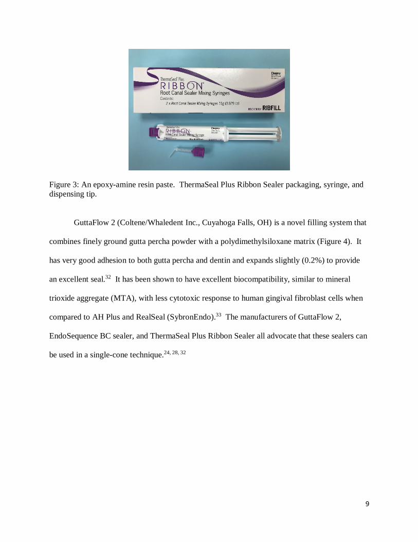
ThermaSeal Plus Ribbon Sealer (Dentsply Tulsa Dental Specialties, Tulsa, OK) is a two-
component paste based on epoxy-amine resin that has long-term sealing and self-adhesive
properties.28 It comes in a mixing syringe to provide a convenient, precise mixture (Figure 3). A
study by Pinna et al. showed that AH Plus (another name for ThermaSeal Plus) did not have the
long-term cytotoxic effects that Pulp Canal Sealer (SybronEndo) possessed.29 Although good in
the long-term, moderate to severe cytotoxicity was still present for the first week after mixing.
This is likely due to the epoxy resin component of the sealer that is released after setting.30 The
sealer’s adhesive properties and adaptability to the root canal walls have been cited as reasons
for its excellent sealing abilities and resistance to bacterial coronal leakage.31
9
Figure 3: An epoxy-amine resin paste. ThermaSeal Plus Ribbon Sealer packaging, syringe, and dispensing tip.
GuttaFlow 2 (Coltene/Whaledent Inc., Cuyahoga Falls, OH) is a novel filling system that
combines finely ground gutta percha powder with a polydimethylsiloxane matrix (Figure 4). It
has very good adhesion to both gutta percha and dentin and expands slightly (0.2%) to provide
an excellent seal.32 It has been shown to have excellent biocompatibility, similar to mineral
trioxide aggregate (MTA), with less cytotoxic response to human gingival fibroblast cells when
compared to AH Plus and RealSeal (SybronEndo).33 The manufacturers of GuttaFlow 2,
EndoSequence BC sealer, and ThermaSeal Plus Ribbon Sealer all advocate that these sealers can
be used in a single-cone technique.24, 28, 32

10
Figure 4: A polydimethylsiloxane paste with finely ground gutta percha powder. GuttaFlow 2 packaging, syringe, and dispensing tip.
After obturation of the RCS, the next major goal of endodontic treatment is sealing the
crown of the tooth to prevent future recontamination of the sealed root canals.4 This is a critical,
often under-valued component to proper endodontic treatment. Research reported by Ray and
Trope in 1995 showed that the quality of the coronal restoration had more impact on the presence
of periapical inflammation than quality of the root filling.34 These findings make it critical to
restore the crown of the tooth as expeditiously as possible.35 If the tooth cannot be restored
immediately, a temporary filling material should be used to minimize possible leakage. The
temporary restoration should be a minimum of four millimeters in thickness and should not be
expected to last more than three weeks.4
If the patient delays having a permanent restoration placed, if a restoration is lost or its
integrity becomes compromised, or if the root canal filling material is exposed through tooth
fracture or recurrent decay, the complication of re-contamination of the RCS arises.4, 5 When
one of these instances occur and the coronal portion of the RCS is exposed to oral flora, it is
difficult to decide whether retreatment of the root canal is necessary. This decision should be

11
made based on the length of time it takes for the entire root canal system to become
contaminated. Previous in-vitro studies have used fluid filtration36, 37 and dye leakage1-3 to
evaluate the seal of endodontic materials, but bacterial leakage has been shown to be a more
clinically relevant model.6 Bacterial penetration studies have utilized a multitude of singular
species38-40 or fresh human saliva.5, 41, 42
An in-vitro bacterial leakage model has been utilized to evaluate the leakage of numerous
sealers and obturation techniques.5, 39, 40 The model consists of a split-chamber, with the
obturated tooth connecting the two spaces. If bacteria are introduced to the top chamber, the
only way they can reach the bottom chamber is through the tooth. To determine when bacterial
contamination of the bottom chamber has occurred, the tip of the obturated tooth is submerged in
a broth that can become turbid once contaminated with bacteria. A different type of media was
utilized in Fransen’s study, which made bacterial contamination more identifiable. In the model
described in his study, the tip of the tooth was submerged in Difco Purple Broth Base (Beckton
Dickinson, Sparks, MD).40 When supplemented with a carbohydrate, the Difco Purple Broth
Base is able to elicit a fermentation reaction when bacteria are exposed to it. Acid is produced
by the fermentation reaction, and as the pH of the broth drops below 6.8, the indicator changes
from purple to yellow color.43 This color change provides a more distinct and abrupt
determination of leakage compared to the assessment of turbidity. The bacterial leakage model,
although effective in determining when full bacterial penetration of the canal has occurred, does
not allow quantification or localization of the bacteria in the RCS. Being able to localize how far
bacteria have penetrated into a RCS at different time points would be beneficial in determining
the need for full retreatment.
12
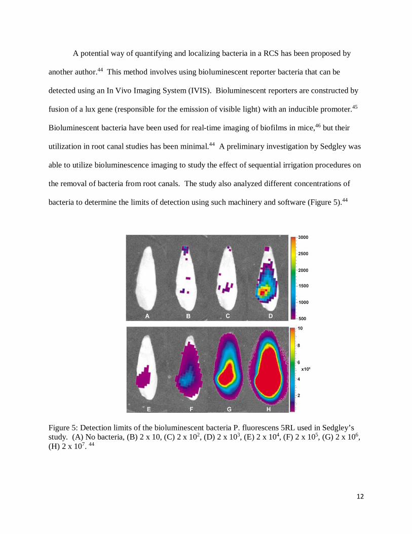
A potential way of quantifying and localizing bacteria in a RCS has been proposed by
another author.44 This method involves using bioluminescent reporter bacteria that can be
detected using an In Vivo Imaging System (IVIS). Bioluminescent reporters are constructed by
fusion of a lux gene (responsible for the emission of visible light) with an inducible promoter.45
Bioluminescent bacteria have been used for real-time imaging of biofilms in mice,46 but their
utilization in root canal studies has been minimal.44 A preliminary investigation by Sedgley was
able to utilize bioluminescence imaging to study the effect of sequential irrigation procedures on
the removal of bacteria from root canals. The study also analyzed different concentrations of
bacteria to determine the limits of detection using such machinery and software (Figure 5).44
Figure 5: Detection limits of the bioluminescent bacteria P. fluorescens 5RL used in Sedgley’s study. (A) No bacteria, (B) 2 x 10, (C) 2 x 10², (D) 2 x 10³, (E) 2 x 10⁴, (F) 2 x 10⁵, (G) 2 x 10⁶, (H) 2 x 10⁷. 44

13
It should be noted in the figure that the bioluminescent images in G and H are larger than
the tooth. This is due to the photons being emitted in all directions from the bacteria. A clear
outline of the root canal space would not be expected with this type of imaging.44
The bacterial population associated with failing endodontic cases has been shown to be
different from that of primary necrotic cases. Gram-positive facultative anaerobes typically
predominate failing endodontic cases, with Streptococcus, Enterococcus, Staphylococcus,
Actinomyces, and Lactobacillus species being the most commonly isolated.47, 48 Although
Enterococcus faecalis is frequently isolated from re-treatment cases and used for the study of
coronal leakage,40 many other species have been used, including Streptococcus mutans39 and
Staphylococcus epidermis.38 Staphylococci are non-motile, non-spore forming facultative
anaerobes that grow by either fermentation or aerobic respiration.49 With these qualities,
Staphylococci are able to survive in a coronally unsealed RCS. Staphylococcus aureus ATCC
49525 (Xen36) possesses these qualities as well as a lux gene, which allows it to emit
bioluminescent light.50
Although Sedgley’s study showed the ability of bioluminescence imaging to detect
bacteria in root canal systems, literature showing the ability of this technique to detect bacteria in
obturated canals is lacking. Therefore, the purpose of this experiment was twofold: to
investigate the possibility of using bioluminescence imaging for the evaluation of bacterial
leakage in obturated root canals, and to use an in vitro bacterial leakage model to evaluate the
resistance to bacterial contamination of three endodontic sealers used with the single-cone
technique.

14
Chapter 3
Materials and Methods
Bacteria
Staphylococcus aureus Xen36 was the bacteria used in the experimental procedures of
this study. Sedgley’s study determined that the lower limit of bacterial detection in root canals
by the IVIS is between 2 x 10² and 2 x 10³ cfu/mL. It was decided to use a concentration of
bacteria of 1 x 10⁶, which is similar to what was used in the experimental section of their study.
The inoculum was prepared as follows:
A clinical isolate of Staphylococcus aureus Xen36 was obtained and fresh sub-cultures
were grown on blood agar plates (BAP) (Thermo Scientific, Waltham, MA)(Figure 6). A stock
solution was made from the 2nd sub-culture. 15ml microcentrifuge tubes were labeled with the
organism ID (Figure 7). The stock solutions were prepared as follows:
1st tube – (4.0 McFarland Test) = 109 cfu/ml.
• 10ml Luria-Bertani (LB) broth (Figure 8) was placed in tube 1.
• A 10ul lambda loop was used to scoop bacteria from the BAP.
• The lambda loop with bacteria was placed into the 10ml of LB broth.
• Spun lambda loop with bacteria to disperse bacteria into media.
• Vortexed the solution (Figure 9).
• Incubated the stock solution for 20-24 hours at 37°C.
• Vortexed the solution.
• McFarland Test was performed to verify concentration of solution (Figure 10).

15
108 and 106 cfu/ml dilutions were then prepared from the stock solution of 109 cfu/ml.
2nd tube 108 – Dilution Calculation: (108)(10ml) = (109)(x)
x = 1.0ml of 109 Stock + 9.00ml of LB broth
1/10 dilution
• 10ml of LB broth was placed in tube 2, and 1.0ml from the tube was removed with a
micropipette and discarded.
• 1.0ml of the stock solution in tube 1 (109) was added into tube 2 (108).
• Vortexed the solution.
• McFarland Test was performed to verify concentration of solution.
3rd tube 106 – Dilution Calculation: (106)(10ml) = (108)(x)
x = 0.1ml (100ul) of 108 stock + 9.90ml of LB broth
0.1/10 = 1/100 dilution
• 10ml of LB broth was placed in tube 3, and 100ul (0.1ml) from the tube was removed
with a micropipette and discarded.
• 100ul (0.1ml) of the solution in tube 2 (108) was added into tube 3 (106).
• Vortexed the solution.
• McFarland Test was performed to verify concentration of solution.

16
Figure 6: Second sub-culture of Staphylococcus aureus Xen36 used to create stock solutions.
Figure 7: 15 ml microcentrifuge tubes labeled with each of the stock solutions (109, 108, 106).

17
Figure 8: LB broth used for bacterial growth in stock solutions.
Figure 9: Machine used to vortex stock bacterial solutions.

18
Figure 10: Densitometer used to perform McFarland test on stock bacterial solutions.
Growth was verified the following way:
A sterile pipette was used to remove 0.5ml (500ul) of the bacterial organism (106) from
dilution tube 3, and this was placed onto the center of 1 BAP (Blood Agar Plate), properly
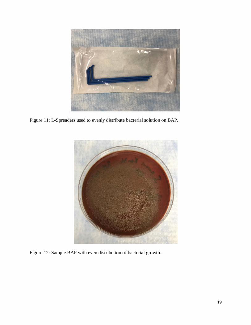
labeled and dated. Using an L-Spreader (Figure 11), the sample solution was spread circularly
around the BAP to give a nice even distribution of sample growth. The sample was placed into a
37°C incubator and removed when sample growth was adequate (Figure 12). Growth was scored
to ensure viability of the sample.
19
Figure 11: L-Spreaders used to evenly distribute bacterial solution on BAP.
Figure 12: Sample BAP with even distribution of bacterial growth.

20
Teeth
Fifty extracted human maxillary anterior teeth, stored in (1 part 8.25% NaOCl, 1 part
glycerin, 3 parts distilled water) to prevent bacterial growth were obtained from the West
Virginia University School of Dentistry Research Labs Tooth Depository (IRB #: T2017.07).
Teeth were decoronated and the root length was standardized to approximately 16 mm (Figure
13). After decoronation, the teeth were immersed in 8.25% NaOCl for 5 hours and then stored in
distilled water. Cleaning, shaping and obturation of the root segments was performed under an
operating microscope (Global Surgical Corporation, St. Louis, MO) (Figure 14). Canal patency
was confirmed with a #10 K-file and working length was determined by subtracting 0.5 mm
from the length at which this file was visible at the foramen. Each canal was instrumented with
ProTaper Universal rotary instruments (Dentsply, Tulsa, OK) according to the manufacturer’s
recommendations to a size F4 (40/06 at D0) (Figure 15). After each file use, each canal was
irrigated with 1 mL of 8.25% NaOCl. A final rinse was performed with 3 mL of 17% EDTA
(Vista Dental Products, Racine, WI) (Figure 16) and the canals were dried with paper points
(Dentsply, Tulsa, OK). After standardization of the instrumentation, the teeth were randomly
divided into three experimental groups (n = 12) and two control groups (5 positive and 5
negative). The final four teeth were used in the IVIS pilot study.

21
Figure 13: Sample of decoronated maxillary anterior teeth (side and incisal views).
Figure 14: Dental operating microscope used for instrumentation and obturation of samples (Global Surgical Corporation, St. Louis, MO).

22
Figure 15: Sequence of hand files and ProTaper Universal rotary files used to instrument canals.
Figure 16: Irrigating solutions and sample irrigation syringe and tip used during cleaning and shaping procedures.

23
Obturation of the three experimental groups was performed with a single-cone technique
using a ProTaper F4 master cone (Dentsply, Tulsa, OK) (Figure 17). The master cone size was
chosen to match the instrumented canal size. The three sealers tested included EndoSequence
BC sealer (Brasseler USA, Savannah, GA), ThermaSeal Plus Ribbon Sealer (Dentsply Tulsa
Dental Specialties, Tulsa, OK), and GuttaFlow 2 (Coltene/Whaledent Inc., Cuyahoga Falls, OH).
This single-cone method of obturation was deemed acceptable by each of the manufacturers.
The sealer for each group was placed on a sterile glass slab and the master cone was
coated in the sealer. The coated cone was gently placed in each canal to obtain a thin coating of
the walls. The cone was withdrawn and more sealer was placed on the apical third of the master
cone. The cone was then carefully seated to full working length. The master cone was seared
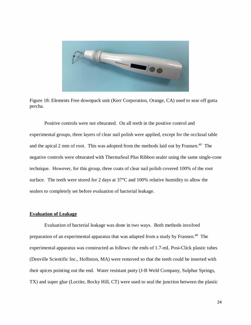
off with an Elements Free downpack unit (Kerr Corporation, Orange, CA) (Figure 18) and
lightly condensed with an endodontic plugger to a level that allowed 14 mm of gutta percha to
remain. This provided an unfilled coronal trough where the bacteria could be introduced.
Figure 17: Final ProTaper Universal file used during instrumentation (F4) and corresponding ProTaper Universal gutta percha points.
24
Figure 18: Elements Free downpack unit (Kerr Corporation, Orange, CA) used to sear off gutta percha.
Positive controls were not obturated. On all teeth in the positive control and
experimental groups, three layers of clear nail polish were applied, except for the occlusal table
and the apical 2 mm of root. This was adopted from the methods laid out by Fransen.40 The
negative controls were obturated with ThermaSeal Plus Ribbon sealer using the same single-cone
technique. However, for this group, three coats of clear nail polish covered 100% of the root
surface. The teeth were stored for 2 days at 37°C and 100% relative humidity to allow the
sealers to completely set before evaluation of bacterial leakage.
Evaluation of Leakage
Evaluation of bacterial leakage was done in two ways. Both methods involved
preparation of an experimental apparatus that was adapted from a study by Fransen.40 The
experimental apparatus was constructed as follows: the ends of 1.7-mL Posi-Click plastic tubes
(Denville Scientific Inc., Holliston, MA) were removed so that the teeth could be inserted with
their apices pointing out the end. Water resistant putty (J-B Weld Company, Sulphur Springs,
TX) and super glue (Loctite, Rocky Hill, CT) were used to seal the junction between the plastic

25
tube and root, and two coats of clear nail polish were placed over the set putty/glue. The cap of a
20-mL liquid scintillation vial (Scientific Specialties Service, Hanover, MD) was modified so
that the plastic tube and tooth assembly could be snugly placed through it. The tube was then
sealed to the cap with super glue and nail polish. The total apparatus was sterilized by a 12-hour
cycle in ethylene oxide gas.
To determine the viability of using the In Vivo Imaging System (IVIS) as a method of
evaluating bacterial leakage, a short pilot study was accomplished. This involved evaluation of
four teeth in the same experimental apparatus. After cleaning and shaping as mentioned earlier,
one tooth was left unobturated and three teeth were obturated with ThermaSeal Plus Ribbon
sealer. The gutta percha in these three teeth was burned off at a level that allowed either 5 mm,
10 mm, or 15 mm to remain. An additional instrumented tooth was placed in a Posi-Click tube
as a control. By omitting the glass vial in this tooth, it could be tested if the glass in the
experimental apparatuses would cause any imaging interference.
0.1 ml of a 1 x 10⁹ cfu/mL culture of Staphylococcus aureus Xen36 was placed in
another Posi-Click tube by itself as an additional control. The root canals in the four samples
and additional control tooth were inoculated with 0.1 mL of the S. aureus Xen36 and a scan was
taken with the IVIS. The IVIS was linked to a data-acquisition computer running
LivingImageTM software, which produced real-time images of each scan (Figure 19). Scans were
taken to show whether apical migration of bacteria could be detected at varying levels in the
root. The scans were taken at Days 0, 3, 7, and 10.

26
Figure 19: In Vivo Imaging System (IVIS) linked to data-acquisition computer.
The remaining forty-six apparatuses were evaluated in the same method as Fransen’s
study. The scintillation vials were filled with Difco Purple Broth Base (Beckton Dickinson,
Sparks, MD) (Figure 20) to a level that allowed approximately 2 mm of the root end to be
submerged. This solution contained a carbohydrate (1% D-Fructose) that the bacteria can
ferment to produce acid. When the pH of the broth drops below 6.8 it changes color from purple
to yellow (Figure 21). The apparatuses were incubated at 37°C for 3 days and the broth was
inspected for a color change to ensure that no contamination occurred before inoculation with
bacteria. 0.1 mL of the 1 x 10⁶ cfu/mL culture of Staphylococcus aureus Xen36 was used to
inoculate the top chamber of the tube/tooth apparatus. The apparatuses were incubated at 37°C
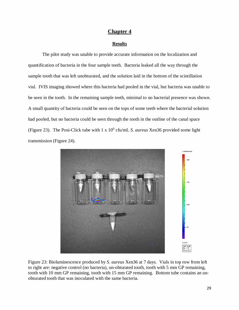
for up to 50 days (Figure 22). Apparatuses were re-inoculated with a freshly prepared 1 x 10⁶
27
cfu/mL solution of Staphylococcus aureus Xen36 every 7 days. The teeth were checked daily to
monitor changes in broth color. Teeth were declared to have fully leaked at the first sign of color
change from purple to yellow.
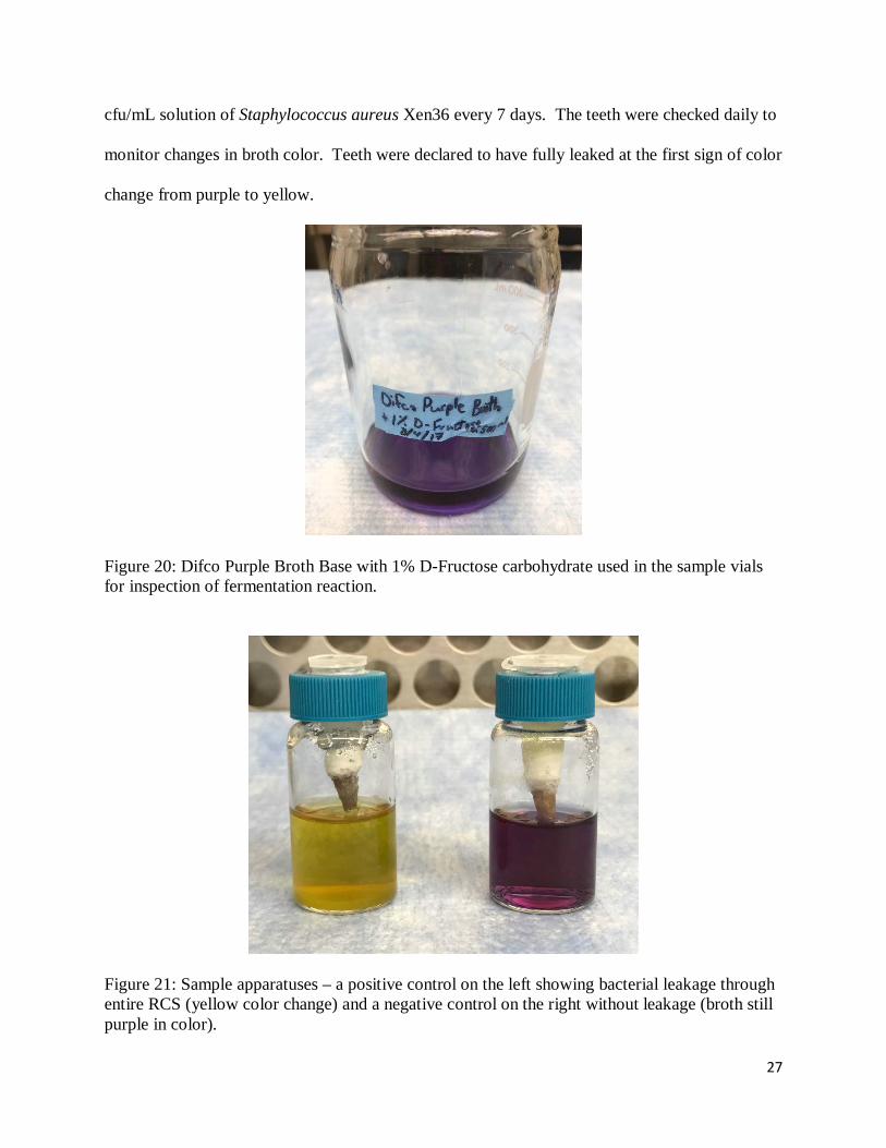
Figure 20: Difco Purple Broth Base with 1% D-Fructose carbohydrate used in the sample vials for inspection of fermentation reaction.
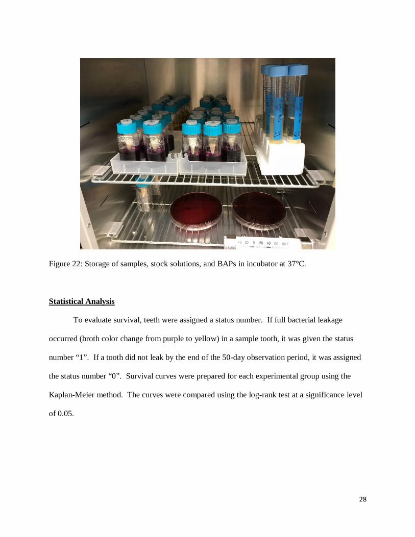
Figure 21: Sample apparatuses – a positive control on the left showing bacterial leakage through entire RCS (yellow color change) and a negative control on the right without leakage (broth still purple in color).
28
Figure 22: Storage of samples, stock solutions, and BAPs in incubator at 37°C.
Statistical Analysis
To evaluate survival, teeth were assigned a status number. If full bacterial leakage
occurred (broth color change from purple to yellow) in a sample tooth, it was given the status
number “1”. If a tooth did not leak by the end of the 50-day observation period, it was assigned
the status number “0”. Survival curves were prepared for each experimental group using the
Kaplan-Meier method. The curves were compared using the log-rank test at a significance level
of 0.05.
29
Chapter 4
Results
The pilot study was unable to provide accurate information on the localization and
quantification of bacteria in the four sample teeth. Bacteria leaked all the way through the
sample tooth that was left unobturated, and the solution laid in the bottom of the scintillation
vial. IVIS imaging showed where this bacteria had pooled in the vial, but bacteria was unable to
be seen in the tooth. In the remaining sample teeth, minimal to no bacterial presence was shown.
A small quantity of bacteria could be seen on the tops of some teeth where the bacterial solution
had pooled, but no bacteria could be seen through the tooth in the outline of the canal space
(Figure 23). The Posi-Click tube with 1 x 10⁹ cfu/mL S. aureus Xen36 provided some light
transmission (Figure 24).
Figure 23: Bioluminescence produced by S. aureus Xen36 at 7 days. Vials in top row from left to right are: negative control (no bacteria), un-obturated tooth, tooth with 5 mm GP remaining, tooth with 10 mm GP remaining, tooth with 15 mm GP remaining. Bottom tube contains an un-obturated tooth that was inoculated with the same bacteria.

30
Figure 24: Bioluminescence produced by S. aureus Xen36 at 10 days. Vials in top row from left to right are: negative control (no bacteria), un-obturated tooth, tooth with 5 mm GP remaining, tooth with 10 mm GP remaining, tooth with 15 mm GP remaining. In the bottom row, an un-obturated, inoculated tooth is on the left and a tube containing 0.1 ml of 1 x 10⁹ cfu/mL S. aureus Xen36 is on the right.
For the traditional bacterial leakage study, all positive control teeth leaked within the first
24 hours. None of the negative control teeth leaked during the entire 50-day observation period.
Data collected for the experimental groups can be seen in the Appendix. The Kaplan Meier
survival curves are shown in Figure 25. After the first 24 hours, three teeth leaked in both the
EndoSequence BC Sealer and GuttaFlow 2 groups compared to four teeth in the ThermaSeal
Plus Ribbon Sealer group. After one week, all three groups had a survival rate of 58.3%. After
50 days, all but one of the teeth in the ThermaSeal Plus Ribbon Sealer group had leaked, and
three teeth survived from each of the EndoSequence BC Sealer and GuttaFlow 2 groups. Table 1
shows the survival rates for each experimental group at different time intervals. The log-rank
31
test showed no statistically significant differences between the three groups with respect to
leakage over time (p = 0.665).
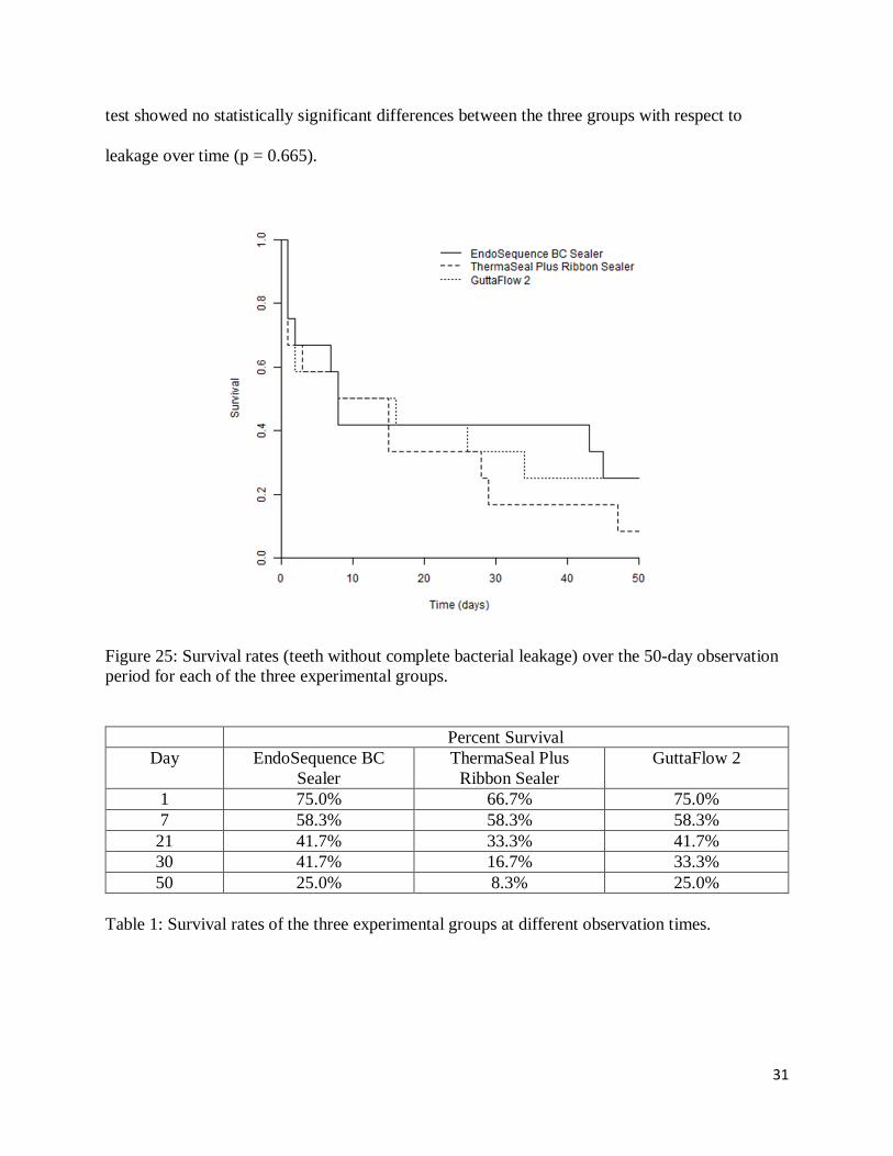
Figure 25: Survival rates (teeth without complete bacterial leakage) over the 50-day observation period for each of the three experimental groups.
Percent Survival Day EndoSequence BC
Sealer ThermaSeal Plus
Ribbon Sealer GuttaFlow 2
1 75.0% 66.7% 75.0% 7 58.3% 58.3% 58.3%
21 41.7% 33.3% 41.7% 30 41.7% 16.7% 33.3% 50 25.0% 8.3% 25.0%
Table 1: Survival rates of the three experimental groups at different observation times.

32
Discussion
The purpose of this project was twofold: to evaluate the resistance of three newly
developed root canal sealers to bacterial leakage in teeth with lost coronal seals and to determine
if bioluminescent bacteria could be utilized in coronal leakage studies to give more spatial and
quantitative information. Based on the findings in Sedgley’s study, it was predicted that similar
detection of bioluminescent bacteria would be found in a bacterial leakage apparatus. This
study, however, was unable to produce similar detection of bacteria in the RCS. Images
produced by the IVIS appeared to only show bacteria in some areas where a larger volume had
pooled and didn’t show any bacteria in the roots of the sample teeth (Figures 23, 24). One
difference in this study compared to that of Sedgley’s was the apical preparation size. In
Sedgley’s study, each experimental tooth was instrumented to a minimum apical size of 60,
while this study completed instrumentation at a 40 (size F4 ProTaper Universal file).44 This
larger apical preparation size may have allowed a larger volume of bacteria to gather in the
canals of Sedgley’s experimental teeth. The experimental apparatus may have been another
factor that limited light transmission. Due to biosafety requirements, the experimental teeth in
this study had to be contained in sealed apparatuses while being imaged in the IVIS, while in
Sedgley’s study, it appears that the teeth were placed directly in the machine’s chamber.
Although images in this study were able to show light transmission through the glass scintillation
vials and plastic Posi-Click tubes, the combination of these materials along with the root
thickness and nail polish covering the roots may have limited transmission. Further studies
looking at different apical preparation sizes and different experimental apparatus designs would
be needed before suggesting that bioluminescent imaging is an appropriate method of analyzing
bacterial leakage in obturated roots.

33
The results from the traditional bacterial leakage model showed that loss of a good
coronal seal could cause re-infection of the entire RCS. The unobturated positive controls all
leaked within 24 hours, showing that bacteria did travel through the RCS quickly when no
barrier was in place to prevent its movement. Since none of the negative controls leaked during
the 50-day observation period, it appears that the experimental apparatuses were properly
constructed and only allowed bacterial leakage through the RCS. Although survival rates of the
experimental groups were different at certain time points, the log-rank test found no statistically
significant differences between the groups with respect to leakage over time. The lack of
statistical significance between the three experimental groups warrants acceptance of the null
hypothesis.
Although no difference was found between the three sealers over time, some general
trends can be shown regarding the single-cone obturation technique. The fact that all three
groups had a survival rate of only 58.3% after the first week shows that the single-cone method
of obturation does not provide resistance to leakage in many teeth. This finding stresses the
importance of timely placement of a high quality coronal restoration after completion of root
canal therapy. If a temporary restoration is placed, waiting longer than the recommended 3-4
weeks to have a definitive restoration could jeopardize the long-term success of the root canal
therapy.
After the first week, leakage in the experimental groups leveled off to some degree. For
example, the experimental teeth in the EndoSequence BC sealer group had no leakage from Day
9 through Day 42. It is likely that the teeth in the EndoSequence BC sealer group that did not
leak early on were able to create a hydroxyapatite layer between the sealer and dentin, and this
layer was able to help resist leakage for a period of time. These findings show that sealers can

34
possibly resist bacterial leakage for some time if factors like sealer thickness and canal anatomy
are ideal. However, it is impossible to know from clinical and radiographic analysis that such
microscopic conditions exist.
Skinner and Himel showed that sealers play a critical role in the seal of the RCS.51
Compared to cold lateral condensation and warm vertical condensation where gutta percha fills
in most irregularities in the RCS, single-cone techniques rely more on the sealer to fill these
spaces. Although greater taper gutta percha cones have been designed to perfectly match the
nickel-titanium rotary files used today,18 the presence of fins and other canal irregularities may
cause a thicker sealer interface with single-cone obturation techniques.52 De-Deus found that a
greater sealer thickness negatively influenced the sealing ability of most sealers.53 The range of
bacterial leakage found in this study could be partially attributed to different sealer thickness in
the experimental teeth.
It is advantageous for a root canal sealer to be able to adhere to the intraradicular dentin
through frictional resistance or micromechanical retention so that the sealer-dentin interface is
retained during tooth flexure or operative procedures.54 AH Plus sealer (equivalent to
ThermaSeal Plus) and iRoot SP (EndoSequence BC sealer) have been shown to produce high
bond strengths with dentin.23, 55 Open epoxide rings in AH Plus can form a covalent bond with
exposed amino groups in the dentin collagen network.56 The calcium silicate composition of
EndoSequence BC sealer releases calcium and hydroxyl ions which can create an apatite layer
when they come in contact with phosphate-containing dentinal fluids. This layer enables a
chemical bond of the sealer to dentin.54, 57 GuttaFlow 2 relies on its slight expansion on setting
and its low solubility for its seal, because it does not chemically bond with dentin.58 The
resistance of some experimental teeth to bacterial leakage are likely due to these properties.

35
The low survival rates at the end of the 50-day observation period show that if an
endodontically treated tooth loses its coronal seal for any extended period of time, re-treatment
of the root canal would be necessary. Since some experimental teeth did not leak by the
conclusion of the observation period, it appears that these sealers do have some capability of
long-term sealing. A longer observation time may be able to show the full potential of these
sealers concerning bacterial leakage resistance. However, the high rate of early leakage shown
in this study seems to make such information clinically insignificant.
The lack of statistical significance in this study may be partially due to both sample size
as well as experimental design. Fransen was also unable to find statistically significant
differences between three different obturation techniques, ActiV GP/Glass Ionomer
Sealer, Resilon/Epiphany, and Gutta-Percha/AH Plus, despite having a larger sample size than
the current study (n = 20 per group).40 On the other hand, Monticelli was able to show
significant differences between groups using the same sample size as the current study (n = 12
per group). In their study, warm vertical compaction with AH Plus had significantly less leakage
over time when compared to two single-cone techniques, ActiV GP and GuttaFlow.39 A larger
sample size in our study would provide greater discriminative power.
A bacterial leakage model utilizing a singular bacterial species, Staphylococcus aureus
Xen36, was used in this study. This bacterium was chosen because it is a facultative anaerobe
that has the ability to survive by fermentation and has bioluminescent capabilities that allow it to
be visible when evaluated with an IVIS. Other authors have utilized a similar bacterial leakage
model with singular bacterial species.38-40 Fresh human saliva has been used in other bacterial
leakage studies5, 41, 42 in an attempt to have a model that provides the proteins, enzymes, and

36
bacterial populations found in normal clinical conditions. However, a solution with a single
bacterial species is easier to standardize for experimental use so that all groups can be compared.
Although bacterial leakage models are more clinically relevant than fluid filtration and
dye leakage studies, they do not provide spatial information of where the bacteria are localized in
the canal. The pilot study using the IVIS was conducted in an attempt to address this problem.
Gutta percha was removed to varying levels in the pilot teeth to see if this type of imaging could
visualize the bacteria at that level. Obtaining such information could be beneficial in providing
guidelines for clinical situations. For example, if the study was able to show that after one week,
teeth obturated with a certain material or technique leaked an average of 3 mm, the need for
retreatment may be avoided. In such a case, just the coronal gutta percha would need removed
for disinfection rather than all of the obturation material. Unfortunately, additional experiments
would be necessary to see if this type of information could be gathered by the IVIS. Further
experiments could be conducted with larger apical preparations in the teeth, different
experimental apparatus designs, and/or the use of different species of bioluminescent bacteria.
The amount of bacterial leakage needed to cause clinical root canal therapy failure is
unknown,59 so it is difficult to determine how much the results of the present study translate into
clinical outcomes. The high prevalence of early contamination and minimal survival at the end
of the 50-day observation period show that teeth obturated with the single-cone technique would
need retreated if they presented with a lost coronal seal. Single-cone obturation techniques with
newer categories of sealers should be analyzed further with animal studies and/or randomized
clinical trials.

37
Chapter 5
Conclusion
Employing a bacterial leakage model, the results of the present study show that in the
absence of a coronal seal, the single-cone method of root canal obturation does not provide long-
term sealing ability in many teeth. Although many contemporary sealers claim to be able to
create an adhesive bond with the dentin and/or gutta percha to better seal the RCS, this appears
to be inadequate to stop bacterial leakage in some teeth. The presence of fins and other
microscopic canal irregularities may increase the sealer thickness and prevalence of voids, which
can create portals for bacterial infiltration. Although survival was different for each of the
experimental groups at certain time points, there were no statistically significant differences
between EndoSequence BC Sealer, ThermaSeal Plus Ribbon Sealer, and GuttaFlow 2 with
respect to leakage over time.
A pilot study using a bioluminescent strain of S. aureus (Xen 36) and an IVIS was also
conducted in an attempt to better localize and quantify the bacterial leakage through the RCS.
Unfortunately, experimentation with this method of imaging was unable to produce adequate
visualization of the bacteria. Further studies should be piloted to see if this type of imaging
could be beneficial for analyzing obturated teeth.

38
Works Cited
1. Swanson K, Madison S. An evaluation of coronal microleakage in endodontically treated
teeth. Part I. Time periods. J Endod 1987;13:56.
2. Madison S, Swanson K, Chiles SA. An evaluation of coronal microleakage in
endodontically treated teeth. Part II. Sealer type. J Endod 1987;13:109.
3. Madison S, Wilcox L. An evaluation of coronal microleakage in endodontically treated
teeth. Part III. In vivo study. J Endod 1988;14:455.
4. Coronal Leakage: Clinical and Biological Implications in Endodontic Success.
Endodontics: Colleagues for Excellence. Fall/Winter 2002.
5. Khayat A, Lee SJ, Torabinejad. Human saliva penetration of coronally unsealed
obturated root canals. J Endod 1993;19:458-61.
6. Michailesco PM, Valcarcel J, Grieve AR, Levallois B, Lerner D. Bacterial leakage in
endodontics: an improved method for quantification. J Endod 1996;22:535-9.
7. Schilder H. Filling root canals in three dimensions. Dent Clin North Am 1967;11:723-
44.
8. ElAyouti A, Achleithner C, Löst C, Weiger R. Homogeneity and adaptation of a new
gutta-percha paste to root canal walls. J Endod 2005;9:687-90.
9. Kakehashi S, Stanley H, Fitzgerald R. The effect of surgical exposures of dental pulps in
germ-free and conventional laboratory rats. Oral Surg Oral Med Oral Pathol 1965;20:340
-9.
10. Sundqvist G. Associations between microbial species in dental root canal infections.
Oral Microbiol Immunol. 1992 Oct; 7(5):257-62.

39
11. Shuping G, Ørstavik D, Sigurdsson A, Trope M. Reduction of intracanal bacteria using
nickel-titanium rotary instrumentation and various medications. J Endod 2000;26:751-5.
12. Usman N, Baumgartner JC, Marshall JG. Influence of instrument size on root canal
debridement. J Endod. 2004;30(2):110-112.
13. Al-Dewani N, Hayes SJ, Dummer PM. Comparison of laterally condensed and low
temperature thermoplasticized gutta-percha root fillings. J Endod 2000;26:733-8.
14. Wu, M.-K. and Wesselink, P.R. Endodontic leakage studies reconsidered. Part 1.
Methodology, application and relevance. Int Endod J. 1993;26:37-43.
15. Canal Preparation and Obturation: An Updated View of the Two Pillars of Nonsurgical
Endodontics. Endodontics: Colleagues for Excellence. Fall 2016.
16. Whitworth J. Methods of filling root canals: principles and practices. Endodontic Topics.
2005;12:2-24.
17. Beatty RG. The effect of standard or serial preparation on single cone obturation. Int
Endod J 1987;20:276-81.
18. Gordon MPJ, Love RM, Chandler NP. An evaluation of .06 tapered gutta-percha cones
for filling of .06 taper prepared curved root canals. Int Endod J 2005;38:87-96.
19. Gillespie WT, Loushine RJ, Weller RN, et al. Improving the performance of EndoREZ
root canal sealer with a dual-cured two-step self-etch adhesive. II. Apical and coronal
seal. J Endod 2006;32:771-5.
20. Sagsen B, Er O, Kahraman Y, Orucoglu H. Evaluation of microleakage of roots filled
with different techniques with a computerized fluid filtration technique. J Endod
2006;32,1168-70.

40
21. Waltimo TM, Boiesen J, Eriksen HM, Ørstavik D. Clinical performance of three
endodontic sealers. Oral Surg Oral Med Oral Pathol Oral Radiol Endod 2001;92:89-92.
22. Li GH, Niu LN, Zhang W, Olsen M, De-Deus G, Eid AA, et al. Ability of new obturation
materials to improve the seal of the root canal system: a review. Acta Biomater
2014;10:1050-63.
23. Nagas E, Uyanik MO, Eymirli A, Cehreli ZC, Vallittu PK, Lassila LVJ, Durmaz V.
Dentin moisture conditions affect the adhesion of root canal sealers. J Endod 2012;38(2):
240-4.
24. EndoSequence BC Sealer. Instructions for Use. Brasseler USA, Savannah, Georgia.
2008.
25. Zhang H, Shen Y, Ruse ND, Haapasalo M. Antibacterial activity of endodontic sealers by
modified direct contact test against Enterococcus faecalis. J Endod 2009;35:1051-5.
26. Zhang W, Li Z, Peng B. Assessment of a new root canal sealer’s apical sealing ability.
Oral Surg Oral Med Oral Pathol Oral Radiol Endod 2009;107:e79-82.
27. Zhang W, Li Z, Peng B. Ex vivo cytotoxicity of a new calcium silicate–based canal
filling material. Int Endod J 2010;43:769-774.
28. ThermaSeal Plus Ribbon Root Canal Sealer. Directions for Use. DENTSPLY Tulsa
Dental Specialties, Johnson City, Tennessee.
29. Pinna L, Brackett MG, Lockwood PE, Huffman BP, Mai S, Cotti E, Dettori C, Pashley
DH, Tay FR. In vitro cytotoxicity evaluation of a self-adhesive, methacrylate resin-based
root canal sealer. J Endod 2008;34(9):1085-8.
30. T.H. Huang, S.J. Ding, T.Z. Hsu, Z.D. Lee, C.T. Kao. Root canal sealers induce
cytotoxicity and necrosis. J Mater Sci Mater Med 2004;15:767-771.
41
31. Haikel Y, Freymann M, Fanti V, Claisse A, Poumier F, Watson M. Apical microleakage
of radiolabeled lysozyme over time in three techniques of root canal obturation. J Endod
2000;26(3):148-52.
32. Hygenic GuttaFlow2. Instructions for Use. Coltene/Whaledent Inc., Cuyahoga Falls, OH.
2013.
33. Mandal P, Zhao J, Sah SK, Huang Y, Liu J. In vitro cytotoxicity of Guttaflow 2 on
human gingival fibroblasts. J Endod. 2014;40(8):1156-9.
34. Ray HA, Trope M. Periapical status of endodontically treated teeth in relation to the
technical quality of the root filling and the coronal restoration. Int Endodont J 1995;
28:12-18.
35. Saunders WP, Saunders EM. Coronal leakage as a cause of failure in root-canal therapy:
a review. Endod Dent Traumatol 1994; 10:105-108.
36. Monticelli F, Sword J, Martin RL, et al. Sealing properties of two contemporary single-
cone obturation systems. Int Endod J 2007;40:374-85.
37. Pommel L, Camps J. Effects of pressure and measurement time on the fluid filtration
method in endodontics. J Endod 2001;27:256-9.
38. Torabinejad M, LJng B, Kettering JD. In vitro bacterial penetration of coronally unsealed
endodonticaliy treated teeth. J Endod 1990;16:566.
39. Monticelli F, Sadek FT, Schuster GS, et al. Efficacy of two contemporary single-cone
filling techniques in preventing bacterial leakage. J Endod 2007;33:310-3.
40. Fransen JN, He J, Glickman GN, et al. Comparative Assessment of ActiV GP/Glass
Ionomer Sealer, Resilon/Epiphany, and Gutta-Percha/AH Plus Obturation: A Bacterial
Leakage Study. J Endod 2008;34:725-7.

42
41. Magura ME, Kafrawy AH, Brown CE Jr, Newton CW. Human Saliva Coronal
Microleakage in Obturated Root Canals: An In Vitro Study. J Endod 1991;17:324-31.
42. Siqueira JF, Rocas IN, Lopes HP, and de Uzeda M. Coronal Leakage of Two Root Canal
Sealers Containing Calcium Hydroxide after Exposure to Human Saliva. J Endod 1999;
25:14-6.
43. Difco and BBL Manual, 2nd Edition.
http://www.bd.com/europe/regulatory/Assets/IFU/Difco_BBL/222810.pdf
44. Sedgley C, Applegate B, Nagel A, and Hall D. Real-Time Imaging and Quantification of
Bioluminescent Bacteria in Root Canals In Vitro. J Endod 2004;30:893-8.
45. Heitzer A, Applegate B, Kehrmayer S, et al. Physiological considerations of
environmental applications of lux reporter fusions. J Microbiol Methods 1998;31:45-57.
46. Francis KP, Yu J, Bellinger-Kawahara C, et al. Visualizing pneumococcal infections in
the lungs of live mice using bioluminescent Streptococcus pneumoniae transformed with
a novel Gram-positive lux transposon. Infect Immun 2001;69:3350-8.
47. Molander A, Reit C, Dahlen G, Kvist T. Microbiological status of root filled teeth with
apical periodontitis. Int Endod J 1998;31:1-7.
48. Mindere A, Kundzina R, Nikolajeva V, Eze D, and Petrina Z. Microflora of root filled
teeth with apical periodontitis in Latvian patients. Stomatologija, Baltic Dental and
Maxillofacial Journal 2010;12:116-21.
49. Harris LG, Foster SJ, and Richards RG. An Introduction to Staphylococcus Aureus, and
Techniques for Identifying and Quantifying S. aureus Adhesins in Relation to Adhesion
to Biomaterials: Review. European Cells and Materials 2002;4:39-60.
50. Staphylococcus aureus ATCC 49525 (Xen36) Technical Data Sheet. Perkin Elmer. 2015.

43
51. Skinner RL, Himel VT. The sealing ability of injection-molded thermoplasticized gutta-
percha with and without the use of sealers. J Endod 1987;13:315.
52. Wu MK, Wesselink PR. A primary observation on the preparation and obturation of oval
canals. Int Endod J 2001;34:137-41.
53. De-Deus G, Coutinho-Filho T, Reis C, Murad C, Paciornik S. Polymicrobial leakage of
four root canal sealers at two different thicknesses. J Endod 2006;32:998-1001.
54. Shokouhinejad N, Gorjestani H, Nasseh AA, Hoseini A, Mohammadi M and Shamshiri
AR. Push-out bond strength of gutta-percha with a new bioceramic sealer in the presence
or absence of smear layer. Aust Endod J 2013;39:102-106.
55. Sagsen B, Ustun Y, Demirbuga S et al. Push-out bond strength of two new calcium
silicate-based endodontic sealers to root canal dentine. Int Endod J 2011;44:1088-91.
56. Carvalho NK, Prado MC, Senna PM, et al. Do smear-layer removal agents affect the
push-out bond strength of calcium silicate-based endodontic sealers? Int Endod J
2017;50:612-19.
57. Sarkar NK, Caicedo R, Ritwik P, Moiseyeva R and Kawashima I. Physicochemical Basis
of the Biologic Properties of Mineral Trioxide Aggregate. J Endod 2005;31(2):97-100.
58. Hammad M, Qualtrough A, and Silikas N. Extended Setting Shrinkage Behavior of
Endodontic Sealers. J Endod 2008;34(1):90-93.
59. Bergenholtz G, Spångberg L. Controversies in Endodontics. Crit Rev Oral Biol Med
2004;15:99-114.

44
Appendix
Data collection for statistical analysis. Treatment 1 – EndoSequence BC Sealer, Treatment 2 – ThermaSeal Plus Ribbon Sealer, Treatment 3 – GuttaFlow 2. Status 1 – Complete bacterial leakage, Status 0 – No bacterial leakage.
Tooth Treatment Time_Days Status 1 1 2 1 2 1 7 1 3 1 43 1 4 1 1 1 5 1 8 1 6 1 50 0 7 1 1 1 8 1 45 1 9 1 50 0 10 1 1 1 11 1 50 0 12 1 8 1 13 2 29 1 14 2 28 1 15 2 3 1 16 2 15 1 17 2 1 1 18 2 8 1 19 2 1 1 20 2 15 1 21 2 47 1 22 2 50 0 23 2 1 1 24 2 1 1 25 3 2 1 26 3 2 1 27 3 16 1 28 3 1 1 29 3 50 0 30 3 26 1 31 3 50 0 32 3 34 1 33 3 1 1 34 3 50 0 35 3 1 1 36 3 8 1


![Endodontic sealers in dentistry - in vitro and in vivo ......ramifications of the root canal system [1]. It is universally accepted that sealers should be able to fill imperfections](https://img.pdfslide.us/doc/110x75/5ed56e6711be98291d042330/endodontic-sealers-in-dentistry-in-vitro-and-in-vivo-ramifications-of.jpg)