Embed Size (px)
Citation preview
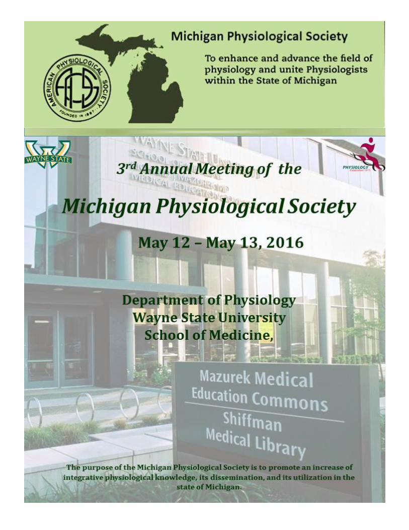

Michigan Physiological Society
Page | 1
Leadership of the MPS 2016
Erica Wehrwein
President (Michigan State University)
Patrick Mueller President-elect
(Wayne State University)
James Poteracki
Secretary/Treasurer (Michigan State University)
Sue Barman
Membership & Fundraising Chair (Michigan State University)
Karen Ball
K-12 Outreach Chair (Alma College)
Nusrat Matin
Trainee Committee Chair (Michigan State University)
Jason Carter
Past President (Michigan Technological
University)
You can find more information about the MPS at our website: https://apsmichigan.org/about/
Michigan Physiological Society
Page | 2
Diamond Sponsor: $2,000
Michigan State University College of Human Medicine (Dr. Aron Sousa-Dean) Wayne State University, Initiative for Maximizing Student Development
(Dr. Joseph Dunbar-Program Director) Wayne State University, Office of the Vice President for Research
(Dr. Stephen Lanier-Vice President)
Platinum Sponsors: $1,000
Henry Ford Health System (Dr. Margot LaPointe-Vice President for Research) Michigan State University College of Natural Sciences (Dr. R James Kirkpatrick-Dean) Michigan Technological University Graduate School (Dr. Jacqueline E Huntoon-Dean); Department
of Kinesiology& Integrative Physiology (Dr. Jason Carter-Chair); and Department of Biological Sciences (Dr. Shekhar Joshi-Chair)
Wayne State University, School of Medicine (Dean Jack Sobel) Wayne State University, School of Medicine, Department of Physiology (Dr. J.-P. Jin-Chair)
Gold Sponsors: $500-999
Michigan State University, Graduate School (Dr. Judith Stoddart- Interim Dean) Michigan State University, Office of the Vice President for Research & Graduate Studies
(Dr. Stephen Hsu-Dean) Wayne State University, School of Medicine, Research Office (Dr. Linda Hazlett-Interim Vice Dean) Wayne State University, School of Medicine, BioMed Graduate Programs
(Dr. Stanley Terlecky- Associate Dean)
Silver Sponsors: $300-499
Wayne State University School of Medicine Cardiovascular Research Institute (Dr. Karin Przyklenk-Director)
Bronze Sponsors: $200-299
Wayne State University Graduate School (Dr. Ambika Mathur-Dean)
Iron Sponsors: $100-199
Mayo Graduate School Biomedical Engineering & Physiology Program
A special
to our generous institutional sponsors!

Michigan Physiological Society
Page | 3
Thanks to the following who donated funds to support the trainee awards program:
American Physiological Society Susan M Barman, PhD – Michigan State University William H Beierwaltes, PhD – Henry Ford Hospital
Steven Cala, PhD – Wayne State University Jason Carter, PhD – Michigan Technological University
Qinghui Chen, PhD – Michigan Technological University Kevin Gordish, PhD – Henry Ford Hospital
Patrick Mueller, PhD – Wayne State University David Pieper, PhD – Southeast Michigan Center for Med Ed (WSU)
Zhiying Shan, PhD – Michigan Technological University Erica Wehrwein, PhD – Michigan State University
Drs. Bonnie Sloane and Douglas R. Yingst (Wayne State University)
Thanks to the following who judged the abstracts to select those giving oral
presentations: William H Beierwaltes – Henry Ford Hospital
Harold Bell – Central Michigan University Qinghui Chen – Michigan Technological University John Durocher – Michigan Technological University
Suzan El Sayed – Oakland University Gregory D Fink – Michigan State University
John Lawrence – Northern Michigan University
Mariela Mendez – Henry Ford Hospital Suresh Palaniyandi – Henry Ford Hospital
David Pieper – Wayne State University Nour-Eddine Rhaleb – Henry Ford Hospital
Naveen Sharma – Central Michigan University Erica Wehrwein – Michigan State University
Thanks to the following who judged the abstracts to select those giving award-eligible poster presentations:
Susan M Barman – Michigan State University John Durocher – Michigan Technological University
Nusrat Matin – Michigan State University Patrick Mueller – Wayne State University
James Poteracki – Michigan State University Erica Wehrwein – Michigan State University
Thanks to the following who judged the oral presentations to select the four trainee award recipients:
Monica McCullough – Adrian University (Chair) Harold Bell – Central Michigan University
Qinghui Chen – Michigan Technological University Nusrat Matin – Michigan State University
David Pieper – Wayne State University
Thanks to the following who judged the poster presentations to select the six trainee award recipients:
Naveen Sharma – Central Michigan University (Coordinator) Qinghui Chen – Michigan Technological University
John Durocher – Michigan Technological University John Lawrence – Northern Michigan University
Nusrat Matin – Michigan State University Suresh Palaniyandi – Henry Ford Hospital
Michigan Physiological Society
Page | 5
Special thanks to the members of the MPS Meeting Site Planning Committee:
Patrick Mueller (Wayne State University) MPS President-elect and Planning Committee Chair Erica Wehrwein (Michigan State University) MPS President Jason Carter (Michigan Tech University) MPS Past President
James Poteracki (Michigan State University) MPS Secretary-Treasurer Susan M Barman (Michigan State University) MPS Membership/Fundraising Chair
Karen Ball (Alma College) MPS K-12 Outreach Chair Nusrat Martin (Michigan State University) MPS Trainee Committee Chair
John Durocher (Michigan Tech University) Awards Chair Kevin Gordish (Henry Ford Hospital) Web coordinator
Special thanks to the members of the MPS K-12 Outreach Committee for organizing the Life Sciences Teacher Workshop
Karen Ball (Alma College) - MPS K-12 Outreach Chair Monica McCullough - Adrian College Dinesh Pal - University of Michigan Dyan Biringer - Onsted High School
Nancy Lefere Jackson - Lumen Christi High School Susan Speirs - Grosse Pointe North High School
Margaret Shain - American Physiological Society
Thanks to the following who judged the poster presentations to select the three trainee award recipients:
William Beierwaltes – Henry Ford Health Systems Qinghui Chen – Michigan Technological University
Sebastien Hayoz – Michigan State University Nusrat Matin – Michigan State University
Monica McCullough – Adrian College Zhiying Shan – Michigan Technological University
Naveen Sharma (Chair) – Central Michigan University
Micah Zuhl – Central Michigan University
…and special thanks to Wayne State University Department of Physiology administrative staff, especially Joanne Kaiser and Christine Cupps for donating time and effort toward meeting
logistics.
Special thanks to those who helped with meeting set-up at Scott Hall and Detroit Historical Museum:
Susan Barman (Michigan State University) Christine Cupps (Wayne State University) Adele Denison (Michigan State University)
Oneita Hunt (Wayne State University) Joanne Kaiser (Wayne State University)
Michael LaBella (Wayne State University) Rochelle LaMacchio (Wayne State University
Patrick Mueller (Wayne State University) Brianna Schick (Wayne State University)
Erica Wehrwein (Michigan State University)
Michigan Physiological Society
Page | 5
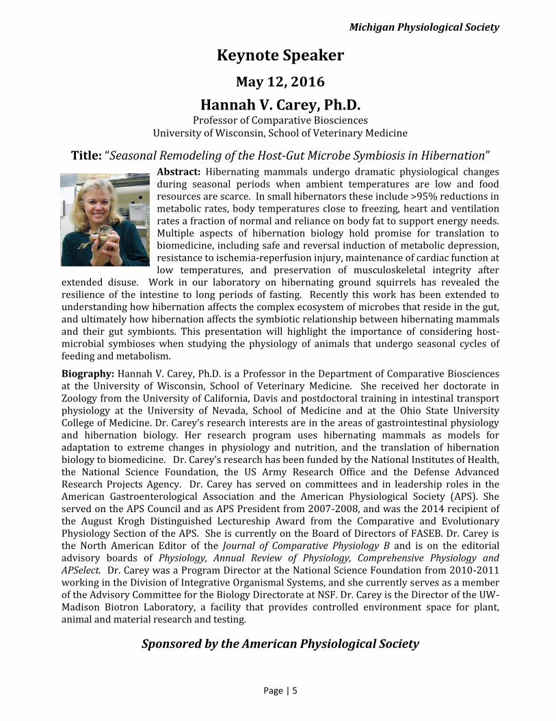
Keynote Speaker
May 12, 2016
Hannah V. Carey, Ph.D. Professor of Comparative Biosciences
University of Wisconsin, School of Veterinary Medicine
Title: “Seasonal Remodeling of the Host-Gut Microbe Symbiosis in Hibernation” Abstract: Hibernating mammals undergo dramatic physiological changes during seasonal periods when ambient temperatures are low and food resources are scarce. In small hibernators these include >95% reductions in metabolic rates, body temperatures close to freezing, heart and ventilation rates a fraction of normal and reliance on body fat to support energy needs. Multiple aspects of hibernation biology hold promise for translation to biomedicine, including safe and reversal induction of metabolic depression, resistance to ischemia-reperfusion injury, maintenance of cardiac function at low temperatures, and preservation of musculoskeletal integrity after
extended disuse. Work in our laboratory on hibernating ground squirrels has revealed the resilience of the intestine to long periods of fasting. Recently this work has been extended to understanding how hibernation affects the complex ecosystem of microbes that reside in the gut, and ultimately how hibernation affects the symbiotic relationship between hibernating mammals and their gut symbionts. This presentation will highlight the importance of considering host-microbial symbioses when studying the physiology of animals that undergo seasonal cycles of feeding and metabolism.
Biography: Hannah V. Carey, Ph.D. is a Professor in the Department of Comparative Biosciences at the University of Wisconsin, School of Veterinary Medicine. She received her doctorate in Zoology from the University of California, Davis and postdoctoral training in intestinal transport physiology at the University of Nevada, School of Medicine and at the Ohio State University College of Medicine. Dr. Carey’s research interests are in the areas of gastrointestinal physiology and hibernation biology. Her research program uses hibernating mammals as models for adaptation to extreme changes in physiology and nutrition, and the translation of hibernation biology to biomedicine. Dr. Carey’s research has been funded by the National Institutes of Health, the National Science Foundation, the US Army Research Office and the Defense Advanced Research Projects Agency. Dr. Carey has served on committees and in leadership roles in the American Gastroenterological Association and the American Physiological Society (APS). She served on the APS Council and as APS President from 2007-2008, and was the 2014 recipient of the August Krogh Distinguished Lectureship Award from the Comparative and Evolutionary Physiology Section of the APS. She is currently on the Board of Directors of FASEB. Dr. Carey is the North American Editor of the Journal of Comparative Physiology B and is on the editorial advisory boards of Physiology, Annual Review of Physiology, Comprehensive Physiology and APSelect. Dr. Carey was a Program Director at the National Science Foundation from 2010-2011 working in the Division of Integrative Organismal Systems, and she currently serves as a member of the Advisory Committee for the Biology Directorate at NSF. Dr. Carey is the Director of the UW-Madison Biotron Laboratory, a facility that provides controlled environment space for plant, animal and material research and testing.
Sponsored by the American Physiological Society
Michigan Physiological Society
Page | 6
MPS 3rd Annual Meeting Thursday - Friday, May 12-13, 2016
Thursday, May 12
1:30 – 2:30 pm REGISTRATION - Wayne State University, School of Medicine, 1358 Scott Hall
2:30 - 3:00 pm WELCOME AND MEETING OPENING, Green Auditorium 3rd Floor Scott Hall (access via main elevators only)
Dr Erica Wehrwein (President) and Dr Pat Mueller (President Elect) Dr Stephen Lanier, WSU Vice President for Research Dr. Jian-Ping Jin, WSU Chair of Physiology
3:00 – 3:45 pm ORAL SESSION #1 (Cell/Molecular) Green Auditorium 3rd Floor Scott Hall Session Co-Chairs: Brandon Coughlin (MSU) and Zeljka Minic (WSU)
3:00-3:15 Vanessa D. Ramseyer (Post-doc) Wayne State University DEVELOPMENTAL REGULATION OF ADIPOCYTE PHENOTYPES IN SUBCUTANEOUS ADIPOSE TISSUE
3:15-3:30 Molly Thorson (Graduate student) Michigan State University SARCOLEMMA WOUNDING ACTIVATES DYNAMIN DEPENDENT ENDOCYTOSIS IN STRIATED MUSCLE
3:30-3:45 Ryan Pettit-Mee (Graduate student) Central Michigan University THE EFFECT OF CALORIE RESTRICTION ON THE ASSOCIATION BETWEEN HEAT SHOCK PROTEIN 90 AND AKT
3:45 - 4:00 pm Break
4:00 – 5:00 pm KEYNOTE ADDRESS: Green Auditorium 3rd Floor Scott Hall
Hannah V. Carey, PhD University of Wisconsin School of Veterinary Medicine, Department of Comparative Biosciences Title: Seasonal Remodeling of the Host-Gut Microbe Symbiosis in
Hibernation
5:00 – 5:15 pm Group Picture, Green Auditorium 3rd Floor Scott Hall
5:15 - 5:30 pm Travel to Evening Events at: Detroit Historical Museum 5401 Woodward Ave, Detroit, MI 48202 (parking on W. Kirby)
-------------------------------------------------------------------------------------------------------------------------------- 5:30 – 7:00 pm Opening Reception, Detroit Historical Museum,
Grand Trunk Passenger Station Lower Level
6:00 – 7:30 pm POSTER SESSION A, Wrigley Room and Grand Trunk Passenger Station, Detroit Historical Museum (Index of presenting author is on page 18; poster abstracts are on pages 20-34)
7:00 – 8:45 pm Strolling Dinner “The Streets of Detroit”, Detroit Historical Museum, Lower Level
7:00 -9:00 pm Live music in the Grand Trunk Passenger Room Detroit Historical Museum, Lower Level
8:45-10:30 pm Museum (viewing open all levels)
Michigan Physiological Society
Page | 7
Friday, May 13
7:30 – 8:30 am REGISTRATION, Main Floor Scott Hall, Room 1358 Continental Breakfast, outside Green Auditorium, Scott Hall 3rd floor (access via main elevators only)
8:25 – 8:30 am MORNING ANNOUNCEMENTS
8:30 – 9:45 am ORAL SESSION #2 (Respiratory/Cardiovascular/Neuro) Green Auditorium, Scott Hall 3rd floor Session Co-Chairs: Andrew Chapp (MTU) and Dragana Komnenov (WSU)
8:30–8:45 Amanda Shoemaker (HS student) and Kevin Steelman (Medical Student) Central Michigan University DISPARITY IN THE EFFECT OF MORPHINE ON EUPNEA AND GASPING AS OBSERVED DURING RESPIRATORY AUTORESUSCITATION FROM ASPHYXIA IN ANESTHETIZED ADULT RATS
8:45-9:00 Joanne F. Garbincius (Graduate student) University of Michigan MODULATION OF METHYLATED ARGININE METABOLISM TO TARGET EXERCISE-INDUCED FATIGUE IN DUCHENNE MUSCULAR DYSTROPHY
9:00-9:15 Andrew R. Kulek (Graduate student) Wayne State University REMOTE ISCHEMIC PRECONDITIONING: DO EXOSOMES PLAY A REQUISITE ROLE IN EVOKING CARDIOPROTECTION?
9:15-9:30 Janice M. Diaz-Ortero (Graduate student) Michigan State University ANGIOTENSIN II-INDUCED HYPERTENSION IS ASSOCIATED WITH PARENCHYMAL ARTERIOLE AND POSTERIOR CEREBRAL ARTERY REMODELING AND REDUCED CEREBRAL PERFUSION
9:30-9:45 Stephen A. Klassen (Graduate student) Brock University THE ROLE OF AORTIC ARCH VASCULAR MECHANICS IN CARDIOVAGAL BAROREFLEX SENSITIVITY
9:45 – 10:00 am Break (Coffee and beverages available outside Green Auditorium 3rd floor)
10:00 - 10:45 am Concurrent Breakout Sessions and Poster Viewing (please refer to page 9 for detailed schedule) Session A: Individual Development Plan,
Tony Nunez (MSU), Green Auditorium Session B: Opportunities to involve undergraduates in research,
Rasheeda Zafar (WSU-IMSD) and Jennifer Tabb (WSU-Re-B.U.I.L.D. Detroit), Mazurek Education Commons, Room 240/241
Session C: Models for Physiologist-Teacher Collaboration (joined by MPS Meeting Participants)
Dinesh Pal (University of Michigan), Margherio Conference Center Session D: poster viewing, 1328 Scott Hall
11:00 – 12:00 pm POSTER SESSION B, Scott Hall Room 1328 (Index of presenting authors is on page 35; poster abstracts are on pages 37-55)
12:00 – 1:00 pm Lunch (box lunch available in the Scott Hall Cafeteria, 1st Floor)
1:15-2:15 pm ORAL SESSION #3 (Neuro) Session Co-Chairs: Travis Wakeham (MTU) and Brandon Sauer (CMU) Green Auditorium, Scott Hall 3rd floor
Michigan Physiological Society
Page | 8
1:15-1:30 Kelsey Kusch (Undergraduate student) Adrian College GLIAL CELL-LINE DERIVED NEUROTROPHIC FACTOR (GDNF) PROTEIN CONTENT IN CRAYFISH
1:30-1:45 Katelyn Norton (Graduate student) The University of Western Ontario REGIONAL CEREBRAL CORTIC REGIONAL CEREBRAL CORTICAL THICKNESS CORRELATES WITH AUTONOMIC OUTFLOW
1:45-2:00 Robert A. Larson (Graduate student) Michigan Technological University INCREASED BRAIN PROINFLAMMATORY CYTOKINES CONTRIBUTE TO AUGMENTED NEURONAL ACTIVITY IN SALT SENSITIVE HYPERTENSION
2:00-2:15 Mark B. Badrov (Graduate student) Western University EFFECT OF SIX MONTHS OF CARDIAC REHABILITATION ON AUTONOMIC FUNCTION IN CORONARY ARTERY DISEASE PATIENTS
2:15-2:30 pm Break (Beverages and snacks) Outside Green Auditorium 3rd floor
2:30-3:30 pm ORAL SESSION #4 (Neuro and Renal) Session Co-Charis: Shibandri Das (MSU) and Isola Brown (MSU) Green Auditorium, Scott Hall 3rd floor
2:30-2:45 Zachery Krease (Undergraduate student) Wayne State University INFLUENCE OF SEDENTARY VERSUS PHYSICALLY ACTIVE CONDITIONS ON THE RELATIONSHIP BETWEEN BLOOD PRESSURE, SYMPATHETIC OUTFLOW AND NEURAL ACTIVITY IN THE ROSTRAL VENTROLATERAL MEDULLA (RVLM)
2:45-3:00 Nusrat Matin (Graduate student) Michigan State University SOLUBLE EPOXIDE HYDROLASE INHIBITION IMPROVES COGNITIVE FUNCTIONS AND DILATION IN PARENCHYMAL ARTERIOLES FROM HYPERTENSIVE RATS WITH CEREBRAL HYPOPERFUSION
3:00-3:15 Ankita B. Jaykumar (Graduate student) Henry Ford Hospital and Wayne State University ALMS1 (ALSTROM SYNDROME 1), A NEW INTERACTING PROTEIN OF NKCC2, REGULATES NKCC2 ENDOCYTOSIS, URINARY CONCENTRATION AND BLOOD PRESSURE
3:15-3:30 Kevin Gordish (Post-doc) Henry Ford Hospital HIGH SALT DIET STIMULATES SALT-SENSITIVE HYPERTENSION IN FRUCTOSE-FED RATS
3:30-3:45 pm Break (Beverages and snacks)/Judges Convene
3:45-4:15 pm BUSINESS MEETING & AWARDS PRESENTATION Green Auditorium, Scott Hall 3rd floor
Michigan Physiological Society
Page | 9
3rd Annual Meeting of the Michigan Physiological Society Life Sciences Teacher Workshop
Margherio Family Conference Center Friday May 13, 2016
7:30 – 8:30 am REGISTRATION, Main Floor Scott Hall, Room 1358 Continental Breakfast, outside Green Auditorium,
Scott Hall 3rd floor (access via main elevators only)
8:30-8:45 am WELCOME AND INTRODUCTIONS, Margherio Family Conference Center Karen Ball, Alma College, MPS K-12 Outreach Chair Erica Wehrwein, Michigan State University, MPS President
8:45-9:45 am Keynote Address: Hannah V. Carey, PhD University of Wisconsin School of Veterinary Medicine, Department of Comparative Biosciences Title: Hibernation: A Cool Way to Spend the Winter
9:45-10:00 am Break (Coffee and beverages-outside Margherio Family Conference Center)
10:00-10:45 am Models for Physiologist-Teacher Collaboration-(please refer to Concurrent Breakout Session for additional options; joined by MPS Meeting Participants) Moderator:
Dinesh Pal (University of Michigan) Panelists:
Sue Speirs (Grosse Pointe North HS) and Pat Mueller (Wayne State Univ) -APS Frontiers in Physiology Professional Development
Julie Della-Moretta (Alma HS) and Maurie Luetkemeier (Alma College) -Alma College/Dow Foundation Cooperative Research Experience (CORE)
Nancy Lefere (Jackson Lumen Christi HS) and Sue Barman (Mich State Univ) -APS Physiology Understanding Week Activities
10:45-11:30 am Career Panel Moderator:
Erica Wehrwein, Michigan State University Panelists:
Valerie Van Ryn, Research Technician, Michigan State Univ Anthony Anzell, Graduate Student, Wayne State Univ Adele Denison, Teaching Faculty, Michigan State Univ Monica McCullough, Private Liberal Arts Faculty, Adrian College Jason Carter, Research Faculty, Michigan Tech Univ
11:30-12:00 pm Lab Demonstration Steve DiCarlo Laboratory
12:00-1:00 pm Lunch (box lunch available in the Scott Hall Cafeteria, 1st Floor)
1:00-1:30 pm Tour of Posters-1328 Scott Hall
1:30-3:30 pm Student Engagement Workshops/Hands-on Activities Sponsored by the American Physiological Society Session Leaders:
Margaret Shain, American Physiological Society Susan Speirs, Grosse Pointe North HS Nancy Lefere, Jackson Lumen Christi HS
3:45 pm BUSINESS MEETING & AWARDS PRESENTATION Green Auditorium, Scott Hall 3rd floor
Michigan Physiological Society
Page | 10
ORAL SESSION #1 ABSTRACTS
DEVELOPMENTAL REGULATION OF ADIPOCYTE PHENOTYPES IN SUBCUTANEOUS ADIPOSE TISSUE
Vanesa D. Ramseyer1, Anelia P. Petkova1 and James G. Granneman1,2 1 Wayne State University, School of Medicine, Center for Molecular Medicine and Genetics, Detroit, MI 2John D. Dingell VA Medical Center, Detroit, MI 48201
Subcutaneous adipose tissue exhibits extensive heterogeneity and plasticity but the mechanisms involved are unknown. At birth, mouse inguinal white adipose tissue (IWAT) is homogeneous and composed of unilocular adipocytes that do not express uncoupling protein 1 (UCP1), a marker of brown adipocyte (BA) cells. However, by the second week of life, about 30% of cells transiently acquire a BA phenotype. The mechanism involved in this phenomena is not known but could be due to recruitment of BA progenitors or conversion of existing cells. We hypothesized that perinatal induction of BA phenotype in mouse IWAT is due to increases in sympathetic innervation and activation of existing cells. IWAT and brown adipose tissue homogenates from 1, 2 and 3 week-old mice were used for quantitative PCR and Western blot analysis. We found that between the first and third week of life, UCP1 protein levels rose by more than 150 fold in IWAT (p<0.02) and this correlated with an increase in tyrosine hydroxylase, a marker of sympathetic innervation (p<0.005 vs week 1). UCP1 protein was observed in multilocular cells at 2 and 3 weeks of age by immunofluorescence. Similarly, phosphorylated perilipin 1, a marker of adrenergic activation, also augmented by weeks 2 and 3 (p<0.02 vs week 1), indicating that by the second week of life IWAT becomes functionally innervated. Interestingly, UCP1 and phosphorylated perilipin were not changed in brown adipose tissue. Direct activation of beta-3 adrenergic receptors, which induce BA phenotype independently of neogenesis, increased UCP1 protein levels by more than 100 fold (p<0.0001) in IWAT from one week-old mice. Adrenergic stimulation also augmented mRNA levels of other BA markers like CideA, PGC1α, Dio2 and Cox8b (p<0.04, p<0.001, p<0.02 and p<0.001 vs vehicle respectively). Altogether these data suggest that during the first week of life white unilocular cells in IWAT have the capability to become BA and this is unmasked by the second week when the tissue becomes functionally innervated. Current experiments are aimed at determining whether sympathetic innervation is necessary for the establishment of BA phenotype in IWAT during development.
SARCOLEMMA WOUNDING ACTIVATES DYNAMIN DEPENDENT ENDOCYTOSIS IN STRIATED MUSCLE
Molly T. Thorson, Joel R. McDade, Daniel E. Michele Michigan State University
Rationale: Mutations in the protein dysferlin have been shown to result in Limb Girdle Muscular Dystrophy 2B and Myoshi Myopathy. In patients and mouse models lacking dysferlin, dysfunctional membrane repair compromises the muscle cells and leads to muscle degeneration and progressive muscle wasting. Preliminary data in our laboratory suggests that sarcolemmal wounding stimulates the endocytosis of dysferlin. Therefore, the objective of this study was to investigate the potential regulators of wound-induced membrane transport. We hypothesized that the activity of dynamin, a GTPase that facilitates endocytosis, may be important for the activation of membrane trafficking following wounding and possibly important for the wound-induced endocytosis of dysferlin. Methods and Results: Much research in the field has focused on measuring membrane disruption by monitoring the uptake of the extracellular, lipophilic dye, FM1-43. However, FM1-43 can also be used as a measure of endocytosis. Therefore, we used FM1-43 dye to track endocytosis in adult skeletal muscle fibers at rest and following acute sarcolemma disruption. We show that while dysferlin recruitment to the wound site is not dependent upon dynamin, initial FM1-43 loading is completely dependent upon dynamin activity. Furthermore, dynamin inhibition significantly reduces wound-induced FM1-43 uptake, suggesting that wound-induced uptake occurs via dynamin-dependent endocytosis. We show, in agreement with others, that dysferlin-deficient muscle fibers show elevated FM1-43 uptake following wounding, which is the basis for the assertion that membrane repair is defective in this model. However, our data suggest that the upregulation of wound-induced endocytosis may contribute to the increased uptake of FM1-43 following wounding in dysferlin-deficient muscle fibers. Conclusions: These data highlight a previously unappreciated role for dynamin-dependent endocytosis in resting and wounded adult skeletal muscle fibers. Importantly, FM1-43 uptake following wounding is considered to be the “gold standard” for membrane repair studies in muscle. However, these data also suggest FM1-43 uptake following wounding may reflect wound-induced endocytosis, in addition to local influx of dye at the wound. Research in our laboratory is exploring the use of other methods of measuring membrane repair in order to distinguish the process of wound-induced endocytosis from local re-sealing of the wound site itself.
Michigan Physiological Society
Page | 11
THE EFFECT OF CALORIE RESTRICTION ON THE ASSOCIATION BETWEEN HEAT SHOCK PROTEIN 90 AND AKT
Lindsey E. Balta, Taylor E. Szczygiel, Ryan J. Pettit-Mee, Micah N. Zuhl, and Naveen Sharma. Central Michigan University, School of Health Sciences, Mount Pleasant, MI
Intro: Heat shock protein 90 (HSP90) is a chaperone protein, which has an essential role in the stress response signaling pathway. Like most heat shock proteins HSP90, is involved in keeping other proteins from denaturing under high stress conditions. Recently, HSP90 has been demonstrated to associate with another central signaling protein, the serine/threonine kinase Akt, suggesting that HSP90 may also be involved with other key cellular processes, particularly glucose metabolism. Therefore, the goal of this study to is characterize the relationship between HSP90 and Akt in physiological conditions where glucose metabolism is upregulated, particularly in the presence high levels of insulin, and during calorie restriction.
Methods: 3-month old rats were placed into 2 experimental groups: ad libitum fed (AL), and calorie-restricted (~60% of AL intake by calories). After 6 months of treatment rats were sacrificed and soleus muscles were excised and treated with or without a physiological dose of insulin in vitro. Muscles were then snap-frozen in liquid nitrogen and processed for analysis using immunoprecipitation and Western blotting.
Results: Insulin treatment compared to no insulin treatment resulted in higher levels of HSP90 associated with Akt. Additionally, in the insulin-treated groups, there was an additional increase in the HSP90 associated with Akt in the CR group compared to the AL group.
Conclusion: We found that in the soleus muscle, HSP90’s association with Akt is elevated with insulin, and is further increased with CR. These data may provide insight on the mechanism for why Akt’s activity and phosphorylation state is elevated during CR, subsequently leading to elevated insulin-stimulated glucose uptake in skeletal muscle. Future directions involve characterizing the association of other chaperone proteins (i.e. HSP25, HSP40, HSP70) with Akt, as well as looking at other highly metabolically active tissues including the liver, heart, and brain.
ORAL SESSION #2 ABSTRACTS
DISPARITY IN THE EFFECT OF MORPHINE ON EUPNEA AND GASPING AS OBSERVED DURING RESPIRATORY AUTORESUSCITATION FROM ASPHYXIA IN ANESTHETIZED ADULT RATS
Amanda Shoemaker, Kevin Steelman, Harold Bell. Central Michigan University College of Medicine, Mount Pleasant, MI
The pre-Bötzinger (preBöt) complex is considered an essential locus for the generation of breathing rhythm. It has been proposed that opioids cause depression of eupneic breathing via their actions on preBöt neurons. We reasoned that opioids should therefore also cause depressive effects on gasping breaths triggered by severe hypoxia. With this in mind, we studied the effect of acute morphine on the morphology and pattern of gasping breathing during respiratory autoresuscitation from asphyxia. Twenty adult-male rats were anaesthetized (pentobarbital) and instrumented to continuously monitor tracheal airflow and femoral artery pressure. Animals had respiratory responses to mild hypoxia and hypercarbia assessed before an episode of transient asphyxia with autoresuscitation. Animals were then randomly assigned to receive either morphine or sham injections before repeating these same tests. Ten animals were assigned to each of the morphine and sham experimental groups. Morphine caused significant depression eupnea with respect to minute ventilation, tidal volume at frequency (p<0.05, all comparisons), and PaCO2 increased from 41.6±5.0 to 63.3±5.8 mmHg following injection (p<0.001). Respiratory chemoreflex responses were also significantly attenuated by morphine (P<0.05 all comparisons). Sham injections caused no change in indices of respiratory status or chemoreflex responses, and PaCO2 was unchanged pre vs. post injection (41.9±8.3 to 42.5±3.6 mmHg, P=0.870). Interestingly, parameters defining the morphology and pattern of gasping breathing were not depressed by morphine. Duration of primary apnea, time to restore eupnea, the number and amplitude of gasping breaths, average and maximum peak flow and volume of gasping breaths were not influenced by injection status or experimental group (ns, all comparisons). Our data show that there is clear disparity in the effect of morphine on eupnea vs. gasping breathing rhythms. These in vivo observations present a novel challenge to the currently accepted paradigm of respiratory rhythm generation.
Michigan Physiological Society
Page | 12
MODULATION OF METHYLATED ARGININE METABOLISM TO TARGET EXERCISE-INDUCED FATIGUE IN DUCHENNE MUSCULAR DYSTROPHY
Joanne F. Garbincius, Lauren E. Merz, Ashley J. Cuttitta, Kaitlynn V. Bayne, Sara Schrade, Emily A. Armstead, Steven E. Whitesall, Louis G. D’Alecy, and Daniel E. Michele University of Michigan
Duchenne muscular dystrophy (DMD) is an X-linked disease caused by mutations in dystrophin and characterized by muscle degeneration, cardiomyopathy, and impaired muscle nitric oxide (NO) production that leads to misregulated muscle blood flow and excessive post-exercise fatigue. Data from our laboratory show that serum levels of the NO synthase inhibitor asymmetric dimethylarginine (ADMA) are elevated in dystrophin-deficient mdx mice and after acute muscle injury, and that infusion of ADMA is sufficient to impair exercise performance in wild-type mice. Therefore, we hypothesized that excessive circulating ADMA impairs NO production and exaggerates exercise-induced fatigue in DMD. To determine the impact of ADMA on fatigue resistance in this disease, female mdx mice were crossed to wild-type males expressing a transgene encoding human dimethylarginine dimethylaminohydrolase 1 (DDAH), an enzyme that degrades ADMA. Voluntary ambulatory activity of transgenic (TG) and non-transgenic (NTG) offspring was measured in open field assays prior to and following acute bouts of treadmill running to assess exercise-induced fatigue. DDAH expression lowered serum ADMA concentration in TG offspring, but failed to attenuate the post-exercise decrease in voluntary activity in mdx males. However, transgene expression resulted in a significant, 20% attenuation of the decrease in voluntary activity following exercise in female mdx carriers. The heart weight/tibia length ratio was significantly reduced in TG versus NTG carriers, although examination of the gastrocnemius and soleus muscles revealed no difference between genotypes in histological markers of dystrophy or ischemic muscle injury. Furthermore, echocardiography revealed a lower resting heart rate, higher stroke volume, and improved myocardial performance in TG versus NTG carriers. We conclude that ADMA contributes to excessive exercise-induced fatigue in dystrophin heterozygous females, possibly by promoting pathological cardiac remodeling and impairing heart performance. These findings emphasize the importance of NO signaling to cardiac function in dystrophinopathies and reveal a new therapeutic target to mitigate the risk of cardiomyopathy in DMD carriers. (Supported by the Muscular Dystrophy Association, the University of Michigan CVRE Training Program, and NIH T32 GM-08322)
REMOTE ISCHEMIC PRECONDITIONING: DO EXOSOMES PLAY A REQUISITE ROLE IN EVOKING CARDIOPROTECTION?
Joseph Wider, Andrew R. Kulek, Vishnu V.R. Undyala, Karin Przyklenk Wayne State University, School of Medicine, Cardiovascular Research Institute, Detroit, MI.
Recent studies consistently report that alcohol consumption increases muscle sympathetic nerve activity (MSNA) in humans. Despite the increased prevalence of hypertension and alcohol consumption in African Americans (AA), no studies have compared MSNA responses to acute alcohol consumption in AA and Caucasians (C). We hypothesized that alcohol consumption would increase MSNA in both AA and C, but that sympathoexcitatory responses would be augmented in AA. Five minutes of supine heart rate (HR), blood pressure, MSNA, and forearm blood flow were recorded in 10 AA (24±1 years) and 11 C (23±1 years) before and 45 min after consumption of 2.5ml of vodka/kg body mass. Increases in estimated blood alcohol content were greater in AA (0.099%) compared to C (0.087%; P<0.05). Contrary to our initial hypothesis, acute alcohol consumption was not augmented in AA. In fact, alcohol tended (P=.14) to increase MSNA more in C (16±3 to 28±5 bursts/100 heart beats) when compared to AA (26±6 to 29±5 bursts/100 heart beats). Forearm vascular conductance (FVC) was not altered by alcohol consumption in AA or C. Sympathetic vascular transduction (i.e., FVC/MSNA) was reduced in C (2.4±0.4 to 1.1±0.1 units; p<0.01), but did not change in AA (1.4±0.3 to 1.3±0.3 units; time×race, p=.027). Acute alcohol elicited similar increases of HR in C and AA. These findings demonstrate that acute alcohol consumption influences MSNA-vascular coupling differently in AA and C, but it remains unclear if the rate of alcohol metabolism is contributing to this difference.
Michigan Physiological Society
Page | 13
ANGIOTENSIN II-INDUCED HYPERTENSION IS ASSOCIATED WITH PARENCHYMAL ARTERIOLE AND POSTERIOR CEREBRAL ARTERY REMODELING AND REDUCED CEREBRAL PERFUSION
Janice M Diaz-Otero, Kelsey Downs, Erika Sarno, William F Jackson and Anne M Dorrance Michigan State University, Department of Pharmacology and Toxicology, East Lansing, MI
Hypertension can cause inward artery remodeling, artery rarefaction, increased vascular resistance and blood brain barrier (BBB) breakdown. These conditions increase the risk of stroke and dementia. The differential effects of Ang II-induced hypertension on the posterior cerebral artery (PCA) and parenchymal arterioles (PAs) have not been investigated. The PAs serve as bottlenecks for the perfusion of the cortex and are important in determining the outcome of stroke. We hypothesized that Ang II-induced hypertension would cause inward remodeling of the PAs and PCAs, artery rarefaction, and blood brain barrier breakdown. PAs and PCAs were collected from 20-week-old male C57Bl/6 mice to assess structure by pressure myography. Data collected at an intralumenal pressure of 60mmHg are presented as mean ± SEM; Sham (n=8) vs. AngII (n=8; 800ng/kg/day for 4 weeks). Ang II increased systolic (148 ± 4 vs 176 ± 6mmHg) and diastolic (115 ± 4 vs. 144 ± 6mmHg) blood pressures (p<0.05). In the PAs, the lumen diameter (37 ± 3 vs. 22 ± 3μm), wall area (830 ± 118 vs. 632 ± 53μm2) and wall stress (199 ± 24 vs 98 ± 22dynes/cm2) were reduced in hypertensive mice (p<0.05). Preliminary studies suggest that Ang II-induced hypertension also impairs myogenic tone and endothelium-dependent dilation in the PAs. In the PCAs, hypertension decreased the lumen diameter (130 ± 5 vs. 97 ± 7μm, p<0.05). The wall/lumen ratio was increased (0.08 ± 0.01 vs 0.14 ± 0.03) while wall stress (362 ± 28 vs 264 ± 33dynes/cm2) was reduced (p<0.05). The decreased cerebral perfusion (836 ± 50 vs. 596 ± 83 perfusion units; p0.05). Ang II-induced artery remodeling was not associated with increased BBB permeability (2 ± 1 vs 3 ± 1 % extravasation Evans Blue; p>0.05). Our results suggest that Ang II-induced hypertension results in remodeling of the PCA and PAs associated with decreased cerebral perfusion; this could have detrimental effects on neuronal function increasing the risk of vascular dementia and stroke.
THE ROLE OF AORTIC ARCH VASCULAR MECHANICS IN CARDIOVAGAL BAROREFLEX SENSITIVITY
Stephen A. Klassen1,2,3, Daniele Chirico1,2, Kylie S. Dempster1,2, J. Kevin Shoemaker3, Deborah D. O’Leary1,2 Brock University, 1Faculty of Applied Health Sciences, 2Brock-Niagara Centre for Health and Well-Being, St. Catharines, Ontario, Canada 3The University of Western Ontario, Neurovascular Research Laboratory, London, Ontario, Canada
Cardiovagal baroreflex sensitivity (cvBRS) measures the efficiency of the cardiovagal baroreflex to modulate heart rate in response to rises or falls in systolic blood pressure (SBP). Given that baroreceptors are located in the walls of the carotid sinuses (CS) and aortic arch (AA), the arterial mechanics of these sites are important contributors to cvBRS. However, the relative contribution of CS and AA mechanics to cvBRS remains unclear. This study employed sex differences as a model to test the hypothesis that differences in cvBRS between groups would be explained by the vascular mechanics of the AA but not the CS. Thirty-six young, normotensive individuals (18 female; 24 ±2 years) were recruited. cvBRS was measured using transfer function analysis of the low-frequency region (0.04-0.15 Hz). Ultrasonography was performed at the CS and AA to obtain arterial diameters for the measurement of distensibility. Local pulse pressure (PP) was taken at the CS using a hand held tonometer while AA PP was estimated using a transfer function of brachial PP. Both cvBRS (25 ±11 vs. 19 ±7 ms/mmHg, p = 0.04) and AA distensibility (16.5 ±6.0 vs. 10.5 ±3.8 mmHg-1 x10⁻³, p=0.02) were greater in females than males. Sex differences in cvBRS were eliminated after controlling for AA distensibility (p = 0.19). There were no sex differences in CS distensibility (5.32 ±2.3 vs. 4.63 ±1.3 mmHg-1 x10⁻³, p = 0.32). The present data demonstrate that AA mechanics are an important contributor to differences in cvBRS.
Michigan Physiological Society
Page | 14
ORAL SESSION #3 ABSTRACTS
GLIAL CELL-LINE DERIVED NEUROTROPHIC FACTOR (GDNF) PROTEIN CONTENT IN CRAYFISH
Kelsey Kusch, Dr. Monica J. McCullough Adrian College, Adrian, MI
Glial cell line-derived neurotrophic factor (GDNF) is a protein that promotes the survival of multiple neurons such as dopaminergic and motor neurons. Humans, among other species such as mice, rats, and frogs, have been shown to express GDNF. However, little research has been previously conducted among invertebrates. Thus this study is aimed at determining if GDNF protein expression is found within the adult crayfish. Analyses of GDNF protein content were examined in various tissue supernatant of male and female crayfish via an enzyme-linked immunosorbent assay (ELISA). All samples were analyzed using an analysis of variance (ANOVA) with a p-value of <0.05 to determine significance. The nervous system produced the highest amount of GDNF protein content which includes, the brain (0.17 ± 0.04pg GDNF/mg tissue weight), eyes (1.07 ± 0.26 pg GDNF/mg tissue weight), optic nerve (3.36 ± 1.06pg GDNF/mg tissue weight), and nerve (2.39 ± 0.93pg GDNF/mg tissue weight). Interestingly, the gills (0.47 ± 0.23pg GDNF/mg tissue weight) and ovaries (1.75 ± 0.78pg GDNF/mg tissue weight) were found to also have higher amounts of GDNF protein content, whereas the flexor and stomach had little or unmeasurable GDNF protein levels, respectively. These trends are similar to that found in mammals. By studying neurotrophic factors in the crayfish, this will allow us to examine regulation of neurotrophic factors in an invertebrate model that can be applied to vertebrate and human models.
REGIONAL CEREBRAL CORTIC REGIONAL CEREBRAL CORTICAL THICKNESS CORRELATES WITH AUTONOMIC OUTFLOW
Katelyn Norton, Mark Badrov, Mark Speechley, Kevin Shoemaker The University of Western Ontario, Neurovascular Research Laboratory, London, Ontario, Canada
Advancing age is associated with dysregulation of autonomic control; a complex series of changes favoring heightened cardiac sympathetic tone with parasympathetic withdrawal and blunted cardiovagal baroreflex sensitivity. An analogous pattern of change is observed in the brain with tissue loss beginning early in the third decade of life and disproportionately high losses in the forebrain and parietal regions known to be associated with cardiovascular control. Despite this functional link between the cortex and efferent physiological responses, the extent to which cortical structure, specifically cortical thickness, correlates with autonomic outflow is not known. The goal of the present study was to investigate whether inter-individual differences in autonomic outflow are related to brain morphology in healthy humans. We tested the hypothesis that baseline values of cardiovagal and sympathetic outflow would be associated with thickness of the cortical autonomic network, specifically the medial prefrontal cortex and insula cortex. A total of 55 healthy, active individuals participated in this study across a range of fitness and age (VO2: 26-81mL/kg/min; 21-73 years; 18 female). Baroreflex sensitivity, heart rate variability, and muscle sympathetic nerve activity were acquired during a laboratory session. Cortical thickness was measured using T1-weighted images acquired at 3T and assessed using Brain Voyager 2.8.2. A priori region of interest analysis revealed a significant association between cortical thickness at the medial prefrontal cortex and markers of autonomic outflow including: ln-high frequency power (slope: -16.01, r2=0.65), SDNN (slope: 22.49, r2=0.22), total power (slope: 2872.65, r2=0.24), burst frequency (slope: 1.07, r2=0.16), burst incidence (slope: -26.33, r2=0.62) and total MSNA (slope: -847.01, r2=0.56). The current results illustrate the potent and persistent effect of preserving cortical mass with advancing age at those regions known to be related to autonomic function and cardiovascular control. The current study is the first to suggest a strong association between cortical structure and autonomic balance in humans. (Supported by CIHR)
Michigan Physiological Society
Page | 15
INCREASED BRAIN PROINFLAMMATORY CYTOKINES CONTRIBUTE TO AUGMENTED NEURONAL ACTIVITY IN SALT SENSITIVE HYPERTENSION
Robert A. Larson, Fengli Zhu, Stephen Berridge, Ana-Lisia Powdhar, Qinghui Chen and Zhiying Shan Michigan Technological University, Department of Kinesiology and Integrative Physiology, Houghton, MI
The hypothalamic paraventricular nucleus (PVN) is a key regulatory center for salt-sensing mechanisms and sympathetic nerve activity (SNA). Augmented neuronal activity in the PVN plays an important role in the development of salt sensitive hypertension (SSHTN); however, the detailed mechanism(s) whereby the PVN influences SNA are not well understood. Recent evidence indicates that brain proinflammatory cytokines (PICs) may contribute to the increased neuronal activity in SSHTN; therefore, we tested the hypothesis that high salt (HS) loading increases expression of PICs in the PVN. To test this hypothesis, 6-week-old Dahl salt sensitive (Dahl S) rats, an animal model for human SSHTN, and age and sex matched normal Sprague Dawley (SD) rats were divided into two groups (n=4~5/group) and fed with either a HS (4% NaCI) or normal salt (NS, 0.4% NaCl) diet. Blood pressure was measured via tail cuff. Five weeks following diet treatment, HS diet induced hypertension in Dahl S rats (HS: 153± 9; vs. NS: 122±2 mmHg, P≤0.05), but not in SD rats (HS: 107± 3; vs. NS: 107±2 mmHg). Cerebrospinal fluid (CSF) was withdrawn for [Na+] and osmolality measurements, and punched PVN tissue was analyzed with real time PCR to assess mRNA levels of genes of interest. HS treatment significantly increased CSF [Na+] in Dahl S rats (HS: 150.6±0.24; vs. NS: 149±0.0 mM, P≤0.05), but not in SD rats (HS: 149.2±0.48; vs. NS: 149.5±0.29 mM). No significant changes in osmolality were observed between HS and NS diet treatment in both strains of rats. HS diet also induced dramatic increases in mRNA levels of PVN PICs including TNFα (8-fold), IL1β (4 -fold), and IL6 (7-fold), as well as Fra1 (4-fold), a chronic marker of neuronal activation. Next, we investigated whether these increases in expression of PVN PICs and Fra1 were induced by increased CSF [Na+]. Adult male SD rats were divided into three groups (n=7~8 per group) and received intracerebroventricular infusion of either hypertonic NaCI (2M, 2µl), mannitol (2M, 4 uL) as an osmotic control, or isotonic NaCI control (0.15 M, 2µl) in anesthetized condition. Three hours following the infusion, rats were euthanized and PVN mRNA levels of PICs and Fra1 were assayed. Results demonstrate that mRNA levels of PICs including TNFα (4.1-fold), IL1β (7.4-fold) and IL6 (11.6-fold), as well as neuronal activation marker Fra1 (4-fold) were significantly increased in hypertonic saline treated rats, but not in mannitol injected rats, compared to isotonic NaCI controls. Finally, we tested whether the increase in PICs expression occurred in neurons. Incubation of hypertonic saline (10 mM NaCI) for 3 hours elicited an increase in IL6 (12-fold) and Fra1 (2.8-fold) mRNA levels in hypothalamic neuronal cultures containing the PVN of neonatal SD rats. These observations, coupled with the important role of PICs in control of neurotransmission, suggest that HS intake induces an inflammatory state in the PVN, which, in turn, augments SNA and contributes to the development of SSHTN. (Support: AHA 11SDG7420029, Zhiying Shan; R15-HL122952, Qinghui Chen)
EFFECT OF SIX MONTHS OF CARDIAC REHABILITATION ON AUTONOMIC FUNCTION IN CORONARY ARTERY DISEASE PATIENTS
Mark B. Badrov1, Katelyn N. Norton1, Sophie Lalande1, Neville Suskin2,4, Cheri L. McGowan5, J. Kevin Shoemaker1,3 Western University, 1School of Kinesiology; 2Department of Medicine (Cardiology) & Program of Experimental Medicine; 3Department of Physiology and Pharmacology; London, ON, Canada 4Cardiac Rehabilitation and Secondary Prevention Program of St. Joseph's Health Care London, London, ON, Canada; 5Department of Kinesiology, University of Windsor, Windsor, ON, Canada
We investigated the effects of a 6-month exercise training-based cardiac rehabilitation (CR) program on autonomic function in patients with coronary artery disease (CAD). Twenty-two CAD patients (4 women; 62±8 yrs, 172±9 cm, 86±13 kg) were studied prior-to and following six months of aerobic and resistance training-based CR. Seventeen healthy aged-matched controls were also studied (6 women; 60±9 yrs, 169±8 cm, 73±14 kg). We evaluated resting blood pressure (BP; manual sphygmomanometry), muscle sympathetic nerve activity (MSNA; microneurography; CAD, n=14), heart rate variability (HRV) in the time- and frequency- (Fast Fourier transform and coarse-graining spectral analysis) domain, and cardiovagal baroreflex sensitivity (BRS; sequence method). At baseline, prior to CR, CAD patients had greater burst frequency (48±8 vs. 36±8 bursts/min; P<0.01) and burst incidence (81±7 vs. 62±10 bursts/100 heart beats; P0.05). Six months of exercise-based CR lowered resting mean arterial BP (86±10 to 83±7 mmHg; P<0.05) and systolic BP (122±16 to 116±11 mmHg; P<0.05) in CAD patients. In CAD patients, CR reduced burst frequency (48±8 to 39±11 bursts/min; P<0.01; d=0.95) and burst incidence (81±7 to 66±17 bursts/100 heart beats; P0.05) or cardiovagal BRS (P=0.21) was changed in CAD patients following CR. In conclusion, six months of exercise training-based CR reduced baseline BP and sympathetic outflow in CAD patients to levels observed in healthy controls. Canadian Institutes for Health Research (CIHR) Team Grant “Physical Activity, Mobility, and Neural Health” (Grant
#217532)
Michigan Physiological Society
Page | 16
ORAL SESSION #4 ABSTRACTS
INFLUENCE OF SEDENTARY VERSUS PHYSICALLY ACTIVE CONDITIONS ON THE RELATIONSHIP BETWEEN BLOOD PRESSURE, SYMPATHETIC OUTFLOW AND NEURAL ACTIVITY IN THE ROSTRAL VENTROLATERAL MEDULLA (RVLM)
Z.A. Krease1, D.J. Huereca2, S.M. Barman3, and P.J. Mueller1 Wayne State University, 1Departments of Physiology and 2Pharmacology, Detroit, MI 3Michigan State University, Dept. of Pharmacology &Toxicology, East Lansing, MI
A sedentary lifestyle is linked to cardiovascular disease (CVD), which has been associated with higher levels of sympathetic nerve activity (SNA) and blood pressure (BP). The rostral ventrolateral medulla (RVLM) plays a major role in the control of BP and SNA. RVLM activity is highly influenced by cardiac- and respiratory-related inputs that are generated on a moment-to-moment basis by changes in blood pressure and ventilation, respectively. The extent to which sedentary or physically active conditions affect these inputs on activity of the RVLM, and its relationship to blood pressure and sympathetic outflow are unknown. Based on previous observations of enhanced sympathoexcitation in sedentary animals, we hypothesized that sedentary conditions augment the relationship between these cardiovascular parameters leading to increased SNA and BP. To test this hypothesis, male Sprague-Dawley rats were divided into two groups: Active rats were allowed in-cage running wheels and sedentary rats were housed without running wheels. After 12-16 weeks, rats were anesthetized and BP, splanchnic SNA and RVLM single unit activity were recorded. Periods of continuous baseline BP, SNA and neuronal activity were examined by frequency domain analysis to determine coherence between each pair of parameters. Time-domain analysis, including pulse-triggered histograms of unit activity and spike-triggered averages of SNA, was used to confirm the relationships identified by frequency domain analysis. Our results showed coherence between RVLM single unit activity and SNA at the frequency of the heart beat in sedentary (0.41 ± 0.07, n=7) and active (0.43 ± 0.07, n=4) rats but there was no statistically significant difference between groups (p=0.88). Similarly, coherence between BP and RVLM unit activity in sedentary (0.57 ± 0.12, n=6) and active (0.44 ± 0.06, n=5) rats did not reach statistical significance (p=0.356. Lastly, coherence between AP and SNA in sedentary (0.62 ± 0.04, n=21) and active (0.55 ± 0.07, n=22) rats was not statically significant either (p=0.725). These data suggest that sedentary conditions do not alter the influence of cardiac related inputs into the RVLM compared to physically active conditions. Other inputs including those related to muscle contraction occurring during exercise may be responsible for the functional and structural neuroplasticity we have observed in our previous studies and contribute to the increased incidence of CVD in sedentary individuals.
SOLUBLE EPOXIDE HYDROLASE INHIBITION IMPROVES COGNITIVE FUNCTIONS AND DILATION IN PARENCHYMAL ARTERIOLES FROM HYPERTENSIVE RATS WITH CEREBRAL HYPOPERFUSION
Nusrat Matin1, Courtney Fisher1, Bruce Hammock2, William F. Jackson1 and Anne M. Dorrance1 1Michigan State University, Department of Pharmacology and Toxicology, East Lansing, MI 2University of California, Department of Entomology & Comprehensive Cancer Center, Davis, CA
Soluble epoxide hydrolase (sEH) converts epoxyeicosatrienoic acids (EETs), arachidonic acid metabolites produced by cytochrome 450 enzymes, into less metabolically active compounds. Studies from our lab showed that bilateral carotid artery stenosis (BCAS), a physiologically relevant model of cognitive impairment, impairs endothelium dependent dilation in penetrating arterioles (PAs) from stroke prone spontaneously hypertensive rats (SHRSPs). We hypothesized that treatment of SHRSPs with BCAS with the sEH inhibitor, trifluoromethoxyphenyl-3 (1propionylpiperidin-4-yl) urea (TPPU) would alleviate cognitive dysfunction and improve endothelium dependent dilation in PAs. Data are shown as mean ± SEM, vehicle vs TPPU, and p <0.05 (an n=3 to 12 in each group). After 8 weeks of BCAS, TPPU treated rats showed improved short-term memory (novel exploration quotient, 90 minutes retention time: 0.5 ± 0.06 vs 0.6 ± 0.04), evaluated by novel object recognition test. TPPU restored endothelial function of PAs from SHRSP with BCAS, as evidenced by increased dilation to carbachol (% dilation at 10-6M: 2.7 ± 2.9 vs 16.5 ± 3.9). Inhibition of EET production with MS-PPOH (10-5M), enhanced dilation in PAs from TPPU treated SHRSP with BCAS (% dilation at 10-5M of carbachol, TPPU vs TPPU+MS-PPOH: 14.9 ± 2.9 vs 23.4 ± 3.5), while dilation in vehicle treated SHRSPs remained unaltered (% dilation at 10-5M, vehicle vs vehicle+MS-PPOH: 2.9 ± 2.9 vs -0.7 ± 5.1, p=0.96). This suggests that inhibiting EET production may unmask a compensatory dilatory mechanism in PAs from TPPU treated rats. Interestingly, PAs from TPPU treated rats were less sensitive to NO as evidenced by a reduction in the EC50 for NO- donor, sodium nitroprusside (logEC50: -7.1±0.14 vs -5.7± 0.2). This reduced sensitivity may be due to an overproduction of NO in PAs from TPPU treated rats. These data suggest that TPPU improves short-term memory after BCAS and is associated with improved endothelium-dependent dilation.
Michigan Physiological Society
Page | 17
ALMS1 (ALSTROM SYNDROME 1), A NEW INTERACTING PROTEIN OF NKCC2, REGULATES NKCC2 ENDOCYTOSIS, URINARY CONCENTRATION AND BLOOD PRESSURE
Ankita Bachhawat Jaykumar1,2, Paulo Caceres1, Gustavo Ares1, William Beierwaltes1,2, Pablo Ortiz1,2 1Henry Ford Hospital, Detroit, MI, 2Wayne State University, Detroit, MI
NaCl absorption by the Thick Ascending Limb (TAL) is mediated by the apical Na/K/2Cl cotransporter, NKCC2. Increased NKCC2 activity and apical trafficking are associated to hypertension. A 150 amino acid region in the carboxyl terminus of NKCC2 (C-NKCC2) was shown to be important for apical trafficking of NKCC2. We hypothesized that proteins which bind to this C-NKCC2 region, play a role in regulating NKCC2 trafficking in the TAL. To identify new TAL proteins that bind C-NKCC2, we performed a proteomics-based screening of TAL proteins which interacted with Glutathione-S-Transferase-C-NKCC2, and identified Alstrom syndrome 1 (ALMS1) as an interacting partner. ALMS1 has been linked to human hypertension in Genome Wide Association Studies. Thus, we hypothesized that ALMS1 is involved in NKCC2 endocytosis in the TAL, sodium reabsorption and blood pressure regulation. First, we confirmed the expression of ALMS1 in TALs by Western blot and immuno-labeling of isolated perfused TALs. To study the effect of ALMS1 deletion on NKCC2 trafficking, we measured surface NKCC2 and found that the percentage of total NKCC2 at the surface in TALs from ALMS1 KO rats was higher (13.8±1.2% vs 9.1±1.0%, p<0.05,n=6). In TALs from ALMS1 KO, percentage of surface NKCC2 internalized in 20 minutes at 37ºC was lower (13.08±1.88% vs 28.22±2.84%, p<0.01,n=5). Urine osmolality was 45% higher in ALMS1 KO rats (2800±37 vs. 1927±167 mOsm/kg H2O, p<0.001,n=5), whereas urinary volume was lower in ALMS1 KO rats (10.1±0.5 vs. 14.4±1.7 ml/day, p<0.05,n=5). ALMS1 KO rats had higher mean arterial pressure (141±5 vs 99±6 mmHg, p<0.001,n=6). Combined, these data indicate that ALMS1 binds and regulates NKCC2 endocytosis and suggest that higher urine concentration and blood pressure in ALMS1 KO rats may be due to increased NKCC2 activity leading to higher sodium reabsorption in the TAL. We conclude that ALMS1 plays a role in NKCC2 endocytosis and blood pressure regulation.
HIGH SALT DIET STIMULATES SALT-SENSITIVE HYPERTENSION IN FRUCTOSE-FED RATS
Kevin L. Gordish1, Pablo A. Ortiz1, Jeffrey L. Garvin2 and William H. Beierwaltes1 1Henry Ford Hospital, Division of Hypertension and Vascular Research, Detroit, MI 2Case Western Reserve University, Department of Physiology, Cleveland, OH
We reported that a 20% fructose diet causes salt-sensitive hypertension. Fructose consumption is associated with increased oxidative stress, which promotes renal salt retention both directly and by reducing nitric oxide (NO) a pronatriuretic factor. We hypothesized that during a high salt diet 20% fructose diet promotes salt-sensitive hypertension by increasing salt retention through decreasing NO. Rats in metabolic cages consumed either: 1) normal salt(0.4% NaCl)for 2 weeks, 2) normal chow for 1 week then high salt (HS;4% NaCl) for 1 week, 3) 20% fructose in drinking water for 2 weeks; 4) 20% fructose for 1 week and then fructose+high salt for 1 week; and 5) 20% glucose for 1 week then glucose+HS for 1 week. Blood pressure (BP), sodium excretion (UNaV) and cumulative sodium balance were measured over 2 weeks and urinary NO (UNOV) and 8-Isoprostane (U8IV) excretion on day 14. Systolic BP was not changed by HS alone, 20% fructose alone or 20% glucose+HS. 20% fructose+HS increased systolic BP from 125±1 to 140±2 mmHg (p<0.001). Cumulative sodium balance was greater in rats consuming fructose+HS than either HS alone, or fructose alone (154.8±5.91 vs. 147.5±1.99 and 133.7±5.69 µEq/Day19; p <0.05). UNaV was lower in rats consuming fructose+HS compared to HS during final week: 5.33±0.21 vs. 7.67±0.31 µmol/24 hrs; p<0.001). NO excretion was 2935±256 µmoles/24hrs in rats fed HS, but reduced by 40% in the 20% fructose+HS group (2139±178 µmoles/24hrs p<0.01). 8-Isoprostane excretion increased by 50-100% with both fructose and HS. Our results suggest fructose predisposes rats to salt-sensitivity and, combined with a HS diet, leads to sodium retention, increased blood pressure, and impaired renal NO synthesis. HS diet accelerates the onset of high fructose-induced hypertension.
Michigan Physiological Society
Page | 18
POSTER SESSION A (Thursday 6:00-7:30 pm) Detroit Historical Museum
**SIGNIFIES THAT THE AUTHOR IS ELIGIBLE FOR A POSTER PRESENTATION AWARD FOR TRAINEES. Eligibility was
based on advanced reviewing of abstracts.
Abst # Presenter (alphabetical by presenters last name)
#28 Abdul-Hafez-EXPOSURE TO HIGH OXYGEN CONCENTRATION DECREASES THE PROTECTIVE ANGIOTENSIN-CONVERTING ENZYME-2 IN HUMAN LUNG EPITHELIAL CELLS
#30 **Ares-STIMULATION OF SWEET TASTE RECEPTORS EXPRESSED IN THE KIDNEY ENHANCE SURFACE NKCC2 LEVELS IN THICK ASCENDING LIMBS (TALS)
#15 **Bakoulas-MEASURING BRAIN ACTIVITY IN CONSCIOUS ANIMALS USING MANGANESE (MN2+) ENHANCED MRI (MEMRI): EFFECT OF NORMALIZATION
#9 **Cummings HYPOXIA ALTERS PROLIFERATION RATES OF NEUROBLASTOMA CELLS #12 **Deliz Rios-Arce-MECHANISM OF ACTION OF LACTOBACILLUS REUTERI ATCC PTA 6475 IN COLON
EPITHELIAL CELLS #29 **El-Chami-MILD INTERMITTENT HYPOXIA WITH SUSTAINED HYPERCAPNIA REDUCES THERAPEUTIC CPAP
AND IMPROVES AIRFLOW IN PARTICIPANTS WITH OBSTRUCTIVE SLEEP APNEA #10 **Fisher -DIABETES-INDUCED INCREASE IN THE NUMBER OF CIRCULATING EXOSOMES AS A POTENTIAL
MECHANISM FOR PATHOGENESIS OF DIABETIC RETINOPATHY #11 Flaherty-EXPLOITING THE WARBURG EFFECT: USING CRISPR/CAS9 TO TARGET GLIOBLASTOMA MULTIFORME
(GBM) CELLS #1 **Fonkoue-ACUTE ALCOHOL CONSUMPTION BLUNTS THE MUSCLE SYMPATHETIC NERVE ACTIVITY RESPONSE
TO MENTAL STRESS IN HUMANS #16 Fyk-Kolodziej-ROSTROCAUDAL CHANGES IN THE PROTEIN EXPRESSION OF GABA A RECEPTOR SUBUNITS IN
THE ROSTRAL VENTROLATERAL MEDULLA (RVLM) OF SEDENTARY AND PHYSICALLY ACTIVE RATS #2 Gunther-REGULATION OF ATP-BINDING, Pi RELEASE AND ADP DISSOCIATION FROM MYOSIN II IN NATIVE
CARDIAC MYOFIBRILS BY THE N-TERMINAL EXTENSION OF CARDIAC TROPONIN T #3 Hiske-QUANTIFICATION OF LARGE SARCOMERIC PROTEINS IN HEART AND SOLEUS MUSCLES FROM
SEDENTARY AND VOLUNTARY EXERCISED RATS #17 **Huber-UPREGULATION OF OREXIN IN THE PARAVENTRICULAR NUCLEUS CONTRIBUTES TO SALT SENSITIVE
HYPERTENSION #18 Keaton-PREGNANCY, DEPRESSION, INFLAMMATION, METABOLITES HOW ARE THESE RELATED? #19 Klem-BASELINE LEVELS OF GDNF PROTEIN IN BLOOD SERUM OF WOMEN #20 Kuang-PHARMACOLOGICAL CHARACTERIZATION OF CHICK FOREBRAIN NEURON BIOSENSOR ON A
MICROELECTRODE ARRAY USING INHIBITORY NEUROACTIVE AGENTS (PART I) #21 Lewis-LIPID COMPARTMENTALIZATION IN SYNAPTIC NERVE ENDINGS #4 **Marti-THE EFFECT OF CHANGES IN FITNESS AND FATNESS ON AORTIC PULSATILE LOAD #5 McCready-RELATIONSHIP BETWEEN MAXIMUM OXYGEN CONSUMPTION AND MUSCLE OXYGENATION
DURING A CROSS COUNTRY SEASON #22 McMahon-EXAMINATION OF CD133 AND CD147 AS CANCER STEM CELL MARKERS #23 Mic-EFFECTS OF EARLY LIFE STAGE EXPOSURE OF ELECTRONIC CIGARETTE AEROSOL ON PROTEIN
EXPRESSION OF GABAA RECEPTOR SUBUNITS IN THE ROSTRAL VENTROLATERAL MEDULLA (RVLM) #24 **Moir-DYNAMIC CEREBRAL AUTOREGULATION IS IMPAIRED IN ADOLESCENTS FOLLOWING CONCUSSION #6 **Munukutla-ALCOHOLISM IN THE DIABETIC CONDITION: A DOUBLE TROUBLE? #7 Pawluk-EFFECT OF HYPERHYDRATION ON THE CARDIOVASCULAR RESPONSES TO AN LBNP PRESYNCOPAL
CHALLENGE #27 **Poteracki-A COMPARISON OF DEVICE-GUIDED AND SELF-PACED BREATHING ON FOREARM VENOUS
OCCLUSION PLETHYSMOGRAPHY #8 Senador-ROLE OF ENDOTHELIAL NITRIC OXIDE IN CONTROL OF PERIPHERAL VASCULAR CONDUCTANCE
DURING MUSCLE METABOREFLEX ACTIVATION #25 Shisheva-THE SAC3-ARPIKFYVE PROTEIN COMPLEX INTERACTS WITH PARKINSON’S-IMPLICATED SYNPHILIN-1:
PREVENTION OF THE FORMATION AND SOLUBILIZATION OF SYNPHILIN-1 AGGREGATES
Michigan Physiological Society
Page | 19
#26 Wigard-ANALYSIS OF TUMOR SPECIFIC PROTEIN EXPRESSION IN GLIOBLASTOMA MULTIFORME (GBMs) TUMORS-THROUGH IMMUNOHISTOCHEMISTRY
#13 Vasquez-GENETIC DIVERSITY OF WATER MITES IN WESTERN LAKE ERIE #14 Van Ryn-A MODEL FOR COURSE-BASED SERVICE LEARNING TO ENHANCE UNDERGRADUATE STUDENT
ENGAGEMENT WITHIN THE COMMUNITY
Michigan Physiological Society
Page | 20
#1 ACUTE ALCOHOL CONSUMPTION BLUNTS THE MUSCLE SYMPATHETIC NERVE ACTIVITY RESPONSE TO MENTAL STRESS IN HUMANS
**Ida T Fonkoue, Brett Gervais, Qinghui Chen, Jason R Carter Michigan Technological University, Department of Kinesiology & Integrative Physiology, Houghton, MI
The sympathoexcitatory response of acute alcohol consumption on muscle sympathetic nerve activity (MSNA) at rest is well documented. Mental stress is a common laboratory stressor that elicits robust increases in blood pressure (BP) and heart rate (HR), and our laboratory has recently established that BP, HR, and MSNA reactivity to mental stress are significantly reproducible within an individual. Acute alcohol consumption has been reported to blunt BP and HR response to mental stress, often referred to as the stress dampening response (SDR), but this SDR response is not universal and mechanisms remain unclear. We hypothesized that acute alcohol consumption would significantly blunt MSNA reactivity and/or augment the forearm vasodilatory response to mental stress. Twenty two young, healthy subjects (23±1 years of age, BMI 26±1 kg/m²) volunteered to be part of this study. BP, HR, MSNA, and forearm blood flow (FBF) were recorded during 5 min of supine rest and five min of mental arithmetic before and 45 min after consumption of 2.5ml of vodka per kg body mass (i.e., a 4-5 drink equivalent). Successful nerve recordings were obtained in 19 of the 22 subjects, and maintained throughout both trials of mental stress (i.e., pre- and post-alcohol) in 16 individuals. At baseline, acute alcohol consumption significantly increased HR (62±3 to 68±3 beats/min; p<0.001) and MSNA (12±2 to 19±2 bursts/min; P<0.001), but did not alter MAP (83±2 to 83±3 mmHg; p=0.848), FBF (2.1±0.1 to 2.3±0.2 ml/100 ml of tissue/min; p=0.223), or forearm vascular conductance (FVC; 0.026±0.002 to 0.028±0.002 ml/100 ml of tissue/min/mmHg; p=0.282). During mental stress, alcohol significantly reduced FBF (Δ1.7±0.3 to Δ1.0±0.3 ml/100 min of tissue/min; p=0.014) and FVC (Δ63±10% to Δ39±10%; p=0.016) reactivity. Acute alcohol consumption did not alter MAP (Δ9±2 to Δ8±2mmHg; p=0.195) and HR (Δ21±3 to Δ17±2 beats/min; p=0.123) reactivity to mental stress, but significantly blunted MSNA reactivity to mental stress when quantified as either changes in MSNA burst frequency (Δ0.1±2 to Δ-2.2±2 bursts/min; p=0.022) or total MSNA (Δ112±10% to Δ21±10%; p=0.005). In conclusion, our findings demonstrate a blunted MSNA reactivity to mental stress that was coupled with a paradoxical blunting of the classic and consistent forearm vasodilation associated with mental stress, suggesting that factors other than MSNA are influencing the alcohol-mediated attenuation of forearm vascular reactivity to mental stress in humans.
#2 REGULATION OF ATP-BINDING, Pi RELEASE AND ADP DISSOCIATION FROM MYOSIN II IN NATIVE CARDIAC MYOFIBRILS BY THE N-TERMINAL EXTENSION OF CARDIAC TROPONIN T
Laura Gunther, Hanzhong Feng, Jian-Ping Jin, Takeshi Sakamoto Wayne State University, School of Medicine, Department of Physiology, Detroit, MI
Cardiac troponin T (cTnT) has an amino-terminal variable region that plays a role in modifying the overall protein conformation and functions in the calcium-dependent regulation of cardiac muscle contraction. Previous studies have shown that the N-terminal domain of cTnT mainly affects the tropomyosin binding site 1 that binds to the head-tail junction of tropomyosins in thin filament, a moiety involved in the cooperativity of the myofilaments upon calcium activation. It has also been shown that in comparison with wild type controls, transgenic mouse hearts over-expressing cTnT lacking the N-terminal variable region (cTnT-ND) had a decreased rate of contraction, which specifically elongates the rapid ejection phase while keeping total ejection time unchanged, suggesting that the altered cTnT molecule affects the actomyosin crossbridge kinetics. In the present study, we employed stopped flow analysis of mant-ATP binding, ADP dissociation and phosphate release in order to investigate which step(s) of the ATPase cycle is regulated by the N-terminal variable region of cTnT. The results showed that cTnT-ND myofibrils exhibited a maximum mant-ATP binding rate constant (k2) significantly lower than that of WT control, while the equilibrium constant K1 for the initial actomyosin complex formation remained unchanged over calcium concentration. ADP dissociation and Pi release from cTnT-ND myofibrils showed no significant change with increasing calcium concentration; however ADP release is slower in cTnT-ND myofibrils when compared to WT control. These results provide novel information regarding the function of the N-terminal variable region in modulating specific kinetic steps and selectively altering systolic but not diastolic function of the heart.
Michigan Physiological Society
Page | 21
#3 THE EFFECT OF CHANGES IN FITNESS AND FATNESS ON AORTIC PULSATILE LOAD
**Hannah L. Marti, Travis R. Wakeham, Mikayla M. Revoyr, and John J. Durocher Michigan Technological University, Houghton, MI
Cardiovascular disease (CVD) is the leading cause of death in the United States. Obesity is associated with increased aortic stiffness, an independent risk factor for CVD. An emerging non-traditional risk factor for CVD, aortic pulsatile load, is thought to be an important contributor to aortic stiffness. Lifestyle interventions are able to effectively reduce risk factors associated with CVD, however, there is debate among researchers if reducing body fat or increasing fitness is more important. Therefore, we examined three lifestyle interventions’ ability to reduce visceral fat, increase fitness, and lessen pulsatile load. Ten obese participants were randomized into one of three groups: aerobic exercise (n=3), combined (75% aerobic, 25% resistance) exercise (n=4), or hypocaloric diet (n=3) for a period of 12 weeks. Participants’ body fat, fitness, and aortic pulsatile load were assessed before and after each intervention. Detailed body fat analyses included dual energy X-ray absorptiometry (DXA) and computed tomography (CT) imaging. Aerobic fitness was determined by peak oxygen consumption (i.e. VO2peak) during a graded exercise test on a treadmill and muscle strength was assessed through resistance training exercises (i.e. bicep curl, leg extension). Aortic pulsatile load was measured as the product of aortic pulse pressure (as assessed via applanation tonometry) and heart rate. Data were analyzed using repeated measured ANOVA with two times (pre vs. post) and group as a between subjects factor. Post hoc analyses utilized paired t-tests. Pearson correlations were performed for changes in pulsatile load vs. changes in other major dependent variables. There tended to be decreases in body weight and visceral fat across all groups. Only the combined training group significantly (P<0.02) increased bicep curl strength. VO2peak did not significantly change across all groups. Aortic pulsatile load tended to increase in the combined training group (Δ249±68 a.u.), while it tended to decrease in the aerobic and diet groups. Changes in visceral fat and changes in aortic pulsatile load from week 0 to week 12 were significantly correlated (r=0.71, n=10, P<0.03), while changes in VO2peak and changes in pulsatile load were not related. Our combined training results suggest that although there are clear benefits in regard to increases in strength, even small volumes of resistance training may contribute to increased aortic pulsatile load in obese individuals. These findings, in conjunction with previous studies, suggest that low-intensity aerobic training may be the best way for obese individuals to simultaneously reduce visceral fat and aortic pulsatile load.
#4 RELATIONSHIP BETWEEN MAXIMUM OXYGEN CONSUMPTION AND MUSCLE OXYGENATION DURING A CROSS COUNTRY SEASON
Trevor McCready and Kaitilyn Arnold Alma College, Alma, MI
The purpose of this study was to examine the relationship between maximum oxygen consumption and muscle tissue oxygenation of collegiate cross country runners throughout their season. Seven collegiate cross country runners (six female and one male, 18.8±0.9 yrs) were studied early season (T1), mid season (T2), directly after their last meet (T3), and one week after their season was completed (T4). Each subject completed a graded exercise test to exhaustion on a motor driven treadmill. Subjects began running at seven mile per hour for two minutes. The elevation of the treadmill was gradually increased to volitional fatigue. A Parvo Medics TrueOne 2400 metabolic measurement system was used to determine oxygen consumption, expired ventilation, and respiratory exchange ratio at each exercise level and at maximum. Maximum oxygen consumption (VO2max) was then determined. Near-infrared spectroscopy (Portamon, Artinis Inc.) was used to measure oxygenated hemoglobin, deoxygenated hemoglobin, total hemoglobin, and tissue saturation index (TSI) for each stage of the graded exercise test. The Portamon was placed over the vastus lateralis and secured. TSI is used as an index of muscle tissue oxygenation. In this study, the slope of the decline in TSI was determined and used to quantify the muscle oxygenation response. TSI data was smoothed via a moving average filter width of 5 seconds. The moving average filter calculates the unweighted mean of the measured data over the filter width. Overall, the TSI deceased at a slower rate during mid season (T2) compared to the early season (T1) testing (Slope: T1=-0.091±0.016; T2=-0.021±0.007). Furthermore, VO2 Max increased between T1 to T2 (VO2 Max: T1=58.5±9.1 ml//kg/min; T2=62.35±12.03 ml/kg/min). Muscle oxygenation declined at the fastest rate during the early part of the cross-country season relative to mid-season. At the same time, VO2max significantly increased between those two time points. Taken together, this suggests that adaptive changes as result of cross country training enables runners to be more efficient at utilizing oxygen probably as a result of adaptive metabolic changes in skeletal muscle.
Michigan Physiological Society
Page | 22
#5 QUANTIFICATION OF LARGE SARCOMERIC PROTEINS IN HEART AND SOLEUS MUSCLES FROM SEDENTARY AND VOLUNTARY EXERCISED RATS
Mark A Hiske, Arjun Chadha, Patrick J Mueller, Charles S Chung Wayne State University, Department of Physiology, Detroit MI
Voluntary exercise is known to ameliorate negative physiologic effects of sedentary behavior such as metabolic dysfunction, hypertension, sympathetic overactivity, and even heart failure. However, the molecular changes in muscle proteins are not fully elucidated. The large sarcomeric proteins titin and (skeletal specific) nebulin modulate muscle force and thin filament stability, respectively. In addition, large proteins, especially titin, are highly susceptible to protein degradation. We sought to quantify protein content in rats undergoing 12 weeks of voluntary exercise. We tested the hypotheses that 12 weeks of voluntary exercise would lead to shorter titin, longer nebulin, and increase the content of both proteins in heart and skeletal muscle. We also tested the hypothesis that the integrity of titin, but not nebulin, is compromised by long term storage. Young male rats (70-100 grams) were obtained and randomized to sedentary or voluntary wheel running cages. After 12 weeks, heart and soleus tissues were flash frozen and stored at -80ºC. Two cohorts were studied: one more recent from Fall 2015 and one stored more long term from Fall 2010. These tissues were solubilized and electrophoresed on large-pore agarose gels to resolve the large proteins (titin >2.9 MDa, nebulin >750 kDa) and myosin heavy chain (MHC ~220 kDa). Preliminary data indicate that when compared, titin mobility was increased by 4% (p=0.04, n=6 per group) and titin degradation (T2/Total Titin) was increased by 2% (p=0.01, n=6 per group) in the long term stored cohort (Fall (2010) compared to the recent cohort (Fall 2015), but titin content (Total Titin/MHC) was constant. These data suggest that cardiac titin is susceptible to degradation during long term storage (i.e. ~5 yrs at -80ºC). In contrast, skeletal muscle titin content, degradation, and mobility along with nebulin content and mobility were unchanged with long term storage. There were no statistically significant changes in protein degradation, content or mobility between exercise and sedentary groups, both including or excluding data from long-term stored tissues. These data corroborate finding in studies using treadmill based exercise training show no change in titin isoform size or content. Future directions are to complete quantification of protein content using variable loading and confirm mobility data using co-electrophoresis with samples of known size. Additional studies may include investigating post-translational modifications in each tissue type. These data suggest that exercise adaptations in muscle do not include the content or size of large sarcomeric proteins, although additional studies are required to confirm this effect.
#6 EFFECT OF HYPERHYDRATION ON THE CARDIOVASCULAR RESPONSES TO AN LBNP PRESYNCOPAL CHALLENGE
Cristyn Pawluk and Morgan Gibson Alma College, Alma MI
PURPOSE: To determine the effects of hyperhydration on heart rate, blood pressure, leg blood volume and forearm blood flow during a lower body negative challenge to presyncope. METHODS: Nine subjects (Age = 20.2 ± 1.3 yrs, Weight = 75.3 ± 21.1 kgs) participated in two graded LBNP (Lower Body Negative Pressure) tests to pre-syncope while forearm blood flow (FBF), heart rate (HR), blood pressure (BP), and leg blood volume were measured. Each subject was tested in a control trial (with no fluid ingestion), and after consumption of a hypervolemia-inducing beverage (NaCl solution of 4.9g per kg of body weight in 10mL of grape flavored Propel). Treatment order was randomized for all subjects. A cumulative stress index (CSI) was subsequently calculated (minutes completed x box pressure) to assess changes in orthostatic tolerance between the control and fluid ingestion LBNP. For all trials in the LBNP chamber, subjects were tested at LBNP levels of -20 mmHg, -40 mmHg, and -60 mmHg for three minutes at each trial, and then an additional increase of -10 mmHg every three minutes until pre-syncopal symptoms appeared. Forearm blood flow was measured using venous occlusion plethysmography. Near-infrared spectroscopy (Portamon, Artinis, Inc.) in the right calf muscle was used to determine lower leg blood volume. RESULTS: There were no significant differences in CSI between the hyperhydration (619.8 ± 55.2mmHg.min) and control conditions (640.0 ± 70.3 mmHg.min). Furthermore, the change in forearm blood flow, lower leg blood volume, and heart rate during the LBNP hyperhydration trial were not different from the control trial. CONCLUSION: Ingestion of a hypervolemia-inducing beverage did not alter orthostatic tolerance or the cardiovascular responses to an orthostatic challenge. This suggests that either a larger volume of fluid or a fluid with a different composition needs to be ingested to improve orthostatic tolerance.
Michigan Physiological Society
Page | 23
#7 ALCOHOLISM IN THE DIABETIC CONDITION: A DOUBLE TROUBLE?
**Srikar Munukutla1, Guodong Pan1, Mandar Deshpande1, Rajarajan A. Thandavarayan2, Prasanna Krishnamurthy2, and Suresh S. Palaniyandi1,3 1Henry Ford Health System, Division of Hypertension and Vascular Research and Department of Internal Medicine Detroit, MI; 2Methodist Research Institute, Center for Cardiovascular Regeneration, Department of Cardiovascular Sciences, Houston, TX; 3Wayne State University, Department of Physiology, Detroit, MI
Eight percent of the US population has been diagnosed with diabetes mellitus (DM). Excessive alcohol consumption concerns the health of more than 50% of all adults. When this heavy-alcohol-drinking population overlaps with DM and its complications, the effects can be dangerous. We provide evidences of alcohol-induced exacerbation of organ damage in diabetic conditions. In certain cases, we have explained how diabetes and alcohol induce similar pathological effects. In explaining the pathological model of "double trouble" we assert the implication of increased oxidative stress as a factor in causing the many complications. Oxidative stress, already a major constituent in the pathogenesis of DM, is heightened due to the accumulation of acetaldehyde, a substrate for ALDH metabolism. Since DM decreases the activity of ALDH, many reactive aldehydes such as 4HNE are not metabolized. In situations when DM and alcohol are concurrent, the resulting synergy leads to a heightened amount of 4HNE due to the inactivity of ALDH as well as the prevalence of acetaldehyde. We maintain that such pathophysiological changes in "double trouble" cause exacerbated complications include those related to heart diseases, liver damage, kidney dysfunction, as well as retinal and neurological impairment. We have found that the complications are especially detrimental in cardiovascular health. In the heart, there is impaired ventricular filling, diastolic dysfunction, lowered EV ejection fraction, as well as LV dilation, which are deadly structural changes. Such damage can lead to an increased prevalence of coronary syndromes and myocardial infarction. Like the aforementioned conditions, others also occur due to pathophysiological damage in the liver, kidney, eyes, and peripheral nervous system. We have found that such conditions conclude with end-stage disorders and even mortality. However, the metabolic, cell signaling, and pathophysiological changes associated with “double trouble” would lead to the identification of novel therapeutic targets. We have asserted the importance of Alda-1, an activator of ALDH2, which can pharmaceutically reduce 4HNE adducts especially in the heart and lead to attenuation of cardiac dysfunction.
#8 ROLE OF ENDOTHELIAL NITRIC OXIDE IN CONTROL OF PERIPHERAL VASCULAR CONDUCTANCE DURING MUSCLE METABOREFLEX ACTIVATION
Danielle Senador, Jasdeep Kaur, Alberto Alvarez, Hanna W. Hanna, Abhinav C. Krishnan, Yasir H. Altamimi and Donal S. O’Leary Wayne State University, Department of Physiology and The Cardiovascular Research Institute, Detroit, MI
The muscle metaboreflex is a powerful pressor reflex induced by the activation of chemically sensitive muscle afferents as a result of metabolite accumulation. During submaximal dynamic exercise, the rise in arterial pressure is primarily due to increases in cardiac output as there is little systemic vasoconstriction. Indeed, in normal animals we have shown a small, but significant, peripheral vasodilation during metaboreflex activation which is mediated at least in part by release of epinephrine and activation of vascular β2 receptors (Kaur et al., Am. J. Physiol. Heart Circ., 2015). We tested whether this vasodilation is in part due to increased release of nitric oxide caused by the rise in cardiac output eliciting endothelium-dependent flow-mediated vasodilation. The muscle metaboreflex was activated via graded reductions in hindlimb blood flow during mild exercise with and without nitric oxide synthesis blockade (L-nitroarginine methyl ester (L-NAME); 5mg/kg). We accessed the role of increased cardiac output in mediating peripheral vasodilation via the slope of the relationship between the rise in non-ischemic vascular conductance (conductance of all vascular beds excluding hindlimbs) vs. the rise in cardiac output. L-NAME increased mean arterial pressure at rest and during exercise. The metaboreflex-induced increases in mean arterial pressure were unaltered by L-NAME; whereas, the increases in cardiac output and non-ischemic vascular conductance were attenuated. However, the slope of the relationship between non-ischemic vascular conductance and cardiac output was not affected by L-NAME indicating that the rise in cardiac output did not elicit vasodilation via increased release of nitric oxide. Thus, although nitric oxide is intrinsic to the vascular tonus, endothelial-dependent flow-mediated vasodilation plays little role in the small peripheral vasodilation observed during muscle metaboreflex activation. (NIH HL55473)
Michigan Physiological Society
Page | 24
#9 DIABETES-INDUCED INCREASE IN THE NUMBER OF CIRCULATING EXOSOMES AS A POTENTIAL MECHANISM FOR PATHOGENESIS OF DIABETIC RETINOPATHY
**Fisher K, Huang C, and Busik J. Michigan State University, Department of Physiology, East Lansing, MI
Activation of pro-inflammatory and pro-angiogenic pathways in the retina contribute to progression of retinal vascular pathology in diabetic retinopathy (DR), a sight threatening disease with few therapeutic options. Exosomes are very small (40-200nm) cell-derived vesicles that are secreted into the extracellular environment and have a special role in transportation of signaling molecules such as lipids, proteins, and miRNA. They can be found in various biological fluids such as urine, blood, or vitreous. Exosomes isolated from plasma of diabetic patients and animal models were shown to induce complement activation and upregulation of inflammatory signaling. The purpose of this study was to determine if the increase in the amount of exosomes may contribute to pathogenesis of diabetic retinopathy in a mouse animal model. Exosomes in plasma of control or STZ induced diabetic C57/B6 mice (3 months) were isolated via ultracentrifugation or ExoQuick purification methods. To determine the size and to quantify the exosomes, Dynamic Light Scattering (DLS) and Static Light Scattering (SLS) methods were used. A novel method for exosome quantification was developed in this study, using a Zetasizer Nano ZS. A standard curve was constructed using a serial dilution of extruded vesicles of known lipid composition (68.3% Glycerophospholipid, 22.7% Sphinoglipid, 4.8% Glycerolipid, and 4.3% Sterol Lipid) and size (50 and 100nm). The lipid composition and size was selected to closely approximate that of plasma exosomes based on our preliminary studies. This standard curve was then used to extrapolate data from circulated exosome samples from control and diabetic mice. As determined by DLS, exosomes isolated from control animals had a diameter of 116.1 ± 20.4 nm (n=10) and exosomes isolated from diabetic animals had a diameter of 113.2 ± 19.3 nm (n=15). Notably, control animals had 7.35x10¹⁰±6.17x10⁹
This increase in the number of exosomes in diabetic animals can contribute to activation of pro-inflammatory pathways in the pathogenesis of diabetic retinopathy.
#10 EXPLOITING THE WARBURG EFFECT: USING CRISPR/CAS9 TO TARGET GLIOBLASTOMA MULTIFORME (GBM) CELLS
Bridget Waas, Melanie Flaherty Northern Michigan University
The protein basigin (CD147) is a transmembrane glycoprotein that is highly expressed in cancer cells, especially in glioblastoma multiforme (GBM) tumors. One of the well-defined roles for basigin, isoform 2, is to act as a chaperone protein for the monocarboxylate transporter family to the plasma membrane. MCT-4 is a hypoxia inducible membrane lactate transporter that has an important role in cell proliferation and tumor cell metabolism. Most cancer cells use anaerobic respiration, which is known as the Warburg effect. As a result, tumor cells accumulate lactate causing an increase in intracellular acidity, which contributes to the cancer cells’ aggressive phenotype. We hypothesize that the loss of basigin in tumor cells will alter the expression MCTs within the tumor cell membrane resulting in an abnormal accumulation of lactate within the cells leading to cell death. We propose to knock-out basigin and MCT-4 expression using the CRISPR/Cas9 technology. Basigin and MCT-4 knock-outs will be generated separately in GBM cell lines using a commercially available plasmid containing the Cas9 nuclease and a specific sequence for basigin or MCT-4. The edited genomic DNA sequences will be determined by DNA sequencing while western blot analysis will be used to measure the expression of basigin and MCT-4 protein in the cells. Real-Time PCR will be used to measure the expression of Basigin and MCT-4 throughout the transition into hypoxia. Finally, changes in the subcellular localization of MCT-4 within the basigin knock out cells will be determined by fluorescent assays and western blot analysis utilizing MCT-4 specific antibodies."
Michigan Physiological Society
Page | 25
#11 HYPOXIA ALTERS PROLIFERATION RATES OF NEUROBLASTOMA CELLS
**Brigid Cummings Greenhills School, University of Michigan
Proliferation rates of human neuroblastoma (NB) cells are altered by the oxygen conditions in which they develop. To investigate the correlation between oxygen availability and growth of neuroblastoma cells, two NB cell lines-- IMR32 and SH-EP1-- were cultured in normoxia and hypoxia for 72 hours. The proliferation rates of each cell line in each condition were analyzed by cell counts of microscopic images. Results supported the hypothesis that hypoxic conditions increase cell proliferation because it mimics condition of cancer in its microenvironment in the body.
#12 MECHANISM OF ACTION OF LACTOBACILLUS REUTERI ATCC PTA 6475 IN COLON EPITHELIAL CELLS
**Naiomy Deliz Rios-Arce Robert A Britton, Laura McCabe, Narayanan Parameswaran Michigan State University, Department of Physiology, East Lansing, MI Baylor College of Medicine, Department of Molecular Virology and Microbiology, Houston, TX
Lactobacillus reuteri ATCC PTA 6475 (LR) is a well-known probiotic that has been shown to be beneficial to the gastrointestinal (GI) system. In addition, LR has been shown to secrete beneficial immunomodulatory factors. In previous studies, we demonstrated that LR can modulate inflammatory genes in the GI tract in mice in vivo. However, the mechanisms of action of LR in the GI system during inflammatory conditions are still poorly understood. In the present study, we investigated the potential role of LR on tumor necrosis factor a (TNFa)-induced colon epithelial cell biology, including expression of inflammatory genes. For this, we pre-treated colon epithelial cells (SW480) with live LR, LR conditioned medium (CM), CM fractions (ranging from 100kD) and heat inactivated CM. Subsequently, we treated the cells with TNFa (10ng/ml) for 6 or 24 hours and examined expression of inflammatory genes. We found that live LR induced IL-8 mRNA expression in a dose dependent manner. We further identified that the less than 3KD fraction of the CM produced by LR is sufficient to induce IL-8 at both the mRNA and proteins levels. While we observed similar effects on MIP2a, TNFa gene expression was not regulated by LR. We further found that the factors that induce IL-8 expression are heat labile. These effects on gene expression were also observed in other intestinal epithelial cell lines including HT29 and CaCo2. The effect on gene expression was not accompanied by cell proliferation. Preliminary examination of signaling pathway activation indicated that LR-CM small factors do not regulate IL-8 production by activation of the MAPK or NF-κB pathways, suggesting other mechanisms. Together, our results confirm that live LR and its secreted factors induce inflammatory genes in epithelial cells that could potentially affect intestinal inflammation without affecting cell growth.
#13 GENETIC DIVERSITY OF WATER MITES IN WESTERN LAKE ERIE
Adrian Vasquez, Milad Qazazi, Andrew Failla, Sanjay Rama, Samuel Randall, Jeffrey Ram Wayne State University, Department of Physiology, Detroit, MI
Water mites are a highly diverse group of arachnids that are morphologically difficult to analyze and for which many species have yet to be described. A recent study of cytochrome oxidase 1 gene (COI) DNA barcode representation of Great Lakes species reported that water mites are severely underrepresented in publicly available databases (Trebitz et al., 2015). To begin remedying this gap in taxonomic knowledge, this study analyzed water mites collected from Maumee River and Maumee Bay (Western Lake Erie) benthic samples in 2012-2013, identified specimens to genus, and determined their COI barcodes. Limnesia sp. yielded a clade with several branches, some within 1% of previous GenBank sequences and another less than 90% similar. Barcodes of Krendowskia sp. and Koenikea sp. are the first for these genera in GenBank. Subsequent studies of fish stomachs reveal that round gobies eat water mites, including Limnesia sp. and Hygrobates sp.; for which barcode analysis is underway. An interesting question is whether native water mites are contributing to sustaining invasive species like the round goby or whether non-native water mites have become part of Great Lakes food web assemblages.
Michigan Physiological Society
Page | 26
#14 A MODEL FOR COURSE-BASED SERVICE LEARNING TO ENHANCE UNDERGRADUATE STUDENT ENGAGEMENT WITHIN THE COMMUNITY
V Van Ryn, JM Poteracki, RJ Haag, M Steury and EA Wehrwein Michigan State University, Department of Physiology, East Lansing, MI
Service-learning initiatives are increasingly recognized across institutions as an innovative approach to enrich learning and promote cognizance of civic responsibility among students. At Michigan State University (MSU), we utilize educational tools, such as service-learning, to inspire dynamic citizen leaders who seek to make a difference both locally and globally. In an upper division physiology laboratory course at MSU, students have the opportunity to partake in a service-learning based honors option that allows them to design and implement an outreach activity. This project fosters a platform that reinforces physiological concepts while also advocating for community engagement. Between 2 and 5 students per semester participate in this self-governed task that is intended to engage and educate groups of K-5 students at outreach events. Throughout the duration of the course, the assignment requires each student to select a physiological area of interest, design and build an activity, construct a tri-fold display of the conceptual information, and write a brief protocol. The project activities are pilot tested for accuracy and clarity prior to inclusion at an event and a short (<1-page) summary is written as a guide. Honors projects are awarded grades following a pass/no pass criteria upon completion. Thus, by implementing an element of service into our course, educational effectiveness is improved through a novel learning opportunity that enables a university to share its valuable resources and simultaneously stimulate a sense of efficacy among students.
#15 MEASURING BRAIN ACTIVITY IN CONSCIOUS ANIMALS USING MANGANESE (MN2+) ENHANCED MRI (MEMRI): EFFECT OF NORMALIZATION
**Konstandinos A. Bakoulas1, Daniel Huereca1, Benjamin Maynard1, Avril Holt2, Bruce Berkowitz2, Patrick Mueller1 Wayne State University School of Medicine, Department of 1Physiology and 2Anatomy and Cell Biology, Detroit, MI
Increased sympathetic nerve activity (SNA) leads to increased blood pressure (BP) and if prolonged can lead to cardiovascular disease (CVD). The rostral ventrolateral medulla (RVLM) is a critical brain region responsible for the regulation of SNA. The ability to measure neuronal activity in conscious animals would provide a significant advancement towards our understanding of the role the RVLM in the development and maintenance of CVD. Systemic administration of the paramagnetic ion manganese (Mn2+) followed by magnetic resonance imaging (MRI) has been used to assess neuronal activity in other brain regions. The technique known as Mn2+-enhanced MRI (MEMRI) has been utilized in several previous studies which typically normalize MRI signals either by factoring in signal intensities in other tissue such as muscle, or less commonly, to scans of the region of interest in the absence of Mn2+ injection. Normalization to other tissues accounts for variances in MRI signals due to dorsoventral or mediolateral location of the region of interest; whereas, normalization to baseline scans allows for determination of baseline signal in the absence of Mn2+ injection, which will vary by brain region. We are not aware of any MEMRI studies in which normalization methods have been applied to scans of the RVLM. Therefore, the purpose of the present study was to test the hypothesis that normalization to two unique muscle regions would produce similar results compared to baseline scans in the absence of Mn2+ injection. One week following baseline MRI scans under Isoflurane without Mn2+, conscious male rats were injected with a single dose of either 33 mg/kg or 66 mg/kg of MnCl2 (i.p., n=5 each group) and scanned again 24 hours later. Signal intensities increased compared to baseline in the RVLM and in lateral and dorsal muscle groups. When expressed as percent change from baseline scans, voxel intensity in the RVLM increased by 7 + 1% and 18 + 2% after 33 mg/kg and 66 mg/kg injection, respectively (p=0.032, dose effect). When normalized to a lateral muscle region at a similar mediolateral depth, RVLM voxel intensity at 24 hours did not exhibit a significant effect of dose (132 + 5% vs. 138 + 3% for 33 mg/kg and 66 mg/kg, respectively; p=0.33). When the RVLM was normalized to a dorsal muscle region with a similar dorsoventral location, a dose effect was also not observed (129 + 4% vs. 131 + 3% for 33 mg/kg vs 66 mg/kg, respectively; p= 0.76). Because normalization of MEMRI scans of the RVLM to non-Mn2+ injected baseline scans demonstrated expected dose-related changes, this normalization may better reflect estimates of neuronal activity in future studies examining the RVLM.
Michigan Physiological Society
Page | 27
#16 ROSTROCAUDAL CHANGES IN THE PROTEIN EXPRESSION OF GABA A RECEPTOR SUBUNITS IN THE ROSTRAL VENTROLATERAL MEDULLA (RVLM) OF SEDENTARY AND PHYSICALLY ACTIVE RATS
Bozena E Fyk-Kolodziej, Maryetta D Dombrowski, Toni A Azar and Patrick J Mueller Wayne State University, School of Medicine, Department of Physiology, Detroit MI
It has been well established that GABA is the major inhibitory neurotransmitter in the rostral ventrolateral medulla (RVLM), the primary brain region regulating sympathetic outflow and blood pressure. GABA, acting primarily through GABAA receptors, regulates tonic activity of pre-sympathetic RVLM neurons. We have reported that GABAergic modulation of sympathoexcitation in the RVLM is enhanced in sedentary versus physically active rats, despite an overall increase in sympathoexcitation in sedentary animals. The purpose of the present study was to test the hypothesis that sedentary conditions enhance protein expression of GABAA receptor subunits within the RVLM. Groups of wheel running or sedentary, male Sprague-Dawley rats (n=3 ea.) were sacrificed after 10 or 12 weeks of their respective conditions. The brainstem was cryosectioned serially at 80 µm and bilateral micropunches were collected in lysis buffer for Western blotting. Post-punched sections were stained with cresyl violet to verify the rostrocaudal boundary of RVLM and orient pooled punches relative to the caudal pole of the facial nucleus (FN). GABAAα1 and GABAAα2 subunit expression was normalized to GAPDH and examined in two caudal (FN -480 µm, FN -240 µm) and two rostral (FN +240 µm, FN +480 µm) regions. GABAAα1 expression was significantly higher in rostral vs. caudal regions in both groups at both time points (p<0.05, main effect, no interaction); however, GABAAα1 expression was significantly lower in sedentary vs. physically active rats (p<0.05, main effect). In contrast, GABAAα2 expression did not exhibit a significant rostrocaudal distribution; and was only lower in sedentary vs. physically active rats after 10 weeks (p<0.05, main effect, no interaction). Interestingly, the lack of difference after 12 weeks appeared to be due to a relative decrease in GABAAα2 expression in active rats compared to 10 weeks and increases in expression in sedentary rats when compared to 10 week sedentary animals. These results suggest that GABAAα1 and GABAAα2 subunit expression is a dynamic and time- and physical (in)activity dependent process. Unless downregulation of these particular subunits enhance inhibitory properties of the chloride-gated GABA receptor, their decreased expression may serve to limit enhanced GABAergic sympathoinhibition observed during our previous in vivo studies. In either case, our studies demonstrate physical (in)activity-dependent neuroplasticity in the RVLM, the major brain regions involved in control of sympathetic outflow and blood pressure. Further studies including examination of other receptor subunits and the time course by which activity-dependent neuroplasticity occurs in the RVLM will be important in elucidating the mechanisms by which a sedentary lifestyle promotes cardiovascular disease via influences on the sympathetic nervous system. (Supported by: HL096787-05; HL096787-05S1; AHA25810010)
#17 BASELINE LEVELS OF GDNF PROTEIN IN BLOOD SERUM OF WOMEN
Zoe Klem, Monica McCullough Adrian College, Exercise Science Department, Adrian, MI
Introduction: Glial Cell Line-Derived Neurotrophic Factors (GDNF) supports the survival of motor neurons. GDNF has been implicated in the development and progression of certain neurocognitive disorders such as Alzheimer's and Parkinson’s Disease, as well as in the progression and potential treatment of various mental disorders and addictions, including; bipolar disorder, depression, and alcoholic patients during withdrawal. Despite this large body of research, baseline levels of GDNF have not been formally established in healthy females primarily due to the large amount of ambiguity in the published research. The purpose of this study is to determine the amount of GDNF protein content in the serum of males and pre-menopausal and post-menopausal women. Methods: Premenopausal and postmenopausal women completed a demographic survey and had a standard venipuncture blood draw. This study was approved by the Institutional Review Board (IRB) at Adrian College. Protein quantification was measured via enzyme-linked immunosorbent assay (ELISA). Correlational analysis was analyzed among the items from the survey. A student’s t-test and one-way ANOVA were used to determine significance (p<0.05). Results: The premenopausal females that were not currently menstruating had an average value of 2,391.5 pg/mL GDNF ± 1636.9 pg/mL GDNF in the serum.The premenopausal females currently menstruating had an average value of 1,043.2 pg/mL GDNF ± 1014.6 pg/mL GDNF in the blood serum. The postmenopausal group had an average value of 63.0 pg/mL of GDNF in their blood serum. Discussion: We found a difference in GDNF protein levels between premenopausal females that were menstruating compared to not menstruating. These observations may indicate that there is a hormonal influence in regulating neurotrophic factor expression in females.
Michigan Physiological Society
Page | 28
#18 UPREGULATION OF OREXIN IN THE PARAVENTRICULAR NUCLEUS CONTRIBUTES TO SALT SENSITIVE HYPERTENSION
**Huber M, Zhu F, Larson R, Cecchettini C, Chen QH, Shan Z Michigan Technological University, Department of Kinesiology & Integrative physiology, Houghton, MI
Orexin and orexin receptors are extensively expressed in the hypothalamic paraventricular nucleus (PVN) and the hyperactivity of PVN orexin contributes to increased sympathetic nerve activity (SNA) and elevated blood pressure in several animal models. However, the role of PVN orexin in the development of salt sensitive hypertension (SSHTN) is unknown. In this study, we tested the hypothesis that PVN orexin expression is increased in SSHTN and contributes to the development of SSHTN via regulating vasopressin secretion. Eight-week-old Dahl salt sensitive (DS) rats were divided into two groups and were fed either a high salt (HS, 8% NaCl) or normal salt (NS, 0.4% NaCl) diet. Six weeks following diet treatment, rats were anesthetized and their mean arterial pressure (MAP) and sympathetic nerve activity (SNA) response to orexin receptor blockade were recorded. In a separate group of rats PVN orexin mRNA levels were measured. The results showed that HS intake induced significant increases in MAP (HS: 146±2 vs. NS: 112±5; n=5; P<0.05) and PVN orexin mRNA levels (HS: 3.8±0.37 vs. NS: 1.0±0.68; n=3; P<0.05). Bilateral PVN microinjection of orexin receptor 1 (OX1R) antagonist SB408124 (30 pmol/50nl) resulted in a greater reduction in arterial blood pressure in DS rats with a HS intake compared to their cohorts with a NS diet (HS: -16±5 vs. NS: -4±4 mmHg; n=5-6; P<0.05). OX1R blockade also increased splanchnic SNA (HS: 48.2±12.6%) vs. NS: 35.82±9%; P=0.2) and renal SNA (HS: 26.7±7.5% vs. NS: 17.4±9.5%; P=0.2) in DS rats with either HS or NS diet. Incubation of orexin A resulted in a dose-dependent increase in vasopressin mRNA levels (100nM: 2.9-fold; 1μM; 5.6 fold; 10μM: 18-fold) in primary cultured neurons from the hypothalamus containing the PVN of neonatal Sprague Dawley rats. These orexin A-induced increases of vasopressin mRNA were abolished by OX1R blockade. We further investigated the role of high salt and orexin on the control of AVP expression in the PVN. Male adult SD rats were divided into three groups and received intracerebroventricular injection of either hypertonic saline (4μm), orexin (0.2nmol), or vehicle control. Three hours following injection, rats were euthanized and brain PVN tissues were punched out and received real time PCR analysis. The results showed that AVP mRNA levels were increased 5.8-fold in hypertonic saline injection rats (n=3), and 7.3-fold in orexin injection rats (n=4) compared to vehicle rats. These results coupled with the evidence that plasma vasopressin is upregulated in DS rats under the condition of HS-induced hypertension suggest that increased PVN orexin expression may be involved in the development of SSHTN through regulating vasopressin secretion.
#19 PHARMACOLOGICAL CHARACTERIZATION OF CHICK FOREBRAIN NEURON BIOSENSOR ON A MICROELECTRODE ARRAY USING INHIBITORY NEUROACTIVE AGENTS (PART I)
Serena Kuang* Oakland University William Beaumont School of Medicine, MI
To develop chick forebrain neuron culture (C-FBN-C) on a microelectrode array (MEA) to act as a biosensor for neuroactive and/or neurotoxic chemical screening, concentrate-response curves were obtained for three well known classic inhibitory neuroactive agents: magnesium ion (Mg2+), tetrodotoxin (TTX), and verapamil (VER). EC50 (the effective concentration that causes 50% inhibition of the maximal effect) of the three agents for C-FBN-C were estimated to be 0.4 mM for Mg2+, 9.8 nM for TTX, and 10.25 μM for VER. Results were compared to rodent counterparts in the literature. Results show that 1) the C-FBN-C responds to the three agents in a pattern similar to that of rodent counterparts in terms of the estimated values of EC50; and 2) the C-FBN-C demonstrates long-term stable reusability as a biosensor for selected neuroactive agents. Results also suggest that C-FBN-C could be used as an alternative biosensor to rodent counterparts in shared sensing domains such as glutamate NMDA receptor blocked by Mg2+, voltage-gated Na+ channel blocked by TTX, and L-type voltage gated Ca2+ blocked by VER.
Michigan Physiological Society
Page | 29
#20 PREGNANCY, DEPRESSION, INFLAMMATION, METABOLITES HOW ARE THESE RELATED?
Sarah Keaton1, Analise Sauro-Nagendra2, Jamie Grit2, Eric Achtyes3,4, Asgi Fazleabas5, Richard Leach5, Lena Brundin2 1Michigan State University, Department of Physiology, East Lansing, MI 2Van Andel Institute, Laboratory of Behavioral Medicine, Center for Neurodegenerative Studies, Grand Rapids, MI 3Michigan State University, Division of Psychiatry and Behavioral Medicine, Grand Rapids, MI 4Pine Rest Christian Mental Health Services, Grand Rapids, MI 5Michigan State University, Department of Department of Obstetrics, Gynecology and Reproductive Biology
Up to 20% of all pregnant women in the USA are plagued by depression in the peripartum period. However, the biological causes are not understood and no specific treatment is clinically available. Increasing evidence indicate that inflammation may be a cause of depression and, and peripartum depression may be a clinical subgroup where inflammatory mechanisms are particularly important due to the placenta biology. Inflammation is thought to induce depression and suicidality through mechanisms at least partially acting on glutamate neurotransmission. The kynurenine pathway is induced by inflammation and leads to the production of metabolites directly acting on NMDA receptors and could thus be the link between inflammation and depressive symptoms. Interestingly, the kynurenine pathway is highly expressed in the placenta as it regulates maternal tolerance to the fetus. We hypothesize that individuals with perinatal depression will have increased inflammation and expression of kynurenine metabolites in blood and placenta. In depression there is impaired activity of aminocarboxymuconate semialdehyde decarboxylase (ACMSD), an enzyme in the kynurenine pathway. This enzyme is pertinent during pregnancy, as the kynurenine pathway is induced in the placenta and normally serves to establish maternal tolerance for the fetus. Multiple patient cohorts exhibiting suicidality and peripartum depression showed increased quinolinic acid (QUIN) levels, a downstream metabolite. This is indicative of impaired biological activity of ACMSD. 7 healthy controls and 4 women diagnosed with depression were enrolled in the ACMSD placental study. Placental tissue was analyzed via Real-time PCR. 4 non-depressive and 4 depressive term placentas were studied to measure QUIN via GC-MS and IL-1B via mesoscale 6000. The expression of ACMSD was higher in placenta from healthy controls than women with a diagnosis of depression. There was a significant correlation between IL-1B and QUIN in term placenta, depressive and non-depressive subjects with a Pearson’s R, p<0.05.This data indicates the enzyme ACMSD is present in term placenta, and expressed to a lower degree in individuals with peripartum depression than in healthy controls. Also, the level of QUIN expressed in the placental tissue correlates with the amount of IL-1B in the same tissue. This study shows evidence that QUIN may be used as a biomarker for peripartum depression, possibly due to ACMSD dysfunction upstream."
#21 LIPID COMPARTMENTALIZATION IN SYNAPTIC NERVE ENDINGS
Kenneth Lewis, Krishna Maddipati, Bhanu Jena Wayne State University, School of Medicine, Department of Physiology, Detroit, MI
Neurotransmission is a critical exocytotic process wherein neurotransmitter-containing synaptic vesicles fuse with the plasma membrane to release their contents. Previous work has shown that altered lipid composition of the plasma membrane can alter integral protein distribution and deter the ability of secretory vesicles to fuse for exocytosis. A specialized organelle, called the porosome, mediates transient fusion and has been shown to possess a unique lipid profile when compared to the pre-synaptic membrane and is disrupted when plasma membrane lipid composition is altered, suggesting it may be involved in lipid-mediated regulation of exocytosis. We hypothesize that, in addition to protein-associated lipids, membrane-associated lipids in synaptic nerve endings play a role in the regulation of exocytosis. In this paper we show, for the first time, that the composition and distribution of lipids differs between the pre-synaptic (or synaptosomal) membrane and synaptic vesicles. Specifically, several species of triacylglycerol (TAG) and sphingomyelin are shown to be significantly enriched in synaptic vesicles when compared to the pre-synaptic membrane. We suggest that the lipid composition of membranes within the neuronal synapse is specifically regulated through either passive or active processes to contribute to regulatory control of exocytosis.
Michigan Physiological Society
Page | 30
#22 EXAMINATION OF CD133 AND CD147 AS CANCER STEM CELL MARKERS
Christopher McMahon, Johnathan Lawrence, Robert Belton, and Robert Winn Northern Michigan University, Marquette, MI
To date, as many as 14,000 patients per year are diagnosed with glioblastoma, the most common and most malignant primary brain tumors. Glioblastoma are characterized by their ability to evade treatment on many fronts; thus a novel approach to curative therapies is imperative. A population of cells with stem cell-like properties are found within glioblastoma and drive their initiation and progression. Identification of extracellular markers which may identify these tumor stem cells is thus paramount. The cell surface glycoproteins CD133 and CD147 were examined as potential markers of cancer stem cells found within glioblastoma. Here we found evidence which shows that formation of neurospheres in U87 glioblastoma cells may be driven by increased expression of CD147, with CD133 potentially playing a role. These findings suggest that relative levels of CD147 expression may be used as a determinant to target cancer stem cells in glioblastoma.
#23 EFFECTS OF EARLY LIFE STAGE EXPOSURE OF ELECTRONIC CIGARETTE AEROSOL ON PROTEIN EXPRESSION OF GABAA RECEPTOR SUBUNITS IN THE ROSTRAL VENTROLATERAL MEDULLA (RVLM)
Marcel E Mic1, Bozena E Fyk-Kolodziej1, Dana Lauterstein2, Judith T Zelikoff2, Moh H Malek3 and Patrick J Mueller1 Wayne State University, 1School of Medicine, Departments of Physiology; 3Eugene Applebaum College of Health Care Sciences, Department of Physical Therapy, Detroit MI 2New York School of Medicine, Department of Environmental Medicine, Tuxedo, NY
It is well documented that elevated blood pressure is correlated to increased prevalence of cardiovascular disease. This correlation is amplified by cigarette smoking, and smoking during pregnancy is known to be detrimental to offspring. However, the effects of maternal exposure to electronic cigarette (e-cig) aerosol on offspring blood pressure and cardiovascular disease is unclear. The RVLM, acting as a tonic source of sympathetic nerve activity, is also thought to be a key brain region in maintaining several forms of hypertension. Sympathoexcitation mediated by the RVLM is typically limited by the inhibitory neurotransmitter GABA acting on GABAA receptors. In addition, more rostral portions of the RVLM have been shown to exhibit structural neuroplasticity, including increased dendritic branching levels, making this region potentially more likely to exhibit changes in GABAA receptor expression in response to various perturbations. We hypothesized that prenatal plus post-natal exposure of mice to e-cig aerosols reduces GABAA receptor expression in mature offspring, leading to less inhibition of the RVLM and increased sympathoexcitation. To test this hypothesis, we examined three groups of 2 month-old male offspring (n=3 each; total of 9 mice) consisting of those that: 1) received filtered air (ctrl); 2) e-cig aerosol alone (e-cig alone); or 3) e-cig aerosol with nicotine (e-cig+nic) via prenatal (throughout gestation) and post-natal exposure (for 3hr/d, 5d/wk). After cryosectioning the brainstem serially (80 µm thickness), bilateral micropunches of RVLM were collected and sections were stained with cresyl violet for further analysis. Landmarks, including the facial nucleus and shape of fourth ventricle, were used to confirm punches examined were taken from the rostral portion of the RVLM. Western Blotting was used to determine protein expression levels of GABAAα1 and GABAAα2 subunits normalized to the housekeeping protein GAPDH. The e-cig+nic group indicated no significant differences from ctrl group expression levels in either GABAA subunit. Interestingly, GABAAα1 receptors exhibited increased expression in the e-cig alone compared to the ctrl group (relative density 1.23+0.08 vs 0.98+0.02, p=0.045). Similarly, GABAAα2 receptors also demonstrated increased expression in the e-cig alone group (relative density 1.20+0.02 vs 0.88+0.08, p=0.017). These results contrast with our hypothesis and might indicate an effect of e-cig aerosol alone which could impact inhibitory transmission of the RVLM. Changes in the relative expression of GABAAα1 and GABAAα2 subunits may also alter the movement of chloride ions through the GABA channel affecting the overall inhibition meditated by GABA. Mechanisms in e-cig alone exposed offspring could occur through epigenetic or genetic effects during maternal e-cig aerosol exposure or could occur postnatally during periods of direct exposure. In either or both cases, the potential for alterations in blood pressure regulation in offspring exposed to e-cig aerosol alone could impact the risk of cardiovascular disease later in life. (Supported by: DL: NYU NIEHS Pilot Project Funds; JTZ: NIH (5–P50-HL-120-163-02); NYU
NIEHS Center of Excellence (5-P30-ES000260-51); MHM: WSU Startup Funds; PJM: HL096787-05; AHA25810010)
Michigan Physiological Society
Page | 31
#24 DYNAMIC CEREBRAL AUTOREGULATION IS IMPAIRED IN ADOLESCENTS FOLLOWING CONCUSSION
**M. Erin Moir1, Kolten C. Abbott1,5, Lindsay Carey2, Sara A. Fischer1, Lisa K. Fischer3, Douglas D. Fraser3,5, and J. Kevin Shoemaker1,4 Western University, 1School of Kinesiology, 2Health and Rehabilitation Sciences-Physical Therapy, 3Schulich School of Medicine and Dentistry, 4Department of Physiology and Pharmacology, London, Ontario, Canada 5Children's Health Research Institute, London, Ontario, Canada
Pediatric populations have the highest incidence of concussions and are at a greater risk for prolonged recovery and long-term difficulties post-injury. The underlying neuropathological changes following concussion are not understood and there exists a strong need to identify objective markers of concussion. Therefore, the purpose of this research was to test the hypothesis that dynamic cerebrovascular autoregulation is impaired in adolescents following a concussive injury. Dynamic cerebral autoregulation was assessed by a sit-to-stand protocol that induces a transient fall in arterial blood pressure (ABP) and elicits a cerebral autoregulatory response. Continuous measures of ABP (finger photoplethysmography) and cerebral blood flow velocity from the right middle cerebral artery (transcranial Doppler ultrasound) were recorded. Cerebrovascular resistance (CVR) was calculated and the rate of regulation (RoR) was determined as the rate of change in CVR to the relative change in ABP upon standing. Twenty-six adolescents diagnosed with a concussion (19 females; 16 ± 1 years; 171 ± 9 cm; 65 ± 13 kg; 53 ± 100 days post-injury) and sixteen healthy controls (7 females; 14 ± 2 years; 171 ± 8 cm; 68 ± 20 kg) performed the testing. The standing-induced fall in ABP was 24 ± 8 vs. 25 ± 9 mmHg in the concussed versus control groups, respectively. Concussed adolescents demonstrated reduced RoR (0.16 ± 0.04 sec-1) compared to healthy controls (0.20 ± 0.06 sec-1) (p = 0.013; 1-β = 0.72). Rate of regulation appears to be a promising marker of concussion and may serve as a useful clinical tool in the diagnosis of concussions, providing information on recovery, and aiding decisions regarding readiness to return to sport. This work was supported by the Children’s Health Research Institute (London, Canada).
#25 THE SAC3-ARPIKFYVE PROTEIN COMPLEX INTERACTS WITH PARKINSON’S-IMPLICATED SYNPHILIN-1: PREVENTION OF THE FORMATION AND SOLUBILIZATION OF SYNPHILIN-1 AGGREGATES
Shisheva A1, Sbrissa D2, Compton L3, Tisdale E4, Chen X1, O. Ikonomov1 Wayne State University, School of Medicine, Departments of 1Physiology, 2Department of Urology, 4Pharmacology, Detroit, MI 3Michigan State University, East Lansing, MI
Mutations in Sac3 (Sac1 domain-containing phosphoinositide phosphatase 3; gene FIG4) are linked to several neurodegenerative disorders. The physical interaction of Sac3 with the scaffolding protein ArPIKfyve prevents the fast degradation of Sac3 by the proteasomal system and prolongs its half-life. Sac3 or ArPIKfyve knockouts in mice cause similar multifaceted phenotypes with predominant neuronal pathology and perinatal lethality. Since neuron-specific proteins associating with the Sac3-ArPIKfyve heterodimer may explain the neuronal pathology triggered by Sac3 dysfunction, we conducted mass spectrometry analysis for brain-derived interactors of Sac3-ArPIKfyve and unraveled the α-synuclein-interacting protein Synphilin-1 (Sph1) as a new component of the Sac3-ArPIKfyve complex. Sph1, a predominantly neuronal protein that facilitates aggregation of α-synuclein, is a major component of Lewy body inclusions in neurodegenerative α-synucleinopathies. Modulations in Sac3/ArPIKfyve protein levels by RNA silencing or overexpression in several mammalian cell lines, including human neuronal SH-SY5Y or primary mouse cortical neurons, revealed that the Sac3-ArPIKfyve complex specifically altered the aggregation properties of Sph1-GFP. This effect required an active Sac3 phosphatase and proceeded trough mechanisms that involved increased Sph1-GFP partitioning into the cytosol and removal of Sph1-GFP aggregates by basal autophagy but not by the proteasomal system. If uncoupled from ArPIKfyve elevation, overexpressed Sac3 readily aggregated, markedly enhancing the aggregation potential of Sph1-GFP. These data identify a novel role of the Sac3-ArPIKfyve in the mechanisms controlling aggregate formation of Sph1 and suggest that Sac3 protein deficiency or overproduction may facilitate aggregation of aggregation-prone proteins, thereby precipitating the onset of multiple neuronal disorders.
Michigan Physiological Society
Page | 32
#26 ANALYSIS OF TUMOR SPECIFIC PROTEIN EXPRESSION IN GLIOBLASTOMA MULTIFORME (GBMs) TUMORS-THROUGH IMMUNOHISTOCHEMISTRY
Amanda Wigard, John Lawrence, Robert Winn, Robert Belton Northern Michigan University, Marquette, MI
GBM tumors are the most aggressive and, unfortunately, the most fatal form of brain cancer. It has been shown that in GBM tumors with IDH1 mutation being expressed, patients have a higher survival rate with full resection of the tumor and chemotherapy. Without this mutation it is thought that tumors have a higher level of the protein Basigin and MGMT present, causing it to be a more aggressive tumor and less responsive to standard care. The objective of this study was to understand the correlation between Basigin, MGMT and IDH1 mutation expression. The expression of these proteins was observed on tissues sections of GBM tumors. Proteins were tagged with a fluorescent antibody and imaged with a confocal microscope. The tissue images were then analyzed using Imaris software.
#27 A COMPARISON OF DEVICE-GUIDED AND SELF-PACED BREATHING ON FOREARM VENOUS OCCLUSION PLETHYSMOGRAPHY
**James M. Poteracki, Kevin L Kelly, Jeff Lin, Katelynn M Adams, Catherine A Kelly, Brian J Wegner, Lauren P Newhouse, Jason S Moser, Erica A Wehrwein Michigan State University, East Lansing, MI
Slow, deep breathing (SDB) is thought to lead to reductions in blood pressure by decreasing vascular-sympathetic coupling. Venous Occlusion Plethysmography (VOP) is used to assess forearm blood flow (FBF) (ml/min/100 dl tissue). We used this technique to determine the vasodilatory effect of device-guided (DG) vs self-paced (SP) slow, deep breathing. We hypothesized that SP would lead to a larger vasodilation response than DG due to a more profound reduction in vascular-sympathetic coupling, and that this would be correlated with psychological evaluations. Young, healthy male subjects (Mean age: 20 yr; DG: n=10; SP: n=11) rested supine for 20 minutes (baseline), followed by 20 minutes of SP or DG slow, deep breathing, then completed 20 minutes of endogenous breathing (recovery). At baseline, there was no difference in breathing rate between SP and DG (14.36±1.03 vs 15.39±1.29 breaths per minute, p=0.27) and respiratory rate was reduced to 8.36±0.74 breaths per minute (BPM) in the SP group (p<0.01 compared to baseline) and to 6.37±0.10 BPM in the DG group (p<0.01 compared to baseline). Respiratory rates between the two groups differed significantly during SDB (p<0.01 between groups). There was a significant increase in SP FBF compared to baseline at the end of the 20 minute intervention (p<0.05),while the DG group saw no significant increases. In DG, there was a strong negative correlation with the cognitive interference scores to VOP responsiveness (-0.77; R2=0.591), as well as between the cognitive failure score and VOP responsiveness (-0.61; R2=0.3693). In contrast, the SP groups had positive correlations when comparing VOP responsiveness to cognitive interference (0.1; R2=0.093) or cognitive failure (0.30; R2=0.011). For subjects in either breathing group, those who had an increase in FBF in response to SDB also had reductions in STAI anxiety score after the intervention whereas those with a decrease in FBF to SDB had an increase in anxiety score during the slow breathing (p=0.04). Overall, male subjects have a variable vasodilator responses to two types of SDB practices related to underlying psychological metrics. Our data underscores the importance of psychological metrics in physiological outcomes related to SDB practices.
Michigan Physiological Society
Page | 33
#28 EXPOSURE TO HIGH OXYGEN CONCENTRATION DECREASES THE PROTECTIVE ANGIOTENSIN-CONVERTING ENZYME-2 IN HUMAN LUNG EPITHELIAL CELLS
Amal Abdul-Hafez, Tarek L. Mohamed1, 1, and Bruce D. Uhal2 Michigan State University, Departments of 1Pediatrics and Human Development, and 2Physiology, East Lansing, MI
Prolonged high oxygen delivery (hyperoxia) to infants is an established cause of bronchopulmonary dysplasia (BPD); a chronic lung disorder of infants that involves alveolar injury with inflammation and fibrosis. Previous work from our laboratory showed that the lung protective enzyme, angiotensin converting enzyme-2 (ACE-2), is decreased in fetal lung fibroblasts in response to hyperoxic gas exposure. This change was mediated by TNF-alpha-converting enzyme (TACE/ADAM17) in a shedding mechanism. However, the hyperoxia effects on alveolar epithelial ACE-2 remain unknown. In this study, we aimed to investigate the effects of hyperoxic gas exposure on the protective ACE-2 enzyme in the human lung alveolar epithelial cell line A549. Cultured A549 cells were exposed to hyperoxia (95% O2) or normoxia (21% O2) for 72 hours in serum-free F-12 nutrient media. Cells were lysed and culture media were collected to test for cellular ACE-2 enzymatic activity and for ACE-2 and TACE/ADAM17 proteins abundance by immunoblotting. Both cellular ACE-2 protein abundance and enzymatic activity were decreased significantly in A549 cells exposed to hyperoxia relative to the normoxia controls. However, no significant change was noted in ACE-2 protein released into the media by the hyperoxic treatment. Moreover, cellular TACE/ADAM17 protein abundance significantly decreased by hyperoxia. These data suggest that hyperoxic exposure of the lung epithelial cells decreases the protective enzyme ACE-2 by mechanisms independent of shedding by TACE/ADAM17.
#29 MILD INTERMITTENT HYPOXIA WITH SUSTAINED HYPERCAPNIA REDUCES THERAPEUTIC CPAP AND IMPROVES AIRFLOW IN PARTICIPANTS WITH OBSTRUCTIVE SLEEP APNEA
**Mohamad El-Chami1,2, Sukhesh Sudan1,2, Arjewan Jassim1,2, Ho-Sheng Lin1,3, Jason Mateika1,2 1John D. Dingell VA Medical Center Research and Development, Detroit, MI Wayne State University, School of Medicine, Departments of 2Physiology, and 3Otolaryngology-Head and Neck Surgery Detroit, MI
Introduction: Intermittent hypoxia (IH) elicits long term facilitation of ventilation and upper airway muscle activity. The latter form of plasticity could contribute to stabilizing the upper airway. Based on this possibility we examined if administration of mild IH reduces the continuous positive airway pressure (CPAP) required to treat obstructive sleep apnea (OSA). Methods: Ten male participants with OSA initially visited the laboratory to confirm the presence of sleep apnea. A second visit was used to determine the therapeutic CPAP (TP). On a third visit, the participants completed a sham protocol (i.e. normoxic and normocapnic conditions), which mimicked the timeframe of the IH protocol implemented in a subsequent visit. During the sham recovery period, the CPAP was reduced in a step-wise fashion to measure the change in flow and ventilation. On a fourth visit, while participants were treated with TP, PETCO2 was increased and sustained 3 mmHg above baseline. Subsequently, twelve 2-minute episodes of hypoxia (PETO2 ≈ 50 mmHg) separated by 2-minute intervals of normoxia were administered. During recovery, the CPAP was reduced in a step-wise fashion until flow limitation was evident. Results: After exposure to IH and sustained hypercapnia, a decrease in TP (Δ CPAP = -4.95 ± 0.5 cmH2O, p < 0.001) without flow limitation was achieved, accompanied by an increase in ventilation (Δ = 1.55 ± 0.5 L/min, p=0.02) and PETCO2 levels that were maintained (p = 0.66). In addition, a similar drop in pressure in both the sham and IH protocol resulted in a decrease in flow that was mitigated after being exposed to IH (42.8 ± 3.9 % vs 15.8 ± 7.6 %, p=0.01). Conclusion: Administration of mild IH decreases the CPAP required to eliminate apneic events. A reduction in CPAP could improve treatment compliance. This possibility coupled with the direct beneficial effects of IH on cardiovascular, metabolic and neurocognitive function suggests that IH may have a multipronged therapeutic effect on treating co-morbid conditions linked to sleep apnea.
Michigan Physiological Society
Page | 34
#30 STIMULATION OF SWEET TASTE RECEPTORS EXPRESSED IN THE KIDNEY ENHANCE SURFACE NKCC2 LEVELS IN THICK ASCENDING LIMBS (TALS)
**Gustavo Ares, Paulo Caceres, Kamal Kaseem and Pablo A. Ortiz Henry Ford Hospital, Department of Internal Medicine, Hypertension and Vascular Research Division, Detroit, MI
The thick ascending limb (TAL) reabsorbs 30% of the filtered NaCl via the Na/K/2Cl cotransporter, NKCC2. NKCC2 activity is regulated by trafficking. We found that the monosaccharide fructose increases surface NKCC2 levels in TALs. This was associated with salt sensitive hypertension induced by fructose in rats. In the tongue, fructose and other monosaccharides are sensed by G-protein coupled sweet taste receptors (T1R2/T1R3). However, the expression and function of T1R2/T1R3 in the kidney or the TAL is unknown. We hypothesize that T1R2/T1R3 are expressed in TALs and their stimulation increases surface NKCC2 levels. We used surface biotinylation and Western blot to measure NKCC2 levels in the apical membrane of rat or mouse TAL suspensions. By Western blot we observed a single band for T1R2 and T1R3 expression in TALs. We then used gurmarin (GUR), a selective inhibitor of T1R2/T1R3 in rodents, to inhibit fructose-stimulated surface NKCC2 expression. We found that treating TALs with GUR completely blocked fructose-stimulated surface NKCC2 expression (GUR = 100%; GUR + fructose= 101 ± 4% n.s.). To directly stimulate T1R2/T1R3 we used acesulfame-K (AceK). In TALs, 20 min treatment with AceK increased surface NKCC2 expression (AceK 0.5 mM= 134 ± 7%, AceK 1mM= 157 ± 15%, p<0.05). In GUR-treated TALs, AceK+ did not stimulate surface NKCC2 (GUR = 100 %; GUR + AceK+ = 94 ± 5, n.s.). To understand the role of T1R2/T1R3 in renal function we obtained T1R2/T1R3 gene knockout mice (KO). Under baseline conditions surface NKCC2 levels were decreased in T1R2/R3 KO TALs (WT: 100, Tas1R2/3 KO: 59±15%, p<0.05) whereas total NKCC2 was not changed (n=4). We concluded that stimulation of sweet taste receptors T1R2/T1R3 increases surface NKCC2 expression in the TAL. Our data suggest that stimulation of T1R2/T1R3 by fructose or high concentrations of glucose could contribute to the development of salt sensitivity.
Michigan Physiological Society
Page | 35
POSTER SESSION B (Friday, May 13)
11:00 – 12:00 pm
Scott Hall Room 1328
**SIGNIFIES AN AUTHOR THAT IS ELIGIBLE FOR THE POSTER PRESENTATION AWARDS FOR TRAINEES- Eligibility was based on submitting an abstract by the early deadline of April 21, 2015 with a trainee as the presenting author.
Abst # Presenter (alphabetical by last name)
#8 **Amarasinghe-STRUCTURAL AND FUNCTIONAL IMPACTS OF NOVEL MUTATIONS IN SLOW SKELETAL MUSCLE TROPONIN T FOUND IN NON-AMISH TNNT1 NEMALINE MYOPATHIES
#19 Anggraini-FIRST MEASUREMENT OF NONLINEAR DYNAMICS OF ACUTE CELL INJURIES IN GLOBAL BRAIN ISCHEMIA #9 Bolinger-PHOSPHORYLATION OF THE TIGHT JUNCTION PROTEIN OCCLUDIN REGULATES EPITHELIAL MONOLAYER
PROLIFERATION AND MATURATION #20 **Brown-REGULATION OF THE ANTIOXIDANT GLUTATHIONE BY ENTERIC GLIA CELLS #1 **Bryson-PROSTAGLANDIN E2 INHIBITS MONOCYTE CHEMOTACTIC PROTEIN 5 IN MOUSE CARDIAC FIBROBLASTS
VIA EP4 RECEPTOR #2 Bucan-GENDER DIFFERENCES INVOLVING SEROTONIN RECEPTORS IN TYPE I DIABETIC RATS #22 **Chapp-ACETATE, THE METABOLITE OF ETHANOL, INCREASES NEUROINFLAMMATION AND CELLULAR DEATH:
IMPLICATIONS IN ETHANOL NEURODEGENERATION #21 **Chapp-INTRANEURONAL ACIDIFICATION WITH ACETIC ACID, AN ETHANOL METABOLITE, INCREASES
EXCITABILITY OF CENTRAL NUCLEUS OF AMYGDALA NEURONS WITH AXON PROJECTING TO ROSTRAL VENTROLATERAL MEDULLA (CEA-RVLM)
#28 Choudhury-CHARACTERIZATION OF THE PHOSPHOINOSITIDE 3 KINASE (PI3K) INTRACELLULAR SIGNALING PATHWAY IN HUMAN RENAL CELL CARCINOMA
#32 **Coughlin-PROTECTIVE EFFECTS OF IL-6 ON HUMAN MÜLLER CELLS UNDER HYPERGLYCEMIC CONDITIONS #3 Cross-PERIPHERAL MICROVASCULAR FUNCTION FOLLOWING EXERCISE TRAINING IN OLDER ADULTS #23 **Das-RENAL SYMPATHETIC NERVE RESPONSES TO CHRONIC AND ACUTE STRESSOR IN CONSCIOUS RATS #25 **Delvalle-Dorta-TACHYKININ ACTIVATION ON ENTERIC GLIA: A NOVEL MECHANISM OF ENTERIC NERVOUS
SYSTEM DYSFUNCTION IN IRRITABLE BOWEL SYNDROME #33 Duncan-CHARACTERIZATION OF THE EFFECTS OF ZERUMBONE IN HUMAN RENAL CELL CARCINOMA ON THE
ACTIVATION OF THE JANUS KINASE PATHWAY #5 Feng-ABNORMAL SPLICING IN THE N-TERMINAL VARIABLE REGION OF CARDIAC TROPONIN T IMPAIRS SYSTOLIC
FUNCTION OF THE HEART WITH PRESERVED FRANK-STARLING COMPENSATION #4 Feng-MICE WITH DOUBLE KNOCKOUT OF CALPONIN 1 AND CALPONIN 2 DEMONSTRATE CONTRACTILITY
MODIFICATIONS IN VASCULAR SMOOTH MUSCLE #10 Ginter-SLOW SKELETAL TROPONIN DEFICIENCY RESULTS IN MYOPATHY AND FAST FIBER COMPENSATORY
HYPERTROY IN AGING MOUSE SLOW TYPE SKELETAL MUSCLE #26 Huereca-DEVELOPING MANGANESE-ENHANCED MRI AS A NON-INVASIVE MEASURE OF IN VIVO NEURONAL
ACTIVITY IN THE ROSTRAL VENTROLATERAL MEDULLA (RVLM) OF SEDENTARY AND PHYSICALLY ACTIVE RATS #15 Joshi-OLE OF OXIDATIVE STRESS AND TETRAHYDROBIOPTERIN SYNTHESIS ON ENOS CATALYZED NITRIC OXIDE AND
SUPEROXIDE PRODUCTION: A COMPUTATIONAL ANALYSIS #30 Komnenov-5HT MODULATION OF AROUSAL AND CHEMOREFLEX RESPONSES IN INTACT AND SCI MICE #13 Liu-DELETION OF CALPONIN 2 IN MACROPHAGES IN ANTI-INFLAMMATORY AND ATTENUATES THE DEVELOPMENT
OF ATHEROSCLEROSIS #12 Liu-ROLE OF CALPONIN 2 IN FIBROBLAST ADHESION #24 Mendez-RENAL CORTICAL HYDROGEN PEROXIDE (H2O2) STIMULATES RENIN RELEASE FROM JUXTAGLOMERULAR
(JG) CELLS AND INCREASES BLOOD PRESURE: ROLE OF JG CELL NOX4 #27 Minic-DIFFERENTIAL NEURAL CONTROL OF THE EFFERENT SYMPATHETIC NERVE ACTIVITY DURING AUTONOMIC
DYSREFLEXIA IN A RAT MODEL OF SPINAL CORD INJURY #17 Naik-FUNCTIONAL RECONSTITUTION OF THE INSULIN-SECRETING POROSOME COMPLEX IN LIVE CELLS #6 **Pan-ALDA-1 ATTENUATES ACUTE ISCHEMIA-REPERFUSION MEDIATED CARDIAC DAMAGE IN ALDEHYDE
DEHYDROGENASE 2 MUTANT DIABETIC MICE #31 Patel-MECHANISTIC INVESTIGATION OF REVERSAL OF ENDOTHELIAL DYSFUNCTION IN HYPERGLYCEMIA #37 **Pop-PopROLE OF 5HT IN CENTRAL SLEEP APNEA
Michigan Physiological Society
Page | 36
#35 **Pradhan-EXCESS VEGF SIGNALING DURING EARLY PLACENTAL DEVELOPMENT RESULTS IN EXPANSION OF THE SFLT1-PRODUCING TROPHOBLAST CELL LAYER AND PREECLAMPSIA LIKE SYMPTOMS
#11 Rajagopalan-VIMP RECRUITS UBIQUITIN E3 LIGASE RNF5 TO THE ENDOPLASMIC RETICULUM FOR CFTR DEGRADATION
#29 Sauer-MECHANICAL VENTILATION ENABLES RESTORATION OF EUPNEIC BREATHING FOLLOWING TRANSIENT ASPHYXIA UNDER URETHANE ANESTHESIA
#38 Shortreed-THE SYNTHESIS AND EFFICACY OF ARTIFICIAL 3D CHITOSAN-ALGINATE SCAFFOLDS FOR IN VITRO CELL CULTURE
#14 Steury-G PROTEIN-COUPLED RECEPTOR KINASE 2'S EFFECT ON TNFΑ SIGNALING MODULATES WOUND HEALING IN INTESTINAL EPITHELIAL CELLS
#36 Talreja-RNA SEQUENCING REVEALS THAT SUSTAINED INFLAMMATION IN SARCOIDOSIS IS LINKED TO DYSREGULATED PHAGOCYTOSIS, OXIDATIVE PHOSPHORYLATION AND PROTEASOME PATHWAYS IN PERIPHERAL MONOCYTES
#16 Turkette-GLYCATION OF INSULIN-LIKE GROWTH FACTOR-1 UNDER HYPERGLYCEMIC CONDITIONS IMPAIRS BINDING TO RECEPTOR
#34 Wains-IMPACT OF AROUSAL THRESHOLD ON BREATHING EVENT FREQUENCY AND DURATION IN PARTICIPANTS WITH OBSTRUCTIVE SLEEP APNEA
#7 **Wakeham-PREDICTING MUSCLE SYMPATHETIC NERVE ACTIVITY AND MEAN ARTERIAL BLOOD PRESSURE RESPONSES TO MENTAL STRESS
#18 Woods-ALTERED PROTEIN EXPRESSION OF PANCREATIC BETA CELL ER CO-CHAPERONES IN DIABETES

Michigan Physiological Society
Page | 37
#1 PROSTAGLANDIN E2 INHIBITS MONOCYTE CHEMOTACTIC PROTEIN 5 IN MOUSE CARDIAC FIBROBLASTS VIA EP4 RECEPTOR
**Timothy D. Bryson1,2, David Szandzik1, and Pamela Harding1,2 1Henry Ford Health System, Division of Hypertension and Vascular Research, Detroit, MI 2Wayne State University, Department of Physiology, Detroit MI
The lipid mediator prostaglandin E2 (PGE2) signals through four receptor subtypes (EP1, EP2, EP3 and EP4) and has either pro or anti-inflammatory actions depending on the receptor stimulated. All receptor subtypes are expressed in the heart, but their individual roles in physiological and pathological conditions remain unclear. We have shown that aged, male cardiomyocyte-specific EP4 knockout mice have a phenotype of dilated cardiomyopathy coupled with elevated mRNA levels of the pro-inflammatory chemokine monocyte chemotactic protein 5 (MCP-5) in the left ventricle. This suggests MCP-5 may be regulated by PGE2 via its EP4 receptor and involved in cardiac remodeling. We thus hypothesized that PGE2 via the EP4 receptor inhibits MCP-5 in cardiac fibroblasts, a key cell type in cardiac remodeling. Primary cultures of cardiac fibroblasts from C57Bl/6 mice (16-21 weeks) were treated with vehicle, PGE2 (1 µM) or EP4 receptor agonist (CAY10598; 1µM), with or without lipopolysaccharide (LPS; 10 µg/µL), a known stimulator of MCP-5 secretion. MCP-5 secretion was determined by ELISA at different time points after LPS treatment. MCP-5 secretion was stimulated 2.3-fold at 4 hr post treatment (p<0.05, n=7), persisting for 24 hours; this increase was completely attenuated with PGE2 (p<0.05) and EP4 agonist treatment. To determine if the decrease in MCP-5 secretion resulted from an effect of PGE2 on gene expression, MCP-5 mRNA was assessed with RT-PCR. MCP-5 mRNA increased 3.8 fold at 1 hr after treatment (n=4), persisting for 24 hr; this increase was mitigated by PGE2 treatment after 2 hr by 48% (n=5) and by EP4 agonist treatment after 2 hr by 17%. These data suggest that PGE2 acts through its EP4 receptor to inhibit MCP-5 production. As MCP-5 functions to recruit monocytes and macrophages to sites of inflammation, PGE2 may exert a cardioprotective effect via a reduction of inflammatory cell migration during cardiac pathology.
#2 GENDER DIFFERENCES INVOLVING SEROTONIN RECEPTORS IN TYPE I DIABETIC RATS
Jonathan Bucan, Marisa Cervantes, Zachary Walker, Bernie LePage, Subha Bhaskaran, Amy Banes-Berceli Oakland University, Rochester, MI
Diabetes mellitus is an important topic of research. In both Type I and Type II diabetes vascular and renal dysfunction are some of the many negative impacts observed in diabetic patients. Vascular dysfunction contributes to high blood pressure, atherosclerosis, kidney disease, and renal failure. Data shows in male diabetic rodent models increases in the plasma levels of serotonin (5-HT). Previous data in male Japanese patients also demonstrated that inhibition of 5-HT2A receptors reduced proteinuria observed in diabetic patients. This study did not examine the 5-HT receptors expression levels in the tissues and whether these were different in males and females. Therefore, we hypothesized that increased levels 5-HT receptors may be the cause of the vascular damage observed in diabetics; elevated levels of 5-HT and 5-HT receptors may lead to increased vasoconstriction, a symptom that can be detrimental to vascular and kidney function over time. We used male and female Sprague-Dawley rats (300-325g) and made them diabetic with Streptozotocin. At 14 days and 28 days post-onset of diabetes we euthanized the animals, harvested tissues and blood vessels for Western blot and myograph analysis. At day 14, there was no significant differences in contractile responses in the blood vessels from either the male or female control vs diabetic rats in the thoracic aorta, renal artery, femoral artery or superior mesenteric artery. However, there was a leftward shift in the response curve to 5-HT in the vessels from the males of both groups compared to the females. At day 28, there was an increased contractile response to 5-HT in the aorta from the diabetic rats compared to control from male and females. The females also showed increased contraction to 5-HT in the superior mesenteric artery at 28 days. There was no change observed in the renal artery or femoral artery from either sex at 28 days. We observed at days 14 and 28 in the aorta of the male rats that there was no differences in 5-HT2A or 5-HT2B receptor levels. In the kidney cortex at day 14 we observed increased expression of both 5-HT2A and 5-HT2B receptor levels. At day 28 only the 5-HT2B receptor in the kidney cortex from the diabetic rats was increased. There was no difference in the 5-HT2A receptor levels. In the aorta from the 14 day female rats we saw greater expression of the 5-HT2A receptor in the diabetic rats compared to control and less expression of the 5-HT2B receptor in the aorta from the diabetic rats compared to control. At 28 days we observed no difference between groups in the 5-HT2A receptor levels but a small increase in the levels of the 5-HT2B receptor in the aortas from the diabetic rats. In the cortex from the female rats we saw increased levels of the 5-HT2A receptor at both 14 and 28 days and increased levels of the 5-HT2B receptor levels at 28 days. These data clearly show altered expression of 5-HT receptor during the development of diabetes and clear sex differences as well.
Michigan Physiological Society
Page | 38
#3 PERIPHERAL MICROVASCULAR FUNCTION FOLLOWING EXERCISE TRAINING IN OLDER ADULTS
Jeff Cross1, Brad Riedinger2, David Hurley3, Ewan Williams4, Ronald Meyer2, George Abela2, Jill Slade2 1Oakland University, 2Michigan State University, 3Northwestern University, 4University of Birmingham, UK
The study aimed to assess changes in peripheral microvascular function in older adults following aerobic exercise training. Twenty five healthy, sedentary older adults (60-80 years old) participated in a randomized 12-week graded treadmill walking intervention (control (CON), n=12, 66±4 yrs, BMI=30±5; exercise (EX), n=13, 68±6 yrs, BMI=29±5); EX performed 45 minutes of uphill walking at 70% heart rate reserve 4 days a week. MRI measured blood-oxygen-level dependent (BOLD) responses of the soleus were used to evaluate microvascular function; brief (1s) maximal plantar flexion contractions were performed. In addition, blood flow in the popliteal artery was measured by ultrasound following brief maximal plantar flexion contractions. Whole body maximal oxygen consumption (VO2max) using a modified Balke protocol was used to confirm training induced cardiorespiratory adaptations. Phosphorus magnetic resonance spectroscopy (31P MRS) of the calf was used to confirm muscle oxidative capacity; the phosphocreatine recovery time constant was assessed following 30-s of plantar flexion to confirm improvements in muscle metabolic adaptations. Repeated measures ANOVA was used to assess differences between groups with significance at p <0.05. Peak post-contraction BOLD response in the soleus increased by ~30% in EX (PRE= 103.5±1.4%, POST=104.5±1.6%) compared to CON (PRE: 103.6±1.9%, POST: 103.6±1.7%), p<0.05. Peak post-contraction blood flow in the popliteal artery increased by 20% in EX (PRE: 248±76.8 ml/min, POST: 298±103.9 ml/min) compared to control (PRE: 251±88 ml/min, POST: 259±80 ml/min), p<0.05. VO2max increased by 10.5% for EX (PRE: 18.83±2.85 ml/kg/min, POST: 20.79±2.71 ml/kg/min) compared to a 5% decrease in CON (PRE: 21.92±3.98 ml/kg/min, POST: 20.81±3.86 ml/kg/min). Muscle oxidative capacity (tau,s) of the plantar flexors improved by 34% (PRE: 40.2±14.0s, POST: 30.1±10.6s, p<0.05) compared to CON (PRE: 35.4±6.6s, POST: 34.7±9.4s), p<.0.05. CONCLUSION: Aerobic exercise significantly improved BOLD microvascular responses in the soleus of older adults. In addition, peak flow following brief contraction was also improved suggesting increased small vessel vasodilation of the calf. (Supported by NIH AG042041)
#4 MICE WITH DOUBLE KNOCKOUT OF CALPONIN 1 AND CALPONIN 2 DEMONSTRATE CONTRACTILITY MODIFICATIONS IN VASCULAR SMOOTH MUSCLE
Han-Zhong Feng1, Katsuhito Takahashiand2 and J.-P. Jin1 1Wayne State University, Department of Physiology, Detroit, MI 2Osaka Medical Center for Cancer and Cardiovascular Diseases, Department of Medicine, Osaka, Japan
Calponin is an actin filament associated regulatory protein in smooth muscle and non-muscle cells. Calponin 1 is specifically expressed in smooth muscle cells and calponin 2 is expressed in both smooth muscle and multiple non-muscle cell types. The role of calponin 1 in the regulation of smooth muscle contraction has been extensively investigated. Calponin 1 gene knockout mice demonstrated minor but clearly detectable changes in smooth muscle contractility, especially in the urogenital system. Here we compared the impacts of single and double knockout of calponin 1 and calponin 2 genes in mice on smooth muscle contractility. The results showed that calponin 1 or calponin 2 single knockout did not significantly alter the passive tension-active tension relationship in aortic smooth muscle and the mice exhibit normal blood pressure. Calponin 1/calponin 2 double knockout mice survived well into adulthood in the absence of environmental stress. However, the double knockout mice showed higher passive tension and less KCl- and norepinephrine-induced active tension in endothelium-removed aorta rings. The double knockout mice also showed lower systolic and diastolic blood pressures compared to gender-matched wild type controls. The results suggest that calponin 1 and 2 both contribute to the regulation of vascular smooth muscle contractility. The minimized phenotypic effects of calponin 1 and calponin 2 single knockout mice indicate a potential mutual compensation between the two isoforms in smooth muscle cells. Supporting this notion, analysis of protein expression in various types of smooth muscle showed compensatory increases of one isoform when the other was deleted. The results implicate that calponins are promising molecular targets for modifications of smooth muscle contractility and especially blood pressure. Combined inhibition of the expression and function of the two calponin isoforms may provide a new approach to the development of new treatment of hypertension.
Michigan Physiological Society
Page | 39
#5 ABNORMAL SPLICING IN THE N-TERMINAL VARIABLE REGION OF CARDIAC TROPONIN T IMPAIRS SYSTOLIC FUNCTION OF THE HEART WITH PRESERVED FRANK-STARLING COMPENSATION
Han‐Zhong Feng1, Guozhen Chen2, Changlong Nan2, Xupei Huang2 and Jian‐Ping Jin1 1Wayne State University, Department of Physiology, Detroit, MI 2Florida Atlantic University, Charles E. Schmidt College of Medicine, Boca Raton, FL
Abnormal splice-out of the exon 7-encoded segment in the N-terminal variable region of cardiac troponin T (cTnT-ΔE7) was found in turkeys and, together with the inclusion of embryonic exon (eTnT), in adult dogs with a correlation with dilated cardiomyopathy. Overexpression of these cTnT variants in transgenic mouse hearts significantly decreased cardiac function. To further investigate the functional effect of cTnT-ΔE7 or ΔE7+eTnT in vivo under systemic regulation, echocardiography was carried out in single and double-transgenic mice. No atrial enlargement, ventricular hypertrophy or dilation was detected in the hearts of 2-month-old cTnT-ΔE7 and ΔE7+eTnT mice in comparison to wild-type controls, indicating a compensated state. However, left ventricular fractional shortening and ejection fraction were decreased in ΔE7 and ΔE7+eTnT mice, and the response to isoproterenol was lower in ΔE7+eTnT mice. Left ventricular outflow tract velocity and gradient were decreased in the transgenic mouse hearts, indicating decreased systolic function. Ex vivo working heart function showed that high afterload or low preload resulted in more severe decreases in the systolic function and energetic efficiency of cTnT-ΔE7 and ΔE7+eTnT hearts. On the other hand, increases in preload demonstrated preserved Frank-Starling responses and minimized the loss of cardiac function and efficiency. The data demonstrate that the N-terminal variable region of cardiac TnT regulates systolic function of the heart.
#6 ALDA-1 ATTENUATES ACUTE ISCHEMIA-REPERFUSION MEDIATED CARDIAC DAMAGE IN ALDEHYDE DEHYDROGENASE 2 MUTANT DIABETIC MICE
**Guodong Pan Henry Ford Health System, Hypertension and Vascular Research Division, Detroit, MI
Aldehyde dehydrogenase 2 (ALDH2), a mitochondrial enzyme in heart, can remove 4-hydroxy-2-nonenal (4-HNE), a toxic by-products of oxidative stress induced by diabetes and ischemia-reperfusion (I/R) injury. A common inactivating mutation of ALDH2 (termed ALDH2*2) was found in 8% of the world's population, which causes lower ALDH2 activity in mutation carriers. We hypothesized that Alda-1, the only known activator of both ALDH2 and ALDH2*2 mutation, is able to protect heart from I/R injury in diabetic mice with/without ALDH2*2 mutation. Adult male ALDH2*2 mutant and C57B6 wild-type (WT) mice at 3-4 months of age were made hyperglycemic with streptozotocin injection (150 mg/kg. i.p.). Three weeks after injection, Alzet osmotic pumps were implanted subcutaneously to deliver Alda-1 (10 mg/kg) or vehicle. Mice were sacrificed after one day of pump implantation. Hearts were isolated and subjected to 30-minute ischemic followed by 90-minute reperfusion in a Langendorff apparatus. The basal myocardial ALDH2 activity in diabetic ALDH2*2 mutant was significantly lower than in diabetic WT mice (0.50±0.23 vs 0.83±0.08 mmol/min/μg, -39.8%, p<0.05). Alda-1 significantly increased myocardial ALDH2 activity in both ALDH2*2 (1.17±0.38 mmol/min/µg, +134.0%, p<0.05) and WT (1.46±0.40 mmol/min/µg, +75.9%, p<0.05) diabetic mice. Compared with vehicle, Alda-1 significantly improved left ventricular pressure (LVP), and decreased infarcted areas (IA) both in ALDH2*2 (LVP: 4.30±2.03 vs 15.77±8.99 mmHg, +266.7%, p<0.05; IA: 75.17%±9.49 vs 40.46%±7.20, -46.2%, p<0.05) and WT (LVP: 14.22±7.92 vs 21.96±4.32 mmHg, +54.4%, p<0.05; IA: 42.44%±8.60 vs 28.61%±8.55, -32.6%, p<0.05) subjected to I/R injury. Western-blots showed that Alda-1 decreased levels of 4-HNE protein adducts, and increased levels of mitochondrial complex V in both ALDH2*2 and WT mice. Our data suggest that one-day Alda-1 treatment can confer cardio-protective effects against I/R injury in ALDH2*2 diabetic mice possibly accelerating the detoxification of toxic 4-HNE and thereby protecting mitochondria.
Michigan Physiological Society
Page | 40
#7 PREDICTING MUSCLE SYMPATHETIC NERVE ACTIVITY AND MEAN ARTERIAL BLOOD PRESSURE RESPONSES TO MENTAL STRESS
**Travis R. Wakeham, Ida A. Fonkoue, Min Wang, Jason R. Carter, and John J. Durocher Michigan Technological University, Houghton, MI
Mental stress consistently increases mean arterial pressure (MAP), but changes in muscle sympathetic nerve activity (MSNA) during mental stress can vary widely between subjects. The MAP responsiveness to mental stress has been reported to be augmented in both prehypertensive and obese adults when compared to normotensive and normal weight adults, respectively. Cardiovascular reactivity to mental stress has been suggested as a predictor of hypertensive risk (i.e., cardiovascular reactivity hypothesis), yet the major contributing factors to the MSNA and MAP responses to mental stress remain unclear. Predictive independent variables include factors such as resting blood pressure, sex, and body mass index (BMI). Therefore, we used a regression selection method with an algorithm that employed both forward selection and backward elimination to determine the best explanatory variable for the changes in MSNA (ΔMSNA) and MAP (ΔMAP) during mental stress. Predictive independent variables included resting systolic arterial blood pressure (SAP), diastolic arterial blood pressure (DAP), mean arterial blood pressure (MAP), heart rate, sex, age, and BMI in 97 adults (67 males and 30 females). Heart rate, MSNA, and MAP were simultaneously and continuously recorded during a 5-10 min supine baseline and 5 min of mental stress (via mental arithmetic). The strongest predicting variable for ΔMSNA responses to mental stress was BMI (p<0.02), which explained 3.5% of the variance; BMI and ΔMSNA were significantly correlated (p<0.04). In contrast, the strongest predicting variable for ΔMAP responses to mental stress was resting SAP (p<0.02), which explained 5.6% of the variance; resting SAP and ΔMAP responses to mental stress were significantly correlated (p<0.01). We conclude that BMI is the predominant factor that contributes to the ΔMSNA response to mental stress, while resting SAP is the most important contributing factor to ΔMAP responses. Our findings reinforce the importance of both BMI and resting blood pressure in neural cardiovascular reactivity studies.
#8 STRUCTURAL AND FUNCTIONAL IMPACTS OF NOVEL MUTATIONS IN SLOW SKELETAL MUSCLE TROPONIN T FOUND IN NON-AMISH TNNT1 NEMALINE MYOPATHIES
**Chinthaka Amarasinghe, J.-P. Jin Wayne State University, Department of Physiology, Detroit, MI
The troponin complex plays a central role in the allosteric function of sarcomeric thin filaments by enacting conformational changes during the Ca2+-regulated contraction and relaxation of striated muscle. Troponin T (TnT) is the thin filament anchoring subunit of the troponin complex and has two tropomyosin-binding sites for the incorporation of troponin into the sarcomeric structure. A nonsense mutation in exon 11 of the slow skeletal muscle troponin T (ssTnT) gene (TNNT1) truncating the polypeptide chain at Glu180 in was found to cause nemaline myopathy in the Amish, an autosomal recessive disease with severe lethal phenotype. More TNNT1 mutations have recently been reported in nemaline myopathies with similar recessive phenotypes. Here we engineered proteins representing the mutant ssTnT to investigate their impact on tropomyosin binding and integration into the thin filament regulatory system. Like the Glu180X mutation, two novel nonsense mutations in exon 9 and exon 11 truncate the ssTnT protein at Ser108 and Leu203, respectively, to delete the C-terminal region tropomyosin-binding site 2. A splicing site mutation causes a deletion of a 39 amino acid segment from the middle region tropomyosin-binding site 1. To understand the molecular mechanisms for these TNNT1 mutations to cause nemaline myopathy, we expressed and purified the mutant ssTnT proteins and analyzed their tropomyosin-binding affinity using solid phase protein binding assays. The results demonstrated that Ser108X has similarly decreased tropomyosin-binding as that of Glu180X(p<0.05). Deletion of the exon 8-encoded segment also leads to a significant decrease in tropomyosin-binding affinity compared to that of intact ssTnT(p<0.05). We recently showed that the affinity of tropomyosin-binding site 1 varies across slow, fast and cardiac isoforms of TnT with slow TnT site 1 showing the weakest tropomyosin-binding affinity determined by the isoform-specific N-terminal variable region. Therefore, the N-terminal variable region-based conformational and functional modulation may be targeted for developing treatments for TNNT1 myopathies. While Leu203X has preserved both tropomyosin-binding sites 1 and 2, its recessive myopathy phenotype suggests that the incorporation into troponin complex may be required for high affinity binding of TnT to tropomyosin, which may be a protective mechanism to avoid dominantly negative effect of the Leu203X mutation.
Michigan Physiological Society
Page | 41
#9 PHOSPHORYLATION OF THE TIGHT JUNCTION PROTEIN OCCLUDIN REGULATES EPITHELIAL MONOLAYER PROLIFERATION AND MATURATION
Mark T Bolinger1, Aniket Ramshekar1, Helen V Waldschmidt1, Scott D Larsen1, Maria C Bewley2, John M Flanagan2, David A Antonetti1 1University of Michigan, Ann Arbor, MI; 2Pennsylvania State University, State College, PA
Barriers against the external environment are crucial for sustaining. In mammals, epithelial barriers form following convergent growth and development of cell-cell junctions. The characteristics of these junctions greatly influence the attributes of the tissues they compose, and vary considerably based on the location and function of the tissue. At least four types of epithelial cell-cell junctions exist, the most apical of which is known as the tight junction (TJ). Barrier forming tissues are often semi-permeable in nature owing to TJ architecture and the organization of the over 40 involved proteins. These can be transmembrane or cytosolic, and are involved in cell signaling in addition to their barrier functions. A specific transmembrane protein known as occludin is highly phosphorylated on its C-terminal coiled-coil, and certain sites have been found to regulate specific aspects of TJ function, including the response to certain cytokines. Recently, our laboratory discovered a novel phosphosite at serine 471 is located at a contact site with an important scaffolding protein and central organizer of the TJ, known as zonula occludens-1. Phosphoinhibitory, serine to alanine (S471A) point mutant MDCK cell lines demonstrate that S471A monolayers are poorly organized with mislocalized TJ proteins, increased small solute flux, and decreased electrical resistance compared to WT occludin (WT Occ) or phosphomimetic, serine to aspartic acid (S471D) lines. Additionally, S471A monolayers are composed of fewer, larger cells than controls, and exhibit proliferative arrest almost immediately following confluency, in contrast to control lines which go through several additional rounds of proliferation. This phenotype can be recapitulated with a cell cycle inhibitor, demonstrating that confluent proliferation or cell packing is necessary for barrier maturation. G-protein coupled receptor kinase (GRK) was confirmed to be an S471 kinase by inhibitor experiments from a bioinformatically compiled candidate kinase list, and GRK inhibitors were able to recapitulate the disrupted monolayer and low resistance barrier of S471A lines. In conclusion, the novel phosphosite, occludin S471, is an important contributor to confluent proliferation, cell packing, barrier resistance, and monolayer maturation, and plays a part in the barrier regulatory function of occludin.
#10 SLOW SKELETAL TROPONIN DEFICIENCY RESULTS IN MYOPATHY AND FAST FIBER COMPENSATORY HYPERTROY IN AGING MOUSE SLOW TYPE SKELETAL MUSCLE
Geoffrey Moore Ginter, Han-Zhong Feng and Jian-Ping Jin Wayne State University, Department of Physiology, Detroit, MI
Troponin T plays an essential role in regulating the calcium-mediated interaction of actin and myosin in skeletal muscle contraction. A nonsense mutation of codon Glu180 results in a lethal autosomal recessive nemaline myopathy (Amish nemaline myopathy, ANM), which is characterized by a complete loss of slow skeletal troponin T (ssTnT). Previous studies have demonstrated a significant atrophy of slow type fibers in ANM models of ssTnT knockout (ssTnT-KO) mice, leading to a marked decrease in resistance to fatigue and reduced ability to recover contractile force. ANM is lethal in infants, but due to an increased proportion of fast skeletal muscle fibers relative to slow type fibers, the myopathic changes are reduced in slow type muscles of young ssTnT-KO mice, suggesting the compensatory effects of fast fiber in slow type muscle with slow fiber deficiency. ANM mice can survive into old age, so our research focuses on the effects of ssTnT deficiency on aging muscle contractility and composition in the soleus, a slow skeletal muscle, extensor digitorum longus (EDL), a fast skeletal muscle, and diaphragm, which contains a mixed distribution of fast and slow fibers in ssTnT knockout mice. The results demonstrated a significant decrease in fatigue resistance and recovery in the soleus muscle of 1 year-old knockout mice, which is consistent with the loss of slow skeletal fiber size seen in younger mice. The quadratus femoris muscle, another slow type muscle, and soleus both showed dramatically atrophic and centralized-nuclear fibers in slow fibers with significant hypertrophic fast type fibers in ssTnT-KO mice as compared with that in WT mice. However, the fast type muscle EDL of old ssTnT-KO mice showed no functional loss and myopathy changes as compared with old WT. Our results demonstrated the ssTnT-KO mice with slow fiber atrophy and increase of muscle fatigue further developed into myopathy during aging. The compensatory hypertrophy of fast fibers suggested a potential therapeutic target to compensate for the dysfunction of slow fibers. Further studies will focus on the mechanism of slow fiber myopathy and fast fiber compensatory responses to the dysfunction of slow fibers in slow type muscle.
Michigan Physiological Society
Page | 42
#11 VIMP RECRUITS UBIQUITIN E3 LIGASE RNF5 TO THE ENDOPLASMIC RETICULUM FOR CFTR DEGRADATION
Carthic Rajagopalan; Hongguang Wei; Hou, Xia; Sun, Fei Wayne State University, Department of Physiology
Cystic fibrosis (CF) is a lethal autosomal recessive inherited disease caused by mutations in the gene encoding the cystic fibrosis transmembrane conductance regulator (CFTR), which functions as a cAMP-dependent Cl channel at the apical membrane of epithelial cells, including those lining the airways. A critical issue in CF disease is the inability of the most common CFTR mutation, deletion of the phenylalanine residue at position 508 (F508del CFTR), to achieve the native, folded state required for its export from the endoplasmic reticulum (ER) and traffic to the epithelial cell apical membrane. Instead, F508del protein is exclusively retained in the ER and degraded by the ubiquitin-proteasome system, a process called ER-associated protein degradation (ERAD). However, F508delCFTR mobilized experimentally (for example, low culture temperature) to the plasma membrane retains substantial CFTR Cl channel function; therefore, understanding of molecular mechanisms that regulate F508del CFTR degradation during its early biogenesis is a critical step to achieve effective and selective mobilizing the mutant CFTR from the ER. Previously, our lab demonstrated that Derlin-1 promotes the efficient degradation of CFTR folding mutants including F508del CFTR. Here we report that VCP-interacting membrane protein (VIMP) is an ERAD component playing important role to degrade misfolded CFTR. Over-expression of VIMP reduced the steady-state levels of WT and F508del CFTR by 70 and 95%, respectively. In cycloheximide chase experiments, VIMP expression resulted in a shorter half-life of F508del CFTR from 40 min to 20 min. Conversely, silence of endogenous VIMP expression increased F508del CFTR expression by more than 5-fold in human airway epithelial cells. Co-immunoprecipitation and immunostaining revealed that VIMP interacts with F508del CFTR and co-localizes with the mutant protein in the ER region of airway epithelial cells. Since Derlin-1 has been reported as a key regulator of CFTR degradation, we asked whether VIMP plays any role in Derlin-1 mediated CFTR degradation. Double silence experiment was performed. Compared with single silences as a control, the double silence of Derlin-1 and VIMP did not result in a significant increasing F508del CFTR expression level, suggesting that VIMP and Derlin-1 utilize same pathway for CFTR degradation. Two ubiquitin E3 ligases, RNF5 and CHIP, have been proved to conjugate ubiquitin to the mutant CFTR protein for proteasomal degradation. We found VIMP interacts with ubiquitin E3 ligase RNF5, but not Ubox E3 ligase CHIP in CFBE-41o-ΔF cells. Over-expression of both VIMP and RNF5 virtually eliminated F508del CFTR expression and VIMP-mediated the mutant CFTR degradation was abolished by RNF5 dominate negative, indicating VIMP utilizes RNF5 to ubiquinate the mutant protein for the proteasomal degradation. A small chemical molecule (VX-809) facilitates a small but significant amount of F508del CFTR folding and maturation. Inhibition of F508del CFTR degradation by silence of VIMP expression increased VX-809 mediated the mutant protein maturation and potentiated the mutant protein function. Taken together, our data demonstrate that VIMP utilizes RNF5 to degrade misfolded F508del CFTR and is a novel target for restoration of the mutant protein function.
#12 ROLE OF CALPONIN 2 IN FIBROBLAST ADHESION
Ahila Manivannan, Rong Liu, M.Moazzem Hossain, and J.-P. Jin Wayne State University, Department of Physiology, Detroit, MI
Calponin is a family of actin filament-associated regulatory proteins expressed in smooth muscle and in many non-muscle cell types. Calponin 2 is the isoform expressed in fibroblasts, regulating multiple actin cytoskeleton-based functions such as cell adhesion, migration and cytokinesis. Calponin 2-knockout (KO) mouse fibroblasts exhibited diminished adhesion to culture substrates compared to that of wild type (WT) cells. The cellular mechanisms are not fully understood. In the present study, skin fibroblasts isolated from neonatal WT and calponin 2 KO mice were cultured on coverslips and fixed at 2 hours, 4 hours, 8 hours, and 24 hours after seeding. Immunofluorescence staining and confocal microscopic imaging were performed to examine the structure of actin skeleton, the localization of calponin 2 and its relationship with myosin, tropomyosin, and a focal adhesion protein, paxillin. The results show that the spreading area of calponin 2 KO fibroblasts is significantly smaller than that of WT fibroblasts at 2 and 4 hours after seeding, while the difference diminishes at later time points. Actin stress fiber development in WT fibroblasts occurred as early as 2 hours after seeding in contrast to 4 hours in calponin 2 KO cells. In fully spread cells, calponin 2 was co-localized with stress fibers but not paxillin at the focal adhesion sites. WT and KO fibroblast cells cultured on hard and soft stiffness gels showed that spreading area in both groups was reduced on soft gel culture. Myosin motor inhibition by blebbistatin treatment showed that WT cells were effected earlier than that on calponin 2 KO cells. These novel findings indicate that calponin 2 enhances fibroblast adhesion via stabilizing actin cytoskeleton.
Michigan Physiological Society
Page | 43
#13 DELETION OF CALPONIN 2 IN MACROPHAGES IN ANTI-INFLAMMATORY AND ATTENUATES THE DEVELOPMENT OF ATHEROSCLEROSIS
Rong Liu J.-P. Jin Wayne State University, Department of Physiology, Detroit, MI
Arterial atherosclerosis is an inflammation disease. Macrophages play a central role in the pathogenesis and progression of atherosclerosis. Substrate adhesion and cell mechanical tension influence macrophage differentiation into pro- or anti-inflammatory phenotypes. Calponin is an actin-filament-associated protein that inhibits myosin-ATPase and cellular traction force, and stabilizes actin cytoskeleton and enhances cell adhesion. Encoded by the Cnn2 gene, calponin isoform 2 is expressed in macrophages. The development of atherosclerosis lesions in apolipoprotein E knockout mice was effectively attenuated when accompanied by global Cnn2 gene knockout as well as myeloid cell-specific Cnn2 gene knockout. Studies of peritoneal macrophages of Cnn2-/- and wild type (WT) mice demonstrated that the deletion of calponin 2 increases cell motility and phagocytosis, whereas weakens cell adhesion. Cnn2-/- macrophages produced lower levels of pro-inflammatory cytokines than that of WT macrophages in both adherent and non-adherent cells, while after adhesion the secretion of pro-inflammatory cytokine IFN-γ was decreased and anti-inflammatory IL-4 increased in Cnn2-/- macrophages, compared to that of WT macrophages. The up-regulation of pro-inflammatory cytokines in foam cells produced by loading acetylated-low-density lipoprotein in culture was also attenuated in Cnn2-/- foam cells. Deletion of calponin 2 removes an inhibition of myosin-motors and increases the dynamics of actin cytoskeleton, which form a foundation for faster migration, enhanced phagocytosis and may be the basis of reduced pro-inflammatory cytokine production, corresponding to the attenuated inflammatory lesion and the development of atherosclerosis. These findings suggest a novel therapeutic approach to the treatment of coronary heart disease and other inflammatory diseases.
#14 G PROTEIN-COUPLED RECEPTOR KINASE 2'S EFFECT ON TNFΑ SIGNALING MODULATES WOUND HEALING IN INTESTINAL EPITHELIAL CELLS
Michael D Steury, Laura McCabe, Narayanan Parameswaran
Michigan State University G protein-coupled receptor kinases (GRKs) are serine/threonine protein kinases critical in the regulation of GPCR lifecycle. Recently GRKs have been shown to have a large interactome and can play a critical role in cell signaling pathways as well as in consequent cell physiological processes. In this study, we examined the role that GRK2 in TNFα signaling in colon epithelial cell biological processes including wound healing, proliferation, apoptosis, and gene expression. Using a siRNA based knockdown approach in SW480 cell line, we find that GRK2 mediates rate of wound closure under basal conditions. GRK2 did not affect either apoptosis or proliferation of these cells. Interestingly, upon examining the levels of various gene transcripts, we observed that matrix metalloproteinases (MMP) 7 and 9 as well as urokinase plasminogen activator (uPa) (proteins involved in cell migration) was markedly upregulated in GRK2 knockdown cells. To assess the mechanism by which GRK2 affects these physiological processes, we examined the role of GRK2 in TNFα-induced MAPK and NFkB pathways. Our results demonstrate opposing roles of GRK2 in MAPK and NFkB pathways-while GRK2 mediates NFkB pathway, it inhibits ERK pathway. Our results further demonstrate that GRK2-mediated regulation of MMP9 is via the ERK pathway. Together, these data suggest that GRK2 plays a critical role in cellular migration and/or wound healing by modulating MMP7, 9 and uPA levels potentially via differential regulation of MAPK and NFkB signaling pathways. Our results provide a novel role for GRK2 in TNFα signaling in colon epithelial cells
Michigan Physiological Society
Page | 44
#15 ROLE OF OXIDATIVE STRESS AND TETRAHYDROBIOPTERIN SYNTHESIS ON ENOS CATALYZED NITRIC OXIDE AND SUPEROXIDE PRODUCTION: A COMPUTATIONAL ANALYSIS
Sheetal Joshi, Sapatarshi Kar, Mahendra Kavdia Wayne State University, Detroit, MI
Endothelial nitric oxide synthase (eNOS) when bound to its co-factor, tetrahydrobiopterin (BH4) produce the vasodilator nitric oxide (NO). Oxidative stress in endothelial cells causes oxidation of BH4 to dihydrobiopterin (BH2). Binding of BH2 to eNOS results in eNOS uncoupling and increased superoxide (O2•-) production from eNOS catalysis. The extent of eNOS uncoupling depends on the biopterin ratio (the ratio of BH4 to oxidized biopterins) in endothelial cells. Changes in endothelial cell oxidative stress levels can dynamically change the biopterin ratio by oxidation of BH4 and by changes in activity of BH4 producing enzyme GTPCH. There is a lack of understanding about the individual and collective effects of oxidative stress and BH4 synthesis on biopterin ratio. In present study, we have developed a computational model for eNOS catalysis based on biochemical pathways of eNOS for NO and O2•- production. The model accounts for temporal changes in BH4 concentration and biopterin ratio in addition to endothelial cell based oxidative stress and GTPCH based BH4 production. The model predicts, increased endothelial cell oxidative stress causes eNOS catalyzed NO production to switch between steady state and oscillatory profiles. BH4 production from augmented GTPCH activity could mitigate these oscillations and restore eNOS coupling only under moderate oxidative stress conditions. The results indicate that oxidative stress initiates temporal perturbations in biopterin ratio that switches eNOS from coupled to an uncoupled state at moderate and high oxidative stress conditions. The model results provide valuable quantitative information about the mechanisms promoting endothelial dysfunction.
#16 GLYCATION OF INSULIN-LIKE GROWTH FACTOR-1 UNDER HYPERGLYCEMIC CONDITIONS IMPAIRS BINDING TO RECEPTOR
Turkette T, Rhinesmith T, and Root-Bernstein R Michigan State University, Department of Physiology, East Lansing, MI
Glycation (non-enzymatic glycosylation) is the stochastic addition of a molecule of glucose to a protein. This reaction has previously been shown to alter the functionality of proteins such as insulin. Insulin is highly homologous to its own insulin receptor (IR), to insulin-like growth factor-1 (IGF-1), to the IGF-1 receptor (IGFR) and to the IGF binding proteins (IGFBPs). Our lab is therefore investigating the glycation of these proteins. In this particular study, we sought to test the hypotheses that (1) IGF-1 glycates under hyperglycemic conditions; and (2) IGF-1 has diminished receptor-ligand interaction that might affect its functionality. IGF-1 was exposed to 0, 20, 60, or 200 millimolar glucose in vitro for between 0 and 5 days. Samples were then analyzed for glycation using MALDI-TOF. It was found that IGF-1 glycated at all concentrations of glucose tested with the rate of glycation being dependent upon the concentration of glucose. The specific location of the adduct(s) on IGF-1 was then determined using LC/MS/MS. Using UV spectrophotometry, we then compared glycated IGF-1 vs. non-glycated IGF-1's ability to bind peptide fragments that closely resemble the IGF-1 binding region of the IGFR. UV absorbance spectra were obtained at decreasing concentrations for the IGF-1 receptor peptides and, separately, for IGF-1 alone. The sum of these spectra was then compared to combinations of the two peptides at the same concentrations. According to Beer’s law, the sum of the spectra (“expected values”) should be the same as the observed values of the combined peptides unless the two molecules interact. The plot of the deviations from expected value as a function of concentration provides a binding curve from which the binding constant can be calculated. Binding was further verified using capillary electrophoresis, in which binding causes changes in peak height and mobility of interacting molecules. IGF-1 signaling is involved in a plethora of physiological processes and alterations in this signaling have been suggested to play a role in a number of pathologies. Our findings form the basis for novel theories of diabetes onset and development of common diabetic complications and co-morbidities.
Michigan Physiological Society
Page | 45
#17 FUNCTIONAL RECONSTITUTION OF THE INSULIN-SECRETING POROSOME COMPLEX IN LIVE CELLS
Akshata R. Naik1, Sanjana P. Kulkarni1, Kenneth T. Lewis1, Douglas J. Taatjes2, Bhanu P. Jena1 1Wayne State University, Department of Physiology, Detroit, MI 2University of Vermont, College of Medicine, Department of Pathology & Laboratory Medicine, Burlington, VA
Supramolecular cup-shaped lipoprotein structures called porosomes embedded in the cell plasma membrane mediate fractional release of intravesicular contents from cells during secretion. The presence of porosomes, have been documented in many cell types including neurons, acinar cells of the exocrine pancreas, GH-secreting cells of the pituitary, and insulin-secreting pancreatic β-cells. Functional reconstitution of porosomes into artificial lipid membranes, have also been accomplished. Earlier studies on mouse insulin-secreting Min6 cells report 100-nm porosome complexes composed of nearly 30 proteins. In the current study, porosomes have been functionally reconstituted for the first time in live cells. Isolated Min6 porosomes reconstituted into live Min6 cells demonstrate augmented levels of porosome proteins and a consequent increase in the potency and efficacy of glucose-stimulated insulin release. Elevated glucose-stimulated insulin secretion 48 hours after reconstitution, reflects on the remarkable stability and viability of reconstituted porosomes, documenting the functional reconstitution of native porosomes in live cells. These results, establish a new paradigm in porosome-mediated insulin secretion in β-cells.
#18 ALTERED PROTEIN EXPRESSION OF PANCREATIC BETA CELL ER CO-CHAPERONES IN DIABETES
James Woods, Xuequn Chen Wayne State University, School of Medicine, Department of Physiology, Detroit, MI
In genome-wide association studies, most of the diabetes-associated genes are enriched in the pancreatic beta cells. Pancreatic beta cell failure plays a central role in the development and progression of both type 1 and type 2 diabetes (T2D). In beta cells, the endoplasmic reticulum (ER) is a central organelle producing proinsulin, prohormone processing enzymes and many other cargo proteins. The correct folding and export of these cargo is pivotal to insulin biogenesis, glucose stimulated insulin secretion and beta cell survival. Therefore, ER homeostasis is critical for normal beta cell functions and is maintained by the delicate balance between protein synthesis, folding, export and degradation. In diseased states, genetic and environmental diabetogenic factors, such as gluco- and lipo- toxicity, perturb ER homeostasis, induce ER stress and cause beta cell death. BiP, the Hsp70 molecular chaperone in the ER, plays a central role in beta cell for protein folding, particularly proinsulin folding. Up to seven members of the Hsp40/DnaJ family proteins are also found, in our proteomics study, in beta cell ER. These proteins function as important co-chaperones to bind client proteins and deliver them to BiP. Deletion of DnaJ proteins such as DNAJC3 causes ER stress in mice and diabetes in human. Despite of their importance as a protein family in beta cell ER homeostasis and diabetes, it is not well understood whether each individual DnaJ member plays an unique role in regulating a specific subset of client proteins in the ER. As the first step to address this problem, we carried out studies to determine the protein expressions of three ER co-chaperones, DNAJB11, DNAJC3 and DNAJC10, in control and diabetic mouse and human islets. In this project, we included a widely used T2D mouse model, the leptin receptor deficient db/db mice. To include both the compensation phase and the decompensation phase of the diabetes progression in our experiments, we characterized the age-dependent parameters including body weights, plasma glucose and plasma insulin, in the db/db and lean mice. We then determined the protein expressions of the three ER co-chaperones using immunostaining in the pancreatic islets of the lean and db/db mice at different ages (6 weeks, 16 weeks and 34 weeks). Furthermore, protein expressions of these ER co-chaperones were also determined in human islets incubated with normal (5 mM) and high glucose (25 mM) for 24, 48 and 72 hours respectively to mimic the diabetes-causing glucotoxic condition. Our preliminary results showed that among the three beta cell ER co-chaperones, the expression of DNAJC3 was the most highly induced in both db/db mouse islets and high glucose treated human islets. Whether DNAJC3 is also induced in T2D human islets is currently under investigation.
Michigan Physiological Society
Page | 46
#19 FIRST MEASUREMENT OF NONLINEAR DYNAMICS OF ACUTE CELL INJURIES IN GLOBAL BRAIN ISCHEMIA
Fika Tri Anggraini1,3, Doaa Taha2, Zhi-Feng Huang2, Donald J. DeGracia1 Wayne State University, Departments of 1Physiology, 2 Physics and Astronomy, Wayne State University, Detroit, MI 3Faculty of Medicine-Andalas University, Department of Physiology, Padang, West Sumatera, Indonesia
We mathematically modeled acute cell injuries by a nonlinear dynamical competition between total damage, D, and total induced stress responses, S [1]. This approach explains cell death in terms of D and S attractor states, and uses bistable solutions to explain, predict, and control therapy. Practical application of the theory requires measuring D and S. We hypothesized that changes in polysomal mRNAs, and protein aggregates (PA) would estimate S and D, respectively. These were isolated from dissected hippocampal CA1 and CA3 from male Long Evans rats subjected to 10 min normothermic global cerebral ischemia by two-vessel occlusion plus hypotension and reperfused for 1, 8, 16, 24, 48, & 72 hours. Five animals were pooled per region per replicate for each time point with 3 replicates per time point. To measure S, polysomal RNA was analyzed on Rat Gene 2.0 ST Arrays (Affymetrix). Gene expression differentials, relative to controls, for ~30,000 probe sets were expressed as multi-dimensional distances and plotted vs. time. To estimate D, PA were detected by anti-ubiquitin Western blot and density plotted vs time. Time plots of PAs and polysome mRNA levels were fit to solutions of the nonlinear dynamic model. The measured time courses allowed refinement of the theory by indicating that the velocity parameter in the theory, v, was a function of time, t, and the decay parameter, k, depended on |D S|. The refined model allowed the first-4.5x and 2X greater in CA3 compared to CA1. Velocity scaled with time 4X greater in CA3 compared to CA1. These results provide a quantitative estimate of how much stronger CA3 is compared to CA1 and of the intrinsic dynamics of ischemia on these two brain regions. Our results suggested a proof of principle of the feasibility of our approach, allowing us to envision its application to solving the as-yet-intractable problem of neuroprotection following brain ischemia and reperfusion. (References: [1] DeGracia, D. J., Huang, Z.-F., and Huang S. (2012). JCBFM (32):1000–1013)
#20 REGULATION OF THE ANTIOXIDANT GLUTATHIONE BY ENTERIC GLIA CELLS
**Isola Brown, Brian Gulbransen, Michigan State University, East Lansing, MI
Enteric glia are a unique type of peripheral neuroglia that regulate neuronal health and activity in the enteric nervous system (ENS). Intestinal inflammation significantly alters glial phenotype, which can contribute to enteric neuropathies. We recently discovered that the activation of enteric glia by neuron danger cues drives neurodegeneration via mechanisms involving oxidative stress (Brown et. al, 2016). Yet other data show that the ablation of glia leads to neurodegeneration and altered gastrointestinal (GI) motility patterns (Aubé et. al, 2006; Abdo et. al, 2010). This suggests that glia can contribute to neurodegeneration through several mechanisms including active signaling and loss of glial neuroprotective factors such as reduced glutathione (GSH). Here, we test the hypotheses that enteric glia are the primary source of GSH in the ENS and that changes in glial GSH content and/or production contribute to neurodegeneration during inflammation. We measured cellular content of reduced/oxidized glutathione (GSH/GSSG) using antibodies against free GSH/GSSG. We measured neuronal survival and localized GSH synthesis proteins using immunohistochemistry and inhibited GSH synthesis using Buthionine Sulfoximine (BSO), an inhibitor of the GSH synthesis enzyme glutamyl-cysteine ligase (GCL). We induced inflammation in vivo using 2,4-dinitrobenzene sulfonic acid (DNBS) and in situ using the P2X7 receptor (P2X7R) agonist BzATP. GCL, the first and rate limiting GSH synthesis enzyme that produces γ-glutamylcsytiene, is primarily expressed in enteric glia. Meanwhile, glutathione synthetase, the second GSH synthesis enzyme that converts γ-glutamylcsytiene and glycine to GSH, is primarily localized to enteric neurons. The localization of GSH synthesis proteins was not altered during DNBS colitis in mice. However, neuronal oxidized glutathione (GSSG) content increased (p<0.001) with no corresponding change in neuronal reduced glutathione (GSH), increasing the ratio of neuronal GSSG/GSH (p<0.05). The neuronal GSSG/GSH ratio did not increase during an in situ model of neuroinflammation. Inhibition of glial GSH synthesis with BSO decreased myenteric neuron density (p<0.01) but did not potentiate P2X7R mediated neuron loss or alter the GSH/GSSG ratio in surviving neurons. Our data suggests a novel neuroprotective role for enteric glia as inhibition of the GSH-producing glial enzyme GCL is sufficient to drive enteric neurodegeneration in the absence of other pathological stimuli.
Michigan Physiological Society
Page | 47
#21 INTRANEURONAL ACIDIFICATION WITH ACETIC ACID, AN ETHANOL METABOLITE, INCREASES EXCITABILITY OF CENTRAL NUCLEUS OF AMYGDALA NEURONS WITH AXON PROJECTING TO ROSTRAL VENTROLATERAL MEDULLA (CEA-RVLM)
**Andrew D. Chapp, Kyle M. Driscoll, Zhiying Shan, Jason R. Carter, Qing-Hui Chen. Michigan Technical University, Department of Kinesiology & Integrative Physiology, Houghton, MI
Evidence indicates weak organic acids are capable of causing changes in neuronal activity and a decrease in intracellular pH due to intracellular transport of the organic acid. We have recently reported that bath application of acetate caused an increase in CeA-RVLM neuronal excitability which contributes to enhanced sympathetic outflow in response to ethanol challenge. We hypothesized that intraneuronal acidification due to intracellular transport of acetic acid may be a contributing factor. Under brain slice preparation the role of intraneuronal acidification with the ethanol metabolite, acetic acid was investigated. This was done by comparing the action potential firing rate, depolarizing input resistance (Rin) as well as voltage threshold (Vt) for firing action potentials between control intracellular recording solution (pH = 7.35) and 7.5 mM acetic acid loaded intracellular recording solution (pH = 5.20). In current-clamp recordings, graded current injections evoked graded increases in spike frequency. Maximum discharge was evoked by +250 pA injections and averaged (19 + 1 Hz, n = 7) vs. (31 + 4 Hz, n = 8) for 7.5 mM acetic acid (p < 0.05). NMDAR blocker, memantine (30 µM) had no effect on reduction of neuronal excitability with intracellular loaded acetic acid (31 + 3 Hz, n = 6). Intraneuronal acidification with 7.5 mM acetic acid significantly (p < 0.05) altered the passive membrane properties of CeA-RVLM neurons compared to control. Rin averaged (731 + 64 MΩ) vs. (531 + 48 MΩ) respectively. Likewise, 7.5 mM acetic acid caused a hyperpolarizing shift in Vt compared to control neurons; (-49.28 + 1.37 mV) vs. (-43.83 + 1.76 mV). In cultured neurons incubated with a pH sensitive probe, increases in bath application of acetate (7.5 and 37.5 mM) significantly (p < 0.05) increased the corrected fluorescence intensity of the neurons compared to control; indicative of a decrease in intraneuronal pH. This data suggests that extracellular acetate leads to intraneuronal acidification via transport of acetic acid which has profound effects on CeA-RVLM membrane properties that enhance neuronal excitability and may contribute to the mechanism underlying alcohol associated sympathetic activation and hypertension. (AHA 16PRE27780121, Andrew Chapp, R15-HL122952, Qinghui Chen)
#22 ACETATE, THE METABOLITE OF ETHANOL, INCREASES NEUROINFLAMMATION AND CELLULAR DEATH: IMPLICATIONS IN ETHANOL NEURODEGENERATION
**Andrew D. Chapp, Kyle M. Driscoll, Zhiying Shan, Qing-Hui Chen Michigan Technological University, Kinesiology & Integrative Physiology, Houghton, MI
Upregulation of neuroinflammatory signaling molecules following ethanol consumption has been identified as a key cellular mechanism to contribute to neuronal degeneration. Evidence indicates that ethanol excitotoxicity occurs during ethanol withdrawal/clearance, times at which acetate concentrations are still elevated. Studies have demonstrated that acetate is a very active and powerful metabolite of ethanol, which may be one of the key components to underlie the mechanisms of neuronal excitotoxicity/apoptosis induced by ethanol consumption. Here, we tested the hypothesis that acetate increases neuroinflammatory cytokines, which may contribute to neuronal excitotoxicity/apoptosis and enhanced cellular death. Whole brain neuronal cultures incubated with acetate (2 mM) for 3 hrs. increased mRNA levels significantly (p < 0.05 vs control) of tumor necrosis factor alpha (TNFα), interleukin-1β (IL-1β) and fos related antigen 1 (fosl1); 2 fold, 8 fold and 9 fold respectively. Whole brain cell cultures were incubated with: control, acetate (2 mM), ethanol (2 mM and 50 mM), and acetate (2 mM) in the presence of dexamethasone (150 µM, steroid and anti-inflammatory), for 24 hrs. and then stained with NucBlue (DNA stain) and propidium iodide (nuclear and chromosome counterstain for dead cells) and analyzed via flow cytometry (5000 cells) for live:dead ratios. The percent of live:dead cells are as follows: control (56.6:43.4), 2 mM acetate (43.9:56.1), 2 mM ethanol (60.3:39.7), 50 mM ethanol (54.9:45.1), 2 mM acetate in the presence of 150 µM dexamethasone (55.6:43.4). Ethanol at low (2 mM) and high (50 mM) doses for 24 hrs. had very little effect on live:dead percent ratios relative to control, while acetate (2 mM) drastically increased cellular death. Co-administration of dexamethasone and acetate attenuated cellular death compared to acetate alone. This data suggests that physiological circulating levels of acetate (2 mM) following ethanol consumption and metabolism lead to increased neuroinflammation which enhances whole brain cellular apoptosis/death and may be the underlying cause of alcohol associated neurodegeneration. (Funding support: AHA 16PRE27780121, Andrew Chapp, AHA 11SDG7420029, Zhiying Shan; R15-HL122952, Qinghui Chen).
Michigan Physiological Society
Page | 48
#23 RENAL SYMPATHETIC NERVE RESPONSES TO CHRONIC AND ACUTE STRESSOR IN CONSCIOUS RATS
**Shibandri Das, Haiping Chen, and Noreen F. Rossi Wayne State University, School of Medicine, Department of Internal Medicine, Division of Nephrology, Detroit, MI
Depression is a non-traditional risk factor for cardiovascular morbidity and mortality. Chronic mild unpredictable stress paradigm (CMS) is a validated model of depression associated with cardiovascular dysfunction. We hypothesized that rats exposed to CMS will have higher baseline mean arterial pressure (MAP), heart rate (HR) and renal sympathetic nerve activity (RSNA), enhanced responses to acute stressors, and a diminished response to the sympathoinhibitory action of gamma aminobutyric acid (GABA) within the paraventricular nucleus (PVN). Male Sprague Dawley rats were equipped with telemetry transmitters for hemodynamic monitoring and renal nerve telemetry transmitters during CMS or standard housing (control). A subgroup was equipped with cannula directed to PVN. Baseline RSNA was obtained for 3 days after CMS or control. They were then subjected to air jet stress and nasopharyngeal reflex testing. Baseline RSNA was 0.80 ± 0.05 uV.s in control vs 1.20 ± 0.14 uV.s in CMS rats (P < 0.05). Peak responses to either air jet stress or nasopharyngeal stimulation were higher in CMS vs control. The area under the curve (AUC = peak X duration) for air jet stress was 82% greater for air jet stress and 362% greater for nasopharyngeal stimulation in CMS rats. Microinjection of 9 nmol muscimol, a GABAergic agonist, into the PVN resulted in a greater decrease in MAP in control rats (P < 0.05) as did HR and RSNA (P < 0.05 for both vs CMS). These results support the concept that rats subjected to a chronic stress paradigm that models depression display a more robust response to acute stress. Furthermore, they exhibit a reduced response to GABA receptor activation in the PVN. Together, these findings suggest that there is enhanced sympathetic tone and neuroexcitability in the CMS model due to impaired GABAergic sympathoinhibition which may contribute to increased cardiovascular risk.
#24 RENAL CORTICAL HYDROGEN PEROXIDE (H2O2) STIMULATES RENIN RELEASE FROM JUXTAGLOMERULAR (JG) CELLS AND INCREASES BLOOD PRESURE: ROLE OF JG CELL NOX4
Mariela Mendez Henry Ford Hospital, Department of Internal Medicine, Hypertension and Vascular Research Division, Detroit, MI
Renin plays an essential role in blood pressure control and the development of hypertension. Enhanced hydrogen peroxide
(H2O2) levels in the kidney are associated with hypertension. This is thought to be due to direct stimulation of tubular salt
transport and enhanced vascular reactivity. However, we previously found that H2O2 can directly stimulate renin release
from renal juxtaglomerular (JG) cells suggesting that this pathway may also be responsible for the hypertension. However, it
is not clear whether enhanced H2O2 in the renal cortex stimulates renin release and increases blood pressure. We
hypothesized that enhancing renal cortical H2O2 stimulates renin release and increases blood pressure. To enhance renal
cortical H2O2 in mice, we placed a subcapsular renal catheter connected to an osmotic minipump to reach a concentration
of 1 µM, and measured blood pressure by radio telemetry and plasma renin concentration (PRC). Two days after infusion of
H2O2, systolic blood pressure increased by 22±2 mmHg (p<0.05) and PRC doubled (from 110± 7 to 214±39 ngAngI/hr/ml,
p<0.05). In control mice, with renal cortical infusion of saline, blood pressure and PRC were not different from baseline,
indicating that renal cortical H2O2 increased renin release and blood pressure. In vivo, H2O2 produced by surrounding cells or
endogenously produced, could enhance renin release. We found that in freshly isolated mouse JG cells, decreasing
endogenous H2O2
endogenously produced H2O2 tonically stimulates renin release. To start dissecting the enzymatic sources of H2O2
production, we studied the NADPH oxidase isoforms present in JG cells. By Western blot, we found that NOX1, NOX2 and
NOX4 are expressed in JG cell lysates (n=3). By immunofluorescence and confocal imaging, we observed that NOX4 isoform
is highly expressed in JG cell renin granules (n=3). Treating JG cells with apocynin (120 μM), a general inhibitor of NADPH
oxidases, decreased renin release by 28±7% (p<0.04). To identify whether NOX4 mediates the endogenous H2O2
production, we used adenovirus mediated gene silencing of NOX4. Silencing NOX4 in JG cells decreased renin release by 34
2O2 is a potent stimuli for renin release in vivo. Endogenously produced
H2O2 from NOX4 expressed in mouse JG cells stimulate renin release. Our data suggest that enhanced renal cortical reactive
oxygen species may induce hypertension by enhancing renin release. NOX4 inhibition in JG cells might be a candidate for
renin release and blood pressure control. (SDG12070304; RO3DK105300-01)
Michigan Physiological Society
Page | 49
#25 TACHYKININ ACTIVATION ON ENTERIC GLIA: A NOVEL MECHANISM OF ENTERIC NERVOUS SYSTEM DYSFUNCTION IN IRRITABLE BOWEL SYNDROME
**Ninotchska Delvalle-Dorta, Brian Gulbransen Michigan State University, East Lansing, MI
The Enteric Nervous System (ENS) is the major neural regulator of the gastrointestinal (GI) tract. Enteric circuits involve precise interactions between neurons and glia and abnormal ENS activity is considered a key contributor to gut motility disorders like irritable bowel syndrome (IBS). However, the role of neuron-glia interactions in functional GI motility disorders is not understood. Tachykinins are important excitatory neurotransmitters in the ENS that contribute to IBS pathophysiology. In the spinal cord, substance P (SP) and neurokinin A (NKA) release by hyperactive neurons drives reactive gliosis. We hypothesize that the activation of enteric glia by tachykinins plays an important role in neuroinflammation during IBS. We tested our hypothesis using immunohistochemistry (IHC) and calcium imaging recordings in tissue from 5-8 week old C57BL/6 male mice. Our results show that both NKA and SP are present in varicosities that surround enteric glia in the myenteric plexus. We found that this population of glial cells primarily express neurokinin-2 receptors (NK2R) (n=3 animals) which preferentially bind NKA over SP. NK1R labeling in the myenteric plexus was primarily neuronal. However, we could not exclude the possibility of expression on glia. We did not find evidence of NK3R expression in the myenteric plexus. We tested the breadth of responsiveness of myenteric glia to tachykinins such as SP and NKA in whole-mount preparations loaded with the calcium indicator dye Fluo-4. Our results show that enteric glia produce robust calcium transients in response to stimulation with NKA but not to SP (n=62 glial cells from 3 animals). To determine if tachykinin receptor activation contributes to neurodegeneration during inflammation, we stimulated enteric glia with either SP or NKA and measured neuronal packing density within myenteric ganglia. Our results show that treatment with NKA significantly reduces neuronal survival (p=0.0416, n=4 animals) while treatment with SP had no effect (p=0.7171, n=3 animals). In conclusion, our results provide strong evidence that the activation of glial NK2Rs by NKA is a novel mechanism of neuroinflammation in the ENS.
#26 DEVELOPING MANGANESE-ENHANCED MRI AS A NON-INVASIVE MEASURE OF IN VIVO NEURONAL ACTIVITY IN THE ROSTRAL VENTROLATERAL MEDULLA (RVLM) OF SEDENTARY AND PHYSICALLY ACTIVE RATS
Daniel Huereca1, Avril Holt2, Bruce Berkowitz2, Patrick Mueller1 Wayne State University School of Medicine, Departments of 1Physiology and 2Anatomy and Cell Biology, Detroit, MI
Elevated sympathetic nerve activity (SNA) is implicated as a major risk factor for cardiovascular disease (CVD). We have previously demonstrated greater SNA in sedentary versus physically active rats following direct and indirect activation of the RVLM, the primary regulator of SNA. We have also reported changes in structure among spinally projecting neurons in the RVLM, including greater dendritic branching in sedentary versus physically active rats. These studies indicate that sedentary conditions promote structural and functional changes within the RVLM linked with increased SNA. The aim of the present study was to develop a new method, manganese-enhanced magnetic resonance imaging (MEMRI), to compare changes in RVLM activity over time in sedentary versus physically active rats. MEMRI utilizes both the paramagnetic properties of manganese (Mn2+) as well as its uptake by active neurons. Mn2+ is a T1 MRI contrast agent that enters active neurons via calcium channels, thus, greater manganese uptake indicates greater neuronal activity. We hypothesized that the enhanced SNA in sedentary versus physically active rats is due to greater activity of bulbospinal RVLM neurons. To evaluate the influence of sedentary and physically active conditions on in vivo neuronal activity of the RVLM longitudinally, five week old male Sprague Dawley rats (n=4) were anesthetized (2% isoflurane) and RVLMs were imaged on a 7T small animal MRI (Bruker ClinScan). One week later, changes in T1 were evaluated 24 hours after an i.p. injection of manganese (66mg/kg MnCl2). Two rats were then each assigned randomly to a sedentary or physically active group (spontaneous wheel running). After five weeks (24 hours after a 2nd injection of MnCl2), Mn2+ uptake was greater in the RVLM of sedentary versus physically active rats, regardless of the method used for collection and analysis of the MRI data; T1 weighted (+16.3% vs +6.8% signal increase) or T1 mapping (1/T1=0.81 vs 0.69). Furthermore, physically active rats had similar or decreased Mn2+ uptake after versus before wheel running (6.8 vs 11.0% change), while sedentary rats appeared to have enhanced Mn2+ uptake after versus before five weeks of sedentary conditions (16.3 vs 7.4% change). These initial findings support our hypothesis that sedentary conditions promote exaggerated neuronal activity in the RVLM, as indicated by greater Mn2+ accumulation in sedentary versus physically active rats. Enhanced RVLM activity due to sedentary conditions may contribute to greater SNA and contribute to the mechanisms by which a sedentary lifestyle increases the risk of developing CVD in humans.
Michigan Physiological Society
Page | 50
#27 DIFFERENTIAL NEURAL CONTROL OF THE EFFERENT SYMPATHETIC NERVE ACTIVITY DURING AUTONOMIC DYSREFLEXIA IN A RAT MODEL OF SPINAL CORD INJURY
Zeljka Minic1,2,3, Donal S. O’Leary2,3, Harry G. Goshgarian1 Wayne State University School of Medicine Departments of 1Anatomy and Cell Biology and 2Physiology; 3Cardiovascular Research Institute, Detroit, MI
Autonomic dysreflexia (AD) occurs following spinal cord injury and is characterized by profound increases in blood pressure (BP) and heat rate (HR) in response to viscerosensory stimulation. AD is known to be mediated, in part, by increased renal sympathetic nerve activity (RSNA); however, the involvement of other sympathetic outputs that influence BP and HR have not been investigated. The goal of the present study was to examine the involvement of adrenal sympathetic nerve activity (ASNA) in the rat model of AD. Thirty days following transaction at T3 spinal segment (T3tx), animals were anesthetized with urethane, tracheotomized and artificially ventilated with room air. Left femoral artery and vein were cannulated for monitoring of BP and infusion of anesthesia, respectively. Adrenal and renal sympathetic nerves were accessed via flank incision, cut distally and placed on bipolar stainless steel electrodes. Colorectal distension (CRD) was performed by inflation of an 8French catheter for 1 min. Responses were analyzed as a percent change from baseline. CRD induced an average increase in BP of 23±6.9% (n=3) with the maximum response occurring 92±4 seconds following the onset of the stimulus. Similarly, HR was increased by 48±8bpm with the maximum response occurring 128±5 seconds after the onset of CRD. ASNA and RSNA were increased by 23±7% and 16±3%, respectively, and the average maximum responses occurred 47±8 seconds following the onset of CRD. The AD response to CRD was virtually abolished following ganglionic blockade using hexamethonium (20mg/kg). Importantly, AD responses in BP, HR, and nerve activity were not elicited in uninjured animals nor acutely following T3tx.These data demonstrate, for the first time, that CRD increases both ASNA and RSNA with adrenal sympathoactivation prevailing. Activation of adrenal efferent sympathetic output is a novel finding and may have implications in the development of new therapeutic avenues for treatment of AD.
#28 CHARACTERIZATION OF THE PHOSPHOINOSITIDE 3 KINASE (PI3K) INTRACELLULAR SIGNALING PATHWAY IN HUMAN RENAL CELL CARCINOMA
Moumita Choudhury1, Tiffany Bellomo1, Jason Hafron2, Bryan Thibodeau3, Timothy Geddes3 George Wilson3, Amy Banes-Berceli1 1Oakland University, Department of Biological Sciences, Rochester MI, Beaumont Health System Departments of 2Urology, and 3Beaumont BioBank, Royal Oak, MI
In the United States, 63,920 new cases of kidney cancer were diagnosed and 13,860 people are estimated to die from Renal Cell Carcinoma (RCC) in 2016 due to poor response to chemotherapy drugs. The Phosphoinositide 3-Kinase (PI3K) pathway is altered in about 30% of RCC samples and inhibition increases chemotherapy-induced tumor cell apoptosis in malignant cells in vitro. While data suggests that PI3-kinase may be involved there is no study investigating its role in the development of the disease and which of the PI3-kinase subunits is involved in the pathogenic actions. Our major focus is on Class I PI3Ks, which are heterodimers composed of a regulatory and a catalytic subunit. The p110 catalytic subunit of PI3K has been found to be mutated frequently in carcinomas of colon, breast and other human cancers that are extremely oncogenic. We hypothesized that altered intracellular signaling in PI3-kinase related pathways leads to development of RCC and of chemoresistance. We analyzed 31 RCC samples from patients with Affymetrix human gene microarrays. The samples were all clear cell renal carcinomas which were verified by a Beaumont Hospital pathologist. The 31 RCC samples that were from clear cell RCC were divided into histological Fuhrman grades: Grade 1 (n=3), Grade 2 (n=11), Grade 3 (n=14), and Grade 4 (n=3). We also have normal (n=14) and benign (n=6) samples. We found no significant increases in mRNA for PTEN. Furthermore, we found increases in the mRNA levels of PI3-K p85α regulatory subunit expression levels were increased in all grades as compared to control. Additionally, we also found that the PI3-K p110β and PI3-K p110Δ levels and were increased in all grades. However, utilizing Western blot analysis we found that PI3-K p85α regulatory subunit expression levels were increased in all grades as compared to control. We also found that the PI3-K p110β levels were increased only in grades 3 and 4 while PI3-K p110Δ catalytic subunit expression levels were increased in all grades. We also observed a significant decrease in grades 2, 3 and 4 of the tumor suppressor PTEN, a phosphatase which regulates PI3-kinase activity. Even with these increases in PI3-kinase subunits there was not an increase in AKT phosphorylation until grade 4. Recent findings have suggested that the phosphatase SHP-1 may regulate both the janus kinase and PI3-kinase pathways. After treating the Caki RCC cell line (ATCC) with the inducer of SHP-1, zerumbone for 24 and 48 hrs we found no significant effects on the levels of AKT phosphorylation. However, we did see significant cell death in the treatment group by 48 hours. This data is in agreement with the biopsy study and suggests that PI3-Kinase is not utilizing AKT as its primary target in RCC cells.
Michigan Physiological Society
Page | 51
#29 MECHANICAL VENTILATION ENABLES RESTORATION OF EUPNEIC BREATHING FOLLOWING TRANSIENT ASPHYXIA UNDER URETHANE ANESTHESIA
Brandon Sauer and Harold Bell Central Michigan University College of Medicine, Mount Pleasant, MI
Respiratory autoresuscitation is normally a very robust process that restores eupneic breathing rhythm following hypoxia-induced respiratory arrest. We have previously shown that urethane anesthesia prevents this response and causes predictable cardiorespiratory collapse and death as a result of even a single episode of severe hypoxia. We hypothesized that death in these animals is a result of absence/delay in transition from respiratory arrest to gasping, as opposed to a fundamental deficit in the cardiovascular component of autoresuscitation. To test this, we studied cardiorespiratory responses to acute transient asphyxia in 20 urethane-anesthetized surgically instrumented adult male rats. Animals were randomly assigned to one of 4 experimental groups. Following transient asphyxia, animals received mechanical ventilation at one of the following onset latencies following respiratory arrest: (1) 70 sec, (2) 90 sec, (3) 110 sec. The fourth group received no intervention. Consistent with our prior study, all five animals in the control group who did not receive intervention with mechanical ventilation experienced fatal respiratory arrest. Only one of these 5 animals was capable of generating spontaneous gasping breaths, with an onset latency of 158 seconds. Compared to the control group, mechanical ventilation following respiratory arrest was effective in restoring rhythmic spontaneous breathing and preventing sudden death when with an onset latency of 70 sec (4/5 animals; p=0.032) and 90 sec (4/5 animals; p=0.032). In contrast, an onset latency of 110 seconds was not effective in restoring breathing (1/5 animals; p=0.556). We conclude that sudden death in response to transient asphyxia under urethane anesthesia is primary a result of delayed or absent transition from apnea to a gasping breathing rhythm, and not an inherent deficit in cardiovascular function. Intervention with mechanical ventilation is most effective when applied with an onset latency of 90 seconds or less post-respiratory arrest.
#30 5HT MODULATION OF AROUSAL AND CHEMOREFLEX RESPONSES IN INTACT AND SCI MICE
D. Komnenov1,2, J.Z. Solarewicz1,2, Fareeza Afzal1,2, D. Kuhn1,3, J.H. Mateika1,2 1John D. Dingell VA Medical Center, Research and Development, Detroit, MI Wayne State University, Departments of 2Physiology; and 3Psychiatry & Behavioral Neuroscience, Detroit, MI
Purpose: The present investigation was designed to examine if modulation of serotonin (5HT) via genetic depletion impacts on the arousal response (AR) and chemoreflex sensitivity (CS) to hypercapnia and hypoxia. This study also investigated if spinal cord injury (SCI) leads to blunting of the AR and CS to hypoxia and hypercapnia. Methods: Telemetry transmitters were surgically implanted in wild type mice (Tph2+/+) (n=4) and tryptophan hydroxylase 2 knockout mice (Tph2-/-) (n=5) to measure electroencephalography, diaphragmatic muscle activity, core body temperature and gross motor activity. Following recovery, mice were placed in whole body plethysmographs and exposed to episodes of hypercapnia (7 % carbon dioxide) and hypoxia (10 % oxygen) to determine the AR and CS to hypercapnia and hypoxia during non-rapid eye movement (NREM) sleep. Following these measures, a C2 hemisection of the spinal cord was completed and measures of the AR and CS were acquired 4 and 14 days after hemisection. Results: The AR and CS to hypercapnia and hypoxia were blunted in Tph2-/- compared to Tph2+/+ intact mice (AR hypercapnia – 31.3 ± 5.0 vs. 8.3 ± 4.4 s; hypoxia – 35.0 ± 1.6 vs. 18.6 ± 2.6 s; P < 0.05) (CS hypercapnia – 4.4 ± 0.6 vs. 6.9 ± 0.5; hypoxia – 1.8 ± 0.2 vs. 2.2 ± 0.2 ml/min/% O2 or CO2; P < 0.05). Likewise, the AR and CS to hypercapnia were blunted inTph2+/+ (AR hypercapnia – 33.8 ± 8.7 s and 40.8 ± 18.4 s, 4 and 14 days after SCI, respectively; CS hypercapnia – 4.5 ±1.0 and 5.3 ±1.3 ml/min/% CO2, 4 and 14 days after SCI, respectively; P < 0.05) but not Tph2-/- mice following SCI. Conclusions: We conclude that genetic depletion of 5HT in a spontaneous breathing non-anesthetized sleeping mouse model results in blunting of the AR and CS to hypercapnia and hypoxia. Moreover, SCI leads to blunting of the AR and CS, which may be the result of 5HT neuronal plasticity. Blunting of the AR and CS to hypoxia and hypercapnia could exacerbate breathing events during NREM sleep following SCI.
Michigan Physiological Society
Page | 52
#31 MECHANISTIC INVESTIGATION OF REVERSAL OF ENDOTHELIAL DYSFUNCTION IN HYPERGLYCEMIA
Hemang Patel1, Juan Chen1, and Mahendra Kavdia1 1Wayne State University, Detroit, MI
Progression of endothelial dysfunction has emerged as a significant risk factor for diabetes related complications such as retinopathy, nephropathy, neuropathy and vascular dementia. At elemental level, endothelial dysfunction results from and into disproportionate changes in vasoactive reactive oxygen species (ROS). Superoxide (O₂‾), nitric oxide (NO) and hydrogen peroxide (H₂O₂) regulates the redox state of dysfunctional endothelium. Increased O₂‾ levels rapidly react away with NO to reduce its bioavailability, which sets the stage for endothelial dysfunction in hyperglycemia. Simultaneous regulation in H₂O₂ levels supports either vasoconstrictive or vasorelaxative processes. Several therapeutic strategies had limited success to manage endothelial dysfunction and results from these strategies have shown a lack of understanding of the redox interplay. We studied the regulation of overall redox state and endothelial function during hyperglycemia. We measured O₂‾, NO, and H₂O₂ levels using dihydroethidium (DHE), DAF-FM diacetate and Amplex Red assay, respectively, during sodium nitrite (NaNO₂), superoxide dismutase (SOD) and catalase treatments in hyperglycemia treated HUVECs. Hyperglycemia significantly increased O₂‾ and H₂O₂ levels and decreased NO levels in HUVECs. Treatments to exogenous SOD and NaNO₂ decreased O₂‾ levels and significantly increased NO levels in hyperglycemic-HUVECs. Similarly, treatment with catalase also induced similar changes in O₂‾ and NO levels. Our results suggest that besides the O₂‾ and NO, regulation of H₂O₂ levels also serves a viable therapeutic alternative for improving the redox state of hyperglycemic-HUVECs. These results also suggest that production of NO, O₂‾ and H₂O₂ is tightly regulated in hyperglycemia-induced endothelial dysfunction. The indirect manipulation of the fate of O₂‾ may hold a key to reverse and manage the hyperglycemic-endothelial dysfunction.
#32 PROTECTIVE EFFECTS OF IL-6 ON HUMAN MÜLLER CELLS UNDER HYPERGLYCEMIC CONDITIONS
**B. Coughlin, D.J. Feenstra, S. Mohr Michigan State University, Department of Physiology, East Lansing, MI
Chronic inflammation and cell death seem to be involved in the induction and progression of diabetic retinopathy. Interleukin-6 (IL-6), a pleiotropic cytokine, is known to participate in acute and chronic inflammatory events. Its actions depend on cellular environment, cell type, and receptor expression promoting protective as well as detrimental effects. We have discovered that IL-6 protects Müller cells from hyperglycemic insult. In contrast, effects of IL-6 on the vasculature have been associated with angiogenesis, one of the detrimental events in diabetic retinopathy. Therefore, this study aimed to better understand the effects of IL-6 on different retinal cell types and to identify potential differences in IL-6 receptor expression by these cells. Primary human Müller cells (hMC) were isolated from retinas of non-diabetic donors. hMCs (1x10⁶ cells) were treated with normal (7.8mM) and high (25mM) glucose for 48 hours in the presence or absence of recombinant IL-6 (1-10 ng/ml). Cell death was assessed by trypan blue exclusion. Flow cytometry was used to determine expression levels of receptors such as glycoprotein 130 (GP130), and the membrane-bound interleukin-6 receptor (mIL-6R). Statistical analysis was done using ANOVA followed by Tukey test.Treatment of hMCs with IL-6 (2ng/ml) significantly attenuated high glucose-induced GAPDH nuclear accumulation (an early parameter for cell death) by 64% ± 7.5% (p<0.05; n=6). Consequently, IL-6 (2ng/ml) treatment significantly reduced high glucose-induced cell death from 18.6% ± 3.6% to 9.4% ± 3.2% (p<0.05; n=6). hMCs express GP130. Expression levels of GP130 did not alter under high glucose conditions (n=3). hMCs also express the membrane bound IL-6R. In contrast, HRECs expressed GP130 but lack mIL-6R expression indicating that IL-6 action on these different cell types is mediated by differences in receptor expression. Our results suggest that IL-6 exerts diverse effects by activating different receptor combinations depending on the cell type affected. Therefore, in order to develop therapies for diabetic retinopathy targeting IL-6, a more complete understanding of IL-6 signaling in the diabetic retina is necessary.
Michigan Physiological Society
Page | 53
#33 CHARACTERIZATION OF THE EFFECTS OF ZERUMBONE IN HUMAN RENAL CELL CARCINOMA ON THE ACTIVATION OF THE JANUS KINASE PATHWAY
Anthony Duncan1, Tiffany Bellomo1, Zach Walker1, Aws Polina1, Jason Hafron2, Amy Banes-Berceli1 1Oakland University, Department of Biological Sciences, Rochester MI 2Beaumont Health System, Department of Urology, Royal Oak, MI
In the United States, Renal Cell Carcinoma (RCC) accounts for 9 out of 10 cases of kidney cancer. 13,860 people are estimated to die from RCC in 2016 due to poor response to chemotherapy drugs. Approximately 33% of the patients present with metastatic disease and of those initially treated by surgical resection 40–50% will develop recurrent metastatic disease. Development of new therapies is limited because the molecular mechanisms of RCC and the chemoresistance are poorly understood. However, we and others have shown that alterations in the levels of the Janus Kinase (JAK2) and Signal Transducers and Activators of Transcription (STAT) pathway may be involved as it has been implicated in invasiveness and cell survival in RCC cell lines. We hypothesize that altered activation of the JAK/STAT/SHP-1 pathway contributes to development of RCC and the chemoresistance observed. To test this hypothesis, we treated the RCC cell line (ATCC) for 24 and 48 hours with the JAK2 inhibitor AG490 to directly inhibit JAK2 and with zerumbone, an inducer of phosphatase SHP-1 activity, which indirectly decreases JAK2 phosphorylation and activation. At 24 hours, we saw no significant decrease in the active phosphorylated JAK2 or STAT3 proteins in either the AG490 or zerumbone treatment groups. However, there was a significant increase in the activation of SHP-1 as measured by phosphorylation of the tyrosine 536 site and a decreased serine 591 phosphorylation. At 48 hours of treatment, there was a significant decrease in the active phosphorylated JAK2: 50% of the total JAK2 in the control group was phosphorylated, but there was no phosphorylated JAK2 present in the AG490 or Zerumbone treatment groups. While 80% of the total STAT3 was phosphorylated in the control group, only 60% of the STAT3 was phosphorylated in both AG490 and Zerumbone treatment groups. These data are consistent with the phosphorylation data of SHP-1. These data suggest that Zerumbone not only had similar effects inactivating JAK2 and STAT3 at 48 hours, but Zerumbone had this effect via the activation of the SHP-1 protein, which is involved in deactivation JAK2 as well as the PI3-kinase pathway. A Trypan Blue stain showed that at 24 hours, the control group had 95% viability while both the AG490 and zerumbone treatment groups had 85% viability. At 48 hours, there an even larger decrease in cell viability between the three groups: the control group had 96% viability, the AG490 treated group had 47% viability, and the zerumbone treated group had 43% viability. These data suggest that Zerumbone may be a more promising therapy than a JAK2 inhibitor alone for patients with RCC.
#34 IMPACT OF AROUSAL THRESHOLD ON BREATHING EVENT FREQUENCY AND DURATION IN PARTICIPANTS WITH OBSTRUCTIVE SLEEP APNEA
Soheeb Wains1,2, Mohamad El-Chami1,2, Jason H. Mateika1,2 1John D. Dingell VA Medical Center, Research and Development, Detroit, MI 2Wayne State University, School of Medicine, Department of Physiology, Detroit, MI
Introduction: Sleep apnea is characterized by upper airway collapse. Increases in the duration and frequency of breathing events are evident in N2 compared to N1 sleep. These differences may be the result of variations in the arousal threshold in N1 compared to N2 sleep. Methods: Thirteen male participants with obstructive sleep apnea slept for 3 hours in the evening, morning and afternoon. In between sleep sessions participants were placed in a dimly lit laboratory. During this time interval participants watched a movie (120 min), read (90 min) and rested quietly (90 min). Breathing events were identified and the following measures obtained i) rate of change of inspiratory pressure throughout each event ii) maximal inspiratory effort immediately preceding the termination of an event and iii) the ventilatory response following each event. These measures were used to identify differences in the arousal threshold in N1 compared to N2 sleep. Results: Upper airway inspiratory pressure associated with the last respiratory attempt (P ≤ 0.04) and the associated decrease in oxygen saturation (P ≤ 0.002) was greater in N2 compared to N1 sleep. Following event termination tidal volume was greater (P ≤ 0.03) and the partial pressure of end-tidal carbon dioxide was less (P ≤ 0.05) in N2. The rate of change of inspiratory effort was similar in N2 and N1. The increase in respiratory effort immediately before and following event termination in N2 was coupled to an increase in event duration and frequency (P ≤ 0.004). Conclusion: The rate of change of inspiratory effort was similar in N1 and N2, but the inspiratory effort and event duration was greater in N2 indicative of an increased arousal threshold. The increased duration and elevated respiratory stimuli (i.e. oxygen saturation) lead to an increase in ventilation and resultant hypocapnia that propagated breathing events in N2
Michigan Physiological Society
Page | 54
#35 EXCESS VEGF SIGNALING DURING EARLY PLACENTAL DEVELOPMENT RESULTS IN EXPANSION OF THE SFLT1-PRODUCING TROPHOBLAST CELL LAYER AND PREECLAMPSIA LIKE SYMPTOMS
**Pradhan BS1, Fan X2, Dhal S1, Nayak N1 1Wayne State University, School of Medicine, Detroit, MI 2Chinese Academy of Sciences, Shenzhen Institutes of Advanced Technology, Shenzhen, China
Preeclampsia is a major cause of maternal morbidity and mortality throughout the world, yet its pathogenesis is poorly understood. Recent studies show that excess vascular endothelial growth factor (VEGF) production throughout pregnancy can stimulate excess placental fms-like tyrosine kinase-1 (sFlt1) production and induce symptoms of preeclampsia. However, the mechanisms of sFlt1 overproduction in the placenta and the timing of this effect have not been clearly determined. In this study, by examining human placenta and placental bed biopsies from preeclamptic pregnancies and using a highly responsive placenta-specific, doxycycline (Dox)-inducible gene expression system in the mouse, we demonstrate that transient VEGF overexpression specifically in early placental development leads to symptoms of preeclampsia, identifying for the first time the mechanism and a critical stage in gestation when excess VEGF could cause symptoms of preeclampsia. sFlt1-e15a mRNA was found to be more abundant than sFlt1-i13 in both villous and extravillous trophoblasts (EVTs). sFlt1-i13 transcripts were expressed primarily in EVTs in the basal plate and were significantly increased in preeclampsia. There were significant increases in cellularity, proliferation index, and thickness of the sFlt1-expressing EVT cell layer in the basal plate in preeclampsia. Because excess VEGF signaling has been implicated in the stimulation of placental sFlt1 production, we examined the effect of transient VEGF overexpression during early pregnancy in mice. VEGF was overexpressed in the placenta during days 6-9 of pregnancy using a Dox-inducible (TetOn3G), placenta-specific gene expression system developed in our laboratory. VEGF overexpression led to a decrease in fetal size, an increase in placental size, and dramatic expansion of the spongiotrophoblast layer. Interestingly, these animals had significantly increased blood pressure and placental and serum levels of sFlt1 (49.7±8.6 vs 25.6±6.5 ng/ml) and proteinuria and focal glomerular endotheliosis in the kidney, all typical signs of preeclampsia in women. These results suggest that expansion of the sFlt1-i13-producing EVT layer in the basal plate in preeclampsia is the primary contributor to the increase in sFlt1 level in preeclampsia and that excess VEGF signaling during early placental development plays a role in the pathogenesis of preeclampsia through expansion of the sFlt1-producing EVT population in the basal plate.
#36 RNA SEQUENCING REVEALS THAT SUSTAINED INFLAMMATION IN SARCOIDOSIS IS LINKED TO DYSREGULATED PHAGOCYTOSIS, OXIDATIVE PHOSPHORYLATION AND PROTEASOME PATHWAYS IN PERIPHERAL MONOCYTES
Jaya Talreja1, Pershang Farshi1, Adnan Alazizi2, Francesca Luca2, Roger Pique-Regi2 and Lobelia Samavati1,2 Wayne State University, School of Medicine, Departments of 1Internal Medicine, Division of Pulmonary, Critical Care and Sleep Medicine, and 2Center for Molecular Medicine and Genetics, Detroit, MI
Sarcoidosis is a systemic granulomatous disease that results in significant morbidity and mortality, primarily from respiratory failure. The mechanism underlying the immuno-pathogenesis of sarcoidosis is not well understood. Exposure to unknown antigens in a susceptible host results in inflammation, characterized by activation of macrophages and monocytes leading to subsequent T cell activation. In the present study we identified the genes and pathways that may be dysregulated in the sarcoidosis via RNA-sequencing. RNA-seq libraries were prepared from mRNA isolated from the peripheral monocytes of 10 healthy controls and 10 sarcoidosis patients. The libraries were sequenced on the Illumina NextSeq500 and RNAseq data obtained was analyzed with Basespace Illumina App and GeneTrail program. We found that 2446 genes are differentially expressed (DFE) in sarcoidosis monocytes as compared to healthy control monocytes. The GeneTrail results of DFE genes showed the enrichment for phagocytosis, lysosome, proteasome, oxidative phosphorylation, and ubiquitination pathways. The results show that genes involved in lysosome, phagosome, oxidative phosphorylation and ubiquitination pathways such as TC1RG1, LAMP2 are upregulated in sarcoidosis monocytes. We also found that genes involved in monocyte activation and leukocyte migration pathway are upregulated in sarcoidosis monocytes as compared to control monocytes. However, the genes involved in the proteasome pathway such as PSME3, PSMD14 are found to be downregulated in sarcoidosis monocytes as compared to control monocytes. Thus, RNA-sequencing results demonstrate that sarcoidosis monocytes exhibit increased phagocytosis and lysosomal gene expression that results in the increased uptake of extracellular particles and lysosomal degradation. However, proteasome degradation is downregulated in sarcoidosis monocytes that results in the accumulation of intracellular proteins and that may be the cause of persistent inflammatory response in sarcoidosis patients. In future we will investigate the specific genes involved in different pathways and their functional mechanisms in relation to sarcoidosis. This dataset will be useful for further investigation of potential targeted drug treatments.
Michigan Physiological Society
Page | 55
#37 ROLE OF 5HT IN CENTRAL SLEEP APNEA
**Alexandru Pop1,2, Dragana Komnenov1,2, Donald Kuhn1,3, Jason Mateika1,2 1John D. Dingell VA Medical Center, Research and Development, Detroit, MI Wayne State University, School of Medicine, Departments of 2Physiology and 3Psychiatry and Behavioral Neuroscience, Detroit, MI
Introduction: Sleep apnea is a disorder that affects 10 % of the population in the United States. There are two types of sleep apnea: central and obstructive sleep apnea. The present study was designed to study central sleep apnea in mice, which is defined by cessation of breathing that is directly linked to elimination of the respiratory rhythm and hence diaphragmatic activity. Serotonin is a known regulator of the respiratory rhythm and arousal state. We are interested in determining the role that serotonin has in the initiation of central sleep apnea over the 24 hour light/dark cycle. Methods: Two types of mice were used: tryptophan hydroxylase 2 knockout mice (TPH2-/-) (n=3) and wild-type mice (TPH2+/+) (n=3). TPH2 is the rate-limiting enzyme in the synthesis of serotonin. The TPH2-/- mouse model lacks serotonin in the central nervous system. However the serotonin neurons remain intact. Telemetry transmitters were surgically implanted in the abdomen of the mice. Following recovery from surgery, measures of arousal state, diaphragmatic activity, movement, and temperature were obtained from spontaneously breathing TPH2+/+ and TPH2-/- mice over a 24 hour period. Results: Apnea occurrence was reduced between 6 a.m. and 6 p.m. (4.2 ± 2.8 events/hr) compared to the rest of the day (11.3 ± 6.4 events/hr) in the TPH2+/+ mice. In contrast, apnea occurrence in the TPH2-/- mice remained relatively constant during the 24 hour cycle (10.1 ± 4.9 events/hr). Moreover, apnea duration index was consistently higher in TPH2-/- compared to TPH2+/+ mice (3.6 ± 0.5 vs. 2.6 ± 0.3 s). Additionally, the number of augmented breaths during sleep was the lowest between 2 p.m. and 6 p.m. in TPH2+/+ animals (6.7 ± 3.9 vs. 11.5 ± 4.9 for the rest of the day), whereas it stayed approximately constant in the TPH2-/- mice throughout the 24 hour cycle (10.4 ± 3.6). Conclusion: Our results reveal a potential role of serotonin in initiation and progression of central apneic events in a cyclic fashion throughout the 24 hour cycle. Moreover, it may also be a regulator of augmented breaths during sleep. Larger sample sizes are required to confirm these results.
#38 THE SYNTHESIS AND EFFICACY OF ARTIFICIAL 3D CHITOSAN-ALGINATE SCAFFOLDS FOR IN VITRO CELL CULTURE
Nicholas A. Shortreed1, Ivan Wiljanen1, Johnathan E. Lawrence1, Robert J. Winn1,2 Northern Michigan University, 1Upper Michigan Brain Tumor Center and 2Graduate Education and Research, Marquette, MI
Glioblastoma multiform (GBM) is the most common primary brain tumor with a median life expectancy of 15-17 months. Targeting GBM cancer stem cells (CSCs) may be beneficial as they are resistant to treatment, can be left behind during surgery, and may be the culprit of disease progression. Research involving CSCs is limited due to the costs associated with culture and rate of cell growth. However, recent studies have demonstrated promising cultures of GBM cells expressing CSC-markers on 3D-scaffolds. Therefore, the aims of this project were to refine the synthesis process for these 3D-scaffolds, and determine their efficacy for in vitro cell culture. Scaffolds were produced utilizing a chitosan-alginate solution cast in 24-well plates. Several synthesis methods were evaluated for suitable growth surfaces, and the most promising scaffolds were selected for cell culture. Cells were seeded at a density of 50,000 cells per well and monitored for viability by fluorescent microscopy. It was observed that these varying synthesis methods yield 3D-scaffolds which support cellular growth. These findings indicate that our refined synthesis protocol for these 3D-scaffolds may be utilized for in vitro cell culture. With additional testing, scaffolds synthesized utilizing this alternate protocol may support future studies involving cancer stem cells.

Michigan Physiological Society
Page | 56
2017 MPS Meeting Attendees
Amal Abdul-Hafez Chinthaka Amarasinghe Fika Anggraini Anthony Anzell Mark Badrov Konstandinos Bakoulas Karen Ball Amy Banes-Berceli Susan Barman Joseph Beatty William Beierwaltes Harold Bell Tiffany Bellomo Stephen Berridge Mark Bolinger Isola Brown Ivory Brown Timothy Bryson Jonathan Bucan Steven Cala Jason Carter Marisa Cervantes Arjun Chadha Andrew Chapp Moumita Choudhury Charles Chung Tamara Coleman Damian Costello Brandon Coughlin Lee Cox Byron Cribley Jeff Cross Brigid Cummings Shibandri Das John Davis Ninotchska Delvalle-Dorta Adele Denison Mandar Deshpande Janice M Diaz-Otero Stephen DiCarlo Maggie Dollar Maryetta Dombrowski Anne Dorrance Kyle Driscoll Anthony Duncan John Durocher Mohamad El Chami
Suzan ElSayed Pershang Farshi Hanzhong Feng Gregory Fink Kiera Fisher Ida Fonkoue Bozena Fyk-Kolodziej Joanne Garbincius Geoffrey Ginter Taania Girgla Kevin Gordish Ian Greenlund Laura Gunther Hanna Hanna Pamela Harding Mark Hiske Md Moazzem Hossain Michael Huber Daniel Huereca Ankita Bachhawat Jaykumar J.-P. Jin Ananya Kar Sarah Keaton Nicole King Shirley Kingsley-Berg Stephen Klassen Zoe Klem Dragana Komnenov Zachery Krease Serena Kuang Andrew Kulek Kelsey Kusch Robert Larson John Lawrence Bernie LePage Peter Levanovich Rong Liu Abe Lovelace Maurie Luetkmeier Heidi Lujan Hannah Marti Nusrat Matin Cheri McGowan Christopher McMahon Mariela Mendez Marcel Mic Zeljka Minic
Erin Moir Patrick Mueller Srikar Munukutla Akshata Naik Nihar Nayak Suresh Palaniyandi Guodong Pan Heather Peterson Ryan Pettit-Mee Aws Polina Alexandru Pop James Poteracki Ana-Lisia Powdhar Bhola Pradhan Carthic Rajagopalan Jeffrey Ram Vanesa Ramseyer Megan Rasaiah Naiomy Rios-Arce Noreen Rossi Kavita Sadwal Brandon Sauer Joshua Scheys Danielle Senador Zhiying Shan Naveen Sharma Assia Shisheva Amanda Shoemaker Nicholas Shortreed Jill Slade Julie Smith Sydney Spiller Kevin Steelman Michael Steury Jaya Talreja Molly Thorson Thomas Turkette Adrian Vasquez Bridget Waas Soheeb Wains Travis Wakeham Zach Walker Erica Wehrwein Amanda Wigand Ivan Wiljanen Sarah Willis James Woods

Michigan Physiological Society
Page | 58
2017 MPS Meeting
Life Sciences Teacher Workshop Attendees
Jane F Cunningham Carrie Dollar Mike Gaule Patrice Hopkins Samantha Lynch Mala Mirchandani Juli Della-Moretta
Peggy Najarian Andrew Pola Christopher Skowronski Kerry Williams Don Wilson Emily Zedan Robert Zynda
SAVE THE DATE!
Please join the Michigan Physiological Society
for our 4th Annual Meeting!
June 8-9th, 2017
Integrative Physiology and Health Science
614 W. Superior St. Alma, Michigan 48801
(989) 463-7111
Visit our website for more information:
https://apsmichigan.org/